R E V I E W A R T I C L E
Open Access
Hypophysitis: Evaluation and Management
Alexander Faje
Abstract
Hypophysitis is the acute or chronic inflammation of the pituitary gland. The spectrum of hypophysitis has expanded in recent years with the addition of two histologic subtypes and recognition as a complication of treatment with immune checkpoint inhibitors. Despite the increased number of published cases, the pathogenesis of hypophysitis is poorly understood, and treatment strategies are diverse and controversial. The diagnosis of hypophysitis generally requires histopathologic confirmation. The presentation and clinical course of hypophysitis varies. Hypophysitis can resolve spontaneously, relapse may occur, and some cases can be refractory to treatment.
Keywords:Hypophysitis, Hypopituitarism, Diabetes insipidus
Abbreviations:ADCC, Antibody-dependent cell-mediated cytotoxicity; CTLA-4, Cytotoxic T-lymphocyte antigen-4; ECD, Erdheim-Chester disease; IIF, Indirect immunofluorescence; LCH, Langerhans cell histiocytosis; MRI, Magnetic resonance imaging; PD-1, Programmed cell death 1
Background
Hypophysitis has gained greater clinical recognition over time. Several histologic variants and causative agents have been identified. Although hypophysitis remains a rare diagnosis, the number of published cases has in-creased substantially and expanded to involve a more gender and age diverse population. The quantity and quality of available information is limited, however, and consensus, especially regarding treatment, has been elu-sive. Prospective studies are necessary to better define optimal diagnostic and management strategies.
Hypophysitis can be classified according to etiology, morphology, and/or histopathology. Etiology refers to primary or secondary cases of hypophysitis. Primary hypophysitis refers to isolated inflammation of the pituit-ary not associated with medications, systemic inflamma-tory disorders, infections, or other diseases. Secondary hypophysitis includes cases associated with immunother-apy (interleukin 2, interferon, and medications targeting cytotoxic T-lymphocyte antigen-4 [CTLA-4] or pro-grammed cell death 1 [PD-1]) [1–6], rupture of sellar cysts (Rathke’s cleft cysts and craniopharyngiomas), and rarely, pituitary adenomas [7–15]. Some authors utilize the term secondary hypophysitis more broadly and also include systemic inflammatory processes which may
involve the pituitary gland (such as sarcoidosis, Wegener’s granulomatosis, Crohn’s disease, Takayasu’s arteritis, Cogan’s syndrome), inflammatory cell proliferative disorders (Langerhans cell histiocytosis [LCH] and Erdheim-Chester disease [ECD]), infections (tubercu-losis, syphilis, Whipple’s disease, mycoses), and tumor-associated inflammatory infiltrate (germinoma).
Morphologic categorization is made according to whether inflammation involves the anterior pituitary gland (adenohypophysitis), posterior gland and stalk (infundibu-loneurohypophystis), or entire gland (panhypophysitis).
Histologic subtypes of hypophysitis include the follow-ing: lymphocytic, granulomatous, xanthomatous, and plasmacytic (Table 1). Occasionally, mixed histology is encountered [16]. Necrotizing hypophysitis has also been proposed as an additional variant, but it has only been reported in 3 cases [17, 18]. Lymphocytic hypophysitis is characterized by diffuse lymphocyte infiltration (primar-ily T cells) of the pituitary gland. Lymphoid follicles can be observed and occasional plasma cells, eosinophils, and fibroblasts may also be present [19]. Granulomatous hypophysitis shows large numbers of multinucleated giant cells and histiocytes with granuloma formation [20, 21]. Xanthomatous hypophysitis demonstrates lipid-laden“foamy” histiocytes without the presence of granulomas [22, 23]. Plasmacytic hypophysitis, also termed IgG4-related hypophysitis, has extensive gland infiltration by plasma cells with a high degree of IgG4 Correspondence:[email protected]
Neuroendocrine Unit, Massachusetts General Hospital and Harvard Medical School, 55 Fruit Street, Boston, MA 02114, USA
positivity [24–26]. Pituitary gland fibrosis and atrophy may occur in later stages of these hypophysitis variants.
Precise usage of the term hypophysitis is important. Loose or inconsistent application (such as grouping ger-minoma-associated inflammation and primary lympho-cytic hypophysitis) can cause reader confusion and suggest inappropriate treatments rather than provide diag-nostic clarification. Unfortunately, such cases have been mixed with primary hypophysitis in some review paper data sets [27]. Unless otherwise stated, further discussion in this manuscript will focus on patients with primary hypophysitis. A caveat exists for IgG4-related hypophysi-tis, which is often a manifestation of systemic disease with involvement of multiple organs. Most authors have not grouped IgG4-related hypophysitis in the general category of secondary hypophysitis, though it may be reasonable to do so. This manuscript does include an examination of IgG4-related hypophysitis. Given the expanding applica-tions of immune checkpoint inhibitors and increasing fre-quency of this form of secondary hypophysitis, brief discussion will also be devoted to immunotherapy-associated hypophysitis.
Epidemiology
The annual incidence of hypophysitis is estimated to be 1 in 7–9 million. Hypophysitis accounts for approxi-mately 0.4 % of pituitary surgery cases (based on a group of large surgical series totaling nearly 10,000 procedures at 5 centers) [28–32].
Lymphocytic hypophysitis was first reported in 1962 [33], and granulomatous hypophysitis was described in the early twentieth century [34, 35]. The first cases of xanthomatous hypophysitis and IgG4-related hypophysi-tis were published more recently in 1998 and 2004, re-spectively [22, 36].
Lymphocytic hypophysitis is the most common histo-logic variant, with over 390 cases reported. Granuloma-tous hypophysitis is the next most frequent subtype, followed by xanthomatous and IgG4-related hypophysi-tis [25]. Lymphocytic hypophysihypophysi-tis was initially thought to occur only in adult women, but cases were
subsequently described in men [37] and children [38– 40]. Lymphocytic hypophysitis does occur more fre-quently in women compared to men (approximately 3:1 ratio of cases [27]), in large part because of its associ-ation with pregnancy [41]. Though recent series have not shown as strong a relationship [42, 43], the majority of cases among reproductive-aged women appear to occur during the end of pregnancy or the first few months after delivery [16, 27]. The incidence of lympho-cytic hypophysitis peaks during the fourth decade of life and is uncommon in children and the elderly.
Granulomatous and xanthomatous hypophysitis also occur more frequently in women (approximately 3:1 ratio of cases), but neither form is linked with preg-nancy. Xanthomatous hypophysitis and lymphocytic hypophysitis have a similar mean age of presentation, but granulomatous hypophysitis is diagnosed more often at a slightly later timepoint in the fifth decade [20, 23]. IgG4-related hypophysitis occurs more fre-quently in men and tends to develop at a more ad-vanced age in the seventh decade of life. IgG4-related hypophysitis also does not have an association with pregnancy [25, 26].
Immunotherapy-associated hypophysitis occurs in up to 10–15 % of patients receiving agents targeting CTLA-4, on average 2–3 months after starting therapy. Older age and male gender may be risk factors for the develop-ment of hypophysitis with anti-CTLA-4 medications. Hypophysitis is comparatively rare following treatment with anti-PD-1 agents. Hypophysitis has also been re-ported very rarely after treatment with interleukin 2 and interferon [6].
Clinical presentation
Patients with hypophysitis present with symptoms re-lated to mass effect from pituitary gland enlargement and pituitary/hypothalamic dysfunction. Headache is the most common presenting symptom, occurring in about half of patients. Visual symptoms due to compression of the optic nerves and/or cranial nerves III, IV, and VI in the cavernous sinuses can occur in a substantial mi-nority of patients [16, 27, 42]. Cavernous carotid ar-tery occlusion is a rare complication of hypophysitis [31, 44–46]. The onset of symptoms, including headache, can be insidious, subacute, or acute even mimicking apo-plexy [42, 47, 48].
The majority of patients with hypophysitis have mul-tiple anterior pituitary hormone deficiencies, and anter-ior panhypopituitarism is not uncommon. The severity of hormone deficiencies may appear to be out of propor-tion to radiographic findings. Serum prolactin levels may be low, normal, or elevated [16, 19, 27, 42]. Unlike what is observed in clinically nonfunctioning pituitary adenomas [49], there is not a clear hierarchy of anterior pituitary
Table 1Histologic subtypes of hypophysitis and patient characteristics
Gender predominance
Association with pregnancy
Mean age of presentation
Lymphocytic Female, ~3:1 Yes 4th decade
Granulomatous Female, ~3:1 No 5th decade
Xanthomatous Female, ~3:1 No 4th decade
Plasmacytic (IgG4-related)
Male, ~2:1 No 7th decade
hormone deficiencies in hypophysitis patients. Hypothal-amic pituitary adrenal axis dysfunction is frequently present. Diabetes insipidus is also common and may occur in up to half of patients [16, 19, 27, 42].
Immunotherapy-associated hypophysitis often presents with headache and anterior hypopituitarism. The degree of pituitary enlargement is typically mild, and compres-sion of the optic apparatus is very rare. Unlike other forms of hypophysitis, diabetes insipidus is extremely unusual in patients with immunotherapy-associated hypophysitis [6].
Diagnosis
The differential diagnosis for primary hypophysitis is broad, and ultimately histopathology (which is not always possible to obtain) is required for confirmation. Alternative diagnostic considerations include anatomic variants (a small/narrow sella turcica with specious pitu-itary enlargement) and congenital malformations, pituit-ary hyperplasia, solid and cystic sellar/suprasellar lesions (such as pituitary adenomas with or without apoplexy, Rathke’s cleft cyst, craniopharyngioma, pituitcyte-derived tumors, hamartoma, dermoid or epidermoid cyst, gang-liocytoma, lipoma), malignancies (central nervous sys-tem germinoma, lymphoma, glioma, metastatic lesions, LCH, ECD), systemic inflammatory disorders (sarcoidosis, Wegener’s granulomatosis, Crohn’s disease, Takayasu’s ar-teritis, Cogan’s syndrome), and infections (tuberculosis, syphilis, Whipple’s disease, mycoses). A thorough evalu-ation is necessary to accurately diagnose hypophysitis, especially in the absence of tissue confirmation. In the lar-gest series of pituitary stalk lesions published to date, only 4 % of pathology-proven diagnoses represented primary hypophysitis (and only one-third of cases were inflamma-tory disorders of any type). Significantly, more than half of the confirmed stalk lesions represented neoplastic pro-cesses, and half of these cases were metastatic lesions [50]. A positive response to glucocorticoids, often interpreted as supporting evidence for hypophysitis, is not specific for inflammatory processes. Glucocorticoids are part of stand-ard treatment regimens for LCH and ECD [51, 52], and temporary treatment responses can be observed in lymph-oma and intracranial germinlymph-omas [53–55]. Treatment re-sponses in the latter are likely due to effects on tumor infiltrating lymphocytes. This lymphoid infiltrate can be significant enough that misdiagnosis can even occur after tissue biopsy due to sampling error [56–58].
Certain radiology findings may support a diagnosis of hypophysitis. These imaging characteristics include homogenous enhancement of the pituitary, diffuse sym-metric gland enlargement, midline stalk thickening, ab-sence of a posterior pituitary bright spot, normal sellar size, dural thickening, parasellar T2-weighted hypointen-sity, and parasellar mucosal thickening. One group
described a radiologic scoring model with an apparent high ability to distinguish hypophysitis from pituitary ad-enomas [59]. This model was not assessed for its discrim-inatory value against other potential diagnoses. Ultimately, radiologic findings are not specific for hypophysitis, espe-cially compared to nonadenomatous sellar lesions.
Diagnostic criteria have been proposed for IgG4-related hypophysitis. These include the following: 1) pituitary histopathology demonstrating mononuclear infiltration with greater than 10 IgG4-positive cells per high-powered field, 2) magnetic resonance imaging (MRI) showing a sellar mass and/or stalk thickening plus biopsy-proven IgG4-related disease at another tissue site, or 3) sellar mass and/or stalk thickening plus a serum IgG4 level > 140 mg/dl and a radiologic and clinical response to treatment with glucocorticoids [24]. These proposed criteria may be inadequate in some circumstances. Recent studies have shown that neither serum IgG4 levels [60, 61] nor IgG4-positive tissue staining [62–65] are necessarily sensitive nor specific for IgG4-related disease. According to more recent international consensus criteria, diagnoses of IgG4-related disease are primarily based upon path-ology demonstrating 2 of 3 major histopathological features (dense lymphoplasmacytic infiltrate, storiform fibrosis [a cartwheel or whirled pattern of fibrosis, at least focally], and obliterative phlebitis) with appropri-ate clinicopathologic correlation. IgG4 serum levels and tissue staining have important secondary roles [66]. Published cases of IgG4-related hypophysitis typ-ically do not comment on the presence or absence of such histologic features [26]. When it is described, storiform fibrosis has been reported in some [67] but not all cases of IgG4-related hypophysitis [68] follow-ing tissue analysis. Obliterative phlebitis has not been reported in any case of IgG4-related hypophysitis. As Ngaosuwan et al. noted, the diagnosis of IgG4-related hypophysitis is difficult without the presence of other organ involvement [69]. Although only a minority of reported IgG4-related hypophysitis cases have in-cluded histopathology, almost all patients had other organ involvement [70].
Patient demographics and coexistent medical conditions may also help focus diagnostic considerations. For ex-ample, intracranial germinomas have a peak incidence in the second decade of life and are extremely rare after the age of 30 [72]. LCH can be diagnosed at any age, but the incidence of this disease progressively declines throughout life [73, 74].
Given the broad differential diagnosis for hypophysitis, caution and close follow up is strongly advised for the treatment of presumed cases lacking histopathologic confirmation.
Treatment
No prospective controlled studies have examined the treatment of hypophysitis, and a limited number of cases detail the natural history of untreated disease. Available retrospective data sets are confounded by reporting and treatment selection biases and likely encompass a het-erogeneous group of diseases due to the lack of histo-logic confirmation in many cases and variable clinical evaluation. Medical therapies differ significantly by the type of agent, dosage, and duration of treatment.
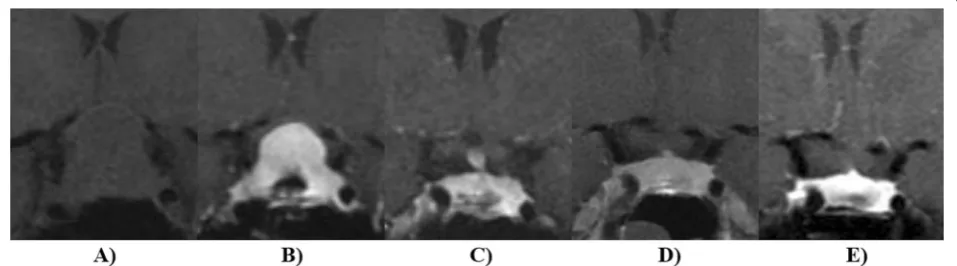
Symptoms from mass effect, such as optic nerve com-pression and other cranial nerve palsies, and severe headache are general indications for the treatment of hypophysitis (Fig. 1). Practice patterns vary for less clinically severe cases. It is unclear whether treatment with immune suppressing medications improves pituit-ary function outcomes compared to supportive therapy.
Spontaneous resolution of pituitary enlargement has been observed in a number of published cases of hypo-physitis [19]. One group recently reported regression of radiologic findings in 15/15 patients receiving supportive therapy [43]. A large recent retrospective review of hypophysitis cases in Germany noted radiologic improve-ment or stability in 16/22 cases without active treatimprove-ment [75]. Pituitary surgery (gross total resection or partial
resection) and glucocorticoid therapy appeared to be somewhat more effective at mass reduction in that study. Surgery (generally patients undergoing gross total resec-tion) was associated with less improvement and greater loss of pituitary function. Patients receiving glucocorticoid therapy had a significant risk of relapsing pituitary en-largement and experienced frequent side effects from treatment. Approximately one-quarter of patients receiv-ing supportive therapy demonstrated improvement in pi-tuitary function, and 82 % of that group had stable or improved function. Similar results were reported in the patients treated with pharmacologic doses of glucocorti-coids. Headache resolution was similar in all three groups [75]. Comparable rates of pituitary function recovery were reported following supportive therapy by Khare et al. [43]. A review by Lupi et al. suggested that pituitary function improvement may occur in approximately one-half of pa-tients treated with glucocorticoids. Importantly, histopath-ology was available in only 22 % of these patients [76]. Similarly, a minority of patients had tissue confirmation in the study from Germany. Moreover, evaluation for sec-ondary causes of hypophysitis was limited in the majority of those patients [42, 75]. Limited clinical evaluations were also frequent in the largest review of granulomatous hypo-physitis cases [20].
Other immunosuppressive agents such as metho-trexate, azathioprine, rituximab, infliximab, cyclospor-ine, and mycophenolate mofetil have been utilized in a small number of patients with hypophysitis [75–82]. Treatment with stereotactic radiosurgery and fraction-ated radiotherapy has been reported in a few patients, typically with refractory disease. Radiation dosages ranged from low levels to higher amounts used to treat pituitary adenomas [31, 83, 84].
Patients with immunotherapy-associated hypophysitis have been treated with physiologic to high-dose gluco-corticoids. Although it is unclear whether pharmacologic
dosages of glucocorticoids improve patient outcomes, higher doses do not appear to negatively impact the anti-tumor efficacy of immunotherapy or patient survival [6]. Improvement of pituitary function occurs in some pa-tients following the resolution of hypophysitis; thyroidal and gonadal axis normalization occurs more frequently than adrenal recovery. The development of hypophysitis may be associated with improved patient survival in mel-anoma patients treated with Ipilimumab [71, 85].
Pathogenesis
The mechanisms underlying the development of hypo-physitis are unknown. Other autoimmune diseases coex-ist in a portion of patients with hypophysitis. Unlike many of these other conditions, pituitary autoantigens in hypophysitis have not yet been clearly identified. Several candidates have been proposed, including growth hor-mone, pituitary gland specific factors 1a and 2 [86, 87], alpha-enolase and gamma-enolase [88, 89], secretogran-nin II [90], chorionic somatomammotropin, CGI-99 [91], and corticotroph-specific transcription factor [92]. Mea-surements of antibodies to these proteins, however, do not have sufficient sensitivity and specificity to be diagnostic-ally useful [16, 93]. Given the lack of clinicdiagnostic-ally validated autoantigens, many studies have utilized indirect im-munofluorescence (IIF) to detect the presence of pituitary autoantibodies. Ricciuti et al. systematically described methodologic limitations of IIF in the assessment of anti-pituitary antibodies, their potential effects on data inter-pretation, and methods to optimize results [94].
The pathogenic role of IgG4 in IgG4-related disease, including hypophysitis, is unclear, and it has been sug-gested that elevation of these antibodies may represent a bystander phenomenon [95]. IgG4 predominance often correlates with immune downregulation, in part due to its ability to participate in fragment antigen binding arm exchange [96].
Immunotherapy agents presumably can activate an autoimmune process directed against unidentified pituit-ary antigens. Pituitpituit-ary autoantibodies were detected in patients who developed hypophysitis following treatment with Ipilimumab (a monoclonal antibody targeting CTLA-4), but these antibodies were not present in pa-tients without hypophysitis. CTLA-4 is also expressed by the pituitary gland, and treatment with Ipilimumab may directly target pituitary cells via activation of the clas-sical complement pathway and antibody-dependent cell-mediated cytotoxicity (ADCC) [97–99]. Pituitary CTLA-4 expression levels appear to vary widely [100] and may affect the risk of developing hypophysitis fol-lowing treatment with Ipilimumab. In support of this hypothesis, hypophysitis has not been reported in pa-tients with germline CTLA-4 mutations, although many of these patients had other severe autoimmune
diseases which can occur following treatment with Ipi-limumab [101, 102]. It is unknown whether PD-1 is expressed by the pituitary gland. Notably, anti-PD-1 agents are IgG4-based antibodies, which can not acti-vate the classical complement pathway and are not ef-fective mediators for ADCC [96, 103–105].
Conclusions
Currently, the diagnosis of primary hypophysitis typically requires a thorough evaluation for other potential neo-plastic lesions, infiltrative diseases, infection, and systemic inflammatory processes plus histopathologic confirmation. In some cases, tissue biopsy may not be feasible. Cases of immunotherapy-associated hypophysitis and lymphocytic hypophysitis associated with pregnancy may potentially be diagnosed with some degree of confidence without sur-gery. Cranial nerve deficits due to mass effect from pituit-ary gland enlargement and severe headache are general indications for treatment with medical therapy and/or sur-gery. It is unclear whether active treatment improves clin-ical outcomes compared to supportive therapy for more mild cases of hypophysitis, and the therapies may be asso-ciated with side effects. Pituitary gland debulking rather than gross total resection is more commonly performed. Glucocorticoids (at variable dosages and duration) are the most frequent choice for medical therapy, though many other immunosuppressive agents have been utilized in the treatment of hypophysitis. Even when treatment is initially successful, disease recurrence is not uncommon. Radi-ation therapy appears promising, especially for refractory cases of hypophysitis, but the available published data consists of only a handful of patients.
Hypophysitis is an increasingly recognized but rare and poorly understood heterogeneous disease. The pathogen-esis of primary hypophysitis is not yet known, and clinic-ally validated disease markers have not been identified. In the absence of more detailed knowledge, various etiologic, morphologic, and histologic categories have been pro-posed, but the clinical utility of such schemas is limited. Inconsistent usage of terminology and variable diagnostic evaluations have also clouded data interpretation. Avail-able studies are largely limited to retrospective series that likely include patients with diverse pathologies. Clinical investigation is constrained by the rarity of the disease. The acquisition of sufficient controlled prospective data will require multicenter collaboration. Until such investi-gations take place, optimal management strategies will remain largely undefined and controversial.
Acknowledgements None.
Availability of data and materials Not applicable.
Competing interests None.
Consent for publication Not applicable.
Ethics approval and consent to participate Not applicable.
Received: 1 July 2016 Accepted: 26 August 2016
References
1. Chan WB, Cockram CS. Panhypopituitarism in association with interferon-alpha treatment. Singapore Med J. 2004;45:93–4.
2. Concha LB, Carlson HE, Heimann A, Lake-Bakaar GV, Paal AF. Interferon-induced hypopituitarism. Am J Med. 2003;114:161–3.
3. Ridruejo E, Christensen AF, Mando OG. Central hypothyroidism and hypophysitis during treatment of chronic hepatitis C with pegylated interferon alpha and ribavirin. Eur J Gastroenterol Hepatol. 2006;18:693–4. 4. Sakane N, Yoshida T, Yoshioka K, Umekawa T, Kondo M, Shimatsu A.
Reversible hypopituitarism after interferonalfa therapy. Lancet. 1995;345:1305.
5. Tebben PJ, Atkinson JL, Scheithauer BW, Erickson D. Granulomatous adenohypophysitis after interferon and ribavirin therapy. Endocr Pract. 2007;13:169–75.
6. Faje A. Immunotherapy and hypophysitis: clinical presentation, treatment, and biologic insights. Pituitary. 2016;19:82–92.
7. Albini CH, MacGillivray MH, Fisher JE, Voorhess ML, Klein DM. Triad of hypopituitarism, granulomatous hypophysitis, and ruptured Rathke’s cleft cyst. Neurosurgery. 1988;22:133–6.
8. Daikokuya H, Inoue Y, Nemoto Y, Tashiro T, Shakudo M, Ohata K. Rathke’s cleft cyst associated with hypophysitis: MRI. Neuroradiology. 2000;42:532–4. 9. Wearne MJ, Barber PC, Johnson AP. Symptomatic Rathke’s cleft cyst with
hypophysitis. Br J Neurosurg. 1995;9:799–803.
10. Hama S, Arita K, Tominaga A, Yoshikawa M, Eguchi K, Sumida M, Inai K, Nishisaka T, Kurisu K. Symptomatic Rathke’s cleft cyst coexisting with central diabetes insipidus and hypophysitis: case report. Endocr J. 1999;46:187–92. 11. Puchner MJ, Ludecke DK, Saeger W. The anterior pituitary lobe in patients
with cystic craniopharyngiomas: three cases of associated lymphocytic hypophysitis. Acta Neurochir (Wien). 1994;126:38–43.
12. McConnon JK, Smyth HS, Horvath E. A case of sparsely granulated growth hormone cell adenoma associated with lymphocytic hypophysitis. J Endocrinol Invest. 1991;14:691–6.
13. Jenkins PJ, Chew SL, Lowe DG, Afshart F, Charlesworth M, Besser GM, Wass JA. Lymphocytic hypophysitis: unusual features of a rare disorder. Clin Endocrinol (Oxf). 1995;42:529–34.
14. Moskowitz SI, Hamrahian A, Prayson RA, Pineyro M, Lorenz RR, Weil RJ. Concurrent lymphocytic hypophysitis and pituitary adenoma. Case report and review of the literature. J Neurosurg. 2006;105:309–14.
15. Holck S, Laursen H. Prolactinoma coexistent with granulomatous hypophysitis. Acta Neuropathol. 1983;61:253–7.
16. Caturegli P, Lupi I, Landek-Salgado M, Kimura H, Rose NR. Pituitary autoimmunity: 30 years later. Autoimmun Rev. 2008;7:631–7.
17. Ahmed SR, Aiello DP, Page R, Hopper K, Towfighi J, Santen RJ. Necrotizing infundibulo-hypophysitis: a unique syndrome of diabetes insipidus and hypopituitarism. J Clin Endocrinol Metab. 1993;76:1499–504.
18. Gutenberg A, Caturegli P, Metz I, Martinez R, Mohr A, Bruck W, Rohde V. Necrotizing infundibulo-hypophysitis: an entity too rare to be true? Pituitary. 2012;15:202–8.
19. Beressi N, Beressi JP, Cohen R, Modigliani E. Lymphocytic hypophysitis. A review of 145 cases. Ann Med Interne (Paris). 1999;150:327–41.
20. Hunn BH, Martin WG, Simpson Jr S, McLean CA. Idiopathic granulomatous hypophysitis: a systematic review of 82 cases in the literature. Pituitary. 2014;17:357–65.
21. Doniach I, Wright EA. Two cases of giant-cell granuloma of the pituitary gland. J Pathol Bacteriol. 1951;63:69–79.
22. Folkerth RD, Price Jr DL, Schwartz M, Black PM, De Girolami U. Xanthomatous hypophysitis. Am J Surg Pathol. 1998;22:736–41. 23. Hanna B, Li YM, Beutler T, Goyal P, Hall WA. Xanthomatous hypophysitis.
J Clin Neurosci. 2015;22:1091–7.
24. Leporati P, Landek-Salgado MA, Lupi I, Chiovato L, Caturegli P. IgG4-related hypophysitis: a new addition to the hypophysitis spectrum. J Clin Endocrinol Metab. 2011;96:1971–80.
25. Caturegli P, Iwama S. From Japan with love: another tessera in the hypophysitis mosaic. J Clin Endocrinol Metab. 2013;98:1865–8.
26. Shimatsu A, Oki Y, Fujisawa I, Sano T. Pituitary and stalk lesions (infundibulo-hypophysitis) associated with immunoglobulin G4-related systemic disease: an emerging clinical entity. Endocr J. 2009;56:1033–41.
27. Caturegli P, Newschaffer C, Olivi A, Pomper MG, Burger PC, Rose NR. Autoimmune hypophysitis. Endocr Rev. 2005;26:599–614.
28. Imber BS, Lee HS, Kunwar S, Blevins LS, Aghi MK. Hypophysitis: a single-center case series. Pituitary. 2015;18:630–41.
29. Buxton N, Robertson I. Lymphocytic and granulocytic hypophysitis: a single centre experience. Br J Neurosurg. 2001;15:242–5. discussion 245–246. 30. Sautner D, Saeger W, Ludecke DK, Jansen V, Puchner MJ. Hypophysitis in
surgical and autoptical specimens. Acta Neuropathol. 1995;90:637–44. 31. Leung GK, Lopes MB, Thorner MO, Vance ML, Laws Jr ER. Primary
hypophysitis: a single-center experience in 16 cases. J Neurosurg. 2004;101:262–71.
32. Honegger J, Fahlbusch R, Bornemann A, Hensen J, Buchfelder M, Muller M, Nomikos P. Lymphocytic and granulomatous hypophysitis: experience with nine cases. Neurosurgery. 1997;40:713–22. discussion 722–713.
33. Goudie RB, Pinkerton PH. Anterior hypophysitis and Hashimoto’s disease in a young woman. J Pathol Bacteriol. 1962;83:584–5.
34. Brissaud HH, Gougerot H, Gy A. Nevrite localised avec troubles trophiques a la suite de coupure de pouce. Rev Neurol. 1908;13:645.
35. Simmonds M. U¨ ber das Vorkommen von Riesenzellen in der Hypophyse. Virchows Arch. 1917;223(3):281–90.
36. van der Vliet HJ, Perenboom RM. Multiple pseudotumors in IgG4-associated multifocal systemic fibrosis. Ann Intern Med. 2004;141:896–7.
37. Guay AT, Agnello V, Tronic BC, Gresham DG, Freidberg SR. Lymphocytic hypophysitis in a man. J Clin Endocrinol Metab. 1987;64:631–4. 38. Levine SN, Benzel EC, Fowler MR, Shroyer 3rd JV, Mirfakhraee M.
Lymphocytic adenohypophysitis: clinical, radiological, and magnetic resonance imaging characterization. Neurosurgery. 1988;22:937–41. 39. Hoshimaru M, Hashimoto N, Kikuchi H. Central diabetes insipidus resulting
from a nonneoplastic tiny mass lesion localized in the neurohypophyseal system. Surg Neurol. 1992;38:1–6.
40. Gellner V, Kurschel S, Scarpatetti M, Mokry M. Lymphocytic hypophysitis in the pediatric population. Childs Nerv Syst. 2008;24:785–92.
41. Landek-Salgado MA, Gutenberg A, Lupi I, Kimura H, Mariotti S, Rose NR, Caturegli P. Pregnancy, postpartum autoimmune thyroiditis, and autoimmune hypophysitis: intimate relationships. Autoimmun Rev. 2010;9:153–7. 42. Honegger J, Schlaffer S, Menzel C, Droste M, Werner S, Elbelt U,
Strasburger C, Stormann S, Kuppers A, Streetz-van der Werf C, et al. Diagnosis of primary hypophysitis in Germany. J Clin Endocrinol Metab. 2015;100:3841–9.
43. Khare S, Jagtap VS, Budyal SR, Kasaliwal R, Kakade HR, Bukan A, Sankhe S, Lila AR, Bandgar T, Menon PS, Shah NS. Primary (autoimmune) hypophysitis: a single centre experience. Pituitary. 2015;18:16–22.
44. Peruzzotti-Jametti L, Strambo D, Sangalli F, De Bellis A, Comi G, Sessa M. Bilateral intracavernous carotid artery occlusion caused by invasive lymphocytic hypophysitis. J Stroke Cerebrovasc Dis. 2012;21:918. e919-911. 45. Ikeda J, Kuratsu J, Miura M, Kai Y, Ushio Y. Lymphocytic adenohypophysitis accompanying occlusion of bilateral internal carotid arteries–case report. Neurol Med Chir (Tokyo). 1990;30:346–9.
46. Melgar MA, Mariwalla N, Gloss DS, Walsh JW. Recurrent lymphocytic hypophysitis and bilateral intracavernous carotid artery occlusion. an observation and review of the literature. Neurol Res. 2006;28:177–83. 47. Minakshi B, Alok S, Hillol KP. Lymphocytic hypophysitis presenting as
pituitary apoplexy in a male. Neurol India. 2005;53:363–4. 48. Husain Q, Zouzias A, Kanumuri VV, Eloy JA, Liu JK. Idiopathic
granulomatous hypophysitis presenting as pituitary apoplexy. J Clin Neurosci. 2014;21:510–2.
50. Turcu AF, Erickson BJ, Lin E, Guadalix S, Schwartz K, Scheithauer BW, Atkinson JL, Young Jr WF. Pituitary stalk lesions: the Mayo Clinic experience. J Clin Endocrinol Metab. 2013;98:1812–8.
51. Cives M, Simone V, Rizzo FM, Dicuonzo F, Cristallo Lacalamita M, Ingravallo G, Silvestris F, Dammacco F. Erdheim-Chester disease: a systematic review. Crit Rev Oncol Hematol. 2015;95:1–11.
52. Monsereenusorn C, Rodriguez-Galindo C. Clinical characteristics and treatment of langerhans cell histiocytosis. Hematol Oncol Clin North Am. 2015;29:853–73. 53. Strowd RE, Burger P, Holdhoff M, Kleinberg L, Okun MS, Olivi A, Pardo-Villamizar
C, Schiess N. Steroid-responsive intracranial germinoma presenting as Holmes’ tremor: importance of a tissue diagnosis. J Clin Neurosci. 2015;22:911–3. 54. Si SJ, Khatua S, Dhall G, Nelson MD, Gonzalez-Gomez I, Finlay JL. Regression
of primary central nervous system germinoma after dexamethasone administration: a case report. Pediatr Hematol Oncol. 2010;27:237–43. 55. Mascalchi M, Roncaroli F, Salvi F, Frank G. Transient regression of an
intracranial germ cell tumour after intravenous steroid administration: a case report. J Neurol Neurosurg Psychiatry. 1998;64:670–2.
56. Gutenberg A, Bell JJ, Lupi I, Tzou SC, Landek-Salgado MA, Kimura H, Su J, Karaviti LP, Salvatori R, Caturegli P. Pituitary and systemic autoimmunity in a case of intrasellar germinoma. Pituitary. 2011;14:388–94.
57. Konno S, Oka H, Utsuki S, Kondou K, Tanaka S, Fujii K, Yagishita S. Germinoma with a granulomatous reaction. Problems of differential diagnosis. Clin Neuropathol. 2002;21:248–51.
58. Endo T, Kumabe T, Ikeda H, Shirane R, Yoshimoto T. Neurohypophyseal germinoma histologically misidentified as granulomatous hypophysitis. Acta Neurochir (Wien). 2002;144:1233–7.
59. Gutenberg A, Larsen J, Lupi I, Rohde V, Caturegli P. A radiologic score to distinguish autoimmune hypophysitis from nonsecreting pituitary adenoma preoperatively. AJNR Am J Neuroradiol. 2009;30:1766–72.
60. Khosroshahi A, Cheryk LA, Carruthers MN, Edwards JA, Bloch DB, Stone JH. Brief Report: spuriously low serum IgG4 concentrations caused by the prozone phenomenon in patients with IgG4-related disease. Arthritis Rheumatol. 2014;66:213–7.
61. Wallace ZS, Deshpande V, Mattoo H, Mahajan VS, Kulikova M, Pillai S, Stone JH. IgG4-Related Disease: Clinical and Laboratory Features in One Hundred Twenty-Five Patients. Arthritis Rheumatol. 2015;67:2466–75.
62. Chang SY, Keogh KA, Lewis JE, Ryu JH, Cornell LD, Garrity JA, Yi ES. IgG4-positive plasma cells in granulomatosis with polyangiitis (Wegener’s): a clinicopathologic and immunohistochemical study on 43 granulomatosis with polyangiitis and 20 control cases. Hum Pathol. 2013;44:2432–7.
63. Nishioka H, Shibuya M, Haraoka J. Immunohistochemical study for IgG4-positive plasmacytes in pituitary inflammatory lesions. Endocr Pathol. 2010;21:236–41.
64. Bando H, Iguchi G, Fukuoka H, Taniguchi M, Kawano S, Saitoh M, Yoshida K, Matsumoto R, Suda K, Nishizawa H, et al. A diagnostic pitfall in IgG4-related hypophysitis: infiltration of IgG4-positive cells in the pituitary of
granulomatosis with polyangiitis. Pituitary. 2015;18:722–30.
65. Ohkubo Y, Sekido T, Takeshige K, Ishi H, Takei M, Nishio S, Yamazaki M, Komatsu M, Kawa S, Suzuki S. Occurrence of IgG4-related hypophysitis lacking IgG4-bearing plasma cell infiltration during steroid therapy. Intern Med. 2014;53:753–7.
66. Deshpande V, Zen Y, Chan JK, Yi EE, Sato Y, Yoshino T, Kloppel G, Heathcote JG, Khosroshahi A, Ferry JA, et al. Consensus statement on the pathology of IgG4-related disease. Mod Pathol. 2012;25:1181–92.
67. Bando H, Iguchi G, Fukuoka H, Taniguchi M, Yamamoto M, Matsumoto R, Suda K, Nishizawa H, Takahashi M, Kohmura E, Takahashi Y. The prevalence of IgG4-related hypophysitis in 170 consecutive patients with
hypopituitarism and/or central diabetes insipidus and review of the literature. Eur J Endocrinol. 2014;170:161–72.
68. Tauziede-Espariat A, Polivka M, Bouazza S, Decq P, Robert G, Laloi-Michelin M, Adle-Biassette H. The prevalence of IgG4-positive plasma cells in hypophysitis: a possible relationship to IgG4-related disease. Clin Neuropathol. 2015;34:181–92.
69. Ngaosuwan K, Trongwongsa T, Shuangshoti S. Clinical course of IgG4-related hypophysitis presenting with focal seizure and relapsing lymphocytic hypophysitis. BMC Endocr Disord. 2015;15:64.
70. Sosa GA, Bell S, Christiansen SB, Pietrani M, Glerean M, Loto M, Lovazzano S, Carrizo A, Ajler P, Fainstein Day P. Histologically confirmed isolated IgG4-related hypophysitis: two case reports in young women. Endocrinol Diabetes Metab Case Rep. 2014;2014:140062.
71. Faje AT, Sullivan R, Lawrence D, Tritos NA, Fadden R, Klibanski A, Nachtigall L. Ipilimumab-induced hypophysitis: a detailed longitudinal analysis in a large cohort of patients with metastatic melanoma. J Clin Endocrinol Metab. 2014;99:4078–85.
72. Jennings MT, Gelman R, Hochberg F. Intracranial germ-cell tumors: natural history and pathogenesis. J Neurosurg. 1985;63:155–67.
73. Howarth DM, Gilchrist GS, Mullan BP, Wiseman GA, Edmonson JH, Schomberg PJ. Langerhans cell histiocytosis: diagnosis, natural history, management, and outcome. Cancer. 1999;85:2278–90.
74. Stalemark H, Laurencikas E, Karis J, Gavhed D, Fadeel B, Henter JI. Incidence of Langerhans cell histiocytosis in children: a population-based study. Pediatr Blood Cancer. 2008;51:76–81.
75. Honegger J, Buchfelder M, Schlaffer S, Droste M, Werner S, Strasburger C, Stormann S, Schopohl J, Kacheva S, Deutschbein T, et al. Treatment of primary hypophysitis in Germany. J Clin Endocrinol Metab. 2015;100:3460–9. 76. Lupi I, Manetti L, Raffaelli V, Lombardi M, Cosottini M, Iannelli A, Basolo F,
Proietti A, Bogazzi F, Caturegli P, Martino E. Diagnosis and treatment of autoimmune hypophysitis: a short review. J Endocrinol Invest. 2011;34:e245–252.
77. Schreckinger M, Francis T, Rajah G, Jagannathan J, Guthikonda M, Mittal S. Novel strategy to treat a case of recurrent lymphocytic hypophysitis using rituximab. J Neurosurg. 2012;116:1318–23.
78. Xu C, Ricciuti A, Caturegli P, Keene CD, Kargi AY. Autoimmune lymphocytic hypophysitis in association with autoimmune eye disease and sequential treatment with infliximab and rituximab. Pituitary. 2015;18:441–7.
79. Lecube A, Francisco G, Rodriguez D, Ortega A, Codina A, Hernandez C, Simo R. Lymphocytic hypophysitis successfully treated with azathioprine: first case report. J Neurol Neurosurg Psychiatry. 2003;74:1581–3.
80. Li HT, Wang ST, Qiu MC. Gynecomastia, obesity and underdeveloped testis and penis: suspected hypophysitis successfully cured with low dose of cyclosporine A. Chin Med J (Engl). 2009;122:2791–3.
81. Ward L, Paquette J, Seidman E, Huot C, Alvarez F, Crock P, Delvin E, Kampe O, Deal C. Severe autoimmune polyendocrinopathy-candidiasis-ectodermal dystrophy in an adolescent girl with a novel AIRE mutation: response to immunosuppressive therapy. J Clin Endocrinol Metab. 1999;84:844–52. 82. Louvet C, Maqdasy S, Tekath M, Grobost V, Rieu V, Ruivard M, Le Guenno G.
Infundibuloneurohypophysitis associated with sjogren syndrome successfully treated with mycophenolate mofetil: a case report. Medicine (Baltimore). 2016;95:e3132.
83. Ray DK, Yen CP, Vance ML, Laws ER, Lopes B, Sheehan JP. Gamma knife surgery for lymphocytic hypophysitis. J Neurosurg. 2010;112:118–21. 84. Selch MT, DeSalles AA, Kelly DF, Frighetto L, Vinters HV, Cabatan-Awang C,
Wallace RE, Solberg TD. Stereotactic radiotherapy for the treatment of lymphocytic hypophysitis. Report of two cases. J Neurosurg. 2003;99:591–6. 85. Eatrides J, Weber J, Egan K, Acierno M, Schell M, Lillienfeld H, Creelan B:
Autoimmune hypophysitis is a marker of favorable outcome during treatment of melanoma with ipilimumab. AACR: Advances in Melanoma: From Biology to Therapy 2014, abstract.
86. Takao T, Nanamiya W, Matsumoto R, Asaba K, Okabayashi T, Hashimoto K. Antipituitary antibodies in patients with lymphocytic hypophysitis. Horm Res. 2001;55:288–92.
87. Tanaka S, Tatsumi KI, Kimura M, Takano T, Murakami Y, Takao T, Hashimoto K, Kato Y, Amino N. Detection of autoantibodies against the pituitary-specific proteins in patients with lymphocytic hypophysitis. Eur J Endocrinol. 2002;147:767–75.
88. Tanaka S, Tatsumi KI, Takano T, Murakami Y, Takao T, Yamakita N, Tahara S, Teramoto A, Hashimoto K, Kato Y, Amino N. Anti-alpha-enolase antibodies in pituitary disease. Endocr J. 2003;50:697–702.
89. O’Dwyer DT, Clifton V, Hall A, Smith R, Robinson PJ, Crock PA. Pituitary autoantibodies in lymphocytic hypophysitis target both gamma- and alpha-Enolase - a link with pregnancy? Arch Physiol Biochem. 2002;110:94–8. 90. Bensing S, Hulting AL, Hoog A, Ericson K, Kampe O. Lymphocytic
hypophysitis: report of two biopsy-proven cases and one suspected case with pituitary autoantibodies. J Endocrinol Invest. 2007;30:153–62. 91. Lupi I, Broman KW, Tzou SC, Gutenberg A, Martino E, Caturegli P. Novel
autoantigens in autoimmune hypophysitis. Clin Endocrinol (Oxf). 2008;69:269–78. 92. Smith CJ, Bensing S, Burns C, Robinson PJ, Kasperlik-Zaluska AA, Scott
93. Falorni A, Minarelli V, Bartoloni E, Alunno A, Gerli R. Diagnosis and classification of autoimmune hypophysitis. Autoimmun Rev. 2014;13:412–6.
94. Ricciuti A, De Remigis A, Landek-Salgado MA, De Vincentiis L, Guaraldi F, Lupi I, Iwama S, Wand GS, Salvatori R, Caturegli P. Detection of pituitary antibodies by immunofluorescence: approach and results in patients with pituitary diseases. J Clin Endocrinol Metab. 2014;99:1758–66.
95. Wallace ZS, Stone JH. An update on IgG4-related disease. Curr Opin Rheumatol. 2015;27:83–90.
96. Vidarsson G, Dekkers G, Rispens T. IgG subclasses and allotypes: from structure to effector functions. Front Immunol. 2014;5:520.
97. Iwama S, De Remigis A, Callahan MK, Slovin SF, Wolchok JD, Caturegli P. Pituitary expression of CTLA-4 mediates hypophysitis secondary to administration of CTLA-4 blocking antibody. Sci Transl Med. 2014;6:230ra245.
98. Romano E, Kusio-Kobialka M, Foukas PG, Baumgaertner P, Meyer C, Ballabeni P, Michielin O, Weide B, Romero P, Speiser DE. Ipilimumab-dependent cell-mediated cytotoxicity of regulatory T cells ex vivo by nonclassical monocytes in melanoma patients. Proc Natl Acad Sci U S A. 2015;112:6140–5.
99. Laurent S, Queirolo P, Boero S, Salvi S, Piccioli P, Boccardo S, Minghelli S, Morabito A, Fontana V, Pietra G, et al. The engagement of CTLA-4 on primary melanoma cell lines induces antibody-dependent cellular cytotoxicity and TNF-alpha production. J Transl Med. 2013;11:108. 100. Faje A, Ma J, Wang X, Swearingen B, Tritos NA, Nachtigall L, Zhang X,
Klibanski A: Cytotoxic T-lymphocyte antigen-4 gene expression in human pituitary adenomas. ENDO 2015, abstract.
101. Kuehn HS, Ouyang W, Lo B, Deenick EK, Niemela JE, Avery DT, Schickel JN, Tran DQ, Stoddard J, Zhang Y, et al. Immune dysregulation in human subjects with heterozygous germline mutations in CTLA4. Science. 2014;345:1623–7.
102. Schubert D, Bode C, Kenefeck R, Hou TZ, Wing JB, Kennedy A, Bulashevska A, Petersen BS, Schaffer AA, Gruning BA, et al. Autosomal dominant immune dysregulation syndrome in humans with CTLA4 mutations. Nat Med. 2014;20:1410–6.
103. Garred P, Michaelsen TE, Aase A. The IgG subclass pattern of complement activation depends on epitope density and antibody and complement concentration. Scand J Immunol. 1989;30:379–82.
104. Michaelsen TE, Garred P, Aase A. Human IgG subclass pattern of inducing complement-mediated cytolysis depends on antigen concentration and to a lesser extent on epitope patchiness, antibody affinity and complement concentration. Eur J Immunol. 1991;21:11–6.
105. Bruhns P, Iannascoli B, England P, Mancardi DA, Fernandez N, Jorieux S, Daeron M. Specificity and affinity of human Fcgamma receptors and their polymorphic variants for human IgG subclasses. Blood. 2009;113:3716–25.
• We accept pre-submission inquiries
• Our selector tool helps you to find the most relevant journal
• We provide round the clock customer support
• Convenient online submission
• Thorough peer review
• Inclusion in PubMed and all major indexing services
• Maximum visibility for your research
Submit your manuscript at www.biomedcentral.com/submit