Decrease in Birth Weight in Relation to Maternal Bone-Lead Burden
Teresa Gonza´lez-Cossı´o, PhD*; Karen E. Peterson, ScD, RD‡; Luz-Helena Sanı´n, MD, MPH*§; Eugenia Fishbein, BS*i; Eduardo Palazuelos, MDi¶; Antonio Aro, PhD‡;
Mauricio Herna´ndez-Avila, MD, ScD*; and Howard Hu, MD, ScD‡#
ABSTRACT. Objectives. Birth weight predicts infant survival, growth, and development. Previous research suggests that low levels of fetal lead exposure, as esti-mated by umbilical cord blood-lead levels at birth, may have an adverse effect on birth weight. This report ex-amines the relationship of lead levels in cord blood and maternal bone to birth weight.
Methods. Umbilical cord and maternal venous blood samples and anthropometric and sociodemographic data were obtained at delivery and 1-month postpartum. Blood-lead levels were analyzed by atomic absorption spectrophotometry. Maternal tibia and patella lead levels were determined at 1-month postpartum with use of a spot-source 109Cd K-X-ray fluorescence instrument. The relationship between birth weight and lead burden was evaluated by multiple regression with control of known determinants of size at birth.
Results. Data on all variables of interest were ob-tained for 272 mother-infant pairs. After adjustment for other determinants of birth weight, tibia lead was the only lead biomarker clearly related to birth weight. The decline in birth weight associated to increments in tibia lead was nonlinear and accelerated at the highest tibia lead quartile. In the upper quartile, neonates were on average, 156 grams lighter than those in the lowest quar-tile. Other significant birth weight predictors included maternal nutritional status, parity, education, gestational age, and smoking during pregnancy.
Conclusions. Our results indicate that bone-lead bur-den is inversely related to birth weight. Taken together with other research indicating that lead can mobilize from bone into plasma without detectable changes in whole blood lead, these findings suggest that bone lead might be a better biomarker than blood lead. Because lead remains in bone for years to decades, mobilization of bone lead during pregnancy may pose a significant fetal exposure with health consequences, long after ma-ternal exma-ternal lead exposure has declined. Pediatrics 1997;100:856 – 862;bone lead, birth weight, pregnancy, fe-tus, epidemiology, x-ray fluorescence.
ABBREVIATIONS. LBW, low birth weight; K-XRF, K-X-ray fluo-rescence.
B
irth weight is a strong predictor of survival and of developmental outcomes in childhood in-cluding growth, morbidity, and cognitive per-formance.1– 4High-level occupational lead exposures have been associated with adverse pregnancy out-comes, but studies of lower-level lead exposure on birth weight in community settings have yielded inconsistent findings across the different anthropo-metric indicators of birth weight.5–7Several well-con-trolled studies failed to show a consistent association of maternal blood-lead levels with preterm delivery, mean gestational age, or birth weight adjusted for gestational age.7However, increased risk of low birth weight [(LBW) ,2.5 kg] and lower mean birth weights suggest that lead exposure may affect birth weight through intrauterine growth retardation rather than prematurity.Such inconsistent findings may be explained at least partially by the fact that all previous studies have relied on measurements of lead in whole blood to assess prenatal exposure. More than 95% of lead in a whole blood specimen is bound to red cells.8Lead that is available to cross the placenta, however, is derived from lead that is in the free state in plasma.9 Evidence is beginning to accumulate that plasma-lead levels may fluctuate significantly without a dis-cernible change in whole blood-lead levels; more-over, plasma lead seems to have a positive correlation with levels of lead in bone.10
This issue is important because bone lead stores can be retained from years to decades despite a de-cline in environmental lead exposure and blood-lead levels. Because pregnancy and lactation stimulate increased bone turnover11 there is concern that mo-bilization of lead from bone into plasma may consti-tute a significant threat to the fetus that is not well estimated by measuring lead in whole blood. Al-though measurement of plasma lead is not feasible for large-scale epidemiologic studies, accurate, in vivo measurements of bone-lead burden can be made with K-X-ray fluorescence (K-XRF).12
Mexico City has a history of severe lead exposure related to lead-glazed ceramics used to prepare and serve food, and to leaded gasoline.13 Because bone lead has a half-life of years to decades, women in their childbearing years who have lived in Mexico City may have a large lead burden that serves as an From the *Centro de Investigaciones en Salud Poblacional, Instituto
Nacio-nal de Salud Pu´cblica, Cuernavaca, Morelos, Me´xico; ‡Departments of Maternal and Child Health and Nutrition, Harvard School of Public Health, Boston Massachusetts; §Universidad Auto´noma de Chihuahua, Chihuahua, Me´xico;iAmerican British Cowdray Hospital, Me´xico City, Me´xico; ¶Sec-retarı´a del Medio Ambiente, Departamento del Distrito Federal, Me´xico City, Me´xico; and #Channing Laboratory, Department of Medicine, Brigham and Women’s Hospital, Harvard Medical School and Occupational Health Program, Department of Environmental Health, Harvard School of Public Health, Boston, Massachusetts.
Received for publication Nov 25, 1996; accepted Mar 24, 1997.
Reprint requests to (M.H.-A.) Instituto Nacional de Salud Pu´cblica, Av Universidad 655, Col Sta Ma Ahuacatitla´n, Cuernavaca, Morelos, Me´xico CP 62508.
endogenous source of fetal exposure and may ad-versely affect fetal growth. This report examines the relationship of blood lead and K-XRF measured ternal bone lead to birth weight, controlling for ma-ternal nutritional status and other determinants of birth weight in a sample of healthy lactating women delivering term infants in Mexico City.
METHODS AND POPULATION
This article presents baseline data of a randomized double-blind calcium supplementation trial to lactating women to test the hypothesis that maternal-to-infant lead mobilization decreases as calcium intake increases. Data collection of such study started at the end of pregnancy and the intervention began at 1-month postpartum.
Sample Selection
We collected data at delivery and 1-month postpartum from women attending any of three hospitals in Mexico City (Mexican Social Security Institute, Manuel Gea Gonza´lez Hospital, and Na-tional Institute of Perinatology) that serve a low-to-moderate in-come population. Baseline information on health status and on social and demographic characteristics was collected from all eli-gible participants. Anthropometric data from the mother and new-born, and umbilical cord and maternal blood-lead levels were gathered within612 hours of delivery. Information on estimated gestational age, based on date of last menstrual period, and char-acteristics of the birth and newborn were extracted from the medical records. Interviewers explained the study to, and ob-tained written consent from, eligible women willing to participate. Exclusion criteria included factors that could interfere with maternal calcium metabolism, medical conditions that could cause LBW, and logistic reasons that would interfere with data collec-tion, which included living in a household outside the metropol-itan area; intention not to breastfeed; delivered a premature neo-nate (,37 weeks); physician’s diagnosis of multiple fetuses; preeclampsia, psychiatric, kidney, or cardiac disease; gestational diabetes; history of repeated urinary infections; family or personal history of kidney stone formation; seizure disorder requiring daily medications; ingestion of corticoids, or blood pressure.140 mm Hg systolic or.90 mm Hg diastolic. One month after delivery (65 days) each mother-infant pair attended the research center for an evaluation that included measurement of maternal bone lead us-ing a spot-source109Cd K-XRF instrument.
The research protocol was approved by the Human Subjects Committee of the National Institute of Public Health of Mexico and the participant hospitals. All participant mothers received a detailed explanation of the study and its procedures used, as well as counseling on reduction of lead exposure.
Questionnaires
An interview questionnaire was used to assess social and de-mographic characteristics, reproductive history, maternal and in-fant perinatal health, inin-fant feeding practices, as well as informa-tion on known risk factors for high blood lead documented in our previous studies.13
Anthropometry
Anthropometry was used to assess the nutritional status of mothers and infants.14Nude newborns were weighed within the
first 12 hours of delivery by experienced obstetric nurses using calibrated beam scales (Oken, Model TD16, Naucalpan, Me´xico) read to the nearest 10 grams. Maternal anthropometric measures were collected by our trained15project personnel and standardized
according to the technique described by Habicht.16 Maternal
height was measured at 1-month postpartum with professional scales (PAME, Puebla, Puebla) read to the nearest mm. Arm and calf circumferences were measured with plastic-covered fabric measuring tapes read to the nearest mm. Standardization exer-cises were performed until the project staff reached imprecision errors equal to or below those reported by Lohman and cowork-ers.15Accepted technical errors were 0.3 cm for arm and calf
circumferences and 0.22 cm for height.
Lead Measurements
Blood samples were analyzed using an atomic absorption spec-trophotometry instrument (Perkin-Elmer 3000, Chelmsford, MA) at the metals laboratory of the American British Cowdray Hospital in Mexico City. Analysis of external blinded quality control sam-ples were provided throughout the study period by the Maternal & Child Health Bureau (MCHB) and the Wisconsin State Labora-tory of Hygiene (WSLH) Cooperative Blood Lead Proficiency Testing Program (PBPTP), which demonstrated good precision and accuracy of the American British Cowdray Hospital determi-nations, with a correlation coefficient of 0.99 and mean difference of 0.17mg/dL compared with MCHB and WSLH PBPTP blanks and spikes.
Bone lead measurements were taken of each mother’s mid-tibia shaft (cortical) and patella (trabecular bone) with a spot-source
109Cd K-XRF instrument constructed at Harvard University and
installed in a research facility at the American British Cowdray Hospital. The physical principles, technical specifications, and validation of this and other similar K-XRF instruments have been described in detail elsewhere.17Briefly, this instrument uses a spot 109Cdg-ray source to provoke the emission of fluorescent photons
from target tissue that are then detected, counted, and arrayed on a spectrum. The net lead signal is determined after subtraction of the Compton background counts by a linear least-squares algo-rithm. The lead fluorescent signal is then normalized to the elastic or coherently scatteredg-ray signal, which arises predominantly from the calcium and phosphorous present in bone mineral. Be-cause the instrument provides a continuous unbiased point esti-mate that oscillates around the true bone lead value, negative point estimates are sometimes produced when true bone lead values are close to zero. The instrument also provides an estimate of the uncertainty associated with each measurement, derived from a goodness-of-fit calculation of the spectrum curves that is equivalent to a single standard deviation. Although a minimal detectable limit calculation of twice this value has been proposed for interpreting an individual’s bone lead estimate, statistical ex-periments have shown that retention of all point estimates makes better use of the data in epidemiologic studies.12For the present
study, 30-minute measurements were taken at the mid-shaft of the left tibia and at the left patella. Analysis of means and standard deviations of phantom-calibrated measurements did not disclose any significant shift in accuracy or precision.
Statistical Analyses
We examined the relationship between birth weight and known determinants of size at birth, as well as potential confound-ers of the lead-birth size relationship. Measured variables in-cluded umbilical cord and maternal venous blood lead and tibia and patella lead as biomarkers of fetal lead exposure. Information on maternal height, arm and calf circumferences at delivery (cm), smoking (current, past, or never), parity (1 vs 21prior deliveries), history of poor reproductive outcomes (yes/no), age and educa-tion (yr), site of delivery (Mexican Social Security Institute/not-Mexican Social Security Institute), infant gender, and gestational age (wk) was also collected.
Univariate and bivariate statistics, tabulations, and distribution plots were examined for all variables. Analyses were conducted using STATA18and SAS-PC Version 6.0419software. An ordinary
least-squares multiple regression procedure was used to estimate the adjusted association of lead to birth weight. Specification of our initial saturated model, based on a biologic paradigm of known determinants of birth weight,20included all the variables
mentioned above. We dropped covariates, one by one, with an associated two-tailed P value of..05. Our final model included only those variables with a statistically significant association (P,
.05) with birth weight. Independent variables were included in their original values, without categorization. Smoothed plots in-dicated that birth weight did not decrease linearly with increasing lead burden. Thus, we analyzed the association of the lead bi-omarkers first with the variables as continuous variables and then with the variables divided into four groups (often quartiles), with the lowest group as the reference category.
RESULTS
because they lived outside the metropolitan area or did not intend to breastfeed their infants. Of the 797 eligible women, 315 (40%) completed the 1-month postpartum evaluations. A final sample of 272 moth-er-infant pairs (34% of the originally eligible women and 86% of those who returned for the study at 1 month) had complete data for all variables of inter-est. Among the eligible women, there were no sig-nificant differences between participants and non-participants in maternal nutritional status, age, education, parity, or infant birth weight or gesta-tional age, or in lead levels in maternal or umbilical cord blood.
Mean, median, and standard deviation of lead lev-els were as follows: maternal blood, 8.9, 8.1, and 4.1
mg/dL, respectively; umbilical cord blood, 7.1, 6.2, and 3.5 mg/dL; maternal tibia, 9.8, 9.1, and 8.9 mg Pb/g bone mineral; patella, 14.2, 13.8, and 13.2 mg Pb/g bone mineral. The numbers of point estimates that were below 0 mg Pb/g bone mineral were 28 (10%) and 36 (13%) for the tibia and patella, respec-tively.
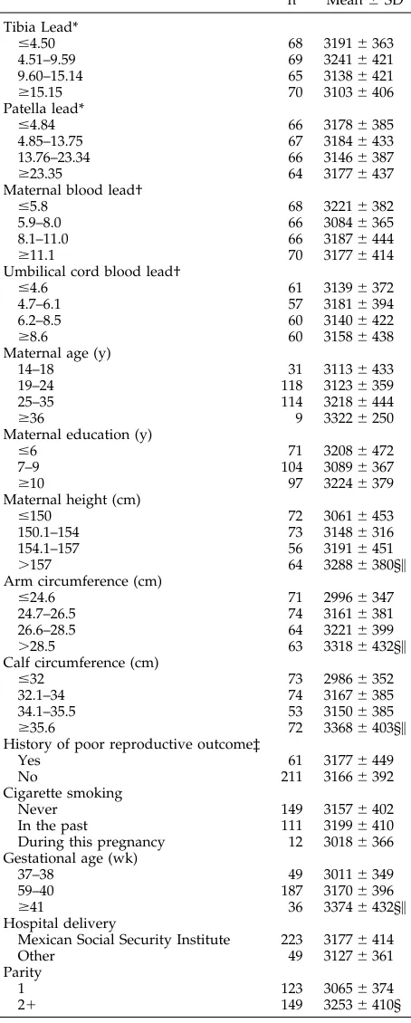
Mean (standard deviation) birth weight was 3168 (6404) grams, and 11 infants (4%) had LBW. Because all newborns had a gestational age of $37 weeks, these 11 neonates were intrauterine growth retarded. Birth weight tended to be higher for neonates of mothers who did not smoke during pregnancy and significantly higher in the group of multiparous women. Birth weight was 3182 (6408) grams and 3155 (6402) grams for boys and girls, respectively (P..05). Data on maternal characteristics and their unadjusted associations with birth weight are pre-sented in Table 1. These crude analyses suggest an inverse association between tibia lead and size at birth. Other lead burden biomarkers were unrelated to the study outcome in a bivariate fashion. Maternal nutritional status, gestational age, and parity were strongly associated with size at birth.
In multivariate regression models, lead was al-ways negatively associated with decreased size at birth, although not all biomarkers with statistical significance. Of the four biomarkers of lead burden, with adjustment for other important determinants of birth weight, only tibia lead level had a statistically significant relationship to birth weight (P , .005; Table 2). The final regression model is presented in the upper panel of Table 2. An increase of 10mg Pb/g bone mineral in tibia is associated with a decrease in birth weight of 73 grams, this estimate increased modestly to 75 grams after correction for measure-ment uncertainty.12 Other significant independent predictors of birth weight include current maternal nutritional status (as estimated by calf circumfer-ence), parity, smoking during pregnancy, education, and newborn gestational age. In the lower panel of Table 2, we present the adjusted change in birth weight associated with quartiles of tibia lead, as de-scribed above. The Figure presents a smoothed curve of the adjusted relationship of these two study vari-ables.
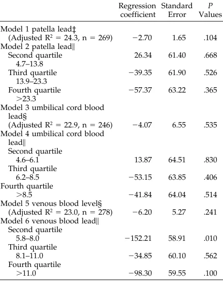
The association between birth weight and the other three biomarkers of lead burden, adjusted for the same variables in the regression model, was also
analyzed. Results show that, although all other ma-ternal lead burden indicators were also inversely related to birth weight (Table 3), these relationships were not statistically significant.
DISCUSSION
The results of this study suggest that lead in bone (as represented by the tibia) is inversely associated with birth weight. This is the first study to evaluate the association between maternal bone-lead burden and birth weight. Using cord blood lead as the bio-logic marker of intrauterine lead burden, other stud-ies in this area have reported an association between blood lead and LBW adjusted for gestational age.21 Their results are in the same direction as ours; ie, birth weight decreases as blood lead increases with an apparent threshold relationship at around 15mg/ dL. However, other studies have failed to find this association22,23 or have found an association in the opposite direction.24
Probably the best explanations for the lack of pos-itive findings in the latter studies may be the type of measurement and control for independent causes of size at birth considered, their statistical power, or the use of blood (as opposed to bone) lead as the biomar-ker to assess lead burden. We collected information on the most relevant predictors of birth weight, and controlled for this information either by design or by statistical modeling. Furthermore, our use of mater-nal bone lead as well as blood lead as biomarkers for fetal lead exposure is a sizable improvement over previous methods because bone lead is an indicator of cumulative rather than recent lead exposure. In addition, as noted above, bone lead might be a better biomarker of plasma lead.
Our data suggest an inverse relationship between tibia lead and birth weight (see Figure). In the upper quartile of tibia lead burden, neonates were on aver-age 156 grams lighter than those in the lowest quar-tile. We selected healthy women for our study so probably the association of lead on birth weight may be larger. Public health problems of this magnitude are seldom as strong, possibly only surpassed by maternal malnutrition, which may explain the more than 50% incidence of LBW in developing coun-tries.20 The impact on birth weight of most supple-mentation trials to pregnant women at high nutri-tional risk have had similar or smaller effects.25–30 Only when food supplements were consumed in most stressful conditions31–32 or during current and previous reproductive cycles,33 is a larger effect on birth weight observed.
impair birth weight through an effect on prenatal bone growth itself in such a way that attained weight at birth may be negatively affected.
On the other hand, evidence that lead deposited in bone serves as a source of blood lead is rapidly growing.41 Ninety-five percent or more of adult’s lead is stored in bone42and it is now recognized that bone, rather than blood, acts as a sink for lead, and that it is a metabolically active depot from which lead moves in and out at a rate determined by factors affecting bone remodeling.43
Neither hospital staff, study personnel, nor study participants were aware of the hypothesis being tested. This prevented that participation in the study or completeness of follow-up were differentially af-fected by birth weight or lead levels. Birth weight data was not directly measured by our project staff, but by trained hospital personnel. However, variabil-ity associated with birth weight in our study was low compared with most,25–27,44although not all29similar reports of hospital deliveries of well-controlled nu-tritional supplementation trials with birth weight as the critical study outcome. This serves as an indica-tion of the adequacy of our birth weight data. Fur-thermore, measurement imprecision was most likely random, and the potential bias would attenuate the observed association. Thus, it is likely that ours is probably an underestimation of the true association between lead burden and size at birth.
Our study may be limited by measurement of bone lead at 1-month postpartum and not during nancy. We did not measure bone lead during preg-nancy because current Mexican human subjects guidelines for medical research prohibit the use of any research procedure that involves radiation in pregnant women, even if the dose delivered is insig-nificant and well below current accepted criteria for all ages of life.45 There are two lines of evidence which suggest that bone lead measurements at 1-month postpartum may adequately reflect those at the beginning of pregnancy: 1) lead in cortical bone (such as the tibia) has a half-life of decades,46and 2) we observed in our data that both patella and tibia lead estimates were correlated to maternal (r5 0.24 and 0.20) as well as to umbilical (r5 0.25 and 0.26) cord blood-lead levels, respectively.
TABLE 1. Birth Weight in Relation to Maternal Lead Burden, Health, and Sociodemographic Characteristics Among Postpar-tum Mexican Women
n Mean6SD
Tibia Lead*
#4.50 68 31916363
4.51–9.59 69 32416421
9.60–15.14 65 31386421
$15.15 70 31036406
Patella lead*
#4.84 66 31786385
4.85–13.75 67 31846433
13.76–23.34 66 31466387
$23.35 64 31776437
Maternal blood lead†
#5.8 68 32216382
5.9–8.0 66 30846365
8.1–11.0 66 31876444
$11.1 70 31776414
Umbilical cord blood lead†
#4.6 61 31396372
4.7–6.1 57 31816394
6.2–8.5 60 31406422
$8.6 60 31586438
Maternal age (y)
14–18 31 31136433
19–24 118 31236359
25–35 114 32186444
$36 9 33226250
Maternal education (y)
#6 71 32086472
7–9 104 30896367
$10 97 32246379
Maternal height (cm)
#150 72 30616453
150.1–154 73 31486316
154.1–157 56 31916451
.157 64 32886380§\
Arm circumference (cm)
#24.6 71 29966347
24.7–26.5 74 31616381
26.6–28.5 64 32216399
.28.5 63 33186432§\
Calf circumference (cm)
#32 73 29866352
32.1–34 74 31676385
34.1–35.5 53 31506385
$35.6 72 33686403§\
History of poor reproductive outcome‡
Yes 61 31776449
No 211 31666392
Cigarette smoking
Never 149 31576402
In the past 111 31996410 During this pregnancy 12 30186366 Gestational age (wk)
37–38 49 30116349
59–40 187 31706396
$41 36 33746432§\
Hospital delivery
Mexican Social Security Institute 223 31776414
Other 49 31276361
Parity
1 123 30656374
21 149 32536410§
*mg Pb/g bone mineral. †mg/dL.
‡ Prior birth weight,2.5 kg, gestational age,37 weeks, stillbirth, or spontaneous abortion.
§ P,.05, Scheffe`. \Test for trend, P,.01.
TABLE 2. Regression of Maternal Tibia Lead on Birth Weight Controlling for Maternal Health and Sociodemographic Charac-teristics Among Postpartum Mexican Women (n5272)
Variables in the Model* Regression Coefficient
Standard Error
P values Tibia lead† 27.29 2.45 0.003 Calf circumference (cm) 40.42 7.81 ,0.001 Parity (151, 2521) 205.87 44.24 ,0.001 Education (y) 17.01 6.98 0.016 Gestational age (wk) 75.49 18.72 ,0.001 Current smokers 2239.61 104.73 0.023 Constant 21576.02 733.59 0.033 Adjusted effect of tibia lead† by quartiles, on birth weight‡
Second quartile
.4.462#9.59
27.57 60.98 0.901
Third quartile
.9.592#15.14
250.86 62.03 0.413
Fourth quartile.15.14 2155.55 61.18 0.012 * Model adjusted R2523.8.
†mg Pb/g bone mineral.
Tibia and patella lead were both inversely related to birth weight, but this association was significant only for tibia lead. The difference in our observed
associations between the two bone lead markers could be attributed to the error measurement as-sociated with patella lead determination. On one side is the fact that patella-lead levels varies faster probably because of its trabecular nature.46 – 47It is also likely that in response to the important cal-cium demand that women experience during preg-nancy and lactation —preferentially affecting tra-becular bone— biologic variability may have increased, conditioning an increase in error mea-surement at this site. On the other side, trabecular bone lead measurement is also measured with a higher degree of error, because of the low mineral content of this bone type. The combination of these factors may explain the observed lack of a statisti-cally significant association between patella lead and birth weight.
Because bone lead has a half-life of years to de-cades, our results suggest that blood-lead levels can-not be used to fully predict risk of associated fetal toxicity. Lead can remain a significant threat to the fetus long after cessation of external lead exposure to women who are pregnant or who will become preg-nant. Bone-lead levels in our study population were three times higher than median postpartum bone levels of women who gave birth in a Boston hospital in 1990 to 1992 (tibia median, 4; patella median, 5mg Pb/g).48 This finding has significant public health implications. Moreover, current regulations govern-ing occupational exposure to lead do not offer any protection to women who might become pregnant in the future.
The finding of neurotoxic effects at increasingly lower lead concentrations21suggests that a fetotoxic effect may still occur among women who have rela-tively low blood-lead levels but that retain high bone-lead burdens. We explored the association of Figure. Adjusted relation between birth weight and tibia-lead levels* (smooth). Adjusted by gestational age, parity, smoking status, calf circumference, and education (year).
TABLE 3. Adjusted* Estimates of the Association† of Mater-nal Venous Blood, Patella, and Umbilical Cord Blood Lead on Birth Weight Among Postpartum Mexican Women
Regression coefficient
Standard Error
P Values Model 1 patella lead‡
(Adjusted R2524.3, n5269) 22.70 1.65 .104
Model 2 patella lead\
Second quartile 26.34 61.40 .668 4.7–13.8
Third quartile 239.35 61.90 .526 13.9–23.3
Fourth quartile 257.37 63.22 .365
.23.3
Model 3 umbilical cord blood lead§
(Adjusted R2522.9, n5246) 24.07 6.55 .535
Model 4 umbilical cord blood lead\
Second quartile
4.6–6.1 13.87 64.51 .830
Third quartile
6.2–8.5 253.15 63.85 .406 Fourth quartile
.8.5 241.84 64.04 .514
Model 5 venous blood level§
(Adjusted R2523.0, n5278) 26.20 5.27 .241
Model 6 venous blood lead\ Second quartile
5.8–8.0 2152.21 58.91 .010 Third quartile
8.1–11.0 234.85 60.10 .562 Fourth quartile
.11.0 298.30 59.55 .100
* Adjusted for the independent variables of model in Table 2. † By quartiles of available observations.
‡mg Pb/g bone mineral. §mg/dL.
tibia lead burden and birth weight at three levels of cord and maternal blood-lead values (,5, 5 to 10, and,10mg/dL; data not shown) and observed that the negative association persisted at the middle level (P,.02 and .07, respectively).
Our data show a negative association between lead burden and birth weight, the latter being one of the strongest predictors of neonatal survival and future health status, growth, and cognitive and school performance. Future research should be di-rected at confirming this finding, and, if con-firmed, at developing strategies to prevent the mo-bilization of lead from bone during conditions of rapid bone turnover such as pregnancy and lacta-tion. Governments and society have been remark-ably slow to recognize the lead burden problem and respond with appropriate lead-abatement pro-grams. In many countries, leaded gasoline contin-ues to be in use, and control over other important sources such as lead-glazed ceramics to prepare and serve food is almost nonexistent or not en-forced. Research in this area provides information needed to be transferred to decision makers to implement measures to effectively eliminate lead from the environment, protecting future genera-tions from its deleterious effects.
ACKNOWLEDGMENTS
Support for this study came from the US NIEHS P42-ES05947 Project 3, NIEHS Center Grant 2 P30 ES 00002, from Consejo Nacional de Ciencia y Tecnologı´a (CONACyT) Grant 4150 M9405, and from CONSERVA, Department of Federal District, Me´xico.
We would like to acknowledge the research assistance of Gail Fleischaker, PhD, Mr Jesus Lozano, Dr Gustavo Olais, and Dr Francisco Cabral from the Instituto Nacional de Perinatologı´a, Dr Dolores Saaverdra, and the late Dr Carlos Ricalde, both from the Manuel GEA Gonzalez Hospital, and also to the late Dr Rodolofo Mun˜oz from the Hospital de Ginecologı´a y Obstetricia No. 4 Luis Castelazo Ayala, Mexican Social Security Institute.
REFERENCES
1. Ashworth A, Feachem RG. Interventions for the control of diarrhoeal diseases among young children: prevention of low birth weight. Bull World Health Organ. 1985;83:183–184
2. Sapenfield WM, Buehler JW, Binkin NJ, Hogue CF, Strauss LT, Smith JC. Differences in neonatal and postneonatal mortality by race, birth weight, and gestational age. Public Health Rep. 1987;102:182–192 3. Barros FC, Huttly SR, Victora CG, et al. Comparison of the causes and
consequences of prematurity and intrauterine growth retardation: a longitudinal study in southern Brazil. Pediatrics. 1992;90:238 –244 4. Binkin NJ, Yip R, Fleshood L, Trowbridge FL. Birth weight and
child-hood growth. Pediatrics. 1988;82:828 – 834
5. Mushak P. Biological monitoring of lead exposure in children: overview of selective biokinetics and toxicology issues. In: Smith MA, Grant LD, Sors AZ, eds. Lead Exposure in Child Development. Boston, MA: Kluwer Academic Publishers; 1989
6. Wong GP, NG TL, Martin TR, Farquharson DF. Effects of low level lead exposure in utero. Obstet Gynecol Surv. 1992;47:285–289
7. Andrews KW, Savitz DA, Hertz-Picciotto I. Prenatal lead exposure in relation to gestational age and birth weight: a review of epidemiological studies. Am J Ind Med. 1994;26:13–32
8. Manton WI, Cook JD. High accuracy (stable isotope dilution) measure-ments of lead in serum and cerebrospinal fluid. Br J Ind Med. 1984;41: 313–319
9. Cavalleri A, Minoia C. Lead level of whole blood and plasma in workers exposed to lead stearate. Scand J Work Environ Health. 1987; 13:218 –220
10. Cake KM, Bowins RJ, Vaillancourt C, et al. Partition of circulating lead between serum and red cells is different for internal and external sources of lead. Am J Ind Med. 1996;29:440 – 445
11. Silbergeld EK. Lead in bone: implications for toxicology during
preg-nancy and lactation. Environ Health Perspect. 1991;91:63–70
12. Hu H, Aro A, Rotnitzky A. Bone lead measured by x-ray fluorescence: epidemiological methods and a new biomarker. Environ Health Persp. 1995;103(suppl 1):105–110
13. Romieu I, Palazuelos E, Herna´ndez-Avila M, et al. Sources of lead exposure in Mexico City. Environ Health Perspect. 1994;102:384 –389 14. World Health Organization. Physical Status: The Use and Interpretation
of Anthropometry. Report of a WHO Expert Committee. WHO Tech-nical Report Series No. 854. Geneva, Switzerland: World Health Organization; 1995
15. Lohman TG, Roche AF, Martorell R, eds. Anthropometric standardiza-tion reference manual. Human Kinetics Book. Champaign, IL: Human Kinetics Publishers; 1988
16. Habicht J-P. Estandarizaci-n de me´todos epidemiol-gicos cuantitativos sobre el terreno. Bol Oficinia Sanit Panam. 1974;76:375–384
17. Aro ACA, Todd AC, Amarasiriwardena C, Hu H. Improvements in the calibration of109Cd K X-ray fluorescence systems for measuring bone lead in vivo. Phys Med Biol. 1994;39:2263–2271
18. Stata Corp. Stata Statistical Software. Release 4.0. College Station, TX: Stata Corporation; 1995
19. SAS Institute. SAS User’s Guide: Statistics. Cary, NC: SAS Institute; 1989 20. Kramer M. Determinants of low birth weight: methodological
assess-ment and meta-analysis. Bull World Health Organ. 1987;65:663–737 21. Bellinger D, Leviton A, Rabinowitz M, Allred E, Needleman H,
Schoenbaum S. Weight gain and maturity in fetuses exposed to low levels of lead. Environ Res. 1991;54:151–158
22. McMichael AJ, Vimpani GV, Robertson EF, Baghurst PA, Clark PD. The Port Pirie cohort study: maternal blood lead and pregnancy outcome. J Epidemiol Community Health. 1986;40:13–25
23. Ernhart CB, Wolf AW, Kennard MJ, Erhard P, Filipovich HF, Sokol RJ. Intrauterine exposure to low levels of lead: the status of the neonate. Arch Environ Health. 1986;41:287–291
24. Factor-Litvak P, Graziano JH, Kline JK, et al. A prospective study of birthweight and length of gestation in a population surrounding a lead smelter in Kosovo, Yugoslavia. Int J Epidemiol. 1991;20:722–728 25. Rush D, Stein B, Susser M. A randomized controlled trial of prenatal
nutritional supplementation in New York City. Pediatrics. 1980;65: 683– 697
26. Viegas OAC, Scott PH, Cole TJ, Mansfield HN, Wharton P, Wharton BA. Dietary protein energy supplementation of pregnant Asian mothers at Sorrento, Birmingham. I: Unselective during second and third trimes-ter. Br Med J. 1982;285:589 –591
27. Viegas OAC, Scott PH, Cole TJ, Eaton P, Needham PG, Wharton BA. Dietary protein energy supplementation of pregnant Asian mothers at Sorrento, Birmingham. II: Selective during third trimester only. Br Med J. 1982;285:592–595
28. Kennedy ET, Kotelchuck M. The effect of WIC supplemental feeding on birth weight: a case-control analysis. Am J Clin Nutr. 1984;40: 579 –585
29. Mora JO, de Paredes B, Wagner M, et al. Nutritional supplementation and the outcome of pregnancy. I. Birth weight. Am J Clin Nutr. 1979;32: 455– 462
30. McDonald EC, Pollitt E, Mueler W, Hsueh AM, Sherwin R. The Bacon-Chow study: maternal nutritional supplementation and birth weight of offspring. Am J Clin Nutr. 1981;34:2133–2144
31. Iyengar L. Urinary estrogen excretion in undernourished Indian preg-nant women. Effect of dietary supplement on urinary estrogens and birth weight of infants. Am J Obstet Gynecol. 1968;102:834 – 838 32. Prentice AM, Whitehead RG, Watkinson M, Lamb WH. Prenatal dietary
supplementation of African women and birth weight. Lancet. 1983;1: 489 – 491
33. Villar J, Rivera J. Nutritional supplementation during two consecutive pregnancies and the interim lactation period: effect on birth weight. Pediatrics. 1988;81:51–57
34. Schwartz J, Angle CR, Pirkle JR, Pitcher H. Relationship between child-hood blood lead levels and stature. Pediatrics. 1986;77:281–288 35. Lauwerys MC, Hauspie RC, Susanne C, Verheyden J. Comparison of
biometric data of children with high and low levels of lead in the blood. Am J Phys Anthropol. 1986;69:107–116
36. Shukla R, Bornschein RL, Dietrich KN, et al. Fetal and infant lead exposure: effects on growth in stature. Pediatrics. 1989;84:604 – 612 37. Shukla R, Bornschein RL, Dietrich KN, Berger O, Hammond PB. Lead
exposure and growth in the early preschool child: a follow-up report from the Cincinnati lead study. Pediatrics. 1991;88:886 – 892
38. Frisancho AR, Ryan AS. Decreased stature associated with moderate blood lead concentrations in Mexican-American children. Am J Clin Nutr. 1991;54:516 –519
childhood. Am J Dis Child. 1989;143:820 – 822
40. Greene T, Ernhart CB. Prenatal and preschool age lead exposure: rela-tionship with size. Neurotoxicol Teratol. 1991;13:417– 427
41. Gulson BL, Mahaffey KR, Mizon KJ, Korsch MJ, Cameron MA, Vimpani G. Contribution of tissue lead to blood lead in adult female subjects based on stable lead isotope methods. J Lab Clin Med. 1995;125:703–712 42. Barry, PSI. A comparison of concentration of lead in human tissues. Br J
Ind Med. 1975;32:119 –139
43. Rabinowitz MB. Toxicokinetics of bone lead. Environ Health Perspect. 1991;91:33–37
44. Mardones-Santander F, Rosso P, Stekel A, et al. Effect of a milk-based food supplement on maternal nutritional status and fetal growth in underweight Chilean women. Am J Clin Nutr. 1988;47:413– 419
45. Todd AC, McNeill FE, Palethorpe JE, et al. In vivo x-ray fluorescence of lead in bone using K X-Ray excitation with 109Cd sources: radiation dosimetry studies. Environ Res. 1992;57:117–132
46. Hu H, Milder F, Burger D. X-Ray fluorescence: issues surrounding the application of a new tool for measuring lead burden. Environ Res. 1989;49:295–317
47. Aufderheide AC, Lorentz E, Wittmers JR. Selected aspects of the spatial distribution of lead in bone. Neurotoxicology. 1992;13:809 – 820 48. Hu H, Hashimoto D, Besser M. Levels of lead in blood and bone of
women giving birth in a Boston hospital. Arch Environ. 1996;:51:52–58 49. Bellinger D, Leviton A, Rabinowitz M, Allred E, Needleman H,
Schoen-baum S. Weight gain and maturity in fetuses exposed to low levels of lead. Environ Res. 1991;54:151–158
This excerpt is from Sir William Osler’s essay, Teaching and Thinking, published in Aequanimitas and Other Addresses, 3rd ed, page 125 (Philadelphia, PA: The Blakiston Company). It was originally presented orally at McGill Medical School, January 8, 1895.
’De´ja` vu’ all over again?
DOI: 10.1542/peds.100.5.856
1997;100;856
Pediatrics
Eduardo Palazuelos, Antonio Aro, Mauricio Hernández-Avila and Howard Hu
Teresa González-Cossi?o, Karen E. Peterson, Luz-Helena Sani?n, Eugenia Fishbein,
Decrease in Birth Weight in Relation to Maternal Bone-Lead Burden
Services
Updated Information &
http://pediatrics.aappublications.org/content/100/5/856
including high resolution figures, can be found at:
References
http://pediatrics.aappublications.org/content/100/5/856#BIBL
This article cites 43 articles, 14 of which you can access for free at:
Subspecialty Collections
http://www.aappublications.org/cgi/collection/lead_sub
Lead
sub
http://www.aappublications.org/cgi/collection/environmental_health_
Environmental Health following collection(s):
This article, along with others on similar topics, appears in the
Permissions & Licensing
http://www.aappublications.org/site/misc/Permissions.xhtml
in its entirety can be found online at:
Information about reproducing this article in parts (figures, tables) or
Reprints
http://www.aappublications.org/site/misc/reprints.xhtml
DOI: 10.1542/peds.100.5.856
1997;100;856
Pediatrics
Eduardo Palazuelos, Antonio Aro, Mauricio Hernández-Avila and Howard Hu
Teresa González-Cossi?o, Karen E. Peterson, Luz-Helena Sani?n, Eugenia Fishbein,
Decrease in Birth Weight in Relation to Maternal Bone-Lead Burden
http://pediatrics.aappublications.org/content/100/5/856
located on the World Wide Web at:
The online version of this article, along with updated information and services, is
by the American Academy of Pediatrics. All rights reserved. Print ISSN: 1073-0397.