University of New Hampshire
University of New Hampshire Scholars' Repository
Master's Theses and Capstones Student Scholarship
Spring 2018
REGULATION OF GABA RELEASE BY
PRESYNAPTIC CAV2.2 IN THE
BASOLATERAL AMYGDALA AND
INFRALIMBIC CORTEX
Maxwell Robert Blazon
University of New Hampshire, Durham
Follow this and additional works at:https://scholars.unh.edu/thesis
This Thesis is brought to you for free and open access by the Student Scholarship at University of New Hampshire Scholars' Repository. It has been accepted for inclusion in Master's Theses and Capstones by an authorized administrator of University of New Hampshire Scholars' Repository. For
Recommended Citation
Blazon, Maxwell Robert, "REGULATION OF GABA RELEASE BY PRESYNAPTIC CAV2.2 IN THE BASOLATERAL AMYGDALA AND INFRALIMBIC CORTEX" (2018).Master's Theses and Capstones. 1178.
REGULATION OF GABA RELEASE BY PRESYNAPTIC CAV2.2 IN THE BASOLATERAL
AMYGDALA AND INFRALIMBIC CORTEX
BY
MAXWELL ROBERT BLAZON
Bachelor of Science in Physiology and Neurobiology, The University of Connecticut, 2016
THESIS
Submitted to the University of New Hampshire
in Partial Fulfillment of
the Requirements for the Degree of
Master of Science
in
Integrative and Organismal Biology
This thesis has been examined and approved in partial fulfillment of the requirements for the
degree of Master of Science in Integrative and Organismal Biology by:
Thesis Director, Dr. Arturo S. Andrade, Assistant Professor of Neurobiology
Dr. Jill A. McGaughy, Associate Professor of Psychology
Dr. Winsor H. Watson, Professor of Zoology
On April 19, 2018
TABLE OF CONTENTS
AKNOWLEDGEMENTS ... v
LIST OF FIGURES AND TABLES ... vi
ABSTRACT ... viii
INTRODUCTION ... 1
Master’s Thesis Research ... 23
CHAPTER 1: CAV2.2-MEDIATED GABA RELEASE FROM CCK+ INTERNEURONS ONTO PYRAMIDAL CELLS IN THE BASOLATERAL AMYGDALA ... 27
Abstract ... 27
Introduction ... 27
Methods ... 30
Results ... 43
Discussion ... 51
Additional Considerations for Future Research ... 56
CHAPTER 2: CB1-MEDIATED MODULATION OF GABA RELEASE FROM CCK+ INTERNEURONS ONTO PYRAMIDAL CELLS IN THE BASOLATERAL AMYGDALA ... 58
Abstract ... 58
Introduction ... 58
Methods ... 61
Additional Considerations for Future Research ... 74
CHAPTER 3: ASSESSING REGULATION OF GABA RELEASE AND INTRINSIC FIRING BY CAV2.2 IN THE INFRALIMBIC CORTEX ... 76
Abstract ... 76
Introduction ... 76
Methods ... 80
Results ... 86
Discussion ... 93
Additional Considerations for Future Research ... 100
OVERALL CONCLUSIONS ... 102
AKNOWLEDGEMENTS
I would like to acknowledge Dr. Arturo Andrade. Thank you for trusting and challenging
me with research projects, grant proposals, and teaching positions. I will apply the level of quality
and thoroughness you have showed me to all that I seek to achieve in my professional career. I
would like to acknowledge Dr. Win Watson, Dr. Katherine Lockwood, and Dr. Robert Ross. I value
the many lessons I learned from you as a teaching assistant, interacting with students and making
important, difficult decisions. I am a better leader as a result, and I will apply this knowledge as a
healthcare provider in the future. I would like to acknowledge Dr. Aaron Bradford. We would not
have accomplished the electrophysiology research we did if not for your help. Your unique insight
and knowledgeability was very much appreciated.
I would like to acknowledge Bri LaCarubba. From creating the mouse model for my
research, to procuring the supplies I needed, to sharing your experience with me, your role cannot
be overstated. I strive to be as integral a part of my future workplace as you were to our lab. I
would like to acknowledge Lexi Bunda. Thank you for your work maintaining the mouse colony
and sharing your expertise in genotyping and behavior with me. Your work ethic and dedication
are enormously admirable. I would like to acknowledge Zachary Tepper and Sumeet Panesar. I
learned a great deal from you both, and your friendship was tremendously helpful when I first
began. Your continued support gave me confidence as I took on my own projects, and your
assistance in those projects helped me to succeed. I would like to acknowledge Julian Brown.
Working with you last summer helped me become a better mentor, and since then your
knowledgeability has surpassed mine. I look forward to hearing about your many future
accomplishments. I would also like to acknowledge the NIH for funding the research contained in
LIST OF FIGURES AND TABLES
INTRODUCTION
Figure 1| Regions of the mouse medial prefrontal cortex
Figure 2| Regions of the mouse amygdala
Figure 3| Regions of the mouse hippocampus
Figure 4| Perisomatic inhibition
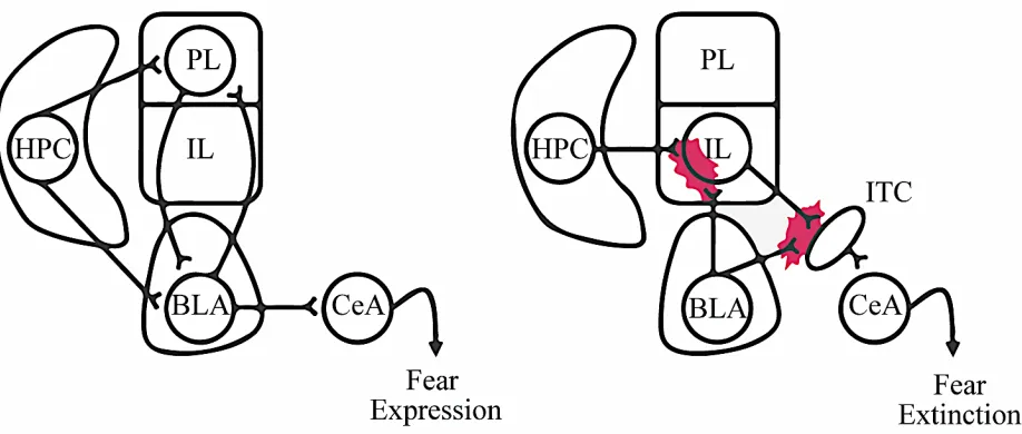
Figure 5| Basic neural connections implicated in fear
Figure 6| Schematic of a voltage-gated calcium channel complex
Figure 7| Proposed mechanism of CaV2.2-mediated anxiogenic signaling
Figure 8| eCB-mediated feedback inhibition system in CA3
CHAPTER 1
Figure 9| Breeding schematics for triple-transgenic mouse models
Table 1| Primer sequences and band sizes for mouse genotyping
Figure 10| CCK expression in CCK-tdT mice and GABAergic CCK expression in
CCK-Dlx5/6-tdT mice
Figure 11| Channelrhodopsin expression in the BLA of CCK-ChR2 and CCK-Dlx5/6-ReaChR mice
Figure 12| Morphophysiology and postsynaptic currents in CCK+IN/P-cell synapses in the BLA
Figure 13| CaV2.2 antagonism reduces IPSC peak amplitude in CCK+IN/P-cell synapses in the
BLA
CHAPTER 2
Figure 15| Effects of CB1 modulation in CCK+IN/P-cell synapses in the BLA
CHAPTER 3
Figure 16| Effects of CaV2.2 antagonism on IPSCs in GABAergic/P-cell synapses in the IL
Figure 17| GABAergic CCK expression in the IL
Figure 18| Effects of CaV2.2 antagonism on IPSCs in CCK+IN/P-cell synapses in the IL
Figure 19| Effects of CaV2.2 antagonism on P-cell intrinsic firing pattern and spike frequency in
ABSTRACT
REGULATION OF GABA RELEASE BY PRESYNAPTIC CAV2.2 IN THE
BASOLATERAL AMYGDALA AND INFRALIMBIC CORTEX
by
Maxwell Robert Blazon
University of New Hampshire, May, 2018
Anxiety is linked to dysregulation of neuronal activity in several brain regions including
the infralimbic (IL) area of the medial prefrontal cortex (mPFC) and the basolateral nucleus of the
amygdala (BLA). Disruptions to the balance of excitatory and inhibitory signaling in these regions
are implicated in anxiety-like behaviors in animals and anxiety disorders in humans. The neuronal
circuitry between excitatory neurons, known as pyramidal cells (P-cells), and inhibitory neurons,
known as interneurons (INs), is a primary target for anxiety modulation at the cellular level. INs,
particularly the subtype that contain the neuropeptide cholecystokinin (CCK+INs), form inhibitory
synapses around P-cells. Through GABAergic neurotransmitter release, CCK+INs regulate P-cell
activity and subsequent anxiety-related neuronal output. Two protein complexes, the N-type
calcium channel (CaV2.2) and the type-1 cannabinoid receptor (CB1), are expressed in the
presynaptic terminal of CCK+INs. CaV2.2 and CB1 have been shown to regulate neurotransmitter
release from CCK+INs in the hippocampus, but it is not known if this role is conserved in the
anxiety-related neuronal circuits of the BLA and IL. CaV2.2 has also been shown to regulate the
intrinsic firing properties of P-cells in the hippocampus and deep cerebellar nuclei, but this role
has not been assessed in the IL. To investigate these neuronal circuits, I used a combination of
pharmacology. In the first chapter of my thesis, I investigated the role of CaV2.2 in
CCK+IN/P-cell synapses in the BLA. In the second chapter of my thesis, I investigated the role of CB1 in
regulating GABA release from CCK+INs in the BLA. In the third chapter of my thesis, I assessed
the regulation of GABA release and intrinsic firing by CaV2.2 in the IL. I present electrophysiology
data which demonstrate GABA release from CCK+INs is partially CaV2.2-dependent in the BLA.
I obtained preliminary electrophysiology recordings which suggest CB1 receptors modulate GABA
release in CCK+IN/P-cell synapses in the BLA. I found that neurotransmitter release from
GABAergic INs is partially CaV2.2-dependent in the IL. However, I found that GABA release
from CCK+INs is not CaV2.2-dependent in this region. Lastly, I observed that CaV2.2 plays a
minimal role in the intrinsic firing properties of P-cells in the IL. My findings suggest that CaV2.2
differentially regulates GABA release and intrinsic firing properties across brain regions,
suggesting there are region-specific implications of CaV2.2 modulation in anxiety-related neuronal
INTRODUCTION
Overview
Fear and anxiety are normal experiences in day-to-day life. Fear often results from an
apparent external danger, while anxiety is a widespread response to an unknown or non-specific
threat. Both trigger appropriate and adaptive responses to risks, be they physical, mental, or
societal (Steimer, 2002). When fear and anxiety become pathological, they can manifest as an
anxiety disorder. Anxiety disorders include panic disorder, generalized anxiety disorder,
agoraphobia, specific phobias, social anxiety disorder, post-traumatic stress disorder,
obsessive-compulsive disorder, and separation anxiety disorder. In these conditions, an individual’s ability
to cope with various stressors is compromised, meaning non-threatening stimuli can cause severe
distress. Anxiety disorders alter numerous physiological processes, facilitating poor physical
health outcomes over time (Kroenke, Spitzer, Williams, Monahan, & Löwe, 2007). The National
Institute of Mental Health reports a 19.1% past-year prevalence of anxiety disorders in U.S. adults
(Harvard Medical School National Comorbidity Survey, 2017). Current medications used to treat
anxiety disorders (benzodiazepines, selective serotonin reuptake inhibitors) cause several adverse
effects (impaired cognition, addiction), thus many patients withdraw from treatment (Baldwin,
Woods, Lawson, & Taylor, 2011). A greater understanding of the neurobiology behind anxiety
disorders is required to produce more effective treatments.
Anxiety is a highly integrated emotional component of cognitive processing facilitated by
the thalamus, locus coeruleus, hypothalamus, hippocampus, amygdalar nuclei, and the prefrontal,
sensory, entorhinal, and association cortices (Steimer, 2002). Classically, these brain areas are
defined as components of the limbic system or ‘emotional brain.’ In animal models, their
anxiety-like behaviors (Steimer, 2002). Of particular importance for their role in modulating
anxiety are the medial prefrontal cortex (mPFC) and the basolateral amygdala (BLA). The mPFC
processes the cognitive components of anxiety (i.e. rumination) and mediates sympathetic nervous
system responses (fight-or-flight). The behavioral responses related to anxiety (i.e. avoidance) are
modulated by the BLA (Johansen, Cain, Ostroff, & Ledoux, 2011). While each area has
independent roles in anxiety, their interconnections are involved in fear expression and the
extinction of conditioned fear (Ferreira, Yousuf, Dalton, & Sheets, 2015; Milad & Quirk, 2002;
Phelps, Delgado, Nearing, & Ledoux, 2004). Research targeting the neural circuits involved in this
disruption may provide insight as to how anxiety is regulated at the cellular level.
The balance between excitatory and inhibitory activity in the mPFC and BLA is disrupted
in anxiety disorders (Ehrlich et al., 2009). Pyramidal neurons (P-cells) are the primary excitatory
cell type in the mPFC and BLA. P-cells receive excitatory inputs through projections from other
brain areas and inhibitory inputs from local interneurons (INs) (Freund, 2003). Multiple types of
INs exist, and the cholecystokinin-containing subtype (CCK+INs) are strongly linked to
anxiety-related behaviors (Truitt, Johnson, Dietrich, Fitz, & Shekhar, 2009). The inhibitory
neurotransmitter gamma-aminobutyric acid (GABA) is released from CCK+INs onto P-cells,
decreasing their excitability (Freund & Katona, 2007). Membrane proteins like voltage-gated
calcium channels (CaVs) and cannabinoid receptors modulate neurotransmitter release (Guo &
Ikeda, 2004). CaVs permit calcium ions to enter the presynaptic terminal which facilitate vesicular
fusion and neurotransmitter exocytosis. Cannabinoid receptors are coupled to CaVs and
downregulate calcium ion entry when activated by cannabinoids (Zamponi & Currie, 2013).
Additionally, CaVs modulate the intrinsic firing properties of P-cells by coupling to
cannabinoid receptor subtypes; the N-type calcium channel (CaV2.2) and the type-1 cannabinoid
receptor (CB1) are known to regulate neurotransmitter release from CCK+INs in the hippocampus
(Szabo et al., 2014). However, the role of CaV2.2 and CB1 in CCK+INs of the BLA and mPFC has
not been described. In my master’s thesis, I aim to elucidate the role of CaV2.2 in CCK+IN/P-cell
synapses in the BLA (CHAPTER 1) and determine if CB1 regulates neurotransmitter release in
this system (CHAPTER 2). Additionally, I will assess the role of CaV2.2 in regulating GABA
release and intrinsic firing properties in the infralimbic region of the mPFC (CHAPTER 3).
The Role of Fear and Anxiety in Survival
Emotions are thought to be the result of natural selection, as they prompt behavioral
responses important for survival (Darwin, 1998). A critical emotion for survival, fear, is defined
as a motivational state that promotes either defensive or escaping behaviors (Gherardi, McFarland,
& Tinbergen, 1988). Exposure to fearful stimuli often correlates with learning. Animals (including
humans) adapt behavioral changes to avoid repeat exposure to fear, particularly if the fearful
stimuli are associated with pain or stress. Fear-related learning can manifest through a closely
related emotion; anxiety (Steimer, 2002). Anxiety involves behavioral and physiological responses
including avoidance, vigilance, and arousal, all intended to protect the animal from danger. While
fear is focused on known dangerous stimuli, anxiety is often a directionless response to an
unknown danger or internal conflict (Ritu, Sandeep, Mamta, & Nirja, 2013).
Fear and Anxiety are Related to the Limbic System
The limbic system, also known as the ‘emotional brain,’ was largely defined by James
thalamus, hypothalamus, hippocampus, and with emotional patterns observed in human
experiments. MacLean refined Papez’s work, highlighting the role of the prefrontal cortex and
subcortical areas like the amygdala and septum in emotional experiences (J. LeDoux, 1996; Roxo,
Franceschini, Zubaran, Kleber, & Sander, 2011). More than a series of brain structures, limbic
areas form a functional system. Feelings of fear and anxiety are complex, involving more than just
emotional components. For example, fear and/or anxiety-related stimuli are processed by the
prefrontal cortex (PFC), which modulates physiological and behavioral responses (via the
amygdala). The amygdala assimilates sensory, contextual, and cognitive information to influence
fear extinction, anxiety-related behavior, and selective attention (J. E. LeDoux, 2009; Steimer,
2002). The integration of cognitive, physiological, and sensory information in the BLA and PFC
illustrates the complexity of fear and anxiety (J. E. LeDoux, 2009).
The Role of the Medial Prefrontal Cortex in Anxiety
The PFC is believed to influence the conscious experiences and thoughts related to anxiety
(J. LeDoux, 1996; Steimer, 2002) as well as module anxiety-like behaviors (Sierra-Mercado,
Padilla-Coreano, & Quirk, 2011). fMRI studies show changes in PFC activity in patients with
anxiety disorders (generalized and post-traumatic), which are behaviorally characterized by the
inability to control fear responses (Sierra-Mercado et al., 2011). Deficient recruitment of the PFC
(ventromedial region) during fear inhibition is observed in patients with generalized anxiety
disorder (Greenberg, Carlson, Cha, Hajcak, & Mujica-Parodi, 2013). Similarly, decreased
activation of the PFC (ventromedial region) in response to trauma-related cues is present in
patients with post-traumatic stress disorder (PTSD) (Shin et al., 2004). In animal research, similar
anxiolytic effects and suppress autonomic nervous system responses (Sullivan & Gratton, 2002).
In mouse models, the PFC is associated with fear-related learning and fear expression, both of
which can be impaired with selective inactivation of PFC sub-regions (Sierra-Mercado, Corcoran,
Lebrón-Milad, & Quirk, 2006; Sierra-Mercado et al., 2011)
The rodent PFC is comprised of multiple sub-regions with specialized functions including
the medial, lateral, and orbitofrontal cortices. The medial prefrontal cortex (mPFC) is the target of
extensive anxiety research (Duvarci & Pare, 2014; Morgan, Romanski, & LeDoux, 1993; Sullivan
& Gratton, 2002). Further divided, the mPFC is comprised into the anterior cingulate cortex (AC),
the prelimbic cortex (PL), and the infralimic cortex (IL) (Figure 1) (Paxinos & Watson, 2007).
The IL and PL play unique roles in anxiety-related processing and differentially connect to the
amygdala. The IL facilitates the storage of extinction learning in target structures (Do-Monte,
Manzano-Nieves, Quinones-Laracuente, Ramos-Medina, & Quirk, 2015). IL-BLA connections
are involved in the extinction of
conditioned fear (Ferreira et al.,
2015; Sierra-Mercado et al., 2011).
Excitatory neurons in the PL project
to the BLA and are involved in the
expression of fear (Sierra-Mercado
et al., 2011; Wang et al., 2015). In
rodents, fear is expressed by
behaviors like freezing. An animal
that displays reduced explorative
behaviors or excessive grooming is
considered to be exhibiting anxiety-like behavior. Stressful stimuli activate the PL and increase
fear expression and anxiety-like behaviors (Hurley, Herbert, Moga, & Saper, 1991; Sullivan &
Gratton, 2002), while selective inactivation of the PL reduces them (Sierra-Mercado et al., 2011).
Abnormal activity in the connections from the PL and IL to the BLA can prompt inappropriate or
deficient response to fear (Greenberg et al., 2013), a characteristic common to anxiety disorders.
The Role of the Amygdala in Processing Fear and Anxiety
The amygdala is an almond-shaped, highly interconnected limbic system structure
implicated in the behavioral output of anxiety. Amygdalar functions and connections are conserved
across many species, suggesting that research findings in animal models like the mouse (Mus
musculus) may have human implications (Janak & Tye, 2015). For non-human primates, lesions
to the amygdala impair their ability to associate a stimulus with positive/non-harmful or
negative/potentially harmful outcomes (Weiskrantz, 1956) and lead to hypo-emotionality
(Aggleton & Passingham, 1981). In rodents, the amygdala facilitates behavioral responses (i.e.
freezing/playing dead in front of predator) by integrating rapid sensory input (i.e. visualization of
a predator) from the thalamus and slower sensory input (i.e. motivation to escape the predator)
from the prefrontal cortex (Steimer, 2002). Furthermore, the amygdala influences cognition,
perception, and associative learning (J. E. LeDoux, 2009; Steimer, 2002). In associative learning,
an animal relates a new response to a particular stimulus, or one stimulus to a second stimulus
(Janak & Tye, 2015). In humans, (Anderson & Phelps, 2001) and rats (Blanchard & Blanchard,
1972), damage to the amygdala hinders the ability to recognize fearful stimuli. Amygdalar
These disruptions are observable in anxiety disorders (Etkin, Prater, Schatzberg, Menon, &
Greicius, 2009).
The amygdala is composed of several nuclei, including the basolateral amygdala (BLA)
and the central amygdala (CeA) (Figure 2). The BLA is further comprised of the basal amygdala
(BA) and lateral amygdala (LA), and is the main source of excitatory output to other brain regions
involved in fear and anxiety. Thalamic and cortical sensory signals project to the LA, which
connects to the BA and CeA (Keifer, Hurt, Ressler, & Marvar, 2015). Direct stimulation of the
BLA is correlated with anxiety-like behavior (Johansen et al., 2010). The BLA can both upregulate
and downregulate freezing behaviors through projections to the PL and IL, respectively, and is
involved in anxiety-related memories via hippocampal connections (Sierra-Mercado et al., 2011).
Opposite of the BLA, the CeA is mainly composed of inhibitory interneurons. Divided into the
centromedial (CEm) and
centrolateral amygdala (CEl),
the CeA is involved in the
translation of BLA signals to
behavioral output (Janak & Tye,
2015). It has been demonstrated
that inhibition of BLA-CeA
connections increases
anxiety-related behaviors while direct
stimulation decreases them (Tye
et al., 2011).
The Role of the Hippocampus in Anxiety-related Learning and Memory
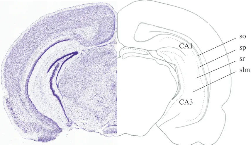
The HPC is divided into ventral and dorsal regions, each of which contains Cornu
Ammonis (CA) regions (CA1 - CA4). Additionally, the CA regions contain a system of laminar
organization (Figure 3). The stratum oriens (SO), stratum pyramidale (SP), stratum radiatum (SR),
and stratum lacunosum-moleculare (slm) possess different cells, circuit organization, and functions
in hippocampal processes. The ventral portion of the hippocampus (vHPC) is implicated in fear
conditioning and anxiety-like behavior through its connections with the mPFC and BLA.
BLA-vHPC connections are necessary for forming memories of environmental context (Goosens, 2001;
Maren & Fanselow, 1995), while vHPC-mPFC connections are required for retrieving memories
when presented with specific context (Sierra-Mercado et al., 2011). Contextual memory and the
memory retrieval process are important for the survival of a species, especially when the context
involves perceived danger or negative emotions (Goosens, 2001). Chronic stress, fear, and anxiety
can disrupt these processes and result in a state of hippocampal dysfunction. There is evidence that
hippocampal dysfunction is both a risk factor for developing and complication of anxiety disorders
(Cominski, Jiao, Catuzzi, Stewart, & Pang, 2014).
Cells and Circuits in Anxiety-related Brain Areas
New and developing pharmacological treatments for anxiety disorders must aim to
decrease activity in the neural circuits that influence anxiety and increase activity in those that
reduce anxiety. Fundamental to these objectives is research which provides a more complete
understanding of the molecular neuroanatomy of the limbic system and the functional
neurophysiology of anxiety disorders. Communication between and within limbic system areas is
facilitated by complex and highly organized neuronal circuits. Similar cell types and circuitry are
thought to exist across limbic regions like the mPFC, BLA, and vHPC (Jinno, 2009). However, it
is not understood if there are underlying functional differences in these circuits, nor what
implications they might have in the neuronal processes of anxiety.
The mPFC, BLA, and vHPC contain a mixture of excitatory (glutamatergic) pyramidal
cells (P-cells) and inhibitory (GABAergic) interneurons (INs). P-cells are characterized by their
piriform morphology, apical dendrites, and long axons which allow for communication across
brain regions. INs innervate local P-cells and are classified by the domain in which they target.
INs that target P-cell bodies (somas/somatas) are termed basket cells (BCs), those that target the
axon initial segments are termed chandelier cells, and those that target dendrites are termed
dendritic inhibitory cells (Freund, 2003). INs can be further categorized by identifying their
intrinsic firing properties and neuropeptide content. Intrinsic firing properties, including action
(Benda & Herz, 2003). Neuropeptide contents are used as a marker of IN subtypes, and indicate
the neurotransmitters, receptors, and functions likely associated with a particular subtype. In the
mPFC, BLA, and HPC, several subtypes of BCs have been identified for their unique roles in
influencing P-cell activity (Barsy, Szabó, Andrási, Vikór, & Hájos, 2017; Freund, 2003; Whissell,
Cajanding, Fogel, & Kim, 2015). BCs containing the neuropeptide parvalbumin (PV+BCs)
process multiple converging stimuli and display fast, non-adapting firing patterns (Nyíri,
Stephenson, Freund, & Somogyi, 2003). Cholecystokinin-containing BCs (CCK+BCs) respond to
slow, asynchronous inputs and display moderate, adapting firing patterns (Bartos & Elgueta, 2012;
Veres, Nagy, & Hájos, 2017). Understanding functional specifics about INs within limbic
structures may help elucidate their complex role in mediating fear and anxiety.
Descriptions of the wiring, innervation, and postsynaptic targets of INs requires a
description of how IN/P-cell networks operate. Across P-cells and IN subtypes, there are variations
in intrinsic firing and synaptic properties (Kullmann, 2011). In general, the synaptic events which
excite INs are faster than those that excite P-cells. This is advantageous for controlling anxiogenic
pathways, as excitation is under strict regulation by INs, particularly BCs (Deleuze, Pazienti, &
Bacci, 2014). This regulation comes in multiple forms including feedforward inhibition. In
feedforward inhibition, a local IN, excited by an afferent projection, delivers an inhibitory signal
to a P-cell. Depending on the cellular properties of the IN, feedforward inhibition can limit the
window for action potential generation or cancel out P-cell excitation (Jasnow, Ressler, Hammack,
Chhatwal, & Rainnie, 2009). In addition to feedforward inhibition, there is feedback inhibition.
feedback and feedforward inhibition decline,
P-cells are able to fire again. The combination
of feedforward and feedback inhibition
establish a neuronal oscillation, also known as
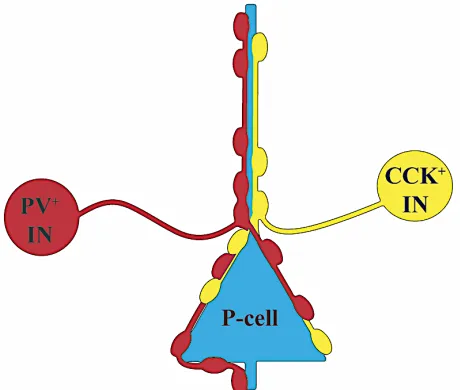
a brainwave (Möhler, 2002). A prominent form
of inhibition in the BLA and mPFC is known as
perisomatic inhibition (Figure 4), wherein
CCK+BCs and PV+BCs synchronize the
activity of local P-cells (Freund & Katona,
2007). The synapses made in perisomatic
inhibitory are highly modifiable by ion
channels, neurotransmitters, and additional extracellular messengers, prompting a closer review of
their underlying molecular components of in anxiety-implicated circuits of the vHPC, mPFC, and
BLA.
In the vHPC, excitatory signaling is transmitted through P-cell projections from CA3 to
CA1. Anxiety research often targets CA3 and CA1, as these sub-regions are major output pathways
to other limbic areas. Several networks of INs exist within these output pathways which can
modulate P-cell activity (Kullmann, 2011). These include fast-spiking PV+BCs, regular-spiking
CCK+BCs, chandelier INs, and Schaffer collateral-associated (SCA) INs (Kullmann, 2011).
Research has shown that while PV+BCs are more abundant overall, CCK+BCs outnumber
PV+BCs in several sub-regions of the hippocampus (Whissell et al., 2015). The most superior
layer of the vHPC, the SO, contains BC bodies and the basal dendrites of P-cells, whose cell bodies
are located the second-most superior layer, the SP (Lacaille & Williams, 1990). The SP is
recognizable by its dense P-cell composition and role in hippocampal output. BCs can be found in
the SP as well; their proximity to P-cells illustrates their known role in perisomatic inhibition (Neu,
Földy, & Soltesz, 2007; Whissell et al., 2015). The layer immediately following the SP, the SR,
contains P-cell projections from CA3 to CA1, termed the Schaffer collaterals. Schaffer collaterals
are the topic of extensive neuropsychiatric research, as they are critical for long-term potentiation
and emotional memory (Kumar, 2011; Stanton, Winterer, Zhang, & Müller, 2005). BCs in CA1
are innervated by Schaffer collaterals, while the dendrites of Schaffer collaterals themselves are
innervated by SCA INs (Kullmann, 2011).
Similar to the SP layer of the vHPC, the mPFC contains mostly P-cells (80-90%) with a
minority of INs (10-20%) spread heterogeneously across a system of laminar organization. In the
human brain, there are six layers (L1-L6) while in rodents there are only five (rodents do not
possess L4) (Heidbreder & Groenewegen, 2003). Across layers, composition and connectivity vary,
but functional characteristics of IN/P-cell connections are relatively conserved. INs wield strong,
differential control over local circuitry and are able to synchronize P-cell spiking throughout the
mPFC (Sparta et al., 2014). In rodents, cells located in L1 are thought to be exclusively GABAergic
INs (Riga et al., 2014). L1 INs receive excitatory input from the thalamus and facilitate dendritic
inhibition of L2/3 P-cells (Cruikshank et al., 2012). BC/P-cell perisomatic inhibitory networks are
seen in L2/3 and L5. P-cells in L2/3 and L5 receive excitatory inputs from the thalamus, the
contralateral mPFC, the vHPC, and the BLA. These P-cells project to various brain areas
depending their location within the mPFC. In the PL, L2/3 P-cells projects to the BLA, while in
the IL, L2/3 and L5 P-cells project to the BLA (Ferreira et al., 2015; Zaitsev, Povysheva,
Gonzalez-Burgos, & Lewis, 2012). P-cells in L2/3 of the PL are involved in fear expression while P-cells in
et al., 2011). It is vital to make these distinctions, as they demonstrate that even within L2/3 of the
mPFC, IN/P-cell connections mediate different anxiety-related processes.
The BLA is comprised of P-cells and multiple IN subtypes including PV+BCs and
CCK+BCs (Ehrlich et al., 2009; Freese & Amaral, 2005). P-cells receive inhibitory input from
local BCs and excitatory input from cortical areas via the external capsule (EC), a white matter
fiber tract containing clusters of P-cell axons (Little & Carter, 2013). Excitation of P-cells in the
BLA correlates with anxiety-like behavior (Johansen et al., 2010). Perisomatic inhibition,
facilitated by CCK+BCs and PV+BCs, has been observed in electrophysiology recordings from
P-cells in the BLA (Freund, 2003). PV+BCs and CCK+BCs receive excitatory signals from local
and distant P-cells (via the EC) and inhibitory signals from local INs (Faber, Callister, & Sah,
underlying molecular components of perisomatic inhibitory synapses in the vHPC, however less
is known regarding similar circuits in the BLA (Barsy et al., 2017; Freund & Katona, 2007;
Trouche, Sasaki, Tu, & Reijmers, 2013). Functional studies on neurotransmitter release in
BC/P-cell synapses are needed to understand how behavioral output associated with anxiety is regulated
in the BLA.
The Neurochemical Correlates of Anxiety - GABA and Glutamate
Neuronal circuits in the BLA, mPFC, and vHPC possess extensive γ-aminobutyric acid
(GABA) and glutamate neurotransmitter systems (Bystritsky, Khalsa, Cameron, & Schiffman,
2013). GABA is the principle inhibitory neurotransmitter in the brain while glutamate is the
principle excitatory neurotransmitter. Balance between GABAergic and glutamatergic activity is
critical for normal fear and anxiety-related processing (Möhler, 2002). Excess GABAergic activity
can be anxiolytic but can also impair cognition. Insufficient GABAergic activity can result in
increased anxiety (Möhler, 2002). Chronic deficits in GABAergic activity contribute to the
pathophysiology of anxiety disorders; anti-anxiety medications including benzodiazepines and
gabapentinoids enhance GABAergic activity (Sills, 2006). Alterations to glutamate transmission
have also been shown to be effective in augmenting anxiety disorders(Dawson & Watson, 2009),
as glutamate is involved in the pathways underlying normal and pathological anxiety states
(Bystritsky et al., 2013).
The effects of neuronal inhibition differ across cell types, circuits, and brain areas. When
P-cells are inhibited, they no longer release glutamate onto their projection targets. On the contrary,
inhibition of INs prevents IN-mediated inhibition; resulting in P-cell disinhibition (Andrási et al.,
to more readily understand its implications in anxiety. GABA binds to the GABAA receptor, a
ligand-gated ion channel. Upon binding, the channel opens and chloride (Cl-) anions flow inward
(U. Rudolph & Antkowiak, 2004). Chloride influx induces electrical events known as inhibitory
postsynaptic currents and potentials (IPSCs and IPSPs). As a result, hyperpolarization, action
potential inhibition, and decreased neurotransmitter release occur (Amir, Michaelis, & Devor,
1999). GABAA receptors are widespread, expressed in multiple cellular domains (i.e. terminals,
somas, and axons) and cell types not exclusively involved in anxiety (Freund, 2003; Freund &
Katona, 2007). In addition to the GABAergic system, the glutamatergic system regulates
anxiety-related neuronal output from P-cells.
Proper glutamatergic transmission is required for anxiety states (Bystritsky et al., 2013).
The glutamate receptors most pertinent to anxiety-related circuits are the AMPA and NMDA
subtypes. As a whole, glutamatergic activity shows cell-type specific dynamics in modulating
anxiety-related neuronal output (Nyíri et al., 2003). For example, long-term potentiation,
implicated in storage of traumatic memories, relies on the NMDA receptors within Schaffer
collaterals of the hippocampus (Harris & Cotman, 1986). Additionally, AMPA receptors on P-cells
in the BLA are redistributed from dendritic stores into spines following severe stress (Hubert, Li,
Rainnie, & Muly, 2014).
Molecular Components of GABAergic and Glutamatergic Neurotransmission
GABA and glutamate neurotransmission are modulated by G protein-coupled receptors
(GPCRs) and voltage-gated calcium channels (CaVs). Located in the transmembrane domain,
GPCRs are signal transduction proteins which interact with molecules in the extracellular and
There are numerus families and subtypes of GPCRs which couple to different subunits. GPCRs in
the mPFC, vHPC, and BLA are coupled to the Gi alpha subunit (Gαi), including metabotropic
GABA and glutamate receptors (Lee et al., 2015; Lenkey et al., 2015; Talani & Lovinger, 2015;
Zoppi et al., 2011). These GPCRs are involved in a variety of anxiety-related physiological
processes including behavior and mood regulation (Swanson et al., 2005). For this reason, Gαi
receptors are the targetof several pharmaceutical anxiety treatments. Gαi receptors are activated by
ligand binding (whether endogenous or synthetic) and exert an inhibitory effect on cAMP and
calcium, two major internal singling molecules. As a result, calcium-mediated processes and
cAMP-dependent signal transduction pathways are inactivated (Scotter, Graham, & Glass, 2009).
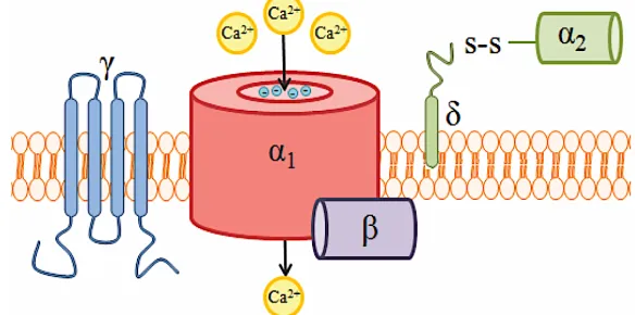
Fundamentally, CaVs are protein complexes consisting of several subunits (α1, β, α2δ, and
γ) which mediate calcium ion entry into a cell (Figure 6). There are two distinct classes of CaVs
based on their voltage-gating properties; high voltage-activated (HVA) and low voltage-activated
(LVA). The phrase ‘voltage-gated’ is derived from the closed state of these channels under resting
membrane potential conditions.
Only a strong enough membrane
depolarization (i.e. an action
potential) will release this
voltage-gate (Stotz, 2001). Within each
class of CaV, subtypes are
determined by the α1 subunit. The
importance of the α1 subunit cannot
be stressed enough, as it forms the
calcium-selective ion channel
through which calcium enters the cell (Williams et al., 1992). The α1 subunit can detect changes
in membrane potential and contains binding sites for pharmaceutical antagonists (Catterall, 2011;
Zamponi, 2016). From ten known α1 subunits there are five known CaV isoforms. The isoforms
and their respective α1 subunit encoding genes (in parenthesis) are as follows: L-type (CaV1.1,
CaV1.2, CaV1.3, CaV1.4), P/Q-type (CaV2.1), N-type (CaV2.2), R-type (CaV2.3), and T-type
(CaV3.1, CaV3.2, CaV3.3) (Catterall, 2011). The remaining CaV subunits serve α1; enhancing its
expression (δ), stabilizing its conformation (β), and regulating its activation and inactivation
kinetics (β, α2δ) (Hannon & Atchison, 2013).
Of the numerous α1 subunits, those encoded by CaV2.1, CaV2.2, and CaV2.3 (P/Q-, N-, and
R-type channels) are most involved in mediating signal transduction in GABAergic and
glutamatergic cells (Catterall, 2011; Zamponi, 2003). Additionally, CaV2 channels have been
shown to modulate neuronal firing through coupling to calcium-activated potassium channels
(Alviña & Khodakhah, 2008; Loane et al., 2007). During membrane depolarization, CaV2 opens
and allows calcium entry into the neuron, which induces numerous physiological events. For
instance, calcium interacts
with SNARE proteins to
fuse neurotransmitter
vesicles to the presynaptic
terminal, where subsequent
exocytosis leads to synaptic
transmission (Südhof,
2012). It has been
demonstrated that calcium
entry through CaV2.1 and CaV2.2 channels initiates the release of glutamate and GABA in fast
synapses. More specifically, it has been discovered that CaV2.2 channels regulate GABA release
from presynaptic CCK+INs in the vHPC (Szabo et al., 2014). Given the role of CCK+INs in
perisomatic inhibition of P-cells, and the role of excess P-cell activity in anxiety-related behaviors
(Figure 7), CaV2.2 channels are a critical research target for anxiety disorders (Kurihara & Tanabe,
2003; Lee et al., 2015; Lee, 2013; Szabo et al., 2014).
Modulation of the N-type Channel and Anxiety Implications
CaV2.2 modulation is a current target for treating neuropathic pain (Hannon & Atchison,
2013) and has emerged as a possible means of treating anxiety (Newton & Messing, 2009).
Previous studies have found that mice lacking CaV2.2 display reduced anxiety levels (Saegusa,
2001), and current research aims to dissect the role CaV2.2 blockers play in anxiety. In nature, a
highly-specific CaV2.2 blocker is found in the venom of the cone snail, Conus magus. Known as
‘w-Conotoxin GIVA’ when isolated, this peptide neurotoxin show affinity only for CaV2.2
(McCleskey et al., 1987). Zinconotide and Leconotide, synthetic w-Conotoxins used to treat
neuropathic pain, have strong yet poorly-understood anxiety correlates (Kolosov, Aurini, Williams,
Cooke, & Goodchild, 2011; Schmidtko, Lötsch, Freynhagen, & Geisslinger, 2010; Scott, Wright,
& Angus, 2002). The GABA analogue drugs Gabapentin and Pregabalin have seen increasing
off-label use to treat anxiety disorders. Typically used to treat seizures and nerve pain, Gabapentin and
Pregabalin target the α2δ subunit, indirectly modulating CaV2.2 (Sills, 2006). By modulating
CaV2.2, the side effects associated with GABA modulation (i.e. benzodiazepines) can be bypassed.
It is important to state that these drugs are not approved as a primary treatment for anxiety disorders.
prompts research into their functional role in anxiety-related neuronal circuits (Lee, 2013; Lotarski
et al., 2011).
CaV2.2 channels are modulated by a variety of GPCRs. One form of modulation is
voltage-dependent inhibition facilitated by the binding of the G beta-gamma complex (Gβγ) to the
pore-forming subunit of CaV2.2 channels (Ikeda, 1996). Two inhibitory mechanisms can occur as a
result. In one mechanism, Gβγ induces a shift in the voltage-dependent activation properties of
CaV2.2 channels, which then require larger-than-normal depolarizations to open (Zamponi &
Currie, 2013). Alternatively, GPCRs can inactivate CaV2.2 through a ‘hinged lid’ mechanism,
closing the ion channel-forming subunit, a1 (Stotz, 2001). In either mechanism, calcium entry
through CaV2.2 is limited or blocked, and subsequent neurotransmitter release from presynaptic
terminals is inhibited. There are a wide variety of GPCRs that exhibit inhibitory effects on CaVs,
but a specific family has been shown to extensively couple with CaV2.2 in anxiety-related brain
areas. Cannabinoid receptors, part of the endocannabinoid system, are a specific family of
primarily Gαi GPCRs heavily implicated in anxiety (Haller, Bakos, Szirmay, Ledent, & Freund,
2002).
The Endocannabinoid System and Anxiety
The endocannabinoid (eCB) system is a network of primarily Gαi GPCRs and ligands
known to modulate synaptic transmission throughout the brain. Of the two cannabinoid receptor
types (CB1 and CB2), CB1 is the major synaptic constituent of the eCB system, heavily expressed
in CCK+INs (Lutz, Marsicano, Maldonado, & Hillard, 2015). CB1 is activated by endogenous
cannabinoid molecules like anandamide (AEA) and 2-arachidonoylglycerol (2-AG), as well as
Cannabinoids act as retrograde feedback
inhibition messengers; postsynaptic cells
synthesize 2-AG/AEA, which bind to CB1
receptors on the presynaptic terminal,
suppressing neurotransmitter release
(Figure 8). CB1-mediated inhibition occurs
in two general forms; phasic and tonic.
Phasic inhibition occurs as a result of
postsynaptic depolarization, thus is
generated in response to specific
postsynaptic signals. Tonic inhibition is the
result of basal CB1 activity, and occurs
irrespective of postsynaptic signals (Lee et al., 2015; Soltesz et al., 2015). Mechanistically, CB1
modulates neurotransmitter release by coupling to and downregulating CaVs (Wilson, Kunos, &
Nicoll, 2001). By inhibiting calcium entry through CaVs, processes like vesicular fusion are
prevented (Catterall, 2011). In the CA3 region of the vHPC, CB1 was shown to specifically couple
to CaV2.2, but it is not known if this coupling is conserved in the BLA.
The eCB system has been shown to differentially modulate synaptic transmission across
different brain regions, cell types, and synapses. Heavy integration of the eCB system has been
observed in the HPC, PFC, and amygdala, where it is thought to have modulatory effects on fear
and anxiety processing (Lutz et al., 2015). At GABAergic synapses, eCB-mediated inhibition is
known as depolarization-induced suppression of inhibition (DSI), and in glutamatergic synapses,
it is known as depolarization-induced suppression of excitation (DSE) (Ramikie & Patel, 2012).
Figure 8| eCB-mediated feedback inhibition system in CA3. Calcium (Ca2+) entry through Ca
V2.2 in the postsynaptic P-cell induces anandamide synthesis. Anandamide binds to cannabinoid receptor 1 (CB1) on the presynaptic cholecystokinin interneuron (CCK+IN) terminal. Ca2+ entry though Ca
DSI and DSE are short-term effects, as eCB-mediated synaptic modulation follows a
negative-feedback loop. Neurons containing CB1 are classified into two groups; high CB1-expressing cells,
which are sparsely distributed in the BLA and cortical areas, and low CB1-expressing cells, which
are more evenly dispersed (Marsicano & Lutz, 1999; Yoshida et al., 2011). Research on
cell-specific CB1 expression has revealed high selectivity for CCK+INs over other INs. In the BLA,
95% of high CB1-expressing cells and 90% of low CB1-expressing cells were CCK+INs
(Marsicano & Lutz, 1999). Contrarily, CB1 expression is largely absent from PV+INs. In addition
to CCK+IN selectivity, CB1 has been shown to couple with CaV2.2. In rat sympathetic neurons,
N-type calcium currents were inhibited by CB1 agonists, inhibition which was then reversed by
CB1 antagonists (Guo & Ikeda, 2004). Furthermore, CB1-CaV2.2 coupling appears to be prevalent
in CCK+INs. In the CA3 region of the vHPC, CaV2.2-dependent GABA release from CCK+IN
terminals was shown to be inhibited with CB1 agonists(Szabo et al., 2014).
It is widely accepted that there is a strong correlation between the eCB system and anxiety,
but this correlation is not fully understood (Lutz et al., 2015). eCB system modulation shows
potential for treating anxiety disorders, a well-known example of which being the anxiolytic
properties of THC (Onaivi, Green, & Martin, 1990). In both humans and rodents, it has been shown
that cannabinoids can influence anxiety-like symptoms in a biphasic manner; low doses produce
anxiolytic effects while high doses produce anxiogenic effects (Moreira, Grieb, & Lutz, 2009; Rey,
Purrio, Viveros, & Lutz, 2012). Research suggests that CB1 modulation has opposite effects on
anxiety-like behavioral output in glutamatergic and GABAergic cells. Knockout mice lacking CB1
in all glutamatergic cells show increased anxiety-like behaviors, while knockout mice lacking CB1
in all GABAergic cells show reduced anxiety-like behaviors (Häring, Kaiser, Monory, & Lutz,
however, and are different when isolating specific brain regions (Rovira-Esteban et al., 2017;
Szabo et al., 2014). Accordingly, more detailed neurophysiological research must be conducted on
the role of CB1 in cell populations and brain regions implicated in anxiety.
Implications of CCK and CCK+INs in Anxiety
CCK and CCK+INs have well-established links to anxiety which originate from their role
in neuronal circuits. CCK, the CCK receptor (CCKB), and CCK+INs are highly expressed in the
limbic system (Vanderhaeghen, Signeau, & Gepts, 1975). As a neuropeptide, CCK is found in
vesicles and displays synaptic properties similar to classical neurotransmitters (Emson, Lee, &
Rehfeld, 1980). CCK is associated with anxiety through its interactions with CCKB. In healthy
patients, CCK administration induces panic attacks (Montigny, 1989) while CCKB antagonists
block these panicogenic effects (Bradwejn, Koszycki, Couetoux du Tertre, van Megen, & et al.,
1994). In rodents, CCK agonists produce anxiety-like behaviors (Frankland, Josselyn, Bradwejn,
Vaccarino, & Yeomans, 1996) while CCKB antagonists attenuate them (Josselyn et al., 1995).
Binding of CCK to CCKB has been shown to modulate the neuronal properties of classical
neurotransmitters (Altar & Boyar, 1989). Interactions exist between CCK and GABA, dopamine,
and endocannabinoid neurotransmission (Altar & Boyar, 1989; Chhatwal et al., 2009; Lanza &
Makovec, 2000).While these interactions remain relatively understudied, they represent potential
underlying mechanisms by which CCK is linked to anxiety.
CCK+INs display a distinct regulatory role over anxiety-like behaviors. Along with
PV+INs, CCK+INs establish GABAergic connections onto the soma of P-cells in several brain
regions including the BLA, HPC, and PFC (Freund & Katona, 2007; Sparta et al., 2014). In the
to prevent anxiety-like responses from occurring in non-threatening situations (Freund, 2003;
Mascagni & McDonald, 2003; Truitt et al., 2009). When CCK+INs in the BLA are lesioned, rats
display increased anxiety-like behaviors (Truitt et al., 2009), a phenotype thought to be the result
of lost GABAergic and CCKergic activity. Additionally, it has been proposed that by acting with
the eCB system, CCK+INs modulate fear inhibition and extinction (Bowers, Choi, & Ressler,
2012). CCK+INs likely modulate fear extinction at the level of the BLA by inhibiting ‘extinction’
neurons (Duvarci & Pare, 2014). CCK+IN synapses are remodeled by fear extinction, displaying
increased CB1 expression and thus decreased inhibition of extinction neurons (Duvarci & Pare,
2014; Trouche et al., 2013). Given their expression of CaV2.2 and CB1, role in perisomatic
inhibition, as well as integration in limbic system structures, CCK+INs are essential targets for
anxiety research.
Master’s Thesis Research
Research Motivations
We are motivated to determine the role of CaV2.2 in GABA release from CCK+INs onto
P-cells in the BLA. It has been demonstrated in the CA3 region of the HPC that GABA release in
CCK+IN/P-cell synapses is eliminated by blocking CaV2.2 (Szabo et al., 2014). If conserved,
CaV2.2 may be a potential target for mediating anxiety at the level of the amygdala. Additionally,
we are motivated to determine the role of CB1 in CCK+IN/P-cell synapses in the BLA. Given
findings which indicate CB1 inhibits CaV2.2 and subsequently GABA release from CCK+INs onto
P-cells in the HPC (Szabo et al., 2014), we are interested in examining this system in the BLA.
CB1 shows promise in the development of novel anxiety treatments, but a greater understanding
extinction and its connections to the BLA motivates us to determine if CaV2.2 regulates presynaptic
transmitter release and intrinsic firing properties in this region of the mPFC. P-cells in the mPFC
receive dendritic inhibition from INs in superficial layers and perisomatic inhibition from proximal
BCs (Riga et al., 2014). However, little is known about the role of CaV2.2 in regulating GABA
release in these inhibitory synapses.
Research Questions
Chapter 1: Is GABA release in CCK+IN/P-cell synapses in the BLA CaV2.2-dependent?
P-cells carry the bulk of information from the BLA to other brain areas implicated in anxiety.
Previous research shows that excitation of P-cells in the BLA increases anxiety-like behaviors
(Johansen et al., 2010). Local INs expressing the peptide cholecystokinin (CCK+INs) synapse onto
the soma of P-cells, forming a system of perisomatic inhibition (Freund, 2003). Previous studies
in the CA3 region of the HPC have shown that the GABA release from CCK+INs onto P-cells is
dependent on CaV2.2 (Szabo et al., 2014). If this circuitry is conserved in the amygdala, Cav2.2
could be a potential pharmacological target for regulating anxiety at the level of the BLA.
Chapter 2: What is the role of CB1 in CCK+IN/P-cell synapses in the BLA? Does CB1
exhibit tonic and/or phasic inhibition of GABA release? The eCB system and anxiety-like behavior
are strongly correlated (Lutz et al., 2015). Similarly, P-cell excitability in the BLA is correlated
with anxiety-like behavior (Davis, Rainnie, & Cassell, 1994). CB1 has been shown to inhibit
neurotransmitter release in both glutamatergic and GABAergic synapses (Azad, 2003) in both
ligand dependent (tonic) and independent (phasic) manners (Farrant & Nusser, 2005; Neu et al.,
2007; Roberto et al., 2010). When GABA release is suppressed, P-cells are disinhibited and more
CCK+IN/P-cell synapses (Yoshida et al., 2011). We are interested if this circuitry is conserved in the amygdala,
and by what means (phasic and/or tonic) CB1 mediates GABA release from CCK+INs onto P-cells
in the BLA.
Chapter 3: Is transmitter release in IN/P-cell synapses in the IL CaV2.2-dependent? Is
GABA release in CCK+IN/P-cell synapses in the IL CaV2.2-dependent? Are intrinsic firing
properties of P-cells in the IL regulated by CaV2.2? Calcium channel physiology is relatively
understudied in the infralimbic area of the mPFC. Our initial goal is to determine if, and to what
degree, CaV2.2 is present in presynaptic IN inputs onto P-cells in the IL. Upon determining the
role of CaV2.2 in IN terminals, we will specifically target CaV2.2 in CCK+INs. Comparisons can
be drawn about the properties of this IL circuit to those in the BLA and HPC. We are also interested
in determining if CaV2.2 modulates the intrinsic firing properties of P-cells in the IL. CaV2.2 makes
a significant contribution to intrinsic firing properties of P-cells in the cerebellum (Alviña &
Khodakhah, 2008), and couples to calcium-activated potassium channels in the HPC to modulate
the after-hyperpolarization phase of action potentials (Loane et al., 2007).
Summary of Findings
Chapter 1: I present fluorescence images depicting CCK+IN expression in the BLA as
well as electrophysiological data on the role of CaV2.2 in CCK+IN/P-cell synapses. Cells were
exposed to a CaV2.2 antagonist (ω-Conotoxin GIVA), following which IPSC peak amplitude
decreased by an average of 51%. Results demonstrate that the release of GABA in CCK+IN/P-cell
synapses in the BLA is partially CaV2.2-dependent.
Chapter 2: I present immunofluorescence images depicting CCK+IN and CB1 expression
electrophysiology data on the sensitivity of GABA release in CCK+IN/P-cell synapses to phasic
and tonic inhibition. A total of two cells were exposed to a CB1 agonist (2-AG) and antagonist
(AM251). IPSC peak amplitude decreased by 26% following exposure to 2-AG and increased 34%
following exposure to AM251. Results can be used to confirm the findings of Rovira-Esteban and
colleagues (2017), who describe tonic and phasic CB1-mediated inhibition of transmitter release
in CCK+IN/P-cell synapses in the BLA.
Chapter 3: I present electrophysiological data on the role of CaV2.2 in regulating GABA
release from general INs, GABA release from specifically CCK+INs, and in P-cell intrinsic firing
properties in the IL. Following exposure to ω-Conotoxin GIVA, IPSC peak amplitude in IN/P-cell
synapses decreased an average of 46% across six cellular recordings. In CCK+IN/P-cell synapses,
IPSC peak amplitude decreased an average of 9% over eight cellular recordings in response to ω
-Conotoxin GIVA. Additionally, ω-Conotoxin GIVA did not significantly affect the firing pattern
and spike frequency of P-cells. These results can be used to demonstrate that 1) GABA release
from INs in the IL is partially CaV2.2-dependent, 2) GABA release from CCK+INs in the IL is not
significantly CaV2.2-dependent, and 3) P-cell firing properties are not significantly modulated by
CHAPTER 1: CAV2.2-MEDIATED GABA RELEASE FROM CCK+ INTERNEURONS
ONTO PYRAMIDAL CELLS IN THE BASOLATERAL AMYGDALA
Abstract
The basolateral nucleus of the amygdala (BLA) is an interconnected limbic system structure implicated in anxiety. Normal function of the BLA relies on a balance between excitatory and inhibitory signaling, which is known to be disrupted in anxiety disorders like post-traumatic stress. Excitatory activity in the BLA is facilitated by glutamatergic pyramidal cells (P-cells), which when directly stimulated produce anxiety-like behavior. P-cells are regulated by inhibitory signaling from local interneurons (INs) including those that contain the neuropeptide cholecystokinin (CCK+). It has been demonstrated that CCK+INs regulate anxiety-like behavior at the level of the BLA, likely due to their inhibitory control over local neuronal circuits. The release of inhibitory neurotransmitter (GABA) from CCK+INs is regulated by voltage-gated calcium channels (CaVs),
of which there are multiple subtypes. In the hippocampus, GABA release from CCK+INs is entirely dependent on the N-type calcium channel (CaV2.2). It is not known if this role of CaV2.2
is conserved in the BLA, representing a potential target by which inhibition of anxiety-related output from the BLA is regulated. In this study, we determined the role of CaV2.2 in GABA release
in CCK+IN/P-cell synapses in the BLA. To visualize the neuroanatomy of CCK+INs, a triple transgenic mouse model (CCK-Dlx5/6-tdT) was generated which labelled CCK+INs with red fluoresce (via the tdTomato fluorophore). To evoke GABA release from CCK+INs, a second triple transgenic mouse model (CCK-Dlx5/6-ReaChR) was generated in which CCK+INs express red-activatable channelrhodopsins. Following optogenetic stimulation, inhibitory postsynaptic currents (IPSCs) were recorded in P-cells. The sensitivity of GABA release to CaV2.2 block was
determined through bath addition of w-Conotoxin GIVA (CTX). Following CaV2.2 block, average
IPSC peak amplitude decreased by 51% (n = 7 cells, SD = 14%, SEM = 5%) compared to control conditions, which was determined to be statistically significant (p = 0.0046, one-tailed paired t-Test). Our results indicate that GABA release from CCK+INs in the BLA is partially CaV
2.2-dependent, contrasting total CaV2.2-dependency reported in the hippocampus. We propose that
CCK+IN/P-cell synapses are differentially regulated by CaV2.2 in the BLA and hippocampus. As
a result, CaV2.2 modulation likely has region-specific implications in anxiety.
Introduction
Fear is the result of an immediate, recognizable threat while anxiety is the result of
perceived, imprecise threats (Steimer, 2002). Both fear and anxiety produce emotional,
physiological, and behavioral changes intended for survival and are products of similar
neurological activity. Anxiety can become dysfunctional however, occurring in inappropriate
disorders in humans and anxiety-like behaviors in animal models. Common in both stations is
abnormal activity in brain regions like the amygdala (Ritu et al., 2013). The amygdala is a critical
brain region for fear-related learning and memory as well as anxiety-related behavioral output
(Phelps et al., 2004; Roozendaal, McEwen, & Chattarji, 2009). Proper function of the amygdala
requires balance between excitatory and inhibitory signaling. This balance is known to be
disrupted by chronic anxiety and is observable in anxiety disorders like post-traumatic stress
(PTSD) (Milad et al., 2009). The molecular components which regulate the balance between
excitatory and inhibitory activity in the amygdala are not fully understood but serve as vital targets
for anxiety research.
The amygdala is composed of multiple nuclei with diverse functions and connections. The
central amygdala (CeA), composed of the lateral nucleus (CeL) and medial nucleus (CeM), is
considered an inhibitory mediator of the basolateral amygdala (BLA) (Capogna, 2014; Janak &
Tye, 2015). The BLA, composed of the lateral nucleus (LA) and basal nucleus (BA), is considered
the main source of excitatory neuronal output from the amygdala to brain regions involved in
anxiety like the hippocampus (HPC) and prefrontal cortex (PFC) (Gale, 2004). The CeA is
predominantly composed of GABAergic cells, containing both local and projection interneurons
(INs). The cytoarchitecture of the BLA is similar to that of the PFC, as a majority (approximately
80%) of neurons are glutamatergic pyramidal cells (P-cells) while a minority (approximately 20%)
are GABAergic INs (Spampanato, Polepalli, & Sah, 2011). It is within the connections established
between these two cell types that neuronal output is modulated.
P-cells are known for their distinct piriform morphology, lengthy apical dendrites, and
far-reaching axonal projections. Within the BLA and PFC, P-cells display similar intrinsic properties
al., 1993). P-cells are targeted by several IN types (Freund, 2003); those which synapse onto the
body of P-cells (soma) are termed basket cells (BCs) (Armstrong & Soltesz, 2012). BCs have a
round morphology and branching dendrites that are involved in perisomatic inhibition in brain
regions like the BLA, PFC, and HPC (Ferreira et al., 2015; Neu et al., 2007; Veres et al., 2017).
Perisomatic inhibition is a form of feedback inhibition wherein P-cell activation stimulates the
release of GABA from presynaptic BC terminals onto the P-cell body, suppressing excitatory
activity (Freund & Katona, 2007). Subtypes of BCs, distinguishable by their firing patterns and
neurochemical markers, differentially regulate P-cells through unique synaptic properties (Duvarci
& Pare, 2014). Of particular interest to anxiety research are cholecystokinin-containing (CCK+)
BCs.
CCK is a neuropeptide with profound anxiogenic and panicogenic properties (Montigny,
1989; Frankland et al., 1996; Rotzinger & Vaccarino, 2003). CCK+INs have been shown to
regulate anxiety-like behavior at the level of the BLA, likely due to their role in neuronal circuits
(Truitt et al., 2009). In the HPC and PFC, it has been demonstrated that CCK+BCs regulate the
activity of P-cells through perisomatic inhibition (Freund & Katona, 2007; Whissell et al., 2015),
however less functional data is available on their connectivity within the BLA (Duvarci & Pare,
2014). It has been suggested that CCK+BCs in the BLA are analogous with cortical CCK+BCs, as
both have relatively slow, adapting spiking patterns and are likely to respond to slow, asynchronous
inputs (Bartos & Elgueta, 2012; Jasnow et al., 2009). However, there are multiple subtypes of
CCK+INs across brain regions with different intrinsic properties (Jasnow et al., 2009). These
differences are thought to be the result of specific ion channel variations (Jasnow et al., 2009).
In multiple brain regions, CCK+INs have been found to express N-type calcium channels
2014). CaV2.2 is a voltage-gated calcium channel (CaV) subtype critically involved in
neurotransmitter release (Szabo et al., 2014). Located in the transmembrane domain, the ion
channel-forming subunit of CaV2.2 (a1B) opens in response to membrane depolarization (Loane et
al., 2007; Williams et al., 1992). Subsequently, calcium ions enter the neuron and interact with the
SNARE protein complex to facilitate synaptic vesicle fusion and neurotransmitter exocytosis
(Südhof, 2012). Functional studies have revealed that in the HPC, GABA release from CCK+IN
is entirely dependent upon CaV2.2. However, the role of CaV2.2 role in regulating neurotransmitter
release from CCK+INs has not been functionally described in the BLA.
Currently, CaV2.2 is a target for treating neuropathic pain (Scott et al., 2002), but shows
promise as a novel target for anxiety disorders (Newton & Messing, 2009). It has been proposed
that the anxiolytic effects of Gabapentin (Lotarski et al., 2011) may be due to α2δ-dependent effects
on CaV2.2 channels (Cassidy, Ferron, Kadurin, Pratt, & Dolphin, 2014; Zamponi, 2016), wherein
neurotransmitter release is suppressed and excitatory synapses are downregulated throughout the
brain (Eroglu et al., 2009). CaV2.2 antagonists have been found to be anxiogenic; administration
of Zinconotide (Prialt) can produce anxiety and panic attacks (McCleskey et al., 1987; Scott et al.,
2002). While it has been shown CaV2.2 is implicated in anxiety-related neurotransmission, the
underlying molecular mechanics are poorly understood and deserve further investigation. Here, I
propose a comprehensive study combining transgenic mouse models, fluorescence microscopy,
brain slice electrophysiology, and pharmacology to test the hypothesis that GABA release in
CCK+IN/P-cell synapses in the BLA is CaV2.2-dependent.
Methods
All procedures were approved by the Institutional Animal Care and Use Committee at the
University of New Hampshire (APPENDIX). To visualize the neuroanatomy of CCK+INs in the
amygdala, we performed intersectional labeling using two molecular markers: CCK and Dlx5/6.
CCK is expressed in both GABAergic and glutamatergic neurons, whereas Dlx5/6 is only
expressed in GABAergic INs. We obtained a triple transgenic mouse line via the breeding
schematic in Figure 9A. Initially, Cck-IRES-Cre mice (012706; The Jackson Laboratory) were
crossed with Dlx5/6-Flpe mice (010815; The Jackson Laboratory). Cre and Flpe are recombinases
expressed under the control of the CCK promoter and the Dlx5/6 promoter, respectively. Progeny
from this initial cross, Cck-IRES-Cre; Dlx5/6-Flpe (abbreviated CCK-Dlx5/6), were dual
transgenic mice with both alleles. Because the Dlx5/6 mutation could not be bread to homozygosity,
we selected for CCK-Dlx5/6 mice that were homozygous for CCK-Cre and heterozygous for
Dlx5/6-Flpe. To optimize the recombination efficiency, male CCK-Dlx5/6 mice were bred to
female reporter mice (Ai65(RCFL-tdT)-D; 021875; The Jackson Laboratory) which contained the
fluorescent protein tdTomato behind two recombinase target-flanked STOP cassettes. The first
STOP cassette was flanked by loxP sites (recognized by Cre) and the second was flanked by FRT
sites (recognized by Flpe). Progeny from this cross were triple-transgenic mice with the genotype
Cck-IRES-Cre; Dlx5/6-Flpe; Ai65(RCFL-tdT)-D (abbreviated CCK-Dlx5/6-tdT). In these mice,
Cre-Lox and Flpe-FRT recombination removed the two STOP cassettes, resulting in tdTomato
fluorescence in tissues that expressed both CCK and Dlx5/6, i.e. GABAergic CCK+INs
(abbreviated CCK+INs). Bright-red fluorescence is observable in the cell bodies and neuronal
processes of CCK+INs throughout the BLA (Figure 10B).
CCK is contained in glutamatergic cells in addition to GABAergic INs (Ma,