Case Report
A Case of Relapsing Polychondritis Initiating with
Unexplained Fever
Kosuke Hirayama,
1Nozomi Iwanaga,
1Yasumori Izumi,
1Satoshi Yoshimura,
1Kazuhiro Kurohama,
2Mai Yamashita,
1Taichi Takahata,
3Ryuta Oku,
4Masahiro Ito,
2Atsushi Kawakami,
5and Kiyoshi Migita
11Department of General Internal Medicine and Rheumatology, Nagasaki Medical Center, Kubara 2-1001-1, Omura 856-8562, Japan
2Department of Pathology, Nagasaki Medical Center, Kubara 2-1001-1, Omura 856-8562, Japan
3Department of Ophthalmology, Nagasaki Medical Center, Kubara 2-1001-1, Omura 856-8562, Japan
4Department of Otolaryngology, Nagasaki Medical Center, Kubara 2-1001-1, Omura 856-8562, Japan
5Department of Rheumatology, Nagasaki University Hospital, Sakamoto 1-7-1, Nagasaki 852-8501, Japan
Correspondence should be addressed to Kiyoshi Migita; [email protected]
Received 19 August 2015; Accepted 9 November 2015
Academic Editor: Thomas R. Chauncey
Copyright © 2016 Kosuke Hirayama et al. This is an open access article distributed under the Creative Commons Attribution License, which permits unrestricted use, distribution, and reproduction in any medium, provided the original work is properly cited.
Relapsing polychondritis (RP) is a rare autoimmune disease affecting the multiple organ system. Here, we describe a case of RP initially presenting with high fever. The patient was referred to our hospital for further examination of fever of unknown origin (FUO). On admission, the patient reported dry cough in addition to fever. On physical examination, her red, swollen ears were noted, attributed on histology to inflammation with auricular perichondritis. She was diagnosed with RP and treated with oral prednisone (50 mg/day); her fever and auricular inflammation resolved. The patient no longer reported cough and body temperature returned to normal and the elevated levels of C-reactive protein (CRP) were normalized. In this case, identification of the origin of fever was a challenge because of unspecific symptoms; however, awareness of the systemic manifestations of RP may lead to the prompt diagnosis and therapeutic intervention.
1. Introduction
Relapsing polychondritis (RP) is a rare systemic autoim-mune disease of unknown etiology that is characterized by recurrent inflammation of the cartilaginous and connective tissues [1]. The most commonly affected cartilages are those of the ears; however, multiple organ involvement, including the eyes, skin, musculoskeletal system, kidneys, cardiovascular system, and central nervous system, is not unusual [2]. Patients with RP typically present with unilateral or bilateral inflammation of the external aspects of the ears. Other frequently involved structures are the cartilaginous portions of the nose, the peripheral joints, and the tracheobronchial tree [3]. Clinically, involvement of the ears and nose is often the key to the diagnosis, because the distribution of the inflammation coincides with areas of cartilaginous tissue [4].
However, patients eventually diagnosed with RP may initially present with general, nonspecific symptoms, such as fever and malaise, which may delay the diagnosis [5]. Here, we describe a patient with RP whose initial manifestation was a high fever.
2. Case Report
A 58-year-old female was referred to our department in January 2014 with spiking fever (>38.5∘C) of 1-month dura-tion. Examinations at local hospitals had not identified the cause of the fever and she had not responded to antibiotic therapy. At this stage, she was referred to our institution with a diagnosis of fever of unknown origin (FUO). Her history was unremarkable and she was not taking regular medications. On admission, the patient complained of chills. She had
2 Case Reports in Medicine
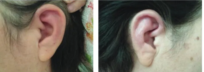
Figure 1: Inflammation of both ears. Obvious redness of the auricularis in this patient.
(a) (b)
Figure 2: Histological findings of biopsy specimen from the left ear. Perichondritis with the mononuclear cells and polymorphonuclear leukocytes at the fibrochondral junction (hematoxylin and eosin, (a) original magnification×50, (b) original magnification×200). The arrows show perichondritis with the presence of mononuclear cells and polymorphonuclear leukocytes at the fibrochondral junction. Basophil’s infiltration was not observed.
a high fever of up to 38.4∘C as well as a nonproductive cough. Otological examination revealed tender and swollen pinnae (Figure 1) with characteristic sparing of the lobule. There was no abnormality of the nose or eyes.
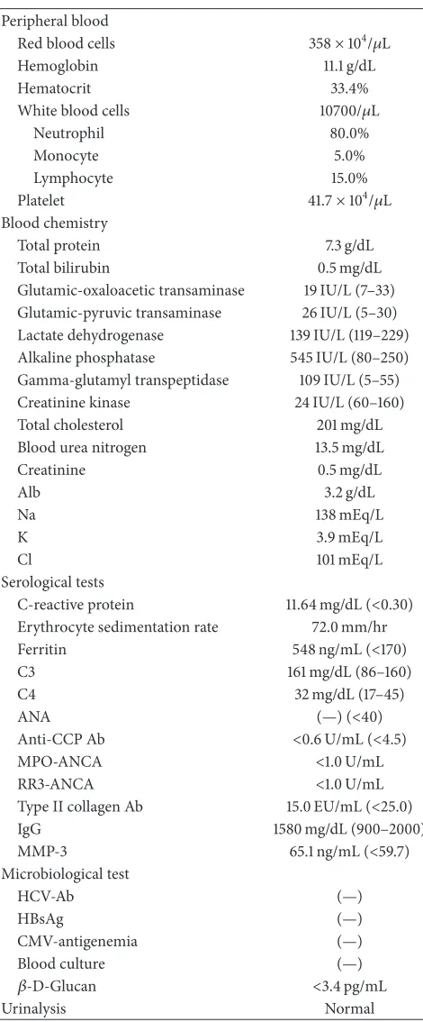
Laboratory findings (Table 1) showed a hemoglobin level of 11.1 g/dL, a total leukocyte count of 10,700/mm3, and elevations of the erythrocyte sedimentation rate and C-reactive protein level. Serology tests for antinuclear antibody and autoantibodies including anti-cyclic citrullinated peptide antibody, PR3-ANCA, and MPO-ANCA were all negative.
There was no tracheal cartilage tenderness, and chest computed tomography showed neither interstitial pneumo-nia nor tracheal stenosis. Neither aortic root dilatation nor aortic regurgitation was observed by echocardiography (data not shown). Although infection and hematological malignancies were excluded, redness of the bilateral auricu-laris suggested auricular perichondritis despite anti-type II collagen antibody negativity. Biopsy specimens were from the skin and the cartilage of the pinna for histopathological study. The histological evaluation showed cellular infiltrates of lymphocytes, neutrophils, and plasma cells, especially at the cartilage-skin interface, and a reduced number of chon-drocytes in areas of cartilage destruction (Figure 2). Other clinical features of RP, such as nonerosive arthritis, ocular inflammation, and nasal chondritis, were not confirmed, whereas the patient fulfilled the McAdam-Damiani-Levine
criteria for the diagnosis of RP [6, 7], according to the presence of one of McAdam’s signs (auricular chondritis) with positive histological confirmation. Based on the pres-ence of polyarthritis, the patient was diagnosed with RP. She was started on 50 mg of prednisolone daily, which led to an improvement in her symptoms. Both the fever and the auricular swelling disappeared. Tapering of her prednisolone dose in combination with the addition of 6 mg methotrexate weekly was recommended due to the suspicion of steroid-related psychiatric symptoms. Three months following her discharge from the hospital stay of 45 days, her corticos-teroids were reduced to 12.5 mg daily and the methotrexate was increased to 8 mg weekly. There was no subsequent flare-up of RP.
3. Discussion
Table 1: Laboratory findings on admission.
Peripheral blood
Red blood cells 358×104/𝜇L
Hemoglobin 11.1 g/dL
Hematocrit 33.4%
White blood cells 10700/𝜇L
Neutrophil 80.0%
Monocyte 5.0%
Lymphocyte 15.0%
Platelet 41.7×10⁴/𝜇L Blood chemistry
Total protein 7.3 g/dL
Total bilirubin 0.5 mg/dL Glutamic-oxaloacetic transaminase 19 IU/L (7–33) Glutamic-pyruvic transaminase 26 IU/L (5–30) Lactate dehydrogenase 139 IU/L (119–229) Alkaline phosphatase 545 IU/L (80–250) Gamma-glutamyl transpeptidase 109 IU/L (5–55) Creatinine kinase 24 IU/L (60–160) Total cholesterol 201 mg/dL Blood urea nitrogen 13.5 mg/dL
Creatinine 0.5 mg/dL
Alb 3.2 g/dL
Na 138 mEq/L
K 3.9 mEq/L
Cl 101 mEq/L
Serological tests
C-reactive protein 11.64 mg/dL (<0.30) Erythrocyte sedimentation rate 72.0 mm/hr Ferritin 548 ng/mL (<170)
C3 161 mg/dL (86–160)
C4 32 mg/dL (17–45)
ANA (—) (<40)
Anti-CCP Ab <0.6 U/mL (<4.5)
MPO-ANCA <1.0 U/mL
RR3-ANCA <1.0 U/mL
Type II collagen Ab 15.0 EU/mL (<25.0)
IgG 1580 mg/dL (900–2000)
MMP-3 65.1 ng/mL (<59.7) Microbiological test
HCV-Ab (—)
HBsAg (—)
CMV-antigenemia (—)
Blood culture (—)
𝛽-D-Glucan <3.4 pg/mL
Urinalysis Normal
ANA: antinuclear antibody, ANCA: antineutrophil cytoplasmic antibody, CMV: cytomegalovirus, HBsAg: hepatitis B surface antigen, HCV: hepatitis C virus, MMP-3: matrix metalloproteinase-3, MPO: myeloperoxidase, RF: rheumatoid factor, and RR3: proteinase 3.
more of the following clinical features: bilateral auricu-lar chondritis; nonerosive, seronegative inflammatory pol-yarthritis; nasal chondritis; ocular inflammation; respiratory
tract chondritis; and cochlear and/or vestibular dysfunction [6].
A striking symptom in our patient was the spiking fever. Although there are no specific laboratory findings in RP, such as positivity for anti-type II collagen antibody, our patient fulfilled one of the McAdam criteria in addition to the typical histological findings. Patients with RP may present with vari-ous signs and symptoms that are often misdiagnosed. Among patients who present with general, unspecific signs, such as fever and progressive malaise, diagnosis and treatment may be significantly delayed [10]. The most common signs of RP are auricular inflammation (89%), nonerosive arthritis (72%), nasal chondritis (11%), and laryngotracheal disease (55%) [11]. Fever is a nonspecific sign that occurs in a wide array of disorders and its origin can be extremely difficult to determine [2]. Patients who present with RP-related fever that is not diagnosed as such may receive a diagnosis of FUO [12]. However, RP has a chronic relapsing course that can be life-threatening if there is airway involvement, and laryngotracheal involvement is a major cause of morbidity and mortality [13, 14]. Therefore, in patients whose only symptoms are prolonged fever and cough, with no pulmonary abnormalities on CT, the differential diagnosis should include RP to improve the likelihood of a timely therapeutic interven-tion to prevent disease progression.
Fever is often caused by the release of endogenous inflammatory cytokines in response to tissue inflamma-tion [15]. An autoimmune reacinflamma-tion against type II collagen stimulates inflammatory cells, especially cytokine-producing macrophages. Glucocorticoid therapy is a fundamental com-ponent in the treatment of RP and its long-term use is recommended for most of these patients [11]. If significant organ involvement is proven, high-dose corticosteroids are often necessary. Severe disease may require treatment with immunosuppressive agents. In patients intolerant or, rarely, unresponsive to steroid therapy or in whom steroid-sparing therapy is required, immunosuppressants such as methotrex-ate, azathioprine, and cyclosporine play a role, particularly when there is severe respiratory or vascular involvement [16, 17]. Airway involvement by RP is considered to be a common course of morbidity and mortality [13], whereas it was reported that laryngotracheal involvement was seen less frequently in an Asian population [18]. Our patient was successfully treated with glucocorticoid and methotrexate, which were selected for their steroid-tapering effects. Asso-ciations with autoinflammatory disorders, such as familial Mediterranean fever (FMF), were reported in RP [19, 20]. In the present case, there was no relapsing periodic fever and sustained high fever was completely cured by steroid therapy; therefore, overlapping FMF in RP seems to be unlikely.
4 Case Reports in Medicine
Conflict of Interests
The authors declare that they have no competing interests.
References
[1] D. E. Trentham and C. H. Le, “Relapsing polychondritis,”Annals
of Internal Medicine, vol. 129, no. 2, pp. 114–122, 1998.
[2] J. F. Molina and L. R. Espinoza, “Relapsing polychondritis,”
Bailliere’s Best Practice and Research in Clinical Rheumatology,
vol. 14, no. 1, pp. 97–109, 2000.
[3] R. Chopra, N. Chaudhary, and J. Kay, “Relapsing polychondri-tis,”Rheumatic Disease Clinics of North America, vol. 39, no. 2, pp. 263–276, 2013.
[4] T. Lahmer, M. Treiber, A. von Werder et al., “Relapsing poly-chondritis: an autoimmune disease with many faces,”
Autoim-munity Reviews, vol. 9, no. 8, pp. 540–546, 2010.
[5] L. Cantarini, A. Vitale, M. G. Brizi et al., “Diagnosis and classi-fication of relapsing polychondritis,”Journal of Autoimmunity, vol. 48-49, pp. 53–59, 2014.
[6] L. P. McAdam, M. A. O’Hanlan, R. Bluestone, and C. M. Pearson, “Relapsing polychondritis: prospective study of 23 patients and a review of the literature,”Medicine, vol. 55, no. 3, pp. 193–215, 1976.
[7] J. M. Damiani and H. L. Levine, “Relapsing polychondritis— report of ten cases,”Laryngoscope, vol. 89, no. 6, part 1, pp. 929– 946, 1979.
[8] A. Sharma, K. Gnanapandithan, K. Sharma, and S. Sharma, “Relapsing polychondritis: a review,” Clinical Rheumatology, vol. 32, no. 11, pp. 1575–1583, 2013.
[9] J. M. Foidart, S. Abe, G. R. Martin et al., “Antibodies to type II collagen in relapsing polychondritis,”The New England Journal
of Medicine, vol. 299, no. 22, pp. 1203–1207, 1978.
[10] E. Letko, P. Zafirakis, S. Baltatzis, A. Voudouri, C. Livir-Rallatos, and C. S. Foster, “Relapsing polychondritis: a clinical review,”
Seminars in Arthritis and Rheumatism, vol. 31, no. 6, pp. 384–
395, 2002.
[11] P. D. Kent, C. J. Michet Jr., and H. S. Luthra, “Relapsing polychondritis,”Current Opinion in Rheumatology, vol. 16, no. 1, pp. 56–61, 2004.
[12] J. N. Avila, S. B. Carvalho, G. Tavares, and R. Garcia, “Fever of unknown origin in a patient with red ears: relapsing polychon-dritis,”BMJ Case Reports, 2014.
[13] A. Ernst, S. Rafeq, P. Boiselle et al., “Relapsing polychondritis and airway involvement,”Chest, vol. 135, no. 4, pp. 1024–1030, 2009.
[14] C. J. Michet Jr., C. H. McKenna, H. S. Luthra, and W. M. O’Fallon, “Relapsing polychondritis. Survival and predic-tive role of early disease manifestations,”Annals of Internal
Medicine, vol. 104, no. 1, pp. 74–78, 1986.
[15] L. Arnaud, A. Mathian, J. Haroche, G. Gorochov, and Z. Amoura, “Pathogenesis of relapsing polychondritis: a 2013 update,”Autoimmunity Reviews, vol. 13, no. 2, pp. 90–95, 2014. [16] K. Yamaoka, K. Saito, K. Hanami et al., “A case of
life-threatening refractory polychondritis successfully treated with combined intensive immunosuppressive therapy with methotrexate,”Modern Rheumatology, vol. 17, no. 2, pp. 144–147, 2007.
[17] J. Park, K. M. Gowin, and H. R. Schumacher Jr., “Steroid sparing effect of methotrexate in relapsing polychondritis,”Journal of
Rheumatology, vol. 23, no. 5, pp. 937–938, 1996.
[18] A. Sharma, P. Bambery, A. Wanchu et al., “Relapsing polychon-dritis in North India: a report of 10 patients,”Scandinavian
Journal of Rheumatology, vol. 36, no. 6, pp. 462–465, 2007.
[19] A. Salihoglu, E. Seyahi, S. Celik, and S. Yurdakul, “Relapsing polychondritis in a patient with familial Mediterranean fever and amyloidosis,”Clinical and Experimental Rheumatology, vol. 26, no. 4, supplement 50, p. S125, 2008.
Submit your manuscripts at
http://www.hindawi.com
Stem Cells
International
Hindawi Publishing Corporationhttp://www.hindawi.com Volume 2014
Hindawi Publishing Corporation
http://www.hindawi.com Volume 2014
INFLAMMATION
Hindawi Publishing Corporation
http://www.hindawi.com Volume 2014
Behavioural
Neurology
Endocrinology
International Journal ofHindawi Publishing Corporation
http://www.hindawi.com Volume 2014
Hindawi Publishing Corporation
http://www.hindawi.com Volume 2014
Disease Markers
Hindawi Publishing Corporation
http://www.hindawi.com Volume 2014
BioMed
Research International
Oncology
Journal of Hindawi Publishing Corporationhttp://www.hindawi.com Volume 2014
Hindawi Publishing Corporation
http://www.hindawi.com Volume 2014
Oxidative Medicine and Cellular Longevity
Hindawi Publishing Corporation
http://www.hindawi.com Volume 2014
PPAR Research
The Scientific
World Journal
Hindawi Publishing Corporationhttp://www.hindawi.com Volume 2014
Immunology Research
Hindawi Publishing Corporation
http://www.hindawi.com Volume 2014
Journal of
Obesity
Journal ofHindawi Publishing Corporation
http://www.hindawi.com Volume 2014
Hindawi Publishing Corporation
http://www.hindawi.com Volume 2014 Computational and Mathematical Methods in Medicine
Ophthalmology
Journal ofHindawi Publishing Corporation
http://www.hindawi.com Volume 2014
Diabetes Research
Journal ofHindawi Publishing Corporation
http://www.hindawi.com Volume 2014
Hindawi Publishing Corporation
http://www.hindawi.com Volume 2014
Research and Treatment
AIDS
Hindawi Publishing Corporation
http://www.hindawi.com Volume 2014
Gastroenterology Research and Practice
Hindawi Publishing Corporation
http://www.hindawi.com Volume 2014
Parkinson’s
Disease
Evidence-Based Complementary and Alternative Medicine
Volume 2014 Hindawi Publishing Corporation