O R I G I N A L R E S E A R C H A R T I C L E
Open Access
Completion thyroidectomy: predicting
bilateral disease
Badr Ibrahim
1, Véronique-Isabelle Forest
1,2, Michael Hier
1,2, Alex M. Mlynarek
1,2, Derin Caglar
3and Richard J. Payne
1,2*Abstract
Introduction:It is not uncommon for patients with indeterminate thyroid nodules to undergo diagnostic
hemithyroidectomy. When the final pathology determines that the nodule is in fact malignant, patients require counseling as to the whether a completion thyroidectomy is necessary.
Objectives:
1. Determine the incidence of well differentiated thyroid cancer (WDTC) in the contralateral thyroid lobe in patients undergoing completion thyroidectomy.
2. Identify features of the malignant tumor in the initial resection that increase the likelihood of malignancy in the contralateral lobe.
Methods:Retrospective chart review of 97 patients who underwent hemithyroidectomy and completion
thyroidectomy in a university’s teaching hospital network between 2006 and 2012. Pathology reports from both surgeries as well as patient and thyroid nodule characteristics were reviewed.
Results:Of the 97 patients, 47 (48 %) had a malignancy in the contralateral lobe. In the contralateral lobe, 42/47 (89 %) of malignancies were papillary microcarcinomas (PMC) and 15/42 (36 %) of the PMC were multifocal. Multifocal malignancies in the initial specimen had a 60 % rate of contralateral malignancy and were found to be a predictor of bilateral disease (p= 0.04) withOR= 2.74 (95 % CI: 1.11–6.79;p= 0.003) in WDTC andOR= 3.59 (95 % CI:1.35 9.48;p= 0.01) in papillary cancer specifically. There was no statistical significant correlation established for the following variables: presence of positive cervical nodes, extrathyroidal extension, positive resection margins, size and angio-lymphatic invasion. Moreover, there was no statistical correlation between any of the variants of papillary thyroid cancer and bilateral disease, even though most aggressive subtypes were found to be bilateral.
Conclusion:In this study, the rate of malignancy in the contralateral lobe was 48 %. Multifocality and presence of an aggressive subtype of papillary thyroid cancer in the initial specimen were found to be more important variables to consider in decision-making regarding completion thyroidectomy than size of the initial tumor alone.
Keywords:Completion thyroidectomy, Thyroid carcinomas, Bilateral disease, Decision making, Patient information
Background
Thyroid nodules are commonly encountered in clinical practice. Palpable nodules have a prevalence ranging from 5.3 to 6.4 % in women and 0.8 to 1.5 % in men in iodine sufficient regions and an incidence rate of 1.7 % in women and 0.9 % in men. Their prevalence increases
with age up to 9.1 % in women older than 75 years old (1, 2). While the majority of these nodules are benign, a thyroid carcinoma will be diagnosed in about 5 % of cases [1, 2]. In their 2009 revised guidelines for the man-agement of patients with thyroid nodules and differenti-ated thyroid cancer, the American Thyroid Association recommends ultrasonography followed by fine needle aspiration biopsy (FNAB) as diagnostic modalities in the work up of thyroid nodules [3]. However, 15–30 % of FNAB will be indeterminate (3) leaving the physician and the patient unable to determine with certainty the * Correspondence:[email protected]
1Division of Otolaryngology Head and Neck Surgery, McGill Thyroid Cancer Center, Jewish General Hospital, 3755 Cote Ste Catherine, Montreal H3T 1E2, QC, Canada
2
Jewish General Hospital–Otolaryngology Head and Neck Surgery Clinic, 3755 Côte-Sainte-Catherine Road, Montreal H3T 1E2, QC, Canada Full list of author information is available at the end of the article
risk of malignancy and therefore, the need for surgery. As a matter of fact, it has been reported in the literature that the risk of malignancy of a nodule with an indeter-minate FNAB ranges from 12 to 42 % [4–8]. For that reason, the 2009 ATA guidelines recommend a diagnos-tic thyroid lobectomy for nodules with indeterminate FNAB as an initial surgical approach [3]. If the definitive diagnosis confirms that the lesion is malignant, they sug-gest that patients undergo a completion thyroidectomy unless the lesion was <1 cm in size, unifocal, intrathyroi-dal, without lymph node involvement, and a low risk tumor [3].
The main reasons for completion thyroidectomy are to optimize radioiodine ablation of the remaining thyroid tissue, to allow for better postoperative follow up of thyroglobulin levels with the aim of detecting recur-rences, as well as to obtain a complete excision of multi-focal disease. Currently, there are no preoperative, intraoperative or postoperative definitive features or methods to ascertain the presence of disease in the contralateral lobe. Therefore, the pathological analysis of the hemithyroidectomy becomes the cornerstone of the decision making process in advising the patients on the need for completion thyroidectomy. This study aims at identifying the rate of bilateral disease on completion thyroidectomy and finding predictors for the presence of malignancy in the contralateral lobe.
Methods Patient selection
After obtaining the combined approval from the McGill University Health Center and Jewish General Hospital Ethics boards for conducting this retrospective chart view, we proceeded with pooling the operating room re-cords of one surgeon from August 2006 to April 2012 of cases of thyroid surgeries. From the 1264 thyroid surgery cases, 115 patients had a completion thyroidectomy. However, after exclusion of patients with incomplete electronic charts and one case where the initial path-ology was benign, 97 cases were included in the present study. These patients had undergone a hemithyroidect-omy followed by a completion thyroidecthemithyroidect-omy for malig-nancy identified in the first resected lobe.
Data collection
Baseline demographic information and dates of surgeries were recorded to calculate age at time of surgery and in-tervals between surgeries. The McGill Thyroid Nodule Score (MTNS) was recorded, when available, for each patient’s initial nodule. The MTNS is a comprehensive assessment tool previously developed by our research group consisting of a 22 variable scoring system that takes into account clinical parameters and laboratory values, imaging results, and cytology. It has been shown
to be an accurate predictor of the risk of malignancy in a given thyroid nodule [9].
Pathology reports of both surgeries were reviewed. Pa-tients were divided into two groups (benign contralateral lobe vs malignant contralateral lobe) based on the re-sults of the final pathology report of the completion thy-roidectomy specimen. The histological type of the tumor and when relevant, the variant of papillary thyroid car-cinoma (PTC), were collected. Also, size, tumor capsule status, lymphovascular involvement, extrathyroidal exten-sion, lymph node metastasis, resection margins, multifo-cality and number of PTC and papillary microcarcinomas (PMC) were recorded. A PMC was defined as a focus of papillary carcinoma less than 1 cm in size. In this study, the presence of an isolated focus of PMC was recorded as a malignant specimen, and multifocality was defined as the presence of more than one malignant focus in the resected thyroid lobe, regardless of size. The presence of a lymph node metastasis was not counted as multifocal dis-ease but rather as a lymph node metastasis. Extrathyroidal extension refers to a tumor that extends into the perithyr-oidal tissue or surrounding soft tissue (excluding lymph node metastasis). A patient with a malignant tumor in each lobe was accounted for as a case of bilateral disease without necessarily having the same histologic type of tumor in each lobe. Details pertaining to other histopatho-logical changes such as chronic lymphocytic thyroiditis were not recorded and a specimen with no cancerous focus identified was recorded as benign.
Statistical analysis
Categorical variables were assessed by means of a Chi-Square test. Age, time to completion, and the MTNS were tested with the studentTtest. Statistical outliers in time to completion were identified by means of a Modi-fied Z-score. A two-tailed Fisher’s exact test was used to assess the relation between different papillary carcinoma variants and bilateral disease. Binary logistic regressions were used to confirm the independent correlations with bilateral disease in subsets of our sample. The calcula-tions were made using SPSS statistics version 21.
Results
Patients demographics
From the 97 patients identified, 77 (79.4 %) were females equally distributed between the group with a benign contralateral lobe and the group with bilateral disease. In comparison only 20 (20.6 %) patients were males with 11 having a benign contralateral lobe and 9 having ma-lignancy. The average age of both groups at the time of the initial surgery was similar: 47.3(±12.9) for the group with a benign lobe on completion and 46.7(±15.1) for the group with a malignant contralateral lobe. The aver-age interval between the initial surgery and the
completion thyroidectomy varied for both groups with 73.2(±21.5) days in the benign completion group and 84.2(±24.2) in the malignancy group (p= 0.05) (Table 1).
Sixteen of the excluded patients had an incomplete chart missing whole or part of the pathology report from the first surgery. All of them however had a completion surgery pathology report. One patient had a total thy-roidectomy misclassified as a completion surgery in the database. The average age of the excluded patients was 55.6 years. They were composed of 16 females and 1 male and had a 65 % malignancy rate at completion (n= 11/17). Thirty five percent (n= 6/17) of these tu-mors were multifocal.
Pathology
In the specimen of the hemithyroidectomy of the initial resection, 75 (77.3 %) nodules exhibited PTC, 14 (14.4 %) PMC (of which 9 (64 %) where multifocal), 6 (6.3 %) follicular carcinoma, 1 (1.0 %) medullary micro-carcinoma, and 1 (1.0 %) Hurthle cell carcinoma. The distribution of the remaining pathological features re-corded after the initial resection is shown in Table 2.
In the completion thyroidectomy specimen, from the 97 cases 46 had a malignancy in the contralateral lobe and 1 case had a malignancy only in a lymph node on the contralateral side for a total of 47 (48 %) malignant cases. Forty-two (43 %) had a PMC, of which 15 (36 %) were multifocal, and 5 (5 %) had a PTC (4 intra-thyroidal and 1 involving a lymph node only) none of which were multifocal. The remaining 50 (52 %) cases exhibited a benign contralateral lobe. As far as lymph node status is concerned, out of the 47 cases of intrathyroidal tumor involvement, 7 (15 %) exhibited malignant contralateral lymph nodes. These 7 cases were constituted of 2 cases with multifocal PMC, 1 unifocal PMC, and 4 cases of PTC in the thyroid gland.
As for extrathyroidal extension (ETE) in the comple-tion specimens, there were 2 cases of PTC with ETE
while there was one case of unifocal PMC with ETE. Predictors of bilateral disease
Our initial screening of the different variables showed that only multifocality had a statistically significant cor-relation with bilateral disease (Table 2). To assess the in-dependence of this correlation and to control for the possible impact of the other studied features on the presence of bilateral disease in well-differentiated thy-roid cancers, a binary logistic regression was done using the following variables: age over 45, female gender, hist-ology, size of the tumor, lymphovascular invasion, lymph node involvement, extrathyroidal extension, tumor cap-sule involvement and multifocality. Multifocality in the initial specimen was the only variable independently as-sociated with bilateral disease with an OR = 2.74 (95 % CI =1.11–6.79; p= 0.03) (Table 3). When analyzing only
Table 1Patient demographics Totaln= 97 Absence of malignancy in the contralateral lobe (n= 50)
Presence of malignancy in the contralateral lobe (n= 47)
p values
Gender
Males 11 9 0.73
Females 39 38
Mean Age at Initial Resection (±SD)
47.6(±12.9) 46.3(±15.0) 0.64
Mean Time To Completion in Days (±SD)
73.2(±21.5) 84.2(±24.2) 0.05
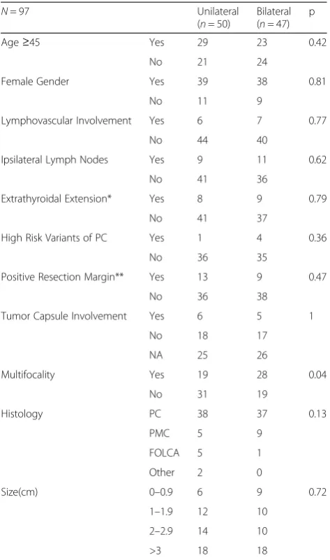
Table 2Features of patients with malignant specimens at first surgery and correlation to bilateral disease at completion
N= 97 Unilateral
(n= 50)
Bilateral (n= 47)
p
Age≥45 Yes 29 23 0.42
No 21 24
Female Gender Yes 39 38 0.81
No 11 9
Lymphovascular Involvement Yes 6 7 0.77
No 44 40
Ipsilateral Lymph Nodes Yes 9 11 0.62
No 41 36
Extrathyroidal Extension* Yes 8 9 0.79
No 41 37
High Risk Variants of PC Yes 1 4 0.36
No 36 35
Positive Resection Margin** Yes 13 9 0.47
No 36 38
Tumor Capsule Involvement Yes 6 5 1
No 18 17
NA 25 26
Multifocality Yes 19 28 0.04
No 31 19
Histology PC 38 37 0.13
PMC 5 9
FOLCA 5 1
Other 2 0
Size(cm) 0–0.9 6 9 0.72
1–1.9 12 10
2–2.9 14 10
>3 18 18
*n= 95 because 2 could not be assessed on pathologic examination; **n= 96 because 1 could not be assessed on pathologic examination
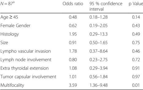
PTC and PMC cases, multifocality in the initial speci-men becomes an even stronger predictor with an OR = 3.59 (95 % CI = 1.36–9.48; p= 0.01) (Table 4). Further-more, in this subgroup, the involvement of ipsilateral lymph nodes at the initial surgery was not a predictor of bilateral disease [OR = 0.80 (95 % CI = 0.24–2.74;p= 0.73)] (Table 4). Also, as shown in Table 5, four out of the 5 cases of high risk variants of PTC (1 sclerosing, 3 tall cells and 1 solid variant) were associated with bilateral disease with OR = 4.00 (95 % CI = 0.43–37.6; p= 0.22). However, this correlation was not statistically significant. None of the low-risk variants were statistically significantly associated with bilateral disease (Table 5).
Discussion
In our present series, the rate of malignancy in the remaining lobe of the thyroid of patients undergoing com-pletion thyroidectomy following a hemithyroidectomy
positive for cancer is 48 % with close to 90 % of those tu-mors in the contralateral lobe being PMC. Among the var-iables analyzed, multifocality of the tumor in the initially resected lobe is the only feature that is consistently associ-ated with bilateral disease [OR 2.74(95 % CI = 1.11–6.79;
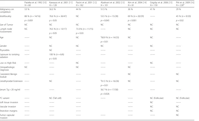
p= 0.03) in well differentiated cancers and 3.59 (95 % CI = 1.36–9.48;p= 0.01) in papillary cancers specifically]. In the literature, the incidence of bilateral carcinoma following CT ranges from 29 to 56.3 % [10–16] placing our rate of 48 % within this range (Table 6). More specifically, when considering only the patients with multifocal disease in the first resected lobe, the rate of bilateral malignancies is in-creased to 60 % as compared to 48 % in our entire series.
Furthermore, our sample demonstrates that size did not correlate with bilateral disease. Considering our pre-vious finding regarding the independent correlation be-tween bilateral disease and multifocality and knowing that 64 % of PMC found on initial resection were multi-focal, it is probably clinically more important to consider the focality of the tumor rather than its size in the deci-sion of proceeding with completion thyroidectomy. In-deed, the results of our study are in line with the ATA guidelines on the management of well differentiated thy-roid cancers according to which completion thythy-roidec- thyroidec-tomy should be offered to all patients with thyroid cancer unless the tumor is 1) < 1 cm in size 2) unifocal 3) intrathyroidal 4) node negative 5) low risk tumor [3], and underlines the need for all 5 conditions to be met simultaneously in order to decide not to proceed with completion thyroidectomy. Moreover, 43 % of the le-sions that were found on completion in our data were PMC and 36 % of them were multifocal. Mantinan et al. followed 91 patients with incidentally found PMC during thyroid surgery for an average of 10 years in an effort to identify predictors of outcomes of incidental PMC. The only independent predictor of recurrence identified in this series was multifocality [17]. This further under-scores the importance of multifocality in deciding on the extent of surgery.
Table 3Odds ratios of candidate predictors of bilateral disease following binary logistic regression in well differentiated cancers
N= 93a Odds ratio 95 % confidence
interval
p Value
Age≥45 0.59 0.24–1.46 0.26
Female gender 0.76 0.25–2.30 0.63
Histology 0.63 0.26–1.54 0.31
Size 0.78 0.48–1.25 0.30
Lympho vascular invasion 1.49 0.33–6.65 0.60
Lymph node involvement 1 0.30–3.26 0.99
Extra thyroidal extension 1.06 0.30–3.73 0.93
Encapsulated tumor 0.90 0.52–1.56 0.69
Multifocality 2.74 1.11–6.79 0.03
R square = 0.111 a
2 patients could not be included in this analysis as extra thyroidal extension could not be assessed on the pathology specimen
Table 4Binary logistsic regression of candidate predictors of bilateral disease in papillary and micropapillary cancer subset
N= 87a Odds ratio 95 % confidence
interval
p Value
Age≥45 0.48 0.18–1.28 0.14
Female Gender 0.62 0.19–2.05 0.43
Histology 1.95 0.29–13.3 0.49
Size 0.91 0.50–1.65 0.75
Lympho vascular invasion 1.78 0.37–8.64 0.46
Lymph node involvement 0.80 0.23–2.75 0.72
Extra thyroidal extension 1.08 0.29–3.94 0.91 Tumor capsular involvement 1.01 0.56–1.84 0.97
Multifocality 3.59 1.36–9.48 0.01
R square = 0.155 a
2 patients could not be included in this analysis as extra thyroidal extension could not be assessed on the pathology specimen
Table 5Papillary cancer variants and bilateral disease Totaln= 76 Absence of malignancy
in the contralateral lobe (n= 37)
Presence of malignancy in the contralateral lobe (n= 39)
Oncocytic 6 5
Classical 2 6
Follicular 24 23
Macrofollicular 3 1
Hyalinizing Trabecular 1 0
Solid 1 0
Sclerosing 0 1
Tall Cell 0 3
Pasieka et al. 1992 [10] N= 60
Kawaura et al. 2001 [11] N= 128
Pacini et al. 2001 [12] N= 182
Alzahrani et al. 2002 [13] N= 101
Kim et al. 2004 [14] N= 81
Grigsby et al. 2006 [15] N= 150
Pitt et al. 2009 [16] N= 228b
Malignancy on completion
53 % 56.3 % 44 % 51.5 % 36 % 41 % 29 %
Multifocality 88 % (n= 14/16) 76.6 % (n= 36/47) NC 53.5 % (n= 15/28) 69 % (n= 20/29) —— 45 % (n= 9/20)
p< 0.001 p> 0.05 p= 0.045 p< 0.001 p= 0.02
Size of Tumor NC NC NC NC NC NC NC
Lymph node involvement
NC 76.5 % (n= 13/17) 73.3.% (n= 11/15) —— NC NC NC
p> 0.05 p= 0.03
Age —— NC NC a60.9 % (n= 14/23) NC NC ——
p= 0.01
Gender —— NC NC NC —— NC ——
Thyroiditis —— NC —— —— —— —— ——
Exposure to ionizing
radiation ——
100 % (n= 6/6) —— —— —— —— ——
p> 0.05
Low vs High Risk —— —— NC —— —— NC ——
Histopathologic Diagnosis
NC —— NC —— NC —— ——
Coexistent Benign Nodule
—— —— —— —— NC —— NC
Extrathyroidal Extension —— NC —— a61.5 % (n= 16/26) NC —— NC
p= 0.01
Serum Tg > 20 ng/ml —— —— —— 56.7 % (n= 17/30) —— —— ——
p= 0.026
PC variant —— NC (Tall cell) —— —— —— NC (Follicular) NC (Follicular)
Soft tissue invasion —— —— —— —— NC ——
Vascular invasion —— —— —— —— NC NC
Resection margins —— —— —— —— NC NC
Tumor capsular
invasion —— —— —— —— —— ——
NC
——Feature not studied,NCNo statistically significant correlation
a
Predictive of cervical lymph node metastasis only
b
Pitt et al. [16]: the study of factors predicting contralateral disease is limited to PMC (<1 cm)n= 70
Journal
of
Otolaryngolo
gy
-Head
and
Neck
Surgery
(2015) 44:23
Page
5
of
As well, we did not perform intra-operative frozen sec-tions to help guide our decision-making regarding the extent of surgery as this practice has been tried several years ago at our institution and did not prove useful. This is in line with multiple series in the literature that prove that frozen sections were of little clinical useful-ness [18–20].
Regarding the involvement of cervical lymph nodes at the initial surgery, 52.3 % of patients with involved lymph nodes had disease in the contralateral lobe. Even if more than half of the patients with positive lymph nodes had bilateral disease, lymph node status was not a statistically significant predictor of bilateral disease in our series. On the other hand, Pacini et al. showed that, in a cohort of 182 patients, 73.3 % of patients with cer-vical lymph node involvement at the initial surgery had bilateral malignancy [12]. However, it is well established that in the presence of involved cervical lymph nodes a total thyroidectomy is warranted to help with radioactive iodine treatment and further follow-up [3]. For this rea-son, at our center, to determine the status of lymph node involvement and also, in an attempt to avoid subsequent surgery, we routinely perform sentinel lymph node bi-opsy using the methylene blue technique [21].
High risk variants have been shown to be more ag-gressive than classical PTC [22] and it is believed that anaplastic carcinoma develops from previously well dif-ferentiated thyroid cancer with tall cell variant being the most common coexistent carcinoma [23]. In fact, a recent review of more than 43,000 cases of papillary thyroid cancer aimed at identifying the prognostic fac-tors for survival in tall cell and diffuse sclerosing sub-types of PTC showed that the incidence of these high risk variants is on the rise and that they are associated with significantly higher disease specific mortality rates [22]. Interestingly, it also showed that most diffuse sclerosing cases are < 1 cm in size and that their sur-vival was unaffected by size. In our study, 4 out of 5 cases with high-risk variants had malignancy in the contralateral lobe. This finding did not reach statistical significance, but we believe that it is due to the small sample size. With thyroid surgery and RAI therapy be-ing independently associated to improved outcome, the authors recommend aggressive treatment with both modalities in all cases of tall cell and diffuse sclerosing subtypes –regardless of size. Indeed, it is of common practice in our center to complete the thyroidectomy in patients with high-risk variants of papillary carcinoma.
This study did not demonstrate that the MTNS could be used as a predictor of bilateral disease. This is not a surprising finding as the MTNS was designed for and is effective to preoperatively predict the likelihood of ma-lignancy in a given thyroid nodule; It was not meant to predict the risk of bilateral disease.
This study represents the second Canadian retrospect-ive review of predictors of malignancy in the second lobe of the thyroid gland at completion surgery. Compared to the data presented by Kawaura et al. [11] in the early 2000’s, our series reaches statistical significance for the relationship between multifocal disease and bilateral ma-lignancies. This, along with the other clinically signifi-cant associations outlined in this paper, can be used to improve communication between surgeons and patients regarding the risk of malignancy in the contralateral lobe of the thyroid and helps guiding both parties in the deci-sion making process for the need and extent of future surgeries.
Lastly, our retrospective study may have been subject to selection bias. However, we had a good sample size and we have subjected our data to multivariate logistic regression to decrease the effect of bias.
Conclusion
The rate of malignancy in the contralateral lobe of the thyroid gland following completion thyroidectomy is 48 %. When multifocal disease is found in the first resected lobe, the incidence of malignancy in the contra-lateral lobe increases to 60 %. Our data shows that mul-tifocality and presence of an aggressive subtype of papillary thyroid cancer in the initial specimen were found to be more important variables to consider in decision-making regarding completion thyroidectomy than size of the initial tumor alone.
Competing interests
The authors declare that they have no competing interests.
Authors’contributions
BI carried out the data collection, data analysis and wrote the manuscript. VIF contributed to writing the manuscript. MH, AM and DC contributed to the design of the study as well as reviewing and correcting the manuscript. RC contributed to the design, data collection, critical analysis of results and correction of the manuscript. All authors read and approved the final manuscript.
Author details
1Division of Otolaryngology Head and Neck Surgery, McGill Thyroid Cancer Center, Jewish General Hospital, 3755 Cote Ste Catherine, Montreal H3T 1E2, QC, Canada.2Jewish General Hospital–Otolaryngology Head and Neck Surgery Clinic, 3755 Côte-Sainte-Catherine Road, Montreal H3T 1E2, QC, Canada.3Department of Pathology, McGill University Health Center Glen site, 1001 Boulevard Decarie, Montreal H4A 3J1, QC, Canada.
Received: 29 November 2014 Accepted: 1 June 2015
References
1. Hegedus L. Clinical practice. The thyroid nodule. N Engl J Med. 2004;351:1764–71.
2. Brito JP, Yarur AJ, Prokop LJ, McIver B, Murad MH, Montori VM. Prevalence of thyroid cancer in multinodular goiter versus single nodule: a systematic review and meta-analysis. Thyroid. 2013;23:449–55.
3. Cooper DS, Doherty GM, Haugen BR, Kloos RT, Lee SL, Mandel SJ, et al. Revised American Thyroid Association management guidelines for patients with thyroid nodules and differentiated thyroid cancer. Thyroid. 2009; 19:1167-1214.
4. Yoon J, Kwak J, Kim EK, Moon HJ, Kim MJ, Kim JY, et al. How to approach thyroid nodules with indeterminate cytology. Ann Surg Oncol. 2010;17:2147–55. 5. Banks ND, Kowalski J, Tsai HL, Somervell H, Tufano R, Dackiw AP, et al. A
diagnostic predictor model for indeterminate or suspicious thyroid FNA samples. Thyroid. 2008;18:933–41.
6. Miller B, Burkey S, Lindberg G, et al. Prevalence of malignancy within cytologically indeterminate thyroid nodules. Am J Surg. 2004;188:459–62. 7. Goldstein RE, Netterville JL, Burkey B, Johnson JE. Implications of follicular neoplasms, atypia, and lesions suspicious for malignancy diagnosed by fine-needle aspiration of thyroid nodules. Ann Surg. 2002;235:656–62. discussion 662–654.
8. Castro MR, Espiritu RP, Bahn RS, Henry MR, Gharib H, Caraballo PJ, et al. Predictors of malignancy in patients with cytologically suspicious thyroid nodules. Thyroid. 2011;21:1191–8.
9. Sands NB, Karls S, Amir A, Tamilia M, Gologan O, Rochon L, et al. McGill Thyroid Nodule Score (MTNS):“rating the risk,”a novel predictive scheme for cancer risk determination. J Otolaryngol Head Neck Surg. 2011;40 Suppl 1:S1–13.
10. Pasieka JL, Thompson NW, McLeod MK, Burney RE, Macha M. The incidence of bilateral well-differentiated thyroid cancer found at completion thyroidectomy. World J Surg. 1992;16:711–6.
11. Kawaura M, Pathak I, Gullane PJ, Mancer K, Irish J, Brown D, et al. Multicentricity in papillary thyroid carcinoma: analysis of predictive factors. J Otolaryngol. 2001;30:102–5.
12. Pacini F, Elisei R, Capezzone M, Miccoli P, Molinaro E, Basolo F, et al. Contralateral papillary thyroid cancer is frequent at completion thyroidectomy with no difference in low- and high-risk patients. Thyroid. 2001;11:877–81.
13. Alzahrani AS, Al Mandil M, Chaudhary MA, Ahmed M, Mohammed GE. Frequency and predictive factors of malignancy in residual thyroid tissue and cervical lymph nodes after partial thyroidectomy for differentiated thyroid cancer. Surgery. 2002;131:443–9.
14. Kim ES, Kim TY, Koh JM, Kim YI, Hong SJ, Kim WB, et al. Completion thyroidectomy in patients with thyroid cancer who initially underwent unilateral operation. Clin Endocrinol (Oxf). 2004;61:145–8.
15. Grigsby PW, Reddy RM, Moley JF, Hall BL. Contralateral papillary thyroid cancer at completion thyroidectomy has no impact on recurrence or survival after radioiodine treatment. Surgery. 2006;140:1043–7. discussion 1047–1049.
16. Pitt SC, Sippel RS, Chen H. Contralateral papillary thyroid cancer: does size matter? Am J Surg. 2009;197:342–7.
17. Mantinan B, Rego-Iraeta A, Larranaga A, Fluiters E, Sanchez-Sobrino P, Garcia-Mayor RV. Factors influencing the outcome of patients with incidental papillary thyroid microcarcinoma. J Thyroid Res. 2012;2012:469397. 18. Kahmke R, Lee WT, Puscas L, Scher RL, Shealy MJ, Burch WM, et al. Utility of
intraoperative frozen sections during thyroid surgery. Int J Otolaryngol. 2013;2013:496138.
19. Huber GF, Dziegielewski P, Matthews TW, Warshawski SJ, Kmet LM, Faris P, et al. Intraoperative frozen-section analysis for thyroid nodules: a step toward clarity or confusion? Arch Otolaryngol Head Neck Surg. 2007;133:874–81. 20. Cetin B, Aslan S, Hatiboglu C, Babacan B, Onder A, Celik A, et al. Frozen
section in thyroid surgery: is it a necessity? Can J Surg. 2004;47:29. 21. Maniakas A, Forest VI, Jozaghi Y, Saliba J, Hier MP, Mlynarek A, et al. Tumor
classification in well-differentiated thyroid carcinoma and sentinel lymph node biopsy outcomes: a direct correlation. Thyroid. 2014;24:671–4. 22. Kazaure H, Roman S, Sosa J. Aggressive variants of papillary thyroid cancer:
incidence, characteristics and predictors of survival among 43,738 patients. Ann Surg Oncol. 2012;19:1874–80.
23. Smallridge RC, Ain KB, Asa SL, Bible KC, Brierley JD, Burman KD, et al. American Thyroid Association guidelines for management of patients with anaplastic thyroid cancer. Thyroid. 2012;22:1104–39.
Submit your next manuscript to BioMed Central and take full advantage of:
• Convenient online submission
• Thorough peer review
• No space constraints or color figure charges
• Immediate publication on acceptance
• Inclusion in PubMed, CAS, Scopus and Google Scholar
• Research which is freely available for redistribution