University of Pennsylvania
ScholarlyCommons
Publicly Accessible Penn Dissertations
1-1-2014
Neuroinflammatory Regulation of Stress Behavior
and Physiology
Alexis Rose Howerton
University of Pennsylvania, alexis.howerton@gmail.com
Follow this and additional works at:http://repository.upenn.edu/edissertations
Part of theAllergy and Immunology Commons,Immunology and Infectious Disease Commons, Medical Immunology Commons, and theNeuroscience and Neurobiology Commons
This paper is posted at ScholarlyCommons.http://repository.upenn.edu/edissertations/1314 Recommended Citation
Howerton, Alexis Rose, "Neuroinflammatory Regulation of Stress Behavior and Physiology" (2014).Publicly Accessible Penn Dissertations. 1314.
Neuroinflammatory Regulation of Stress Behavior and Physiology
Abstract
Neuropsychiatric diseases represent a major public health burden worldwide; due to gaps in our understanding of the pathogenic mechanisms of disease, approximately 30% of patients are refractory to treatment. Activation of neuroinflammatory signaling cascades has shown promise as a contributing factor for disease development. This hypothesis is driven in part by an intriguing overlap of symptoms in patients with neuropsychiatric and immunological disorders. Patients with depression, bipolar disorder, schizophrenia, and autism commonly present with immune dysfunction, while patients with multiple sclerosis, lupus, and rheumatoid arthritis often experience severe mood disturbances. Diversity in presentation of symptoms, however, has posed a research challenge to our mechanistic understanding of this link. In contrast to the complexity of modeling specific diseases, altered sensitivity to stress is a well-documented vulnerability marker across neuropsychiatric disorders. Of relevance to clinical advancement, aspects of stress behavior and physiology can be modeled and measured in animals, where core components of the stress axis are conserved in humans and rodents. Thus, we performed an examination of the neuroinflammatory regulation of stress behavior and physiology. Using a genetic model of stress sensitivity, we report the discovery that anti-inflammatory treatment ameliorates hypothalamic-pituitary-adrenal axis dysregulation, identifying the dorsal raphe (DR) as a locus of heightened responsivity. We then demonstrated sex differences in this brain region in response to the stress neuropeptide, corticotropin-releasing factor, suggesting that differences in its
responsivity may underlie sex differences in vulnerability to stress-related disorders. Finally, we used a transgenic approach to show that neuroinflammation localized specifically to the DR results in dysregulated stress behavior and physiology through interactions with the serotonergic neurotransmitter system. Overall, this work demonstrates that hyper- or hypo-function of the DR, based on genetic susceptibility, sex, or neuroinflammatory insult, can result in altered stress physiology and behavior. Though the DR has previously been identified as a potential locus of dysregulation, here we establish the specific, mechanistic link between risk factors for stress-related disorders. We present evidence of quantitative changes to this brain region and its functional output, and demonstrate that differences in responsivity of the DR may underlie vulnerability to stress-related disorders.
Degree Type
Dissertation
Degree Name
Doctor of Philosophy (PhD)
Graduate Group
Neuroscience
First Advisor
Tracy L. Bale
Keywords
Subject Categories
DEDICATION
This work is dedicated to the loving memory of my Grandpa Hank. Through life’s highs and lows, he saw the best in everyone around him. His eternal optimism was and always
will be a great source of inspiration for us all.
ACKNOWLEDGEMENTS
To my advisor and mentor, Dr. Tracy Bale: Your sharp eye, strong will, and keen
gut has made you a role model for excellence in science. Thank you for sharing this with
me. I have grown tremendously as a member of your lab, and I thank you for having me
as a part of it. To my thesis committee, Drs. Chris Pierce, Teresa Reyes, Judy Grinspan,
and Kelly Jordan-Sciutto: Thank you for your helpful feedback and guidance,
scientifically, professionally, and personally, and for your encouraging attitudes, which
always leave me motivated to work harder and think smarter. To Drs. Sheryl Beck and
Benjamin Rood: Thank you for opening up a whole new world of electrophysiology. To
the “trained immunologists” who helped me along the way, Drs. Chris Hunter, Tajie
Harris, Louise Randall, Mike Cancro, and Ari Glatman Zaretsky: Thank you for sharing
your expertise, for your patience, and for helping me step outside of the brain once in a
while. To current and past members of the Bale Lab, Katie, Stef, Jess, Amanda, Chris,
Diana, Cindy, David, Casey and Alison: You’re a bunch of tough cookies and I’m happy
to have taken this journey alongside you all. Thank you for helping to build such a
supportive, collaborative, and fun research environment. A special thanks to Mr. Chris: I
couldn’t have asked for a better bench-mate, music-listening partner, and grill-master.
And to Ali B.: I’ll miss our semi-regular walks around the block and experimental design
in the conference room. Thanks for sharing your spirited attitude, tenacity, and stellar
editing skills. I wish you luck in your role as The Graduate Student. To The Committee
Lauren, Judy, and Colleen: Thank you for being there through the good stuff and the hard
stuff. I’ll miss closing out NGG events, our regular margarita nights, and our
Zornitsa: I thank you for your friendship through all of our many shared milestones, and
for providing shelter during which time this thesis was written. To my family, Mom, Dad,
and Lindsey: You have been the greatest support team. Thank you for pushing me to
achieve my goals, for measuring my success by my happiness, and for your unconditional
support and love. And lastly, to my husband, Dr. Chris: It’s not often one finds a life- and
dissection-partner in a single person. Thank you for your companionship, encouragement,
and love. Thank you for reading through manuscripts, listening to crazy experimental
ideas over dinner, and for helping me keep perspective when it was most needed. I’m
lucky to have you.
ABSTRACT
NEUROINFLAMMATORY REGULATION OF STRESS BEHAVIOR AND PHYSIOLOGY
Alexis Rose Howerton
Tracy L. Bale
Neuropsychiatric diseases represent a major public health burden worldwide; due
to gaps in our understanding of the pathogenic mechanisms of disease, approximately
30% of patients are refractory to treatment. Activation of neuroinflammatory signaling
cascades has shown promise as a contributing factor for disease development. This
hypothesis is driven in part by an intriguing overlap of symptoms in patients with
neuropsychiatric and immunological disorders. Patients with depression, bipolar disorder,
schizophrenia, and autism commonly present with immune dysfunction, while patients
with multiple sclerosis, lupus, and rheumatoid arthritis often experience severe mood
disturbances. Diversity in presentation of symptoms, however, has posed a research
challenge to our mechanistic understanding of this link.
In contrast to the complexity of modeling specific diseases, altered sensitivity to
stress is a well-documented vulnerability marker across neuropsychiatric disorders. Of
relevance to clinical advancement, aspects of stress behavior and physiology can be
modeled and measured in animals, where core components of the stress axis are
conserved in humans and rodents.
behavior and physiology. Using a genetic model of stress sensitivity, we report the
discovery that anti-inflammatory treatment ameliorates hypothalamic-pituitary-adrenal
axis dysregulation, identifying the dorsal raphe (DR) as a locus of heightened
responsivity. We then demonstrated sex differences in this brain region in response to the
stress neuropeptide, corticotropin-releasing factor, suggesting that differences in its
responsivity may underlie sex differences in vulnerability to stress-related disorders.
Finally, we used a transgenic approach to show that neuroinflammation localized
specifically to the DR results in dysregulated stress behavior and physiology through
interactions with the serotonergic neurotransmitter system.
Overall, this work demonstrates that hyper- or hypo-function of the DR, based on
genetic susceptibility, sex, or neuroinflammatory insult, can result in altered stress
physiology and behavior. Though the DR has previously been identified as a potential
locus of dysregulation, here we establish the specific, mechanistic link between risk
factors for stress-related disorders. We present evidence of quantitative changes to this
brain region and its functional output, and demonstrate that differences in responsivity of
TABLE OF CONTENTS
DEDICATION ... II ACKNOWLEDGEMENTS ... III ABSTRACT ... V TABLE OF CONTENTS ... VII LIST OF TABLES ... IX LIST OF FIGURES ... X
CHAPTER 1: INTRODUCTION ... 1
NEUROPSYCHIATRIC DISEASE: A MAJOR UNMET MEDICAL NEED ... 2
STRESS DYSREGULATION AS A VULNERABILITY MARKER FOR NEUROPSYCHIATRIC DISEASE .... 3
INFLAMMATION AS A RISK FACTOR FOR NEUROPSYCHIATRIC DISEASES ... 8
THE BIDIRECTIONAL RELATIONSHIP BETWEEN STRESS AND NEUROINFLAMMATION ... 10
BRAIN-WIDE, AND REGION-SPECIFIC NEUROINFLAMMATION ... 11
GOALS OF THIS DISSERTATION ... 13
CHAPTER 2: ANTI-INFLAMMATORY TREATMENT AMELIORATES HPA STRESS AXIS DYSFUNCTION IN A MOUSE MODEL OF STRESS SENSIVITY ... 14
ABSTRACT ... 15
INTRODUCTION ... 16
METHODS ... 18
RESULTS ... 23
DISCUSSION ... 25
ACKNOWLEDGEMENTS ... 29
FIGURESANDLEGENDS ... 30
ADDITIONALDATA ... 35
CHAPTER 3: SEX DIFFERENCES IN CORTICOTROPIN-RELEASING FACTOR RECEPTOR-1 ACTION WITHIN THE DORSAL RAPHE NUCLEUS IN STRESS RESPONSIVITY ... 37
ABSTRACT ... 38
INTRODUCTION ... 39
METHODS ... 40
RESULTS ... 52
DISCUSSION ... 56
ACKNOWLEDGEMENTS ... 62
FIGURESANDLEGENDS ... 63
CHAPTER 4: DORSAL RAPHE NEUROINFLAMMATION PROMOTES DRAMATIC BEHAVIORAL STRESS DYSREGULATION ... 72
ABSTRACT ... 73
INTRODUCTION ... 74
METHODS ... 76
RESULTS ... 85
DISCUSSION ... 92
ACKNOWLEDGEMENTS ... 98
CHAPTER 5: GENERAL CONCLUSIONS AND FUTURE DIRECTIONS ... 107
MODELING STRESS DYSREGULATION ... 108
ANTI-INFLAMMATORY DRUGS IN THE TREATMENT OF PSYCHIATRIC DISORDERS ... 110
FOCUSING ON ANATOMICAL LOCI OF VULNERABILITY ... 111
SEX DIFFERENCES AS A MODEL OF VULNERABILITY ... 114
NEUROINFLAMMATORY REGULATION OF SEROTONIN SIGNALING ... 115
GENERAL IMPLICATIONS ... 118
APPENDIX A: SEX DIFFERENCES IN STRESS BEHAVIOR AND PHYSIOLOGY FOLLOWING INFECTION WITH TOXOPLASMA GONDII ... 119
INTRODUCTION ... 120
METHODS ... 121
RESULTS ... 124
DISCUSSION ... 127
ACKNOWLEDGEMENTS ... 129
FIGURESANDLEGENDS ... 130
BIBLIOGRAPHY ... 137
LIST OF TABLES
CHAPTER 3:
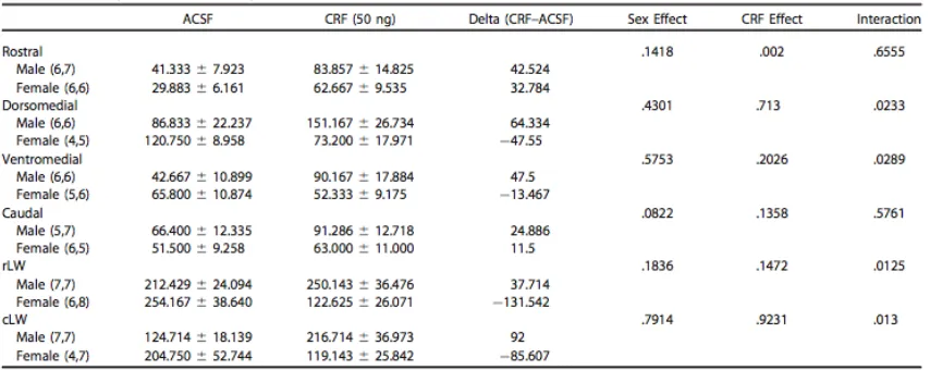
Table 3.1 Quantification of dorsal raphe cFos response to ACSF or CRF infusion ... 66
Table 3.2 Active and passive cell characteristics of 5-HT neurons ... 70
Table 3.3 Clinical trials targeting CRFr1 for the treatment of major depression and
anxiety disorders ... 71
LIST OF FIGURES
CHAPTER 2:
Figure 2.1Anti-inflammatory treatment ameliorates chronic stress-induced corticosterone
elevation in stress-sensitive CRF2-/-mice ... 30
Figure 2.2 Anti-inflammatory treatment prevents chronic stress-induced increases in GFAP in the PVN of stress-sensitive CRF2-/-mice. ... 31
Figure 2.3 Anti-inflammatory treatment prevents chronic stress-induced increases in GFAP in the ventral hippocampus of stress-sensitive CRF2-/-mice ... 32
Figure 2.4 Anti-inflammatory treatment prevents chronic stress-induced increases in GFAP in the medial prefrontal cortex of stress-sensitive CRF2-/-mice ... 34
Figure 2.5 Anti-inflammatory treatment prevents chronic stress-induced increase in cFos in response to an acute stressor in the dorsal raphe of CRF2-/- mice ... 35
Figure 2.6 CRFr2-/- have fewer TPH+ neurons following chronic stress exposure. NSAID treatment rescues these effects. ... 36
CHAPTER 3:
Figure 3.1 DR infusion of the CRFr1 small molecule antagonist NBI 35965 or 50 ng CRF had male-specific effects on HPA responsiveness ... 63
Figure 3.2 Males and females show divergent behavioral responses to CRFr1 antagonism or CRF infusion into the DR ... 64
Figure 3.3 A 50 ng CRF infusion elicited differential patterns of neuronal activation in males and females across DR subregions ... 65
Figure 3.4 CRFr1 is expressed on different cell populations in males and females ... 67 Figure 3.5 Males and females exhibit differences in baseline characteristics, and
divergent responses to bath applied CRF ... 69
CHAPTER 4:
Figure 4.3 IL-1β-mediated neuroinflammation in the DR increases male stress
responsivity ... 102
Figure 4.4 IL-1β-mediated neuroinflammation within the PFC does not induce
manic-like behavior or physiology ... 103
Figure 4.5 IL-1ß-mediated neuroinflammation in the DR results in broad changes in gene expression ... 104
Figure 4.6 IL-1β-mediated neuroinflammation in the DR results in serotonergic
hypofunction ... 105
Figure 4.7 Acute increase in serotonin ameliorates manic-like phenotype in the elevated plus maze ... 106
APPENDIX A:
Figure A.1CRFr2 deficiency resuces female susceptibility to infection with T. gondii. .... 130 Figure A.2Survival curve of male and female mice following infection with T. gondii .... 131 Figure A.3CRFr2 deficiency attenuates weight loss following T. gondii infection ... 132 Figure A.4 Infection with T. gondii increases expression of CRFr2 in the ventromedial
hypothalamus of WT mice. ... 133
Figure A.5CRFr2 deficiency rescues T. gondii-mediated heightened corticosterone response to an acute restraint stress. ... 134
Figure A.6CRFr2 deficiency rescues female-specific behavioral effects of infection with T. gondii ... 135
Figure A.7 Sex differences in basal and T. gonii-induced cytokine and chemokine
CHAPTER 1: INTRODUCTION
The goal of this dissertation is to understand the contribution of
neuroinflammation to the regulation of stress related behavior and physiology, with the
hope of providing insight into specific targetable, pathogenic mechanisms of
stress-related disease. In this dissertation I focus on the bidirectional relationship between stress
exposure/coping behavior, and neuroinflammation, both brain-wide and in potentially
susceptible brain regions. I have specifically focused on the contribution of the
serotonergic dorsal raphe nucleus (DR) as a locus of vulnerability. Using a genetic model
of stress sensitivity, I report the discovery that anti-inflammatory treatment ameliorates
hypothalamic-pituitary-adrenal (HPA) stress axis dysregulation. I then characterize sex
differences in the DR response to the stress neuropeptide, corticotropin-releasing factor
(CRF), demonstrating that sex differences in responsivity may contribute to differences in
vulnerability to stress-related disorders. Finally, I use a transgenic approach to show that
neuroinflammation localized specifically to the DR results in dysregulated stress behavior
and physiology through interactions with the serotonergic neurotransmitter system. This
work has helped gained insight into the potential mechanism underlying the clinical
overlap of neuroimmune and neuropsychiatric symptomology.
The following introductory chapter describes the challenges associated with the
advancement of research in neuropsychiatric disease, providing background of how, by
utilizing different models of vulnerability, one might learn about this family of
stress-related disorders. This includes a general description of psychiatric disease symptom
contribution of the serotonergic DR, I provide extensive background on the known role
for this brain region and neurotransmitter system to respond to, and affect, stress related
behavior and physiology. The introduction then focuses on how the relationship between
these factors has been studied in humans with disease and in animal models. Finally, I
present an overview of the goals of this dissertation, and the potential implications of the
findings presented herein.
Neuropsychiatric disease: a major unmet medical need
Neuropsychiatric diseases represent a major public health burden worldwide,
accounting for 13% of the global burden of disease (Collins et al., 2011). In the United
States, it is estimated that 26.2% of adults suffer from a diagnosable mental disorder
every year (Kessler et al., 2005b; Wang et al., 2005).
The complex clinical manifestations of neuropsychiatric disorders generally fall
in an overlap of domains, where examples include: sadness, confused thinking or reduced
ability to concentrate, excessive fears or worries, extreme mood changes of highs and
lows, withdrawal from friends and activities, significant tiredness or low energy,
problems sleeping, detachment from reality (delusions), paranoia or hallucinations,
extreme feelings of guilt, alcohol or drug abuse, major changes in eating habits, changes
in sex drive, excessive anger or hostility or violence, suicidal thinking, and an inability to
cope with daily problems or stress (MayoClinic, 2012). However, even within a
particular diagnosis, illnesses vary with respect to symptom number, type, intensity, and
duration.
due to the complex nature of risk factors, which include environment, genetics and
gender. This has lead to significant diagnostic and treatment challenges; despite treatment
being fairly common, gaps in our understanding of the pathogenic mechanisms
contributing to disease have left up to 30-50% of patients refractory to treatment (Souery
et al., 2006).
Stress dysregulation as a vulnerability marker for neuropsychiatric disease
Responding to stress is a normal, homeostatic response of the body and brain to a
perceived threat or challenge. It is vital to an organism’s survival, allowing appropriate
allocation of energy and attention. However, an inability to cope with stress exposure is a
well-documented vulnerability marker of neuropsychiatric disease, where patients often
present with altered basal stress hormones, inappropriate feedback following stress
exposure, and a failure to produce adaptive stress coping responses (Breslau and Davis,
1986; Koenig et al., 2002; Nestler et al., 2002). Indeed, in addition to subjective
measurement of behavioral outcome on scales such as the Hamilton Rating Scale for
Depression (HRSD) or the Young Mania Rating Scale (YMRS), the amelioration of
stress hormone dysregulation is an increasingly common readout for the success of
clinical trials in the domain of neuropsychiatry.
Of relevance to clinical advancement, aspects of stress behavior and physiology
can be measured with some reliably in animal models, where core components of the
stress axis are conserved in humans and rodents. Thus, the examination of stress coping
behavior and physiology is a tractable target for an investigation into the etiology and
corticotropin-releasing factor (CRF) and serotonin (5-HT) systems in stress responsivity, and the
biochemical changes that occur with chronic stress exposure.
Corticotropin-releasing factor (CRF)
CRF represents an important link between stress and mood regulation (Vale et al.,
1981; Sutton et al., 1982; Stenzel-Poore et al., 1994; Groenink et al., 2002). The classical
neuroendocrine stress response - activation of the hypothalamic-pituitary-adrenal (HPA)
stress axis - is initiated upon stress-induced release of CRF from the paraventricular
nucleus of the hypothalamus (PVN). In parallel to its hypothalamic release,
extra-hypothalamic CRF serves as an important neuromodulator to orchestrate stress
responsivity within the brain (Bale and Vale, 2004).
Originating largely from the bed nucleus of the stria terminalis (BNST) and
central amgydala (CeA), in addition to the PVN, CRF acts on two G-protein coupled
receptor (GPCR) subtypes, CRFr1, and CRFr2. Characterization of transgenic mice that
have been constructed to lack either CRFr1 or CRFr2 have aided in our understanding of
the role of these receptors in stress related behavior and physiology. CRFr1-/- mice have a
blunted stress response and display reduced anxiety-like behavior (Smith et al., 1998;
Timpl et al., 1998). Conversely, mice lacking CRFr2 are hypersensitive to stress
exposure, displaying augmented HPA stress axis corticosterone levels, increased
anxiety-like behavior, and reduced ability to mount appropriate coping responses to stress
exposure (Bale et al., 2000; Bale and Vale, 2003; McEuen et al., 2008). Because of this,
the two CRF receptors have been described as playing opposing roles as the “gas” and
not strictly dichotomous. This is evidenced by the generation of mice deficient for both
CRFr1 and CRFr2, which exhibit a more blunted stress response compared to CRFr1 null
mice (Bale et al., 2002). Clearly, the cell type and brain region of localization plays a
critical in the response of these receptors to CRF.
Serotonin (5-HT)
Serotonin (5-hydroxytryptamine; 5-HT), a monoaminergic neurotransmitter, is
produced by almost every organ in the body, including the skin, gut, liver, and lungs. In
the brain, its primary sources are the dorsal and medial divisions of the raphe nucleus
(DR), which sits below the cerebral aqueduct at the junction of the midbrain and
brainstem. Despite there being relatively few 5-HT producing neurons, they innervate
much of the brain and regulate many aspects of behavior (Dahlström and Fuxe, 1964;
Steinbusch, 1981; Azmitia and Gannon, 1982). The most studied roles of 5-HT are in the
modulation of arousal, sleep, memory, and mood.
Because selective 5-HT reuptake inhibitors (SSRIs), which target the 5-HT
transporter, SERT, have had some success for the treatment of depression, this
neurotransmitter system has been a major focus of research on mood disorders.
Serotonin turnover rates are reduced in major depression, and increase with recovery
(Asberg et al., 1976; Asberg et al., 1984). Alterations in 5-HT signaling have also been
identified in patients with depression, including reduced 5-HT metabolites in the CSF and
postmortem tissue, and reduced SERT levels (Reddy et al., 1992; Owens and Nemeroff,
1994; Huang et al., 2010; Chatzittofis et al., 2013). Single nucleotide polymorphisms in
enzyme in the synthesis of 5-HT, has also been found correlated with depression (Gao et
al., 2012). Polymorphisms in the promoter region of the 5-HT transporter have also
associated with increased stress-induced depressive symptoms (Ho et al., 2013).
Interestingly, this SERT polymorphism appears to have a greater impact on the
depressive symptoms of women compared to men (Sjoberg et al., 2006).
5-HT also plays an important role in the “manic” side of the behavioral spectrum,
influencing arousal, impulsivity, hedonic and hyper-aggressive behaviors, all of which
are central manifestations of mania (Shiah and Yatham, 2000; Crockett et al., 2010).
Clinical studies of CSF, platelet assessments, and neuroendocrine challenge, and
postmortem tissue provide evidence to support the hypothesis that serotonin deficiency is
involved in mania, and that enhancement of serotonin neurotransmission may exert a
mood-stabilizing effect (Shiah and Yatham, 2000).
Importantly, there are major sex differences in the 5-HT system, which are
thought to contribute to sex differences in neuropsychiatric disease, including the
aforementioned behaviors associated with both depression and mania. Human PET
imaging has revealed a reduced rate of serotonin synthesis (Nishizawa et al., 1997) and
differences in 5-HT binding sites throughout the brains of women compared to men
(Jovanovic et al., 2008). Rodent studies have demonstrated that 5-HT-increasing
pharmacological agents, which are known to activate the HPA axis, show a greater effect
on glucocorticoid production in female rodents compared to males (Goel and Bale, 2010).
Central 5-HT levels and CSF concentrations of the 5-HT metabolite, 5-HIAA, too, appear
Biochemical changes following chronic stress
The consequences of stress are dependent on specific stressor characteristics,
including intensity, controllability, and predictability. Importantly, they are also heavily
dependent on the duration of exposure, which is generally categorized as acute or
chronic. Chronic stress enhances both basal HPA tone and stress responsivity, often
resulting in hypersecretion of glucocorticoids and adrenal hypertrophy. Chronic stress
also impacts central stress circuitry, downregulating glucocorticoid receptors and
reducing glucocorticoid negative feedback efficacy [reviewed in (Herman, 2013)]. Major
stress responsive brain regions such as the hippocampus, amygdala, and prefrontal cortex,
all undergo structural remodeling following chronic stress exposure (Vyas et al., 2002).
Stress-induced changes in dendritic morphology have been observed in the dentate gyrus
of the hippocampus (Wood et al., 2004), amygdala (Vyas et al., 2003), and medial
prefrontal cortex (Liston et al., 2006). Interestingly, cortical changes may be particularly
plastic, as they display substantial recovery by 3 weeks after the termination of stress
exposure (Radley et al., 2004). Similar changes occur in these brain regions with chronic
corticosterone administration, suggesting the effects of chronic stress may be
glucocorticoid-mediated. This possibility is supported by a study that found treatment
with 3-β-hydroxysteroid dehydrogenase, which blocks stress-induced corticosterone,
prevents stress-induced dendritic remodeling in the CA3 region of the hippocampus
(Magarinos and McEwen, 1995).
Importantly, many of the effects of chronic stress in the brain are sex-specific; for
stress has been shown to decrease norepinephrine, dopamine, and 5-HT in the
hippocampus of males whereas increased levels of these neurotransmitter levels have
been observed in females (Sunanda et al., 2000; Beck and Luine, 2002). These findings
are of particular relevance considering the dramatic sex differences in behavior that occur
following chronic stress.
Inflammation as a risk factor for neuropsychiatric diseases
The CNS contains a blood-brain-barrier (BBB) consisting of endothelial cells and
astrocytes, which regulates the permeability of entry into the brain. This includes the
regulation of immune cellular and molecular infiltration from the periphery. The question
of how the brain and peripheral immune system communicate is still largely under
investigation. Since cytokine and chemokine receptors are expressed by both glia and
neurons in the adult brain, a humoral route of communication from the periphery has
been proposed. Additionally, autonomic nerve fibers provide a loop of communication
between the brain and peripheral blood vessels, which contain circulating immune cells.
The brain also contains specialized cells capable of performing immune
surveillance and repair [reviewed in (Ousman and Kubes, 2012)]. Microglia, astrocytes,
and neurons all display some immune function through their ability to produce and
respond to pro- and anti-inflammatory cytokines, express cytokine and chemokine
receptors, and display inducible expression of major histocompatibility complex (MHC).
Microglia are generally considered the “resident immune cell” of the brain, where they
function to survey and clean-up (i.e. phagocytose) damaged or intrusive cells. Microglia
IL-1β, and several chemokines. Additionally, they can serve an anti-inflammatory role
through production of IL-4, IL-10, and TGF-β, as well as BDNF and GDNF. Astrocytes
can play a similar pro- or anti-inflammatory function, though their role as immune cells is
far less well examined. However, posed at the intersection of the BBB, astrocytic release
of cytokines and chemokines can play an important role in regulating BBB permeability
and promoting infiltration of peripheral immune cells.
Activation of inflammatory signaling cascades have been a promising
environmental risk factor for the development of a number of neuropsychiatric disorders.
Schizophrenia, bipolar disorder, depression, and other psychotic illnesses commonly
co-present with immune dysfunction (Muller et al., 2009; Modabbernia et al., 2013).
Furthermore, there is significant co-presentation of primary immunological disorders
with psychiatric manifestations. Notably, patients with synaptic autoimmune encephalitis
are often first admitted into care on the basis of symptoms of mania (Rosenfeld and
Dalmau, 2011), and the neuropsychiatric manifestations of multiple sclerosis, lupus, and
rheumatoid arthritis include depressive episodes, irritability, mood swings, rapid speech,
distractibility, and impulsivity, in addition to cognitive decline (Kwentus et al., 1986;
Bruce, 2008; Meszaros et al., 2012). Consequently, many researchers have hypothesized
that (neuro)immunological mechanisms drive aspects of neuropsychiatric symptomology
[reviewed in (Najjar et al., 2013)]. However, the complex nature of disease presentation,
and the difficulty in modeling these complex disorders in animals, has left advancement
stalled; while there is substantial epidemiological evidence, the understanding of
The bidirectional relationship between stress and neuroinflammation
The first observation that stress impacts immunity came when Hans Selye noted
that chronic stress results in atrophy of the thymus (Selye, 1936). Since then, stress has
historically been considered immunosuppressive. Indeed, glucocorticoids, the steroid
hormone produced by the adrenal cortex in response to stress exposure, play a
well-appreciated anti-inflammatory and immunosuppressive role throughout the body, acting
through both genomic and non-genomic modes of action (Coutinho and Chapman, 2011)
(Silverman and Sternberg, 2012)
Within the brain, however, these stress-induced neuropeptides and hormones can
actually serve a pro-inflammatory role (Sorrells and Sapolsky, 2007; Sorrells et al.,
2009). Acute stress has been shown to enhance NF-κB activity (Madrigal et al., 2001),
increase IL-1β (Nguyen et al., 1998), activate TNF-α (Madrigal et al., 2002), and
upregulate prostaglandins and cyclooxygenase-2 (Madrigal et al., 2003). Importantly,
stress exposure has been clearly documented to increase inflammatory cytokine
production in humans and in rodent models (Reyes et al., 2003; Deak et al., 2005;
Hueston et al., 2011; You et al., 2011).
Conversely, pro-inflammatory cytokines also have the ability to modulate
neurotransmitter systems in the brain to affect stress related behavior and physiology
(Konsman et al., 2002; Johnson et al., 2004; Gadek-Michalska et al., 2013). Most
notably, IL-1β, TNF-α, and IL-6 can all stimulate glucocorticoid release through HPA
axis activation at the level of the hypothalamus. This sequence of events has the potential
to create a dangerous positive feedback loop, whereby increased stress exposure sets a
increases stress responsivity. In a chicken or egg type of scenario, elevated basal
neuroinflammation, due to environmental or genetic influence, could also predispose
individuals to susceptibility for stress-related disorders.
Brain-wide, and region-specific neuroinflammation
Animal models have clearly demonstrated the ability of pro-inflammatory
cytokines, such as interleukin (IL)-1β, interferon-γ, tumor necrosis factor-α, and
lipopolysaccharide (LPS) to modulate arousal and coping behaviors when administered
peripherally (Dantzer et al., 2008). Intriguingly, the same molecules alter discrete aspects
of behavior when delivered intracerebroventricularly, or acutely into specific brain
regions, suggesting that access to and action on particular brain areas may be central to
their effects (Song et al., 2006; Huang et al., 2008; Barrientos et al., 2012; Hayley et al.,
2013).
Toxoplasma gondii (T. gondii)is an intracellular obligate parasite with the ability
to cross the blood-brain-barrier and induce neuroinflammation. In the chronic stage of
infection, neuroinflammation is maintained, while peripheral infection is cleared. As
such, T. gondii is a powerful tool to ask about the impact of brain-wide
neuroinflammation on behavior, both in humans and rodents. What T. gondii infected
individuals have taught us, is that neuroinflammation has a profound ability to increase
impulsivity, incidence of schizophrenia, and risk of suicide (Alvarado-Esquivel et al.,
2013; Ingram et al., 2013; Webster et al., 2013). The hypothesis that the location of the
parasite might influence the behavioral outcomes of infection is not new, and in fact, was
Observations of patient behavior in association with measured loci of insult has
helped to greatly improve our understanding of the impact of brain region-specific
neuroinflammation on behavior. For example, traumatic brain injury (TBI) and stroke
both result in a focal neuroinflammatory response, and commonly present with a host of
neuro-cognitive challenges. TBI, the result of mechanical force applied to the skull and
transmitted to the brain, leads to psychiatric problems in as many as 40% (Kim et al.,
2007) and secondary mania in close to 9% of patients. Post-stroke depression occurs in
roughly 30% of survivors (Ayerbe et al., 2013). As such, it has been proposed that TBI
and stroke impact behavior through disruptions in neurotransmission in relevant
behavior-coordinating brain regions, termed, the “lesion location perspective.” However,
data to support this theory remain inconclusive, largely due to the fact that neuroimaging
methods are not yet able to assess the full extent of the TBI- and stroke-affected areas
with the precision necessary to denote sufficient anatomical specificity. However, with
the current rate of advancement in neuroimaging, it is likely that information will soon be
available.
Clearly the brain is not a homogenous organ, therefore understanding loci of
neural dysfunction may help in our understanding of disease etiology, presentation, and
treatment. Recent studies have found evidence for the presence of focal
neuroinflammation in a number of psychiatric diseases, including autism (Onore et al.,
2012; Theoharides et al., 2013), depression (Hercher et al., 2009), schizophrenia (Cagnin
et al., 2007), and bipolar disorder (Rao et al., 2010). These findings suggest that
anatomical specificity may underlie the epidemiological evidence linking neuroimmune
Goals of this dissertation
It is becoming clear that neuroinflammaiton can have a profound effect on
behaviors associated with neuropsychiatric disease. Furthermore, there is mounting
evidence that impact of neuroinflammation on behavior is driven by brain region
specificity. However, the mechanism by which this occurs remains unknown, in part due
to the complex presentation and trajectory of neuropsychiatric disease symptoms. With
this in mind, the goal of this dissertation was to elucidate the role of neuroinflammation
as a determinant of stress sensitivity. As there are major sex difference in incidence and
presentation of both neuroimmune and neuropsychiatric diseases, this thesis also contains
an examination of sex differences as an additional “model of vulnerability” towards stress
dysregulation. The hope is that a better understanding of the intersection of
neuroinflammation, sex, and stress physiology, at a brain region-specific level of
investigation, may provide the framework to develop more appropriate targets for
CHAPTER 2: ANTI-INFLAMMATORY TREATMENT AMELIORATES HPA STRESS AXIS DYSFUNCTION IN A MOUSE MODEL OF STRESS SENSIVITY
Alexis R. Gerber* and Tracy L. Bale
Department of Animal Biology, Neuroscience Graduate Group, School of Veterinary
Medicine, University of Pennsylvania, Philadelphia, Pennsylvania 19104
This work was originally published in:
Endocrinology. 2012 Oct;153(10):4830-7
ABSTRACT
Dysregulated stress responsivity is a hallmark of neuropsychiatric disease. The
regulation of stress activation and recovery involves tight coordination between neuronal
and glial networks. At a certain threshold of sensitivity, stress exposure can evoke a
neuroimmune response. Astrocytes are potential mediators of these effects, as they are
able to respond to neuroimmune effector molecules and regulate neuronal activity. Mice
deficient in corticotropin-releasing factor (CRF) receptor-2 (CRF2-/-) display increased
stress sensitivity, and are therefore a useful model in which to examine the intersection of
neuroimmune activation and stress pathway dysregulation. We hypothesized that a
component of elevated stress reactivity may involve an engagement of neuroimmune
effectors, including astrocytes. Therefore, we hypothesized that this phenotype may be
rescued by concomitant non-steroidal anti-inflammatory drug (NSAID) treatment. To
examine this, mice exposed to chronic stress were treated with NSAIDs in their drinking
water, and changes in HPA stress axis function were examined. As a correlate of altered
astrocyte function, levels of glial fibrillary acidic protein (GFAP) were measured.
Supportive of our hypothesis, NSAID treatment rescued the HPA stress axis dysfunction
in stress-sensitive CRF2-/- mice, and also reversed the stress-induced increase in GFAP in
stress-regulating brain regions including the paraventricular nucleus of the hypothalamus,
ventral hippocampus, and prefrontal cortex. These findings support the local involvement
of astrocytes in the exacerbation of stress pathway dysregulation. The specificity of these
effects in a stress-sensitive genotype highlights the importance of utilizing a model of
stress dysregulation in the examination of factors that may translate to neuropsychiatric
INTRODUCTION
Stress pathway dysregulation is one of the most pervasive symptoms in
neuropsychiatric disease. Patients with stress-related affective disorders, such as anxiety,
major depressive disorder, and post-traumatic stress disorder often present with altered
basal stress hormones, inappropriate feedback following stress exposure, and a failure to
produce adaptive stress coping responses (Breslau and Davis, 1986; Koenig et al., 2002;
Nestler et al., 2002). Thus, the ability to appropriately respond and adapt to stress at the
physiological, molecular, and cellular levels are necessary to prevent dysfunction and
disease. While complex regulatory mechanisms likely contribute to the development of
neuropsychiatric disease, increasing evidence implicates inflammatory processes in their
pathophysiology (Strous and Shoenfeld, 2006; Dowlati et al., 2010; Dantzer et al., 2011).
Within the central nervous system, astrocytes function as immune effector cells capable
of producing and responding to proinflammatory cytokines, and are intricately involved
in the integration of signals within neuronal networks (Theodosis and Poulain, 1999;
Ullian et al., 2004; Allen and Barres, 2005; Volterra and Meldolesi, 2005; Haydon and
Carmignoto, 2006; Theodosis et al., 2008; Schwarz and Bilbo, 2012). However, how
such inflammatory processes intersect with stress reactivity is unknown.
Stress dysregulation and elevated neuroimmune activation commonly co-present
in psychiatric patient populations, including major depressive disorder and posttraumatic
stress disorder (Pace and Heim, 2011; Raison and Miller, 2011). However, animal models
relevant to neuropsychiatric disease rarely consider this dual phenotype. In healthy
individuals, mild stress exposures do not typically produce neuropsychiatric disease
neuroimmune activation (Munhoz et al., 2006). In susceptible individuals, however,
stressful life events can both precipitate disease onset and exacerbate symptoms
(MacMillan et al., 2009). Thus, a co-presentation of stress dysregulation and
neuroimmune activation may only be present in susceptible individuals. Mice deficient in
corticotropin-releasing factor (CRF) receptor-2 (CRF2-/-) are hypersensitive to stress
exposure, displaying augmented HPA stress axis corticosterone levels, increased
anxiety-like behavior, and reduced ability to mount appropriate coping responses to stress
exposure (Bale et al., 2000; Bale and Vale, 2003; McEuen et al., 2008). These
stress-sensitive mice are therefore a useful ‘susceptible’ population in which to examine the
intersection of neuroimmune activation and stress pathway dysregulation
Therefore, we hypothesized that stress dysregulation, a significant factor in
disease susceptibility, involves activation of neuroimmune factors in stress modulating
brain regions. In addition, the ability to detect such changes may require an appropriate
stress-sensitive animal model in which stress engages a neuroimmune response involving
local astrocytes. The current studies examined changes in the astrocyte cytoskeletal
protein, glial fibrillary acidic protein (GFAP), associated with exposure to chronic stress
in brain regions central to the regulation of stress responsivity: the paraventricular
nucleus of the hypothalamus (PVN), hippocampus, and medial prefrontal cortex
(Keller-Wood and Dallman, 1984; Jankord and Herman, 2008; Radley and Sawchenko, 2011).
To determine if genotypic differences in stress responsivity were related to differences in
inflammatory processes, concomitant NSAID treatment was assessed for its ability to
ameliorate stress dysregulation, as evidenced by hypothalamic-pituitary-adrenal (HPA)
immunoreactivity in key stress-regulatory brain regions.
METHODS Animals
Corticotropin-releasing factor (CRF) receptor-2 deficient (CRF2-/-) mice were generated
in-house on a mixed C57Bl/6:129J background as described (Bale et al., 2000). Male WT
and CRF2-/- littermates (14-17 wks) bred from heterozygous crosses were group housed
under a 12 h light, 12 h dark cycle (lights on at 0600 h), with food and water available ad
libitum. WT and CRF2-/- mice were randomly assigned to control (CTL; N = 7-9 per
genotype) or chronic variable stress (CVS; N = 8-9 per genotype) treatment groups. In
order to determine if inflammation may be involved in the stress dysregulation found
after exposure to chronic stress, an additional group received treatment with NSAID
concomitant with CVS exposure (CVS + NSAID; N = 9 per genotype). All studies were
performed in accordance with experimental protocols approved by the University of
Pennsylvania Institutional Animal Care and Use Committee.
Chronic Variable Stress
Chronic variable stress was comprised of seven mild stressors presented in a randomized
order for 4 weeks beginning at 14-17 weeks of age. Stressors were specifically selected to
not directly perturb thermogenic, metabolic, or pain pathways, as described (McEuen et
al., 2008). Stressors included: overnight wet cage bedding (damp but without standing
water), 15-min acute restraint, cage change (3 x in a single day), overnight exposure to
generator (Brookstone), 15-min predator odor exposure (fox odor, Acros Organics, NJ;
diluted 1:10,000), and 36-h constant light exposure. The HPA stress axis assessment was
administered to all mice and replaced the final stressor. Mice were sacrificed 48 h
following the HPA axis assessment, and brains were processed for immunofluorescence
analysis of GFAP.
Non-Steroidal Anti-inflammatory Drug (NSAID) Treatment
Beginning one week prior to the onset of chronic variable stress and throughout the
course of the study, CVS + NSAID WT and CRF2-/- mice received acetylsalicylic acid
(CVS Pharmacy) at 162 mg/L in their drinking water (available ad lib), resulting in a
dosage of approximately 480 µg/mouse/day (Amateau and McCarthy, 2004).
Acetylsalicylic acid has been shown to remain stable in water, by fluorescent polarization
assay, for up to 48 h. Therefore, water (with or without acetylsalicylic acid) was replaced
every 48 h (Bulckaen et al., 2008).
Hypothalamic-pituitary-adrenal (HPA) axis assessment
To examine the effects of chronic variable stress exposure and NSAID treatment on the
HPA axis stress response, plasma corticosterone levels were determined following a
15-min restraint stress in male WT and CRF2-/- mice (N = 7-9 per genotype/treatment
group). Testing was administered between 0800–1100 h on the final day of CVS by
placing mice in a 50 ml conical tube containing a 50 mm air hole. Tail blood samples (10
µL) were taken at onset and completion of restraint (0 and 15 min, respectively). To
min following the culmination of restraint (30 and 90 min, respectively). Samples were
collected into 5 µL of 50 mM EDTA, centrifuged, and stored at -80 C until analysis.
Plasma corticosterone concentration was measured in duplicate using a 125
I-corticosterone radioimmunoassay kit (MP Biomedicals, Orangeburg, NY). The minimum
detection limit of the assay was 7.7 ng/ml and intra-assay coefficient of variation was
7.3%.
Immunofluorescence and analyses
Whole brains were removed from WT and CRF2-/- mice (N = 4) and frozen on dry ice 48
h after the HPA axis assessment test. Brains were stored at -80 C until cryostat
sectioning. Frozen tissue was cut on a cryostat into 10 µm coronal sections containing the
medial prefrontal cortex (Bregma 1.78 to 1.42 mm), paraventricular nucleus of the
hypothalamus (Bregma -0.58 to -0.94 mm), dorsal hippocampus (Bregma -1.46 to -1.70
mm), and ventral hippocampus (Bregma -2.80 to -3.16 mm). Every third section was
thaw-mounted, air-dried on charged slides (Colorfrost® Plus, Fisher Scientific), and
stored at -80 C. Sections were identified and anatomically matched using the Paxinos and
Franklin mouse atlas (Paxinos and Franklin, 2003). Immunofluorescence for the
astrocyte-specific cytoskeletal protein, GFAP, was performed on 3 sections per animal
per brain region. Slide-mounted fresh frozen sections were fixed for 10 min in ice cold
acetone, washed 3 times in PBS, and then incubated in 3% normal goat serum and 0.25%
Triton X 100 in phosphate buffered saline (NGS-PBST) to block and permeabilize,
respectively. Sections were incubated overnight in rat anti-GFAP polyclonal antibody
washed and incubated for 1 h in goat anti-rat Alexa 568 fluorescent secondary antibody
(1:500; Rockland Immunochemicals, Gilbertsville, PA; cat A-11077) in NGS-PBST.
Slides were mounted with ProLong gold anti-fade reagent containing DAPI to stain
nuclei (Molecular Probes/Invitrogen, Carlsbad, CA), and then cured at room temperature
overnight before image acquisition. Control sections were processed in parallel omitting
either the primary or secondary antibodies.
Regions of interest were captured using a 10 bit cooled QICam digital camera
(QImaging) affixed to a Nikon Eclipse fluorescent microscope at 20X magnification
(medial prefrontal cortex, hippocampus), or 10X (paraventricular nucleus of the
hypothalamus). Slides from each brain region were captured at a uniform exposure time;
however, baseline differences in GFAP between brain regions necessitated different
exposure times for different brain regions in order to remain within linear dynamic range
for semi-quantification. Semi-quantitative fluorescence measurements were made within
a defined region of interest to yield a mean intensity value, using IPLabs for Macintosh
software (BD Biosciences/Scanalytics).
Serum TNFα
Trunk blood was collected, mixed with 15 µL of 50 mM EDTA, and centrifuged at 5000
rpm for 15 min. Plasma was collected and frozen at -80 C. Plasma TNFα levels were
quantified by sandwich enzyme linked immunosorbant assay (R&D, Minneapolis, MN)
according the manufacturer’s instructions. The minimum detection limit of the assay was
Statistical Analyses: Corticosterone Radioimmunoassay
Corticosterone data were analyzed using a repeated-measures MANOVA (genotype x
treatment x time) with time as a within-subjects repeated measure. To assess magnitude
of the stress response, area under the curve (AUC) was calculated separately for stress
response (0 min – 30 min) and recovery (30 min – 90 min) phases. This approach yielded
an integration of peak response with duration of stressor. A 2-way ANOVA was
performed on AUC of the response phase and of the recovery phase. Main effects and
interactions were further explored with Tukey honestly significantly difference (HSD)
post hoc test. Differences were identified at p < 0.05. Statistical analyses were performed
with R software (R Foundation for Statistical Computing, Vienna, Austria). Data are
reported as mean ± standard error of the mean.
Statistical Analyses: Immunofluorescence
In each brain region, the intensity of GFAP immunofluorescence within a defined region
of interest was measured in 3 sections per animal. Models that included all possible
combinations of predictor variables (genotype, treatment, animal) and interaction terms
were fit to mean fluorescence intensity data and the best-fit model was determined
according to Akaike’s Information Criterion (AIC). The best-fit model across each brain
region was a linear mixed model with different intercepts for each mouse. Confidence
intervals that did not bound zero were considered to hold a measurable effect. Statistical
analyses were performed with R software (R Foundation for Statistical Computing,
Vienna, Austria). P values were calculated using the pvalues.fnc package. Data were fit
estimates of the best-fit model. Error bars represent the observed standard deviation of
the respective groups.
RESULTS
HPA axis responses to acute restraint stress
In order to assess HPA axis stress responsivity, corticosterone levels were measured
following a 15-min restraint stress (Fig.1). A repeated measures MANOVA of corticosterone levels in response to an acute restraint stress revealed a main effect of
genotype (CRF2-/- > WT) [F(1, 45) = 5.654; p = 0.02]. There was a significant interaction
between genotype and treatment [F(2, 45) = 3.273; p = 0.048]. Post hoc analysis revealed
this effect to be driven by augmented corticosterone secretion by CVS-treated CRF2
-/-mice. NSAID treatment concomitant with CVS exposure prevented the augmented rise in
corticosterone secretion. A 2-way ANOVA of the area under the curve (AUC) of the rise
(0 min – 30 min) in corticosterone production revealed a main effect of genotype (CRF2
-/- > WT) [F
(1,45) = 6.869; p = 0.012] and treatment (CVS > Control; CVS > CVS +
NSAID) [F(2,45) = 4.166; p = 0.023]. Analysis of AUC during the recovery phase (30 min
– 90 min) revealed effects of genotype (CRF2-/- > WT) [F(1,45) = 3.898; p = 0.056] and
treatment (CVS > Control; CVS > CVS + NSAID) [F(2,45) = 2.657; p = 0.083] that did not
reach statistical significance. As per our a priori objective, AUC of corticosterone
production during the recovery phase were compared between CVS and CVS + NSAID
treatment groups. No significant effect was observed in WT mice; however, NSAID
treatment significantly attenuated the augmented corticosterone response of
Glial fibrillary acidic protein (GFAP) immunofluorescence
Paraventricular nucleus of the hypothalamus (PVN): Immunofluorescence of GFAP
was performed on sections from the caudal and rostral PVN (Fig. 2). In the caudal PVN,
there was a significant interaction [T = -2.185, p = 0.033], whereby NSAID treatment
decreased GFAP in CRF2-/- mice and increased GFAP in WT mice. GFAP did not
significantly differ between genotype or treatment groups in the rostral PVN.
Hippocampus:Immunofluorescence of GFAP was performed on sections from the dorsal
and ventral CA1, CA3, and dentate gyrus of the hippocampus (Fig. 3). In both the CA1
and CA3 subregions of the ventral hippocampus there was a significant interaction [T =
2.138 - 2.16, p = 0.034 - 0.037], whereby CVS increased GFAP selectively in CRF2
-/-mice. The CVS-induced elevation in GFAP protein was prevented with concomitant
NSAID treatment. No differences in GFAP were observed in WT animals across any
treatment group in the ventral CA1, CA3, or dentate gyrus. GFAP did not significantly
differ between genotype or treatment groups in any of the subregions of the dorsal
hippocampus.
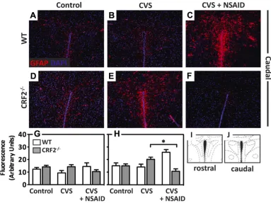
Medial prefrontal cortex: Immunofluorescence of GFAP was performed on sections
from the medial prefrontal cortex (Fig. 4). There was a significant effect [T = 2.048, p =
0.045], whereby CVS increased GFAP selectively in CRF2-/- mice. The CVS-induced
elevation in GFAP protein was prevented with concomitant NSAID treatment. No
differences in GFAP were observed in WT animals across any treatment group.
Serum TNFα
pg/mL (data not shown). No significant differences in TNFα were found between
genotypes [F(1, 38) = 0.02; p = 0.89] or prior exposure to chronic variable stress [F(2, 38) =
0.16; p = 0.85].
DISCUSSION
The aim of the present study was to test the hypothesis that an underlying
component of elevated stress responsivity involves an engagement of neuroimmune
responses during chronic stress exposure. We propose that the ability to detect such
changes requires an appropriately stress-sensitive model. Using CRF2-/- mice as a model
of stress dysregulation, we uncovered several novel findings: 1) NSAID treatment
ameliorated augmented HPA axis corticosterone production characteristic of CRF2-/-
mice exposed to chronic stress; 2) In CRF2-/- mice, GFAP-positive astrocytes increased
with chronic stress exposure in stress coordinating brain regions, paralleling changes in
HPA axis corticosterone; 3) NSAID treatment prevented stress-mediated changes in
GFAP-positive astrocytes.
HPA stress axis activity is a physiological indicator of organismal stress state;
exposure to stress ultimately results in the production of the primary stress hormone,
corticosterone (Whitnall, 1993). Prior exposure to stress can modify HPA axis activity,
augmenting or blunting responsivity to novel stressors (Pfister and King, 1976; Bhatnagar
and Dallman, 1998). While it is necessary to maintain the ability to respond to a novel
stressor, overactive HPA axis activity can be detrimental. We previously reported that
exposure to chronic stress in CRF2-/- mice augmented HPA stress axis corticosterone
study, NSAID treatment during chronic stress completely ameliorated this effect in
CRF2-/- mice. These findings suggest an underlying neuroimmune involvement in the
dysregulated stress response, and may suggest a mechanism by which NSAID treatment
may be efficacious in affective disorder treatment (Muller et al., 2005; Brunello et al.,
2006; Mendlewicz et al., 2006; Muller et al., 2006).
Astrocytes are a promising mediator of the neuroimmune consequence of stress
exposure. In select environments, they can both produce and respond to neuroimmune
effector molecules (reviewed in (Volterra and Meldolesi, 2005)). While numerous
end-feet leave them elegantly poised to respond to and integrate signals within neuronal
networks, perturbations to astrocytes have the potential for widespread impact, including
changes in extracellular ionic homeostasis, local neurotransmitter regulation, and
remodeling of neural circuits (Theodosis and Poulain, 1999; Ullian et al., 2004; Allen and
Barres, 2005; Haydon and Carmignoto, 2006; Schwarz and Bilbo, 2012). Indeed,
alterations in astrocyte structural morphology or function have been associated with
neuropsychiatric illnesses, including depression and schizophrenia (Rajkowska et al.,
1999; Bowley et al., 2002; Toro et al., 2006). Thus, altered astrocyte-mediated plasticity
may explain why CRF2-/- mice are unable to mount an adaptive coping response to stress
exposure (McEuen et al., 2008). Therefore, we examined we examined GFAP
immunoreactivity as a correlate of altered astrocyte function in the PVN, ventral
hippocampus, and prefrontal cortex, stress-coordinating brain regions. Consistent with
our hypothesis, GFAP immunoreactivity increased with chronic stress in CRF2-/- mice
across these three brain regions. These stress-mediated changes were also prevented with
hippocampus was unaffected by stress exposure. As astrocytes are widely heterogeneous
in structure and function, and play distinct roles across different brain regions, these
findings point to a specificity of astrocyte changes that may parallel the specifics of
dysregulation (Kimelberg, 2004; Lee et al., 2006; Cahoy et al., 2008).
The PVN is a primary regulator of corticosterone production and is innervated by
multiple limbic forebrain structures, including the prefrontal cortex and ventral
hippocampus (Herman et al., 2005). These circuits play important roles in determining
stressor intensity and in negative feedback (Jankord and Herman, 2008; Radley and
Sawchenko, 2011). A surprising finding in the PVN was the remarkable increase in
GFAP positive astrocytes in the WT mice exposed to chronic stress and concomitant
NSAID treatment. While the explanation for this effect is not currently known, it
demonstrates important genotypic differences in astrocyte plasticity following stress, and
may suggest a unique interaction of stress and neuroimmune activation in the normal
healthy brain that may be beneficial in stress coping (Munhoz et al., 2006).
CRF receptor-2 is expressed on peripheral immune cells (Lovenberg et al., 1995;
Baigent and Lowry, 2000). Therefore, to ensure genotype effects detected here were not
resultant from inflammatory cytokines produced in the periphery, we measured serum
tumor necrosis factor (TNF)-α in these mice. Levels were below the minimal level of
assay detection (7.7 pg/ml) for all animals. These findings are consistent with studies
using similar models of chronic stress exposure that also failed to evoke a significant
peripheral immune response in the absence of additional immune challenge (d'Audiffret
et al., 2010; Farooq et al., 2012). These data do not preclude the possibility of peripheral
neuroimmune modulation have arisen centrally rather than indirectly through activation
of the peripheral immune system.
Neuroimmune effectors have been clearly documented to activate the stress
response, and cytokine treatment can induce symptoms of depression (Tsagarakis et al.,
1989; van der Meer et al., 1996). Taken together, this suggests a potential role for
immune effectors in stress dysregulation and related phenotypes found in
neuropsychiatric disease. However, results of several investigations examining the
neuroimmune response to stress exposure are conflicting; while in some models stress
drives increases in cytokine production, other models show fewer changes (Bartolomucci
et al., 2003; Reyes et al., 2003; Deak et al., 2005; Hueston et al., 2011; You et al., 2011).
Our studies indicate that these differences may be driven by the sensitivity of the model
utilized. In support of a neuroimmune contribution to the dysregulated stress state,
NSAID treatment prevented dysregulation of HPA stress axis corticosterone secretion in
a model of stress sensitivity. Further, chronic stress resulted in upregulation of GFAP by
astrocytes in the PVN, ventral hippocampus and prefrontal cortex of CRF2-/- mice. The
current studies provide evidence that not only may NSAID treatment be beneficial in the
treatment of disorders associated with dysfunctional HPA stress axis, but that it may do
so through either direct or indirect modification of astrocytes in brain regions involved in
stress regulation. The specificity of these effects to stress-sensitive animals highlights the
importance of utilizing a model of stress dysregulation in the examination of factors that
ACKNOWLEDGEMENTS
We thank J. Fluharty for assistance with animal care and Dr. C. Howerton for assistance
with statistical analysis. These studies were funded by a grant from the NIH MH073030.
FIGURES AND LEGENDS
Figure 2.1 Anti-inflammatory treatment ameliorates chronic stress-induced
corticosterone elevation in stress-sensitive CRF2-/- mice. Corticosterone levels were examined following a 15 min restraint stress (shaded column) in (A) WT and (B) CRF2-/ -mice. Exposure to chronic variable stress (CVS) enhanced corticosterone production in
CRF2-/- mice, and this effect was ameliorated by non-steroidal anti-inflammatory drug (NSAID) treatment. (C) Corticosterone production during the rise phase (0 min - 30 min) was higher in CRF2-/- mice, and was further enhanced during CVS exposure. Concomitant
NSAID treatment during CVS rescued this outcome. (D) NSAID treatment reversed CVS
-induced total AUC of corticosterone production in CRF2-/-mice. Data are presented as mean
Figure 2.2 Anti-inflammatory treatment prevents chronic stress-induced increases in GFAP in the PVN of stress-sensitive CRF2-/- mice. (A-F) Representative immunofluorescence images (10X magnification) of the astrocyte-specific cytoskeletal protein, GFAP (red), counterstained with DAPI (nuclei, blue) in the caudal PVN of (A-C)
atlas images illustrating the brain regions analyzed for (A) ventral and (B) dorsal hippocampus, adapted from the mouse atlas (Paxinos and Franklin, 2003). Boxes highlight region corresponding to image acquisition. (C-H) Representative immunofluorescence images (10X magnification) of GFAP (red) counterstained with DAPI (nuclei, blue) in (C, F) CA1, (D, G) CA3, and (E, H) dentate gyrus of the ventral hippocampus of CVS exposed WT and CRF2-/- mice. (I-K) Bar graphs illustrate semi-quantitative analysis of GFAP immunofluorescence. CVS increased GFAP in both the (I)
CA1 and (J) CA3 subregions of the ventral hippocampus selectively in stress-sensitive
CRF2-/- mice. (K) No differences were observed in the dentate gyrus. (L-Q)
Representative immunofluorescence images of the (L, O) CA1, (M, P) CA3, and (N, Q)
dentate gyrus of the dorsal hippocampus of CVS treated WT and CRF2-/- mice. There
were no significant effects of genotype or treatment on GFAP levels in the (R) CA1, (S)
Figure 2.4 Anti-inflammatory treatment prevents chronic stress-induced increases in GFAP in the medial prefrontal cortex of stress-sensitive CRF2-/- mice. (A-F)
Representative immunofluorescence images (20X magnification) of GFAP (red) counterstained with DAPI (nuclei, blue) in the medial prefrontal cortex (mPFC) of (A-C)
WT and (D-F)CRF2-/- mice for (A, D) control, (B, E) following exposure to CVS, or (C, F) CVS + NSAID. (G) Bar graph illustrates semi-quantitative analysis of GFAP immunofluorescence showing significantly elevated levels in the mPFC following CVS in CRF2-/- mice. Concomitant treatment with NSAID rescued this effect. (H) Atlas image illustrating brain section used for analysis, adapted from the mouse atlas (Paxinos and Franklin, 2003). Data are presented as parameter estimates of the best-fit model + observed standard deviation of respective groups (N = 3-4; *, P < 0.05).