University of Pennsylvania
ScholarlyCommons
Publicly Accessible Penn Dissertations
1-1-2015
The Identification of Novel Mechanisms in
Neuronal Development and Degeneration
Angela Marie Jablonski
University of Pennsylvania, angela.m.jablonski@gmail.com
Follow this and additional works at:
http://repository.upenn.edu/edissertations
Part of the
Biology Commons
,
Molecular Biology Commons
, and the
Neuroscience and
Neurobiology Commons
This paper is posted at ScholarlyCommons.http://repository.upenn.edu/edissertations/1067 For more information, please contactlibraryrepository@pobox.upenn.edu.
Recommended Citation
Jablonski, Angela Marie, "The Identification of Novel Mechanisms in Neuronal Development and Degeneration" (2015).Publicly Accessible Penn Dissertations. 1067.
The Identification of Novel Mechanisms in Neuronal Development and
Degeneration
Abstract
The goal of this dissertation is to further understand two key, broad processes which occur over the course of a neuron's lifetime: its development and possible degeneration in disease. We identify novel components in both of these processes and attempt to understand the functional significance as well as the mechanism each component uses to exert its effects.
We begin with work done focusing on how the neuron's dendritic tree develops. The development of neurons has two phases: (1) a first phase relying on a genetic program and (2) a second phase that uses synaptic activity to guide the fine tuning of connections. We are primarily interested in understanding how neurons develop within the motor system using cues from synaptic activity. One type of activity-dependent
development in the motor system is driven by AMPA receptors assembled with the GluA1 subunit using the scaffolding protein, SAP97. In Chapter 2, we describe our finding that the small protein, CRIPT, functions in the development of the neuron's dendritic tree within the motor system. We show that CRIPT is expressed during the time in development when activity-dependent remodeling is occurring the in spinal cord. Additionally, we show that CRIPT binds to the PDZ3 domain of SAP97 using its C-terminus. Finally, we show that CRIPT is necessary for proper dendritic growth of the motor system and normal motor system responses in vivo.
There are a number of diseases that specifically target motor neurons. One adult-onset disease is Amyotrophic Lateral Sclerosis (ALS). Like many neurodegenerative diseases, it is a multifactorial disease and a contributing factor is the lack of proper protein quality control and the formation of large protein aggregates within motor neurons. In Chapter 3, we focus on how endoplasmic reticulum-associated degradation (ERAD) components might affect mutant proteins in ALS and their toxicity. ERAD is responsible for maintaining the proper folding of proteins that are to be trafficked to the cell surface or secreted from the cell. Although many proteins associated with ALS are not ERAD substrates, a number of ERAD components have been found to interact with ALS-causing mutant proteins and have been found to be mutated in forms of familial ALS. We identify new modifiers within the ERAD pathway of the toxicity of two ALS-linked proteins, mutant TDP-43 and mutant SOD1. We focus on the mechanism of one suppressor of ALS-linked proteotoxicity, the loss of RAD-23. The levels of RAD23 are increased in ALS models and reducing RAD-23 levels can suppress
phenotypes in ALS models, as well as suppress motor neuron toxicity of the mutant proteins. We further show that reduced RAD-23 is able to accelerate the turnover of mutant proteins associated with ALS. Finally, we show that there is a mislocalization of RAD-23 protein and increased RAD-23 protein expression in post mortem spinal cord tissue of ALS patients, suggesting that RAD-23 may be a new novel target in the treatment of ALS.
Collectively, this work identifies and describes novel proteins and mechanisms involved in the development and pathobiology of motor neurons.
Degree Type
Dissertation
Degree Name
Doctor of Philosophy (PhD)
Graduate Group
Neuroscience
First Advisor
Robert G. Kalb
Keywords
ALS, CRIPT, dendrite, development, neurodegeneration, RAD-23
Subject Categories
Biology | Molecular Biology | Neuroscience and Neurobiology
THE IDENTIFICATION OF NOVEL MECHANISMS IN NEURONAL DEVELOPMENT
AND DEGENERATION
Angela Marie Jablonski
A DISSERTATION
in
Neuroscience
Presented to the Faculties of the University of Pennsylvania
in
Partial Fulfillment of the Requirements for the
Degree of Doctor of Philosophy
2015
Supervisor of Dissertation
Signature__________________________
Dr. Robert G. Kalb, M.D.
Professor of Neurology, Perelman School of Medicine at the University of Pennsylvania
Graduate Group Chairperson
Signature__________________________
Dr. Joshua Gold, Ph.D.
Professor of Neuroscience, Perelman School of Medicine at the University of Pennsylvania
Dissertation Committee
Dr. Todd Lamitina, Ph.D. – Visiting Associate Professor of Pediatrics and Cell Biology
Dr. Harry Ischiropoulos, Ph.D - Research Professor of Pediatrics and Systems Pharmacology and
Translational Therapeutics
Dr. Kelly Jordan-Sciutto, Ph.D. - Chair and Professor of Pathology
THE IDENTIFICATION OF NOVEL MECHANISMS IN NEURONAL DEVELOPMENT
AND DEGENERATION
COPYRIGHT
2015
ANGELA MARIE JABLONSKI
This work is licensed under the Creative Commons Attribution- NonCommercial-ShareAlike 3.0 License
To view a copy of this license, visit
iii
DEDICATION
This dissertation is dedicated to the memory of my parents, Frank and Margaret
Jablonski. I would have not reached this point without them. They are and will continue
to be missed every single day.
“What though the radiance which was once so bright
Be now for ever taken from my sight,
Though nothing can bring back the hour
Of splendor in the grass, of glory in the flower
We will grieve not, rather find
Strength in what remains behind;
In the primal sympathy
Which having been must ever be;
In the soothing thoughts that spring
Out of human suffering;
In the faith that looks through death,
In years that bring the philosophic mind.”
iv
ACKNOWLEDGMENT
First and foremost, I would like to thank my advisor, Robert Kalb, for his
guidance, training, and words of wisdom throughout my time here. He knew how to
provide me with the right amount of guidance, while also giving me the needed
independence to develop as a scientist. I am grateful for the countless number of hours
he has spent filling my head with new information. His inspiring enthusiasm for science
has helped me many times throughout my time as a graduate student. Bob, thank you
for helping me develop as a scientist, listening to my crazy ideas (and letting me listen to
yours), and for being so supportive and pushing me to always be better. I am also
grateful to all of the previous and current members of the Kalb lab for providing a
wonderful environment to work in every day.
The projects described in this dissertation would have not been possible without
many collaborators. I would like to specifically acknowledge Brian Kraemer, Jiou Wang,
Hannes Lans, and Lyle Ostow for not only the work that they contributed, but also for
useful conversations. Thank you for making this work so much stronger than it would
have been without you.
I’d also like to thank all of the members of my thesis committee including Todd
Lamitina, Harry Ischiropoulos, Kelly Jordan-Sciutto, and Rick Morimoto for their time and
support. I appreciate the hours that they spent with me in meetings – not only helping
the scientific aspect of my work, but also helping me progress and grow into an
independent researcher and developing further as a person. Thank you for always
answering my questions, meeting with me whenever I needed it, helping me to focus,
v
I am also grateful to the outstanding Neuroscience Graduate Group at the
University of Pennsylvania. This includes the administrators, faculty, and students. I
want to specifically thank Mikey Nusbaum, Rita Balice-Gordon, Josh Gold, and Jane
Hoshi who always seem to create the time necessary to keep the program running and
be tremendously supportive. Thank you to all of you for creating such a fantastic,
challenging, and supportive graduate program. I would also like to acknowledge the
excellent Neuroscience program at Muhlenberg College where I completed my
undergraduate studies. I would like to especially thank my undergraduate advisor,
Jeremy Teissere, who did not only introduce me to neuroscience, research, and take me
to my first Society of Neuroscience conference, but also has remained as an advisor to
me throughout my time as a graduate student. Despite having an increasing number of
students every year, he has always found the time to be tremendously supportive.
Jeremy, accept this tremendous thank you from the bottom of my heart. I still remember
when you wrote “Ph.D.” down when I told you about my interests and declared
Neuroscience as a major. Who knew we would eventually be here?
I also want to thank all of my friends and family, the people I’ve known for
decades and all of the new lifelong friends I made throughout this process. Thank you
for keeping me grounded, making me laugh, and commiserating with me as needed.
There are just too many to name, you all know who you are. Thank you for the G-chats,
listening to me over many happy hours and dinners, and always being there for me.
Unfortunately, there are so many people who have gotten me to this point, but
cannot be physically here to see the final product and celebration. I am extremely
grateful to the family members who supported me, but could not be here to see this
milestone. I want to specifically acknowledge my mom, Peggy Jablonski. I could not
vi
mom, for teaching me that anything is possible with a little bit of hard work and
sacrificing every day for me. I know how happy you were to hear I was done, and you
will still be here for the big day in your own way. I also am grateful to my father, Frank
Jablonski. Dad, I can still hear you saying: “if you end up at a place like Penn, you better
be ready to work hard.” I have never stopped hearing your voice say that since you’ve
been gone, and it has kept me going. I’m also grateful to my grandmothers – Lottie
Jablonski and Tushie Burns – two more women who modeled how to remain strong in
times of adversity. You will all always be in my heart, and so will home and Shenandoah.
Last, but not least, I wish to thank my husband, David. I don’t know how I would
have done the past five years without him. I appreciate the support he gave me
throughout my training and the sacrifices he made over the past five years. He has been
the one to keep our lives running when I was unable (literally). He has unconditionally
supported me when I needed someone there. He was always there for the good and the
bad (not just the bad, the absolute worst), and was always there to listen to me. I will
never be able to repay him for how much he truly helped me throughout my time in
graduate school. David, you have accepted me completely as a person since the day we
met. I am so grateful I found someone I could always be my crazy self with. It’s a
privilege to stand beside you in this journey and in life. I love you, always.
Throughout my training, I was supported by the NIH Systems Integrative Biology
vii
ABSTRACT
THE IDENTIFICATION OF NOVEL MECHANISMS IN NEURONAL DEVELOPMENT
AND DEGENERAITON
Angela Marie Jablonski
Dr. Robert Gordon Kalb, M.D.
The goal of this dissertation is to further understand two key, broad processes
which occur over the course of a neuron’s lifetime: its development and possible
degeneration in disease. We identify novel components in both of these processes and
attempt to understand the functional significance as well as the mechanism each
component uses to exert its effects.
We begin with work done focusing on how the neuron’s dendritic tree develops.
The development of neurons has two phases: (1) a first phase relying on a genetic
program and (2) a second phase that uses synaptic activity to guide the fine tuning of
connections. We are primarily interested in understanding how neurons develop within
the motor system using cues from synaptic activity. One type of activity-dependent
development in the motor system is driven by AMPA receptors assembled with the
GluA1 subunit using the scaffolding protein, SAP97. In Chapter 2, we describe our
finding that the small protein, CRIPT, functions in the development of the neuron’s
dendritic tree within the motor system. We show that CRIPT is expressed during the time
in development when activity-dependent remodeling is occurring the in spinal cord.
C-viii
terminus. Finally, we show that CRIPT is necessary for proper dendritic growth of the
motor system and normal motor system responses in vivo.
There are a number of diseases that specifically target motor neurons. One
adult-onset disease is Amyotrophic Lateral Sclerosis (ALS). Like many
neurodegenerative diseases, it is a multifactorial disease and a contributing factor is the
lack of proper protein quality control and the formation of large protein aggregates within
motor neurons. In Chapter 3, we focus on how endoplasmic reticulum-associated
degradation (ERAD) components might affect mutant proteins in ALS and their toxicity.
ERAD is responsible for maintaining the proper folding of proteins that are to be
trafficked to the cell surface or secreted from the cell. Although many proteins
associated with ALS are not ERAD substrates, a number of ERAD components have
been found to interact with ALS-causing mutant proteins and have been found to be
mutated in forms of familial ALS. We identify new modifiers within the ERAD pathway of
the toxicity of two ALS-linked proteins, mutant TDP-43 and mutant SOD1. We focus on
the mechanism of one suppressor of ALS-linked proteotoxicity, the loss of RAD-23. The
levels of RAD23 are increased in ALS models and reducing RAD-23 levels can suppress
phenotypes in ALS models, as well as suppress motor neuron toxicity of the mutant
proteins. We further show that reduced RAD-23 is able to accelerate the turnover of
mutant proteins associated with ALS. Finally, we show that there is a mislocalization of
RAD-23 protein and increased RAD-23 protein expression in post mortem spinal cord
tissue of ALS patients, suggesting that RAD-23 may be a new novel target in the
treatment of ALS.
Collectively, this work identifies and describes novel proteins and mechanisms
ix
TABLE OF CONTENTS
DEDICATION ... III
ACKNOWLEDGMENT ... IV
ABSTRACT ... VII
LIST OF TABLES ... XII
LIST OF FIGURES ... XIII
CHAPTER 1: INTRODUCTION ... 1
NEURONAL DEVELOPMENT ... 1
ACTIVITY-DEPENDENT DEVELOPMENT ... 1
GLUR1 AND SAP97 PROMOTE DENDRITE GROWTH ... 5
CRIPT: A CANDIDATE EFFECTOR FOR GLUA1-DEPENDENT DENDRITE GROWTH ... 9
MOTOR NEURON DISEASE: AMYOTROPHIC LATERAL SCLEROSIS ... 10
GENETIC MODELS OF MOTOR NEURON DISEASE ... 12
ENDOPLASMIC REICULUM-ASSOCIATED DEGRADATION AND ALS ... 15
RAD-23: A MULTI-FUNCTIONAL PROTEIN... 18
STATEMENT OF MOTIVATION AND HYPOTHESES ... 21
CHAPTER 2: IDENTIFYING THE DOWNSTREAM MACHINERY OF GLUA1
AND SAP97 IN DENDRITE DEVELOPMENT ... 23
SUMMARY ... 23
INTRODUCTION ... 24
MATERIALS AND METHODS ... 26
RESULTS ... 35
x
FIGURE LEGENDS ... 47
CHAPTER 3: LOSS OF RAD-23 PROTECTS AGAINST MODELS OF MOTOR
NEURON DISEASE BY ENHANCING MUTANT PROTEIN DEGRADATION .. 59
SUMMARY ... 59
INTRODUCTION ... 60
MATERIALS AND METHODS ... 62
RESULTS ... 74
DISCUSSION ... 88
TABLE LEGENDS ... 91
FIGURE LEGENDS ... 93
CHAPTER 4: GENERAL CONCLUSIONS AND FUTURE DIRECTIONS ... 121
ACTIVITY-DEPENDENT DEVELOPMENT ... 121
THE IDENTIFICATION OF OTHER MEDIATORS OF GLUA1-MEDIATED DENDRITE GROWTH ... 123
UNRESOLVED QUESTIONS REGARDING CRIPT’S ROLE IN DENDRITE GROWTH ... 124
THE IMPORTANCE OF ERAD COMPONENTS IN ALS ... 125
TARGETING RAD-23 IN NEURODEGENERATIVE DISEASE ... 128
HOW REDUCED RAD-23 ACCELERATES MUTANT PROTEIN TURNOVER AND MITIGATES TOXICITY ... 131
LOSS OF RAD-23 AS A PROTECTOR AGAINST PROTEOTOXICITY ... 133
A LINK BETWEEN NUCLEOTIDIE EXCISION REPAIR AND NEURODEGENERATIVE DISEASE ... 135
FINAL REMARKS ... 137
APPENDIX 1: OTHER COMPONENTS IN THE GLUA1/SAP97/CRIPT
COMPLEX TO PROMOTE DENDRITE GROWTH ... 138
SUMMARY ... 138
INTRODUCTION ... 138
xi
RESULTS ... 143
DISCUSSION ... 145
FIGURE LEGENDS ... 147
APPENDIX 2: OTHER MODIFIERS OF MOTOR NEURON DISEASE MODELS
... 151
SUMMARY ... 151
INTRODUCTION ... 151
MATERIALS AND METHODS ... 154
RESULTS ... 157
FIGURE LEGENDS ... 160
xii
LIST OF TABLES
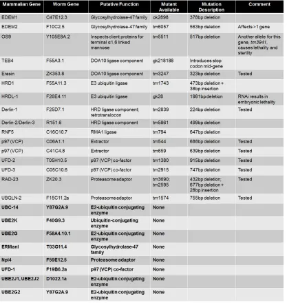
Table 3.1. Mammalian ERAD genes and their predicted orthologs in C. elegans. 102
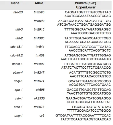
Table 3.2. List of primers used in genotyping C. elegans strains. ... 103
Table 3.3. A list of the C. elegans strains used in this study. ... 104
Table 3.4. Description of human ALS cases used for staining and expression analysis. ... 118
Table 3.5. Description of control cases used for staining and expression analysis. ... 119
xiii
LIST OF FIGURES
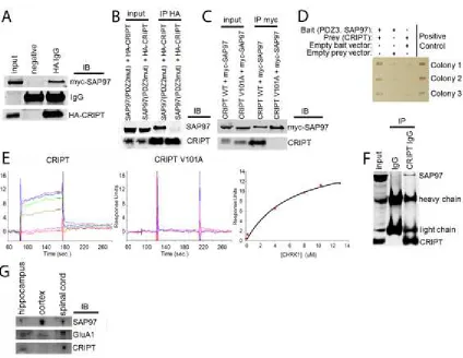
Figure 2.1. CRIPT is bona fide PDZ3 and SAP97 interacting protein. ...51
Figure 2.2. CRIPT is necessary for the dendritic growth of spinal cord neurons. .52
Figure 2.3. CRIPT is not sufficient to promote dendritic growth of spinal cord neurons. ...53
Figure 2.4. CRIPT functions downstream of SAP97 in promoting dendrite growth. ...54
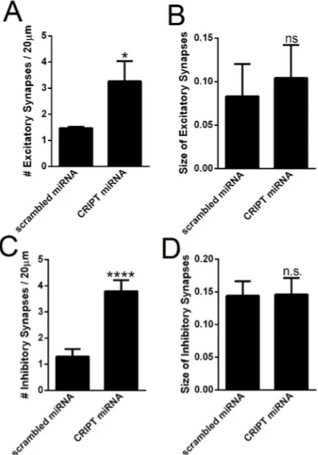
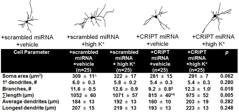
Figure 2.5. CRIPT knockdown increases the number of excitatory and inhibitory synapses, but does not change synapse size. ...55
Figure 2.6. CRIPT’s function in dendritic growth is independent from synaptic activity. ...56
Figure 2.7. Loss of cript decrease dendrite branch number in vivo. ...57
Figure 2.8. Loss of cript results in a mechanosensory defect in vivo in young adults. ...58
Figure 3.1. Locomotor defects of C. elegans models of ALS and their modification by the loss of ERAD and UPS genes. ... 106
Figure 3.2. Loss of rad-23 in C. elegans protects against models of ALS via an effect in the nervous system. ... 107
Figure 3.3. Loss of rad-23 in C. elegans protects against neurodegeneration following expression of mutTDP-43 in vivo. ... 108
Figure 3.4. RAD-23 protein is expressed throughout the worm, including the worm nervous system. ... 109
Figure 3.5. Loss of RAD-23 in C. elegans protects against aging decline and proteotoxicity. ... 110
Figure 3.6. Loss of rad-23 does not suppress the C. elegans mutTDP-43 locomotor deficit via known pathways. ... 111
Figure 3.7. Knockdown of RAD-23 orthologs in mammalian motor neurons
protects against toxicity of mutSOD1 or mutTDP-43. ... 112
Figure 3.8. hR23A expression is increased in the spinal cord of mutSOD1 mice at P90 and P120. ... 113
Figure 3.9. Manipulations of RAD-23 expression change TDP-43 and SOD1
xiv
Figure 3.10. Loss of RAD-23 accelerates turnover through the proteasome and autophagy by increasing ubiquitination. ... 115
Figure 3.11. Loss of rad-23 reduces TDP-43 and SOD1 insolubility. ... 116
Figure 3.12. hR23A and hR23B are aberrantly expressed in human ALS tissue. . 117
Figure A1.1. Association of GluA1 with SAP97 is activity-dependent. ... 149
Figure A1.2. Successful tool development to identify new SAP97 PDZ3 domain binding partners in neurons. ... 150
Figure A2.1. Overexpression of wild type (wt-) and mutant (mut-) TDP-43 (M337V) in the nervous system causes an accumulation of normally degraded
ubiquitinated substrates in a non-cell autonomous manner. ... 162
Figure A2.2. Knockdown of ufd-3, but not rad-23, in C. elegans reduces
ubiquitinated GFP abundance in mutTDP-43 background. ... 163
Figure A2.3. Expression of constitutively spliced XBP1 in the nervous system enhances toxicity of ER stress. ... 164
Figure A2.4. Stabilization of p53 by treatment with tenovin-1 protects motor
neurons from toxicity caused by mutSOD1 (G85R). ... 165
Figure A2.5. Only ubiquitous knockdown of ubql-1 enhances the mutSOD
locomotor deficit. ... 166
I
CHAPTER 1: INTRODUCTION
This thesis attempts to understand two major events in the life of the nervous
system: the development of the nervous system and its possible degeneration, with a
key emphases on the motor system. Here, I will outline the critical information needed to
understand the progress made in these two areas.
NEURONAL DEVELOPMENT
The development of the nervous system is roughly divided into two parts: (1) an
activity-independent phase outlined by a genetic program and (2) an activity-dependent
phase. Together, these phases produce the adult nervous system with the correct
connectivity and physiology to produce meaningful behavior. This work primarily focuses
on the second phase and the role of activity-dependent development.
ACTIVITY-DEPENDENT DEVELOPMENT
Activity-dependent development during pre- and postnatal life is an important
mechanism for the specification of synaptic phenotype and connectivity. Here, we will
review some of the key experiments undertaken in the visual system that highlight the
cell biological processes underlying activity-dependent development. These introductory
remarks provide the context for thinking about the role of activity-dependent processes
in motor system development.
In the mammalian visual system, retinal ganglion cells (RGC) project to the
lateral geniculate nucleus (LGN) of the thalamus. Thalamocortical (TC) connections from
2
segregated from each other in both the LGN and visual cortex (Katz and Shatz, 1996;
Mooney et al., 1996). For example, in adult cats and primates, TC afferents projecting to
layer four of the visual cortex are organized into discrete right and left eye patches,
referred to as ocular dominance columns (OCD) (Shatz, 1996). This circuitry
arrangement subserves high acuity vision (Butts et al., 2007).
In their classic work, Hubel and Weisel showed that this pattern of visual cortex
innervation is not present at birth, as right and left eye afferents demonstrate extensive
overlap (Hubel et al., 1977). Most remarkably, they showed that the segregation of right
and left eye afferents is driven by visual experience (Hubel et al., 1977). Monocular
deprivation of one eye (by suturing the eyelid shut) during a discrete period in early
postnatal life leads to a dramatic shift in TC innervation of the visual cortex (Hubel et al.,
1977). The afferents from the nondeprived eye innervate a larger territory of the visual
cortex, while those from the deprived eye innervate a smaller territory (Hubel et al.,
1977). This experience-dependent shift in ocular dominance leaves a permanent imprint
on visual system organization (Hubel et al., 1977).
How does environmentally evoked synaptic activity lead to changes in synaptic
strength and connectivity? Substantial evidence supports the view that synapses will
undergo strengthening and stabilization when the activity of pre- and postsynaptic
elements is coincident. This model of synaptic plasticity was originally suggested in
theoretical work by Daniel Hebb and has been most rigorously tested in long-term
potentiation (LTP) paradigms (Hebb, 1949). Many forms of LTP depend upon the
activation of N-methyl-d-aspartate receptors (NMDA-Rs). NMDA-Rs are believed to be
3
postsynaptic elements (Seeburg et al., 1995). The ionic mechanism underlying this
process has been linked to the voltage-dependent block of NMDA-Rs by Mg2+ (Shaywitz
and Greenberg, 1999). Patterned afferent input, sufficient to remove the Mg2+ block,
allows NMDA-Rs to conduct Ca2+ influx, which inactivates several protein kinases,
including Ca2+/calmodulin-dependent protein kinase II (CamKII) (Seeburg et al., 1995).
CamKII is necessary for the maintenance of LTP and phosphorylation of
2-amino-3-(3-hydroxy-5-methyl-isoxazol-4-yl) propanoic acid receptor (AMPA-R) subunits to increase
their conductance (Shaywitz and Greenberg, 1999). These and other calcium-activated
processes drive the observed synaptic plasticity (Shaywtiz and Greenberg, 1999; Luthi
et al., 2004).
In the visual system, activity-dependent processes drive large-scale alterations in
the architecture of axons and dendrites. How do activity-dependent changes in synapses
control the growth and distribution of axons and dendrites? Vaughn proposed the
synaptotropic hypothesis: dendritic branches are formed near active synapses and
synapse stabilization consequently stabilizes dendrites (Vaughn, 1989). Original in vivo
work from the Haas lab implicates beta-neurexin (NRX) and neuroligin-1 (NLG1) in this
process, in which NMDA-R–dependent synapse maturation was required for persistent
NRX-NLG1 function in dendritogenesis (Chen et al., 2010). Blocking synaptogenesis
thereby blocks dendrite outgrowth stabilization. The linkage of synaptic plasticity to
neurite architecture is a fundamental principle in developmental neuroscience and
provides an explanation for earlier observations that the size and complexity of the
dendrite tree controls the qualitative and quantitative nature of the afferent input
(Oswald, 1989). In studies of rabbit ciliary ganglia, the number of ganglion cell primary
4
1981). This is not true of some of the cells in the neonate, where the initial set of inputs
is confined to the cell body, allowing only one axon to survive (Hume and Purves, 1981).
It is hypothesized that the complexity of some cells allows for a higher number of
afferents to innervate the ganglion cell (Hume and Purves, 1981). Thus, a
competition-based model of synapse formation holds for dendrite growth.
While many studies implicate AMPA-Rs in the control of dendrite growth, a
consistent picture has yet to emerge. Blocking AMPAergic transmission in retinotectal
neurons decreases synapse stabilization, and subsequent dendrite growth and
stabilization (Haas et al., 2006). Conversely, Casticas et al. (2011) showed that
enhanced conductance of Ca2+-permeable AMPA-Rs inhibited neurite outgrowth in
dissociated chick retinal neurons (Casticas et al., 2001). Outside of the visual system,
blockade of AMPA-Rs in chick motoneurons has also been seen to increase dendritic
outgrowth in chick motoneurons, but only at certain time points in embryonic
development (Ni and Maratin-Caraballo, 2010). It is unclear what role NMDA-Rs played
in these processes because blocking AMPA-Rs will prevent NMDA-R activation.
In the central nervous system (CNS), it is understood that activation of AMPA-Rs,
sufficient to relieve the voltage-dependent magnesium block of NMDA-Rs, drives
activity-dependent plasticity, synaptic stabilization, and patterned innervation (Peng et
al., 2009). Less understood is the extent to which NMDA-R–independent mechanisms
can drive activity-dependent developmental processes. Next, we describe work in the
spinal cord showing how AMPA-Rs assembled with the GluA1 subunit can promote
5
GLUR1 AND SAP97 PROMOTE DENDRITE GROWTH
Neonatal motor neurons express a very high level of GluA1 (both mRNA and
protein). GluA1’s properties can be modified by alternative splicing and editing at the
glutamine/arginine “Q/R” site. The GluA1 expressed during this developmental period
contains the “flip” alternatively spliced exon and is unedited in the Q/R site (Jakowec et
al., 1995a; Jakowec et al., 1995b). Previous work has shown that neonatal motor
neurons express Ca2+ permeable AMPA receptors (as one would expect if they were
enriched with GluA1(Q)) (Jakowec et al., 1995b).Taken together with the
electrophysiological data, it suggests that many AMPA-R are assembled of GluA1
homomers at this point in development in motor neurons.
The unusually high level of GluA1 expression by neonatal motor neurons raises
the possibility that AMPA receptors assembled with GluA1 play a special role in
activity-dependent motor system development. To examine this notion, we began by asking
whether manipulation of GluA1 influenced spinal neuron dendritic architecture. Several
approaches were taken. First, we found that knockdown of GluA1 expression inhibited
dendrite growth. Conversely, overexpression of GluA1 in spinal neurons in vitro
stimulated dendritic growth; this growth effect was blocked by the AMPA-R antagonist,
CNQX (6-cyano-7-nitroquinoxaline-2, 3-dione) (Zhang et al., 2008). Second, we
compared the effects of two types of overexpressed GluA1 in motor neurons in vivo. We
used a version that robustly passes current (GluA1(Q)) and compared that with a version
that passes very little current (GluA1(R)). Only overexpressed GluA1(Q) stimulated
dendritic branching (Jeong et al., 2006). These results suggest: 1) that the activity of
AMPA-R assembled with GluA1 is a crucial step for dendrite growth and 2) that this
6
permeability of AMPA-R assembled with GluA1 controls the dendritic growth process
(Jeong et al., 2006).
One interpretation of the above results is that overexpression of GluA1 enhances
neuronal depolarization, thereby promoting NMDA-R mediated events. We think this is
not true for a number of reasons. First, our in vivo observations were made in juvenile
rodents at a time when motor neurons do not express NMDA-R (Steganga and Kalb,
2001). It is possible that prior in situ hybridization and immunohistological studies were
insufficiently sensitive to detect NMDA-R in juvenile motor neurons. To address this
possibility, we expressed GluA1(Q) in juvenile animals and simultaneously treated them
with the NMDA-R antagonist, MK-801 (Inglils et al., 2002). We know that MK-801 was
administered in an effective dose because LTP could not be evoked in these animals
(Inglis et al., 2002). Nonetheless, MK-801 did not block the pro-dendrite growth actions
of overexpressed GluA1(Q) (Inglis et al., 2002). Second, we undertook in vitro
pharmacological studies. Administration of MK-801 did not block the dendrite growth
promoting actions of GluA1(Q). In contrast, administration of the L-type calcium channel
blocker, nifedipine, did block the GluA1(Q) effect (Kalb et al., 2013). Taken together, all
of these results suggest that GluA1 is sufficient to promote dendrite growth in an
NMDA-R-independent manner.
The work described above primarily focuses on the effects of GluA1 on dendrite
architecture in vitro. What about in vivo? To address this question, studies of the GluA1-/-
mouse have been informative (Zhang et al., 2008). Analysis of the dendritic tree
revealed that motor neurons from GluA1-/- animals are smaller and less branched at P10
and P23 (Zhang et al., 2008).This suggests that GluA1-/- motor neurons develop over a
7
size and complexity of the motor neuron dendrite tree effect motor circuitry and behavior
of the animal? To study the innervation of motor neurons within the segmental spinal
cord, a recombinant Pseudorabies virus engineered to express green fluorescence
protein (PRV-GFP) was employed. PRV-GFP labeling experiments revealed a distinct
pattern of interneuronal connectivity in the spinal cord of GluA1-/- mice in comparison to
WT mice. The greatest difference between genotypes was found in the number of
contralaterally located interneurons, especially in Rexed’s lamina VIII (Zhang et al.,
2008). The stunted dendrite tree and change in interneuron connectivity correlate with a
locomotor defect in the GluA1-/- animals. In comparison to WT counterparts, GluA1
-/-mice showed poorer performance in grip strength, treadmill, and rotarod at P23 and
adulthood (Zhang et al., 2008). This suggests that GluA1 is not only important for
dendrite growth, but also for patterning segmental spinal cord circuitry and motor
behavior. Furthermore, changes in development during the postnatal period lead to
deficits throughout adulthood (Zhang et al., 2008).
By what molecular mechanism does the activity of AMPA-R assembled with
GluA1 control the morphology of motor neuron dendritic architecture? A series of
experiments have indicated that the multi-domain scaffolding protein,
synapse-associated protein of 97 kDa molecular weight (SAP97), interacts with GluA1 and plays
a key role in this process (Zhou et al., 2008). The C-terminal seven amino acids of
GluA1 physically interact with the second PDZ domain of SAP97.
AMPA-R physiology and synaptic plasticity is entirely normal when the physical
interaction between GluA1 and SAP97 is disrupted (Kim et al., 2005). This was
demonstrated using mice in which the wild type allele of GluA1 was replaced by a
8
SAP97 (GluA1∆7 mice) (Kim et al., 2005). Contrary to expectations, GluA1 also traffics
normally to the cell surface in these mice, suggesting GluA1 chaperones SAP97 to
synapses – not the vice versa. This was demonstrated further using biochemical and
imaging methodologies (Kim et al., 2005). However, normal elaboration of motor neuron
dendrites requires SAP97. This was demonstrated in vitro: knockdown of SAP97
decreases the total size of the dendritic tree and prevents the pro-dendrite growth effect
of GluA1 overexpression (Kim et al., 2005) This was further confirmed in vivo – a smaller
dendritic tree was found in the GluA1∆7 mice (where SAP97 does not traffic to the cell
surface) as well as in mice with a conditional deletion of SAP97 from motor neurons
(Kim et al., 2005). Finally, co-overexpression of GluA1 and SAP97 in vitro has a
synergistic pro-dendrite growth effect; this only depends on their co-localization at the
plasma membrane, not their physical association (Kim et al., 2005). This was
demonstrated using a membrane-targeted version of SAP97 in conjunction with
GluA1∆7 or a membrane-targeted version of SAP97 with mutations in its PDZ2 domain
that make it incapable of binding GluA1 (Kim et al., 2005).
Together, these experiments suggest that the endogenous GluA1 and SAP97
complex is the necessary platform upon which GluA1-containg AMPA-R activity is
translated into a signal(s) that stimulates dendritic growth and branchingin the spinal
cord.
How do GluA1 and SAP97 work together to promote dendrite growth? One
hypothesis is that GluA1 and SAP97 help form the assembly of a multi-protein complex
during GluA1-containing AMPA-R activity to activate downstream effectors capable or
stimulating dendritic growth. At this time, the molecular mechanism by which SAP97 and
9
evidence that the PDZ3 domain of SAP97 is crucial for these pro-growth effects by
SAP97 and GluA1 (unpublished observations). Overexpression of SAP97 with a
mutation in the PDZ3 domain no longer increases dendrite growth. Thus, it is logical to
ask whether proteins that bind to the PDZ3 domain of SAP97 are part of the machinery
to translate activity into growth. This is one current avenue for research and key
downstream players in the GluA1-mediated process are beginning to be identified. One
protein identified in this thesis work is CRIPT.
CRIPT: A CANDIDATE EFFECTOR FOR GLUA1-DEPENDENT DENDRITE GROWTH
CRIPT (cysteine-rich-interactor-of-PDZ-three) was originally identified as a small
12 kDa protein that was localized to excitatory synapses. It is believed to be a
crosslinking proteins between proteins in the post-synaptic density (i.e. PSD-95) and
microtubules because it could be co-immunoprecipitated with tubulin and PSD-95
(Niethammer et al., 1998; Passafaro et al., 1999). It could also be co-immunoprecpitated
with NR2B and Chapsyn-110 from synaptosomal preparations (Niethammer et al.,
1998). Furthermore, overexpression of CRIPT in heterologous cells was shown to cause
a redistribution of PSD-95 to microtubules (Niethammer et al., 1998).
We chose to focus on CRIPT as a possible mediator of GluA1- and
SAP97-driven dendrite growth because it was found to selectively bind to the third PDZ domain
(PDZ3) of PSD-95 previously. CRIPT contains a canonical PDZ-binding sequence at its
terminus (-QTSV). Furthermore, CRIPT is conserved from mammals to plants (i.e.
human and Arabidopsis thaliana CRIPT share 78% similarity in their amino acid
sequences), suggesting an important functionality (Niethammer et al., 1998). CRIPT was
10
of plasticity, such as the hippocampus (Niethammer et al., 1998). Furthermore, it is likely
that a protein involved in dendritic remodeling would interact with the cytoskeleton, such
as microtubules, which CRIPT has been found to do (Niethammer et al., 1998).
Up until now, no definitive function of CRIPT has been established. For example,
although interference of CRIPT’s interaction with PSD-95 disrupts PSD-95’s localization
with microtubules (Passafaro et al., 1999), it had no effect on the clustering or number of
NMDA-R. This work, therefore, proposes a novel function for CRIPT in the dendrite
growth of spinal cord neurons. This work can be found in Chapter 2.
MOTOR NEURON DISEASE: AMYOTROPHIC LATERAL SCLEROSIS
A number of neurodegenerative diseases are known to selectively target the
upper motor neurons in the cortex and lower motor neurons in the brainstem and spinal
cord. These diseases are known as motor neuron diseases (MND), but it is still not
completely understood why these diseases target this subset of neurons.
The disease we focused on in this work is Amyotrophic Lateral Sclerosis (ALS),
popularly referred to in the United States as Lou Gehrig’s disease. ALS is commonly
classified into two subtypes: (1) familial (fALS) and (2) sporadic (sALS), depending
respectively on if the disease is or is not caused by a single genetic and inherited
mutation. Familial ALS accounts for roughly 5-10% of cases and has been linked to a
number of gene mutations. The most prevalent of these is the C9ORF72 mutation,
although new mutations continue to be found. The disease tends to present itself earlier
than some other neurodegenerative diseases, anywhere between 50 to 75 years of age,
and disease progression lasts for three years on average. The disease has a worldwide
11
the disease than women (1.2-1.5:1 ratio) (Cronin et al., 2007). The disease can
sometimes be associated with non-motor systems in a form of frontotemporal dementia
(FTD). Postmortem tissue from both ALS and FTD patients reveals the presence of
protein aggregates, strongly supporting the existence of a pathophysiological continuum
between these two disorders (Forman et al., 2004). In fact, it is estimated that 15% of
FTD patients meet the criteria to be diagnosed with ALS (Ringholz et al., 2005). The
prevalence of protein aggregates within motor neurons in both sporadic and familial ALS
tissue also points to a lack of protein quality control in the disorder, which is a major
focus of the work discussed in this dissertation.
Patients with ALS exhibit progressive neuromuscular weakness, atrophy, and
eventual death. Most patients will die from respiratory failure. The biggest motivation for
studying the disease is that it has no known cure. The only drug known to modify the
course of ALS is Riluzole, a drug that is thought to act by reducing the burden of
glutamate excitotoxicity, but only achieves a modest improvement in survival (Miller et al,
2007). The lack of treatment is in part due to the multifactorial nature of the disease.
Contributing pathogenic processes include glutamate excitotoxicity, protein misfolding,
inflammation, oxidative stress, endoplasmic reticulum (ER) stress, mitochondrial
dysfunction, impaired axonal transport, impaired endosomal trafficking, as well as
dysregulated transcription and RNA processing (Ferraiuolo et al., 2011; Rothstein,
2009).
Due to the lack of promising treatment, there is an urgent need for ALS research.
One of the obstacles in studying the disease is that most of the cases are sporadic and
most research done in the laboratory to model and study the disease utilizes genetic
12
GENETIC MODELS OF MOTOR NEURON DISEASE
In order to identify new modifiers of motor neuron disease, we utilized several
models of motor neuron disease in the small genetically tractable nematode,
Caenorhabditis elegans (C. elegans). Some of the mutations found in fALS include
mutations in copper-zinc superoxide dismutase 1 (SOD1), TAR DNA-binding protein of
43 kDa molecular weight (TDP-43), fused in sarcoma (FUS), vesicle-associated
membrane protein-associated protein B (VAPB), ubiquilin-2 (UBQLN2), and many more
(Ferraiuolo et al., 2011). Here, we focused on models based on mutations commonly
found in familial forms of ALS, mutant SOD1 (G85R), which counts for 20% of fALS
cases (or 2% of total cases) (Wang et al, 2009) and mutant TDP-43 (M337V) (Liachko et
al., 2010). Mutations in TDP-43 account for roughly 5% of fALS cases (Kabashi et al.,
2008; Sreedharan et al., 2008; van Deerlin et al., 2008). It is important to note, however,
that TDP-43 inclusions can be found in the tissue of both fALS and sALS patients
(Baralle et al., 2013).
SOD1 was the first gene for which a causative mutation for ALS was found
(Deng et al., 1993; Rosen et al., 1993). SOD1 is a ubiquitously-expressed cytosolic
enzyme highly conserved across species involved in dismutating superoxide by binding
cooper and zinc ions and this property suggests that SOD1 helps to prevent oxidative
stress (Ratovitski et al., 1999). Other functions for SOD1 have recently been identified
and include roles in metabolism (Reddi and Culotta, 2013), copper buffering (Wei et al.,
2001), the nitration of proteins (Beckman et al., 1993), and zinc homeostasis (Wei et al.,
2001). Over 100 different mutations have been identified in SOD1 in fALS, and research
has demonstrated that mutant SOD1 induces disease through a toxic gain-of-function
13
and there appears to be no correlation between the level of SOD1 dismutase activity and
severity of the disease (Ratovitski et al., 1999). In addition, SOD1-null animals lack any
overt ALS symptoms (Flood et al., 1999; Fischer et al., 2012). Meanwhile, mutant
(G85R) SOD1 transgenic mice have the same survival times on both wild type (two
copies of the SOD1 gene) and SOD1-null backgrounds (Brujin et al., 1998).
Furthermore, mutant SOD1 that is enzymatically inactive due to depletion of copper
loading was shown to be still capable of causing motor neuron degeneration
(Subramaniam et al., 2002).
SOD1 is believed to induce its toxic effects in a both cell autonomous and
non-cell autonomous manners (Ilieva et al., 2009). For example, expression of mutant SOD1
in astrocytes is sufficient to induce motor neuron death (Marchetto et al., 2008) and
overexpression of mutSOD1 (G85R) in the nervous system of C. elegans is sufficient to
cause a profound locomotor deficit (Wang et al., 2009). Certain studies have also found
that overexpression of wild type SOD1 at high levels can also induce neurodegenerative
symptoms including mitochondrial damage in the axons within the spinal cord, axonal
degeneration itself, and eventually a moderate loss of spinal motoneurons at 2 years of
age (Jaarsma et al., 2000). This suggests that the concentration of SOD1 must normally
be controlled to prevent its toxicity
TDP-43 is a 414 amino acid protein encoded by the TARDP gene on
chromosome 1. TDP-43 has been found to participate in many functions including
transcription, mRNA processing, mRNA splicing, RNA transport, and stress granule
formation (Ling et al., 2013). It has not been established yet whether loss or corruption of
any of these functions has a link to TDP-43’s role in ALS. TDP-43 has two RNA binding
14
models of ALS, TDP-43 is found in the cytoplasm, where it is often found as an insoluble
species. These insoluble species of TDP-43 contain a hyperphosphorylated form of
TDP-43 and a C-terminus cleaved fragment (Neumann et al., 2006). At least 40
mutations in TDP-43 have been found in ALS and expression of different mutations in
mutant TDP-43 (mutTDP-43) in the nervous system have had conflicting results on
neuronal viability. For example, in one model, expressing mutant TDP-43 (Q331K) or
mutant TDP-43 (M337V) in the mouse central nervous system showed large scale motor
neuron degeneration (Arnold et al., 2013). Meanwhile, overexpressing other forms of
mutant TDP-43 (G348C or A315T) displayed only mild neuronal loss (Swarup and
Julien, 2011; Verbeeck et al., 2012). Recently, it has been found the expressing
mutTDP-43 in astrocytes is sufficient to induce motor neuron death and ALS-like
symptoms in rats (Tong et al., 2013). It is still unclear as to what the pathogenic effect of
the mutant protein is and if it is a gain of function, loss of function, or both (Ling et al.,
2013). However, overexpression of wild type TDP-43 in mouse and C. elegans models
has also been shown to be toxic. Therefore, like SOD1, it appears TDP-43 levels need
to be controlled and lack of the autoregulation of TDP-43 expression levels can lead to
neurodegeneration (Igaz et al., 2011). Therefore, it has been proposed that perhaps
what the mutations in TDP-43 do is increase the stability and thereby the abundance of
the protein, which itself is toxic.
Both the mutTDP-43 and mutSOD1 models of ALS in C. elegans show severe
locomotor deficits and the mutTDP-43 model also shows death of the GABAergic motor
neurons (Wang et al., 2009; Liachko et al., 2010). Furthermore, both of these proteins
have been shown to aggregate and form detergent insoluble proteins. Therefore, a high
15
common theme in both models is a lack of protein quality control. In addition, both
models also show an increase in the ER unfolded protein response. Because ER stress
and proteostasis have been a recurrent theme in ALS research (Matsus et al., 2013;
Musaro, 2013), we chose to focus on the ER-resident process, ERAD (endoplasmic
reticulum-associated degradation). ERAD and the unfolded protein response (UPR) are
also known to be tightly linked; for example, ERAD requires an intact UPR and UPR
induction leads to increased ERAD capacity (Travers et al., 2000).
ENDOPLASMIC REICULUM-ASSOCIATED DEGRADATION AND ALS
ERAD (endoplasmic reticulum-associated degradation) is a conserved biological
process used in the quality control of proteins that are destined to be trafficked to the cell
surface or to be secreted from the cell. One cause of ER stress is the accumulation of
misfolded proteins within the ER. In order to ensure this does not happen, ERAD is
employed. This process can be roughly divided into a series of steps including: (1)
recognition of the misfolded substrate; (2) retrotranslocation of the substrate from the ER
lumen, (3) ubiquitination, and (4) degradation. If a lesion or misfolding is identified, that
substrate is eventually targeted for ERAD after a number of opportunities to fold
correctly by a number of recognition factors, such as YOS-9. From there, the protein is
pumped out of the ER through the retrotranslocon channel, believed to mostly be
comprised of DERLIN-1. This process requires energy and is driven by the AAA
ATP-ase pump, CDC-48 (also known as VCP). Once the substrate is pumped through the
ER, it is ubiquitinated and destined for the proteasome. Ubiquitination requires a number
of steps and the action of several classes of proteins, including an E1 ubiquitin-activating
enzyme, an E2 ubiquitin-conjugating enzyme, and an E3 ubiquitin-ligase. A number of
16
there, the substrate is shuttled to the proteasome where it is ultimately degraded
(Vembar and Brodsky, 2008).
Both sporadic and familial ALS cases have been found to have an abnormal ER
stress response (Ilieva et al., 2007; Atkin et al., 2008; Hetz et al., 2009; Ito et al., 2009).
There have been a number of links made between ERAD and ALS in the past couple of
decades. For example, the ERAD component, VCP, has recently been found to be
mutated in forms of fALS (González-Pérez et al., 2012). In addition, ERAD components
have been found to erroneously interact with non-ERAD ALS-causing proteins. For
example, DERLIN-1, believed to be the major constituent of the retrotranslocon channel
used to pump misfolded substrates from ER (Lilley and Ploegh, 2004; Ye et al., 2004),
has been shown to interact with mutant SOD1 (Nishitoh et al., 2008).
There have been conflicting studies regarding the role of ERAD and the UPR in
ALS. In past work, mutSOD1 has been shown to accumulate within the ER and cause
activation of the unfolded protein response (UPR) (Urushitani, et al., 2008). Inactivation
of the PERK pathway within the UPR has been previously found to dramatically hasten
the onset of disease in mutSOD1 (G85R) mice and mutSOD1 aggregation (Wang et al.,
2011). This suggested that the UPR might be important to reduce proteotoxic burden by
inhibiting the synthesis of more protein and more mutSOD1 which would continue to
accumulate and aggregate (Wang et al., 2011). Activation of ER stress and thereby the
UPR could go on to inhibit future protein synthesis until the current load of misfolded
proteins could be cleared from the cell.
However, more recent research suggests that this might not be the case. For
17
neuronal toxicity in vivo in C. elegans (Vaccaro et al., 2013). This has also been
translated into mammalian systems where XBP1-deficiency in the nervous system has
been found to extend the lifespan of mutSOD1 mice (Hetz et al., 2009). Surprisingly, this
appears to be a consequence of accelerated mutSOD1 turnover through accelerated
autophagy (Matsus et al., 2009). Not only was this seen with knockdown of XBP1, but
also by knockdown of a critical ERAD component, EDEM, in a cell culture system (Hetz
et al., 2009). The major output of the ER stress response pathway is to inhibit further
protein synthesis and this is regulated by phosphorylated-eIF2α. Recently, it was shown
that inhibitors of phosphorylated-eIF2α could actually attenuate toxicity due to the
aggregation of mutTDP-43 (Kim et al., 2014).
Therefore, one possible way to reduce toxicity of mutant proteins linked to ALS
might be to accelerate their clearance or the clearance of other misfolded proteins. It is
not intuitive that this can be done by the reducing activation of the ER stress and UPR
pathways. Up until now, clearance of these large protein aggregates from neurons has
been challenging. Forced expression of ubiquitin-proteasome components has not been
sufficient to clear misfolded proteins from the cell in several experiments. For example,
overexpression of ubiquilin-1 bound to ubiquitinated forms of mutTDP-43 and actually
recruited TDP-43 to detergent-resistant cytoplasmic aggregates and no evidence is
present that these aggregates went on to be destroyed (Kim et al., 2009). Therefore,
overexpressing UPS components do not simply act in the destruction of misfolded
proteins, and may have other functions in the cell.
Taken together, all of these reports suggest that ERAD and the UPR has a
significant influence on ALS, although the meaning of that influence is still unresolved.
18
ERAD components should be detrimental in models of motor neuron disease, there are
clear examples where this is not the case. This suggests that there is still much insight to
be gained into how to target ERAD and the UPR in ALS and neurodegenerative disease.
It also warrants our investigation into the contribution of the ERAD pathway in ALS. This
is further discussed in Chapter 3. The studies we performed in Chapter 3 describe a loss
of function approach, although much of what we know to date has come from
overexpression and in vitro studies. This is one unique advantage to the work described
in this dissertation.
RAD-23: A MULTI-FUNCTIONAL PROTEIN
In this thesis work, we specifically focused on one suppressor that was identified
in our candidate gene screen approach, RAD-23. It is noteworthy that the CDC-48
interactors, UFD-2 and UFD-3 (Rumpf and Jentsch, 2006), were also identified as
suppressors in our candidate gene approach in Chapter 3 and are known to interact with
RAD-23 (Kim et al., 2004).
RAD-23 is a medium-sized multifunctional protein containing 400 amino acids
with no enzymatic activity. It contains two UBA domains, one UBL domain, and one XPC
domain (Chen et al., 2001). It is conserved from mammals to yeast (where it has been
most extensively studied). In mammals, there are two genes that are homologous to the
yeast and C. elegans RAD-23. These mammalian orthologs include hR23A and hR23B
and are highly similar to one another. RAD-23 is able to bind to the ubiquitin chain on
substrates through its ubiquitin-associated domains (UBA) (Bertolaet et al., 2001;
Wilkinson et al., 2001) and to the proteasome through its ubiquitin-like element domain
19
ubiquitin-proteasome adaptors in ubiquitin (Ub)-mediated proteolysis. Other proteins in
this class include DSK2, DDI1, and UBIQUILIN. RAD-23 is also sometimes considered a
scaffolding protein due to the large number of protein-protein interactions capable of
each of its domains. RAD23’s function in the delivery of ubiquitinated substrates to the
proteasome make it an essential component of ERAD, although RAD-23 has also been
shown to interact with the protein glycanase, PNG-1, which deglycosylates substrates to
help allow for their ultimate degradation (Kim et al., 2006; Habibi-Badabi et al., 2010).
Unlike its substrates, it is also accepted that RAD-23 is a very stable protein that is not
degraded by the canonical proteasomal pathway because it lacks any effective initiation
region (Fishbain et al., 2011) and because its C-terminal UBA domain acts as cis-acting
stabilization signal (Heinen et al., 2011).
Mice that are null for either hR23A or hR23B are viable, although the double
knock-out mouse is embryonic lethal (Ng et al., 2003). As mentioned, RAD-23 has been
previously shown to have an important role in protein turnover, although this role has
been recently found to be more complex than initially anticipated. In some studies, loss
of RAD-23 has been found to inhibit the turnover of a subset of proteins, such as
glycoproteins (Chen et al., 2001). Meanwhile, other studies have found that RAD-23
might act to stabilize other substrates, namely p53 (Brignone et al., 2003) and XPC-1
(also known as RAD-4) (Ortolan et al., 2004). For example, hR23A and hR23B
physically interact with XPC-1 and this interaction allows for stabilization of XPC-1
following the detection of DNA damage (Ortolan et al., 2004). Initial studies into the
function of RAD-23 in yeast suggested that RAD-23 inhibited the formation of
Lys48-linked polyubiquitin chains and this inhibition was counteracted by the ubiquitin
20
Varadan, et al., 2005). Furthermore, studies suggest that RAD-23 binding to the ubiquitin
chain acts to protect the ubiquitinated substrate from ubiquitin chain elongation and
deubiquitination (Raasi et al., 2003). However, studies in yeast also suggested that
RAD-23 could promote the binding of ubiquitinylated substrates to the proteasome
(Chen et al., 2002). This controversy in the literature is still unresolved, although it
suggests that RAD-23 may be both a facilitator and inhibitor of proteasomal degradation.
This property of RAD-23 may provide efficient and selective proteasomal degradation
within the proteome of the cell (Dantuma et al., 2009). Regardless, it is clear that
RAD-23 has opposing effects on different client substrates and there must be some logic or
evolutionary advantage as to why some clients of RAD-23 are degraded and others are
given stability.
Unlike many other ubiquitin receptors, RAD-23 also has a separate function in
nucleotide excision repair (NER). DNA damage is repaired by two separate pathways
within NER: 1) global genomic NER (GGR) and (2) transcription-coupled NER (TC-NER)
(Zhang et al., 2009; Le May et al., 2010). Both GGR and TC-NER are similar, although
TC-NER refers to an incidence where DNA damage is not recognized until the stalling of
RNA polymerase II at the site of a lesion during transcription (Marteijn et al., 2014). NER
is a process by which lesioned DNA is excised by a complex and removed so that it can
be repaired, allowing cells to cope with damage of their genomic content caused by
intrinsic and extrinsic factors. The distinguishing feature of NER is that it senses
structural distortions in the double helix, rather than specific base modifications like other
forms of DNA repair (Dip et al., 2004).For example, one extrinsic factor that NER
protects against is damage caused by ultraviolet (UV) light which can inflict
21
NER can be broadly separated into a series of steps: 1) lesion recognition; 2)
unwinding of the DNA, 3) dual incision and excision of stretches of the damaged DNA;
and 4) repair synthesis of the removed strand by DNA polymerase and ligation.
RAD-23’s role in NER is reliant upon its binding partner, XPC-1 (also known as RAD-4) where
both proteins are responsible for the recognition of photolesions of DNA. XPC-1 and
RAD-23 together bind damaged DNA and induces bending of the double helix so
incision can take place and the lesioned DNA can be subsequently removed (Marteijn et
al., 2014).
Given the progress made in this work in establishing a link between RAD-23 and
ALS, this work also highlights a sometimes overlooked, but intimate, link between the
ubiquitin/proteasome system (UPS) and DNA repair. It also establishes a never before
identified and novel role for RAD-23 in the stabilization of disease-causing mutant
proteins.
STATEMENT OF MOTIVATION AND HYPOTHESES
This thesis focuses on two separate processes affecting motor neurons:
development and degeneration. In Chapter 2, we aim to identify how GluA1 and SAP97
translate synaptic activity of GluA1-containing AMPA receptors into dendrite growth. Up
until now, the mechanisms by which plasticity governed in non-NMDA receptor settings
is grossly under studied. Furthermore, by understanding the molecular components that
promote dendrite growth, we might gain insight into how to manipulate them to promote
regeneration following damage. We hypothesized that the small protein, CRIPT,
interacts with SAP97 to promote dendrite growth because of its C-terminal
22
In the Chapter 3, we aim to understand the involvement of ERAD components in
genetic models of ALS. Given the state of the literature within regards to whether loss of
components are beneficial or detrimental, we hoped to bring insight into this topic. We
also hoped to identify new targets in the treatment of ALS. Given the documented
increase of ER stress levels in ALS found in the literature and number of ERAD
components found to be mutated in fALS, we hypothesized that there may be an
overload of involvement by ERAD and UPR components in neurodegeneration.
Therefore, we utilized a loss of function approach using mutated ERAD and UPR
components to test that hypothesis. We successfully found that loss of several ERAD
components could suppress toxicity of ALS-causing proteins and focused on the loss of
one suppressor, RAD-23. We found that there are increased levels of RAD-23 in ALS
and these levels inhibit the ubiquitination and clearance of misfolded proteins that cause
ALS. Future work will be required to determine if these findings are ALS-specific or may
23
CHAPTER 2: IDENTIFYING THE DOWNSTREAM MACHINERY OF GLUA1 AND
SAP97 IN DENDRITE DEVELOPMENT
SUMMARY
The dendritic tree is a key determinant of how neuronal information is processed.
In the motor system, the dendritic tree of spinal cord neurons undergoes dramatic
remodeling in an activity-dependent manner during early postnatal life. This leads to the
proper segmental spinal cord connectivity that subserves normal locomotor behavior.
This mechanism for establishing dendrite architecture in mammalian motor neurons
relies on AMPA receptors assembled with the GluA1 subunit and is independent of
NMDA receptors. The dendrite growth promoting activity of GluA1-containing AMPA
receptors depends on its intracellular binding partner, SAP97, and SAP97’s PDZ3
domain. We show here that CRIPT is a bona fide SAP97 PDZ3-domain binding partner
and is necessary for the dendritic growth of mammalian spinal cord neurons. We further
show that CRIPT has a well conserved ortholog in the nematode, Caenorhabditis
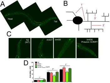
elegans, and two loss of function alleles of CRIPT result in decreased branching of the
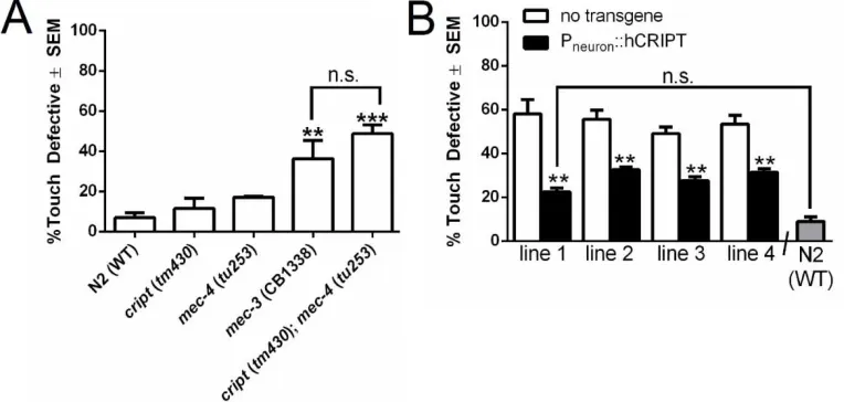
well-studied PVD neuron in vivo. Loss-of-function cript in C. elegans leads to a functional
consequence rendering cript mutants touch defective. Finally, we show that the
mechanism by which CRIPT promotes dendritic growth is independent of synapse
destabilization. Understanding the mechanisms that drive dendritic growth might be
24
INTRODUCTION
Activity-dependent development occurs throughout the neuroaxis, including the
spinal cord during early postnatal life (Inglis et al., 2002; Haas et al., 2006; Ni and
Martin-Caraballo, 2010). During early postnatal life, motor neurons express a very high
level of GluA1 and this coincides with the period of extensive dendrite growth and
remodeling (Jakowec et al., 1995; Jeong et al., 2006). These alterations in dendritic
architecture are confined to a critical period and occur independent of NMDA
(N-methyl-D-aspartic acid) receptors (Jakowec et al., 1995; Inglis et al., 2002; Jeong et al., 2006).
Activity-dependent growth of the dendritic tree in spinal cord neurons is driven by
the GluA1 subunit of AMPA (2-amino-3-(3-hydroxy-5-methyl-isoxazol-4-yl)propanoic
acid) receptors and its intracellular binding partner, synapse associated protein of 97
kDa (SAP97) (Zhang et al., 2008; Zhou et al., 2008). Animals lacking the GluA1 subunit
or SAP97 in motor neurons have a stunted dendritic tree and suffer locomotor
impairments that persist throughout life (Zhang et al., 2008; Zhou et al., 2008). All of the
dendrite promoting actions of GluA1 are mediated by SAP97 (Zhou et al., 2008).
Trafficking of SAP97 to the cell surface is dependent on the interaction of SAP97 with
GluA1, but GluA1 is trafficked to the cell surface independently of GluA1’s interaction
with SAP97 (Zhang et al., 2006). SAP97 is a scaffolding protein with several
protein-protein interacting modules, including SH3, L27, GUK, and 3 PDZ domains. Preliminary
studies indicate that the PDZ3 domain of SAP97 is responsible for promoting dendritic
growth (unpublished observations; Jablonski and Kalb, 2013).
To understand the molecular mechanism by which GluA1 and SAP97 promote
25
of SAP97? and 2) are such proteins able to modify the architecture of the dendritic tree?
Here we focus on cysteine-rich-interactor-of-PDZ-three (CRIPT), a 12 kDa protein that
localizes to excitatory synapses and links proteins such as postsynaptic-density protein
of molecular weight 95 kDa (PSD95) to microtubules (Niethammer et al., 1998;
Passafaro et al., 1999). Given CRIPT’s described interaction to PSD95’s PDZ3 domain
and its localization pattern, we investigated a potential role for CRIPT in GluA1 and
26
MATERIALS AND METHODS
Antibodies
The following antibodies were used: immunoprecipitation of SAP97 (1:50;
Thermo Pierce); immunoblotting and immunoprecipitation of CRIPT (1:500; Protein Tech
Group); immunoprecipitation and immunoblotting of HA-tag (1:50 for IP and 1:1000 for
IB; Convance); immunoblotting of SAP97 (1:1000; NeuroMab); immunoprecipitation and
immunoblotting of the myc-tag (1:50 for IP and 1:1000 for IB; Cell Signaling);
immunostaining of GFP (1:250; Sigma); immunostaining secondary antibody against
GFP (1:500; Alexa Fluor 594). The following primary antibodies were used in
quantitative inhibitory synapse studies: pre-synaptic marker GAD 65/67 (Rabbit
anti-glutamate decarboxylase 65 & 67, Millipore # AB1511), and postsynaptic marker Alpha1
GABA-A receptor (Rabbit anti-Alpha1 GABA-A receptor, Clone N95/35 antibody
NeuroMab #75-136). Secondary antibodies were Alexa Fluor 633 Goat Anti-Mouse IgG
(H+L) (Invitrogen # A-21050) and Alexa Fluor 568 Goat Anti-Rabbit IgG (H+L) Antibody
(Invitrogen # A-11011). GFP green fluorescence was sufficiently strong that
enhancement with additional anti-GFP staining was unnecessary.
Mixed spinal cultures
Mixed spinal cord neuron cultures were prepared as previously described (Jeong
et al., 2006). They were maintained in glia-conditioned medium supplemented with
trophic factors (Alomone Labs at 1.0ng/mL): human neurotrophin-3, human
neurotrophin-4, human brain-derived neurotrophic factor, human cardiotrophin-1, human
glial-derived neurotrophic factor, and rat ciliary neurotrophic factor. One half of the
27
Heterologous cells and transfection
HEK293 cells were maintained in Dulbecco’s Modified Eagle’s medium (DMEM)
(Invitrogen) supplemented with 10% fetal bovine serum (FBS), 1% penicillin, and 1%
streptomycin. Cells were transfected (Lipofectamine 2000; Invitrogen) per
manufacturer’s protocol when they were ∼75% confluent and maintained in DMEM with
10% FBS until lysed 48 hours post transfection.
Immunoprecipitation experiments
For immunoprecipitation experiments, lysates were made in 1% NP-40 lysis
buffer (25mM Tris-HCl pH 7.4, 150mM NaCl, 1mM EDTA, 1% NP-40, 5% glycerol;
150μL per 60mm dish) supplemented with fresh protease inhibitor cocktail (Sigma). After
2 washes in ice-cold 1X phosphate-buffered saline (PBS), cells were lysed in lysis
buffer, sonicated (20% strength, 10 seconds), and centrifuged at 14,000 rpm for 10
minutes (4˚C) to remove cellular debris. 5% of the lysate was saved for input, and the
remaining lysate was used for immunoprecipitation. For immunoprecipitation,
DynaBeads Protein G (Invitrogen) were pre-cleared in PBS/1% Tween and were bound
to the antibodies (4 μg of antibody) (30’ at room temperature (RT)). Lysate was then
added, incubated for 1 hour at RT, and the lysate and bead mixture was washed in lysis
buffer. Proteins were then boiled in 1% BME and SDS loading buffer. Proteins were
immunoblotted following standard western blotting technique using 4-12% Bis-Tris gels
(Invitrogen).
Surface plasmon resonance preparation
The PDZ3 domain of SAP97 was amplified by PCR and the PCR product was