University of Pennsylvania
ScholarlyCommons
Publicly Accessible Penn Dissertations
1-1-2016
A Study of the Role of Gata6 in Definitive
Endoderm Specification and Β-Cell Functionality
by Genome Engineering of Pluripotent Stem Cells
Amita Tiyaboonchai
University of Pennsylvania, [email protected]
Follow this and additional works at:
http://repository.upenn.edu/edissertations
Part of the
Cell Biology Commons, and the
Developmental Biology Commons
This paper is posted at ScholarlyCommons.http://repository.upenn.edu/edissertations/2057
Recommended Citation
Tiyaboonchai, Amita, "A Study of the Role of Gata6 in Definitive Endoderm Specification and Β-Cell Functionality by Genome Engineering of Pluripotent Stem Cells" (2016).Publicly Accessible Penn Dissertations. 2057.
A Study of the Role of Gata6 in Definitive Endoderm Specification and
Β-Cell Functionality by Genome Engineering of Pluripotent Stem Β-Cells
Abstract
Human pluripotent stem cells (PSCs) provide a powerful model system for the study of early human development, disease modeling and physiology. We chose to focus our studies on monogenic diabetes using this model system. Within the pancreas, β cells are one of the most critical endocrine cells as loss of this cell type disrupts blood glucose homeostasis, leading to diabetes. Due to the limited availability of primary human cells it is difficult to study them in vitro, especially in the context of genetic disease where patient material is even more difficult to obtain. Here, we characterize endodermal progenitor (EP) derived β-like cells as a model system for studying β cell development and function in vitro. EP cells are a population of endodermal stem cells, which can self-renew and be differentiated into multiple endodermal cell lineages. EP derived β-like cells are mono-hormonal for insulin and express a number of genes important for insulin processing and secretion. By use of both static stimulations and perifusion assays we show that EP derived β-like cells are responsive to both glucose and a number of other know secretagogues. Next, we demonstrate that genome editing with a zinc finger nuclease at the AAVS1 safe harbor locus can generate stable gene expression in PSCs during differentiation. We then combine these tools and describe the use of PSCs as an in vitro model system to study GATA6. Heterozygous mutations in this gene are the leading cause of pancreas agenesis while studies in mice do not replicate the human phenotype. Induced pluripotent stem cells were created from a pancreas agenesis patient with a heterozygous mutation in GATA6. Using genome editing technology, additional stem cell lines with mutations in both GATA6 alleles were generated and demonstrate a severe block in definitive endoderm induction. Re-expression of GATA6 or other GATA family members can rescue this endoderm phenotype. Partial rescue could also be achieved by treatment with a basic fibroblast growth factor. Using the EP cell culture system to bypass the developmental block at the endoderm stage, cell lines with mutations in one or both GATA6 alleles were differentiated into β-like cells. The mutant cells were shown to be functionally defective by failure to secrete insulin upon glucose stimulation. Decrease in retinoic acid concentrations used during the differentiation lead to decreased β-like cell differentiation efficiency of the heterozygous GATA6 mutants suggesting a possible mechanism for the patient phenotypes. These data show that GATA6 plays a critical role in endoderm specification and β-like cell functionality in humans while in mice it is dispensable, highlighting the importance of studying a human system.
Degree Type
Dissertation
Degree Name
Doctor of Philosophy (PhD)
Graduate Group
Biology
First Advisor
Keywords
Beta Cell, Definitive Endoderm, Diabetes, GATA6, Pancreas Agenesis, Stem Cell
Subject Categories
A STUDY OF THE ROLE OF GATA6 IN DEFINITIVE ENDODERM SPECIFICATION
AND β CELL FUNCTIONALITY BY GENOME ENGINEERING OF PLURIPOTENT
STEM CELLS
Amita Tiyaboonchai
A DISSERTATION
in
Biology
Presented to the Faculties of the University of Pennsylvania
in Partial Fulfillment of the Requirements for the
Degree of Doctor of Philosophy
2016
Supervisor of Dissertation:
________________________
Paul Gadue, Associate Professor of Pathology and Laboratory Medicine
Graduate Group Chairperson:
________________________
Michael Lampson, Associate Professor of Biology
Dissertation Committee
Scott Poethig, John H. and Margaret B. Fassit Professor of Biology
Dejian Ren, Professor of Biology
Doris A. Stoffers, Professor of Medicine
Gregory M. Guild, Professor of Biology
A STUDY OF THE ROLE OF GATA6 IN DEFINITIVE ENDODERM SPECIFICATION AND β CELL FUNCTIONALITY BY GENOME ENGINEERING OF PLURIPOTENT STEM CELLS
COPYRIGHT
2016
Amita Tiyaboonchai
ACKNOWLEDGEMENTS
First and foremost, I would like to thank my advisor Paul Gadue. Paul has been
extremely supportive and encouraging throughout my time in the lab. Thank you for your
patience while I learned new techniques, insistence to keep going when experiments did
not work out the way I had expected and for the optimism when I needed it the most. I
am thankful for all the advice and guidance which has helped me in accomplishing my
goals to become a better scientist.
I would also like to thank Debbie French, who has been a great mentor to me. Debbie
has always been enthusiastic and supportive during discussions and provided great
advice throughout my time in the lab. Debbie is also a great proofreader, and I am
grateful for all the times she has helped me look over a poster or some of my writing.
I would like to thank all the past and present members of the Gadue and French Labs.
Everyone has contributed to the lab being a more enjoyable place to be in and have
almost become a second family to me during these past 5 and a half years. I greatly
appreciate Prasuna Paluru, Aline Moreau and Jay Mills for teaching me tissue culture
and molecular biology techniques when I initially started in the lab. As I started working
on endodermal related projects, Xin Cheng was an invaluable teacher who I am forever
thankful for patiently teaching me many differentiation techniques, answering questions
no matter how busy he was and the late night sushi runs.
I am grateful to the fellow graduate students in the lab, Xiuli Sim, Leo Cardenas-Diaz,
discussions whether it be about science or non-science topics, and have a sympathetic
ear for all of my complaints. I am especially thankful to Xiuli for encouraging me to keep
going whenever research was not going so well and also for the enjoyable afternoon tea
sessions to discuss the happenings of the lab. And to Leo, I am grateful for all the help
with so many different experiments, no matter how many other things he had going on
himself. Somdutta, my desk and peppermint patty buddy, I appreciate the company
during the late nights in lab. Thank you to Sid for the interesting discussions as you have
begun to work on the GATA6 project and to Sara for all the tissue culture things you
have helped me with.
Thank you Chia-min Liao for the many conversations and life advice as a scientist and
Lei Ying, for advising and helping me on difficult experiments. Thank you JeanAnn
Maguire for answering my questions, whether it be about science, life or anything else I
may happen to ask. I am grateful to Chintan Jobaliya for keeping our lab running
smoothly (and cost-efficiently) and for listening to my complaints and always having
some kind of entertaining rebuttal. Thank you to Grace Liang for helping with the endless
cell sorts and always telling me everything will be okay, despite not exactly always
knowing what I was doing. Lastly, to Alyssa Groh for always being up to talk about
anything fun.
To all of the friends I made in Philly outside of lab, especially Pun Suriyalaksh, P’Dome
Preechawutthidech, P’Pom Kulwattanaporn, Gift Hannanta-anan, Sun Vasansiri, Thanat
Owlarn, Palm Harinsuit, June Supapannachart and Tee Ramaithitima. Thank you for the
midnight snacks, the 3 hour random discussions over dim-sum, the road trips and the
shopping sprees. I am forever appreciative of the fun times.
None of this would have been possible without the support, love and encouragement of
my immediate and extended family. Mum, Dad and Ohm have always had an
unwavering belief in me that I could achieve my goals and have always encouraged me
to strive to do the best I can. Thank you for always providing a place to fall back on and
coming to visit as often as you do despite the distance that has to be travelled.
Lastly, I want to thank my husband, Lawrence Adijanto for encouraging me to keep
going during the tough times when experiments were not going so great and always
trying to help. Although your ideas may not have been the most practical, they were
always amusing and imaginative. I am forever grateful for having you by my side, for
believing in me and for being a source of constant source of support.
ABSTRACT
A STUDY OF THE ROLE OF GATA6 IN DEFINITIVE ENDODERM SPECIFICATION
AND β CELL FUNCTIONALITY BY GENOME ENGINEERING OF PLURIPOTENT
STEM CELLS
Amita Tiyaboonchai
Paul Gadue
Human pluripotent stem cells (PSCs) provide a powerful model system for the study of
early human development, disease modeling and physiology. We chose to focus our
studies on monogenic diabetes using this model system. Within the pancreas, β cells are
one of the most critical endocrine cells as loss of this cell type disrupts blood glucose
homeostasis, leading to diabetes. Due to the limited availability of primary human β cells
it is difficult to study them in vitro, especially in the context of genetic disease where
patient material is even more difficult to obtain. Here, we characterize endodermal
progenitor (EP) derived β-like cells as a model system for studying β cell development
and function in vitro. EP cells are a population of endodermal stem cells, which can self-
renew and be differentiated into multiple endodermal cell lineages. EP derived β-like
cells are mono-hormonal for insulin and express a number of genes important for insulin
processing and secretion. By use of both static stimulations and perifusion assays we
show that EP derived β-like cells are responsive to both glucose and a number of other
know secretagogues. Next, we demonstrate that genome editing with a zinc finger
nuclease at the AAVS1 safe harbor locus can generate stable gene expression in PSCs
in vitro model system to study GATA6. Heterozygous mutations in this gene are the
leading cause of pancreas agenesis while studies in mice do not replicate the human
phenotype. Induced pluripotent stem cells were created from a pancreas agenesis
patient with a heterozygous mutation in GATA6. Using genome editing technology,
additional stem cell lines with mutations in both GATA6 alleles were generated and
demonstrate a severe block in definitive endoderm induction. Re-expression of GATA6
or other GATA family members can rescue this endoderm phenotype. Partial rescue
could also be achieved by treatment with a basic fibroblast growth factor. Using the EP
cell culture system to bypass the developmental block at the endoderm stage, cell lines
with mutations in one or both GATA6 alleles were differentiated into β-like cells. The
mutant cells were shown to be functionally defective by failure to secrete insulin upon
glucose stimulation. Decrease in retinoic acid concentrations used during the
differentiation lead to decreased β-like cell differentiation efficiency of the heterozygous
GATA6 mutants suggesting a possible mechanism for the patient phenotypes. These
data show that GATA6 plays a critical role in endoderm specification and β-like cell
functionality in humans while in mice it is dispensable, highlighting the importance of
studying a human system.
Table of Contents
Acknowledgements ... iii
Abstract ... vi
Table of Contents ... viii
List of Tables ... xiii
List of Figures ... xiv
List of Abbreviations ... xvi
CHAPTER 1: Introduction and Overview ... 1
1.1 Characteristics and Function of Pancreatic β-cells ... 1
1.1.1 The Pancreas ... 1
1.1.2 Development of the Pancreas ... 2
1.1.3 Insulin Biosynthesis ... 4
1.1.4 Insulin Secretion ... 7
1.2 Diabetes Mellitus ... 10
1.2.1 Characteristics and Types of Diabetes Mellitus ... 10
1.2.2 Type I and Type II Diabetes Mellitus ... 11
1.2.3 Monogenic Diabetes Mellitus ... 12
1.3 GATA6 ... 13
1.3.1 Characteristics of GATA6 ... 13
1.3.2 The Function of GATA6 ... 15
1.3.3 Functional Redundancy of GATA family members ... 17
1.4 Pluripotent Stem Cells ... 18
1.4.2 Human Embryonic Stem Cells ... 20
1.4.3 Induced Pluripotent Stem Cells ... 21
1.4.4 Human Pluripotent Stem Cells as a Model System ... 23
1.4.5 Differentiation of Pluripotent Stem Cells ... 25
1.4.6 Definitive Endoderm and Endodermal Progenitor Cells ... 25
1.4.7 Pluripotent Stem Cell Derived β Cells ... 26
1.5 Overview of Research Goals ... 30
CHAPTER 2: Methods and Materials ... 31
2.1 Maintenance of human pluripotent stem cells ... 31
2.2 Primary human islets (Chapter 3) ... 32
2.3 Vector construction for AAVS1 targeting ... 32
2.4 Gene targeting of the AAVS1 locus ... 32
2.5 PCR screen for zinc finger nuclease construct integration (Chapter 4) ... 33
2.6 Genome editing using CRISPR/Cas9 (Chapter 5) ... 33
2.7 Differentiation of human pluripotent stem cells ... 35
2.8 Endodermal progenitor (EP) cell generation and maintenance ... 37
2.9 Pancreatic β cell differentiation from endodermal progenitor cells ... 38
2.10 Glucose stimulated insulin secretion assay ... 38
2.11 Perfusion of β–like cells (Chapter 3) ... 39
2.12 Electron microscopy (Chapter 3) ... 40
2.13 Flow Cytometry and Cell sorting ... 40
2.14 Reverse Transcription and Quantitative Real Time Polymerase Chain Reaction (qRT-PCR) ... 43
2.15 Southern Blot (Chapter 4) ... 46
2.17 Immunofluorescence staining (Chapter 5) ... 47
2.17 Lentiviral vector generation and transduction (Chapter 5) ... 47
2.19 Microarray and Bioinformatics Analysis (Chapter 5) ... 48
2.20 Statistical Analysis ... 48
CHAPTER 3: The Characterization of Endodermal Progenitor Cell Derived β-like Cells ... 49
3.1 Introduction ... 49
3.2 Results ... 51
3.2.1 Dispase Aggregation ... 51
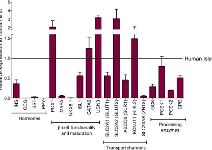
3.2.2 Gene and Protein Expression ... 53
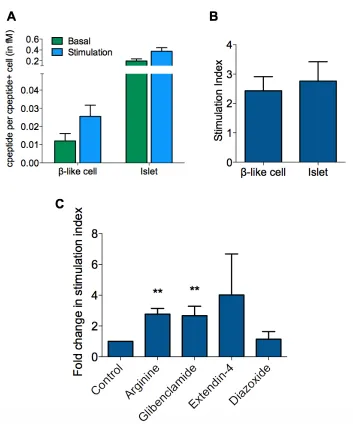
3.2.3 Glucose Stimulation Insulin Secretion ... 55
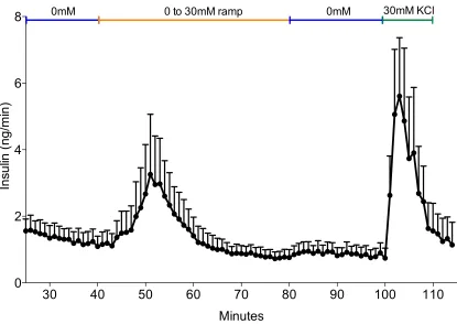
3.2.4 Perfusion ... 58
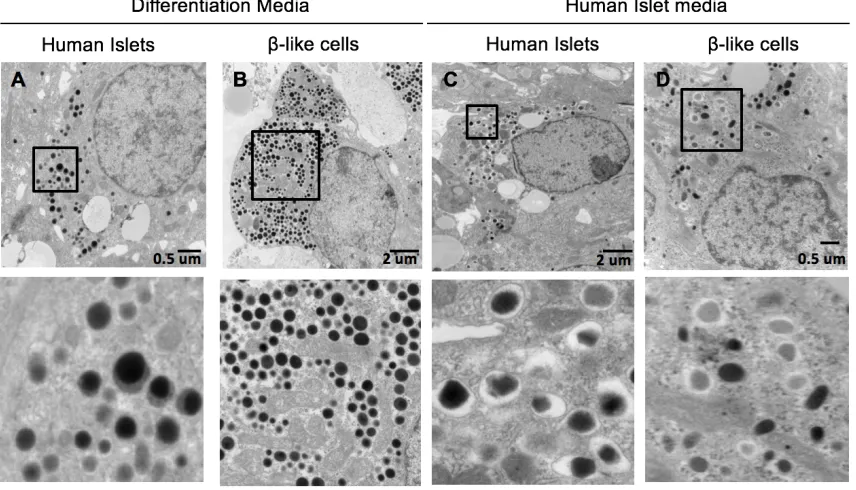
3.2.5 Ultrastructure ... 60
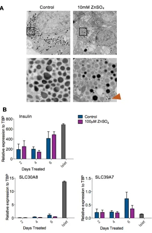
3.2.6 Zinc Sulphate Treatment ... 62
3.3 Discussion ... 65
CHAPTER 4: Utilization of the AAVS1 Safe Harbor Locus for Hematopoietic Specific Transgene Expression and Gene Knockdown in Human ES Cells ... 69
4.1 Introduction ... 69
4.2 Results ... 71
4.2.1 Generation of a CD43 Hematopoietic Reporter Cell Line ... 71
4.2.2 Knockdown of the Myeloid Specific Transcription Factor PU.1 ... 79
4.3 Discussion ... 83
5.1 Introduction ... 86
5.2 Results ... 89
5.2.1 Establishment of GATA6 PSC Lines ... 89
5.2.2 Definitive Endoderm Differentiation ... 97
5.2.3 Partial Rescue of Definitive Endoderm by the Addition of Growth Factor ... 102
5.2.4 Rescue of Definitive Endoderm by GATA6 ... 105
5.2.5 Rescue of Definitive Endoderm by Other GATA Family Members ... 108
5.2.6 Downstream Targets of GATA6 During Definitive Endoderm Specification ... 109
5.2.7 Functional Analysis of GATA6 Mutants as Endodermal Progenitor derived β-like cells ... 111
5.2.8 Gene Expression Analysis of GATA6 Mutant Endodermal Progenitor derived β-like cells ... 115
5.2.9 Retinoic Acid is the Exogenous Signal That Allows the Patient iPS+/indel cells to differentiation in vitro ... 115
5.3 Discussion ... 120
CHAPTER 6: Summary and Speculations ... 127
6.1 Comparing ES based protocols to EP derived β-like cells ... 127
6.2 Culturing EP cells allows for their Maturation ... 128
6.3 The Lack of NKX6.1 Expression in EP derived β-like cells ... 129
6.4 The Function and Relationship of GATA Family Members ... 130
6.6 Downregulation of HNF4α in GATA6 Mutant β-like cells ... 133
6.7 Downstream Targets of GATA6 ... 134
6.8 Overcoming the Limitations of EP derived β-like cells ... 134
6.9 Rescue of Functionality in β-like cells ... 135
6.10 Exploring other GATA6 Patient Mutations and Phenotypes ... 136
Bibliography ... 138
List of Tables
CHAPTER 2
Table 2-1: Primers utilized in generation of GATA6 compound heterozygous
mutations and correction. ... 34
Table 2-2: Conjugated Antibodies for Flow Cytometry ... 41
Table 2-3: Unconjugated Primary Antibodies ... 41
Table 2-4: Secondary Antibodies ... 42
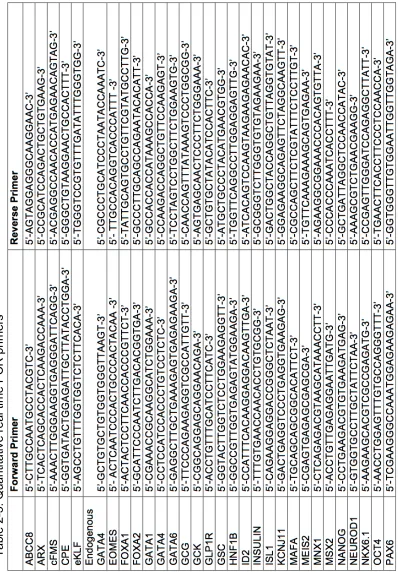
Table 2-5: Quantitative real time PCR primers ... 44
CHAPTER 5 Table 5-1: Table of Pluripotent Stem Cell Lines ... 91
List of Figures
CHAPTER 1
Figure 1-1: Processing of the Insulin Molecule ... 6
Figure 1-2: Insulin Secretion ... 8
Figure 2-3: GATA6 Protein Structure ... 14
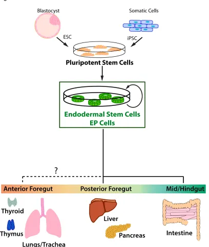
Figure 2-4: Endodermal Progenitor Cells ... 27
CHAPTER 3 Figure 3-1: Dispase Aggregation Enriches for C-peptide Positive β-like Cells .... 52
Figure 3-2: Gene Expression Profile of β-like Cells ... 54
Figure 3-3: Static Stimulation of β-like Cells ... 57
Figure 3-4: Perifusion of β-like Cells ... 59
Figure 3-5: Ultrastructure of β-like Cells ... 61
Figure 3-6: Zinc Sulphate Treatment of β-like Cells ... 64
CHAPTER 4 Figure 4-1: AAVS1 Safe Harbor Gene Targeting ... 72
Figure 4-2: Southern Blot Analysis of Targeted Clones ... 74
Figure 4-3: PCR Analysis of ZNF Integration into Genomic DNA ... 75
Figure 4-4: Generation of a CD43-GFP Reporter Cell Line ... 76
Figure 4-5: CD43-GFP Reporter Expression Neuroectoderm and Endoderm .... 78
CHAPTER 5
Figure 5-1: Generation of Pluripotent Stem Cell Lines with GATA6 Mutations ... 90
Figure 5-2: Targeting the AAVS1 Locus in Mel1-INS-GFP ES+/+ Cells ... 92
Figure 5-3: GATA6 Mutant Pluripotent Stem Cell Lines ... 93
Figure 5-4: Characterization of Patient iPS+/indel CHOP.Panagenesis1 Cells ... 96
Figure 5-5: GATA6 is Required for Definitive Endoderm Differentiation of
Pluripotent Stem Cells ... 98
Figure 5-6: Definitive Endoderm Differentiation Experiments were Repeated in
the ES Cell Background ... 100
Figure 5-7: Differentiation of GATA6 Mutants to Mesoderm and Ectoderm
Lineages ... 103
Figure 5-8: Partial Rescue of Definitive Endoderm Differentiation with Basic
Fibroblast Growth Factor ... 104
Figure 5-9: Rescue of Definitive Endoderm Differentiation by GATA Factor Re-
Expression ... 106
Figure 5-10: Gene Expression Microarray to Identify Downstream Targets of
GATA6 ... 110
Figure 5-11: Generation of Endodermal Progenitor Cells ... 112
Figure 5-12: GATA6 is Dispensable for Differentiation of Pancreatic β-like Cells
but Necessary for Functionality ... 113
Figure 5-13: Gene Expression Analysis of β-like Cells ... 116
Figure 5-14: Retinoic Acid Overcomes Patient iPS+/indel Cell Deficiency During
Differentiation ... 118
Figure 5-15: A Minimum Threshold of GATA4 and GATA6 Must be Met for
List of Abbreviations
AAVS1 adeno-associated virus integration site 1
APC allopycocyanin
bFGF basic fibroblast growth factor
CA chicken actin
CAS9 CRISPR associated protein 9
cDNA complementary
CPE carboxypeptidase E
CRISPR clustered regularly-interspaced short palindromic repeats
DMEM Dulbecco’s minimum essential medium
DNA deoxyribonucleic acid
DPBS Dulbecco’s phosphate buffered saline
ECC embryonal carcinoma cells
ELISA enzyme-linked immunosorbent assay
EP endoderm progenitor
ES embryonic stem
EpiSC epiblast stem cells
FACS fluorescence-activated cell sorting
FBS fetal bovine serum
GCK glucokinase
GFP green fluorescent protein
GLUT1 glucose transporter 1 (also known as SLC2A1)
GLUT2 glucose transporter 2 (also known as SLC2A2)
HRP horseradish peroxidase
HEPES 2-[4-(2-hydroxyethyl)piperazin-1-yl]ethanesulfonic acid
IMDM Iscove’s modified Dulbecco’s medium
iPS induced pluripotent stem
IRES internal ribosome entry site
KATP adenosine triphosphate sensitive potassium channel
Kir6.2 potassium channel (also known as KCNJ11)
KRBH Kreb ringers buffer with HEPES
LIF leukemia inhibitory factor
MEF mouse embryonic fibroblast
MODY maturity onset diabetes of the young
PCSK2 prohormone convertase 2
PCSK1/3 prohormone convertase 1/3
PCR polymerase chain reaction
PDX1 pancreatic and duodenal homeobox 1
PE phycoerythrin
PFA parafolmaldehyde
PP pancreatic polypeptide
PSC pluripotent stem cells
qRT-PCR quantitative real time PCR
RFP red fluorescent protein
RNA ribonucleic acid
ROCK rho-associated kinase
rTTA reverse tetracycline transactivator
SUR1 sulfonylurea receptor (also known as ABCC8)
TALEN transcriptional activator-like effector nuclease
TEM transmission electron microscopy
TRE tetracycline response element
ZFN zinc finger nuclease
ZnSO4 zinc sulphate
CHAPTER 1
Introduction and Overview
1.1 Characteristics and Function of Pancreatic β-cells
β cells reside within the endocrine portion of the pancreas. They are important for the
maintenance of blood glucose and a loss of their functionality leads to diabetes mellitus.
As this thesis will first focus upon the characterization of an in vitro system for the study
of β cells and then apply this system to the study monogenic diabetes caused by GATA6
mutations, we will first review the characteristics and functionality of β cells.
1.1.1 The Pancreas
The human pancreas is an organ consisting of exocrine, ductal and endocrine cells.
Exocrine cells constitute the majority of the pancreatic tissue and function to secrete
digestive enzymes such as amylase, proteases, lipases and nucleases to the duodenum
via the pancreatic duct that runs through the middle of the pancreas. These enzymes
function in nutrient digestion by catalyzing the breakdown of lipids, proteins and
carbohydrates (Shih et al., 2013). Endocrine cells are located in small tight clusters in
structures known as the Islets of Langerhans found scattered throughout the exocrine
tissues. Within the islets of Langerhans in addition to the endocrine cells, there is also
vasculature, neurons and mesodermal derived stromal cells (Pan and Wright, 2011).
Endocrine cells function mostly independent of the exocrine pancreas. Each type of
endocrine cell within the islet produces a specific type of hormone. α-cells secrete
glucagon, β-cells secrete insulin, δ-cells secrete somatostatin, ε-cells secrete ghrelin and
of glucagon when blood glucose is low and secretion of insulin when blood glucose is
high from their respective cell types. δ-cells, ε-cells and γ-cells function to regulate
nutrient metabolism. Human islets are composed of 55% β-cells, 35% α-cells and 10%
δ-cells with all cell types intermixed within the islet architecture. An adult pancreas can
contain between 300,000 and 1,500,000 islets (Brissova et al., 2005).
1.1.2 Development of the Pancreas
In vitro differentiation protocols for pluripotent stem cells (PSCs) to β-like cells are
informed by the events of in vivo development. By mimicking known signaling pathways
that are important at each stage of development in vivo, PSCs can be directed to
differentiate toward a specific lineage. To determine whether a stage of development
has been achieved through in vitro differentiation, markers that are expressed in vivo are
often assayed. Throughout this thesis, different stages of β-cell development will often
be referred to along with their marker expression. Thus, we present here a summary of
pancreatic development.
During early development, the region of the foregut endoderm from which the pancreas
will arise is prepatterned. This region has a lack of expression of sonic hedgehog and
requires the expression of retinoic acid. The presumptive pancreatic endoderm is
characterized by the expression of Pdx1, Ptf1a and Sox9. At this stage the pancreatic
epithelium is tightly surrounded by mesenchyme which highly expresses FGF10. FGF10
regulates expression of Ptf1a and Sox9 and is important for cell proliferation and
expansion of the early pancreatic buds once they form (Pan and Wright, 2011;; Shih et
al., 2013).
The pancreas begins to develop by forming as two epithelial buds, a ventral and a dorsal
pancreatic bud at opposite sides of the foregut endoderm by evagination into the
surround mesenchyme. In both mouse and human, the dorsal bud forms first from the
dorsal foregut endoderm. The emergence of the dorsal bud appears to be regulated by
extrinsic cues from the surrounding vascular endothelial cells. The evagination of the
ventral pancreatic bud along with the liver from the ventral foregut endoderm follows.
The ventral pancreatic bud receives cues form the cardiac mesoderm and the vitelline
veins (Gittes, 2009;; Pan and Wright, 2011). At this stage of development, the majority of
the buds are composed of multipotent pancreatic progenitor cells that express the key
transcription factors Nkx6.1, Nkx6.2 and Ptf1a. Also present in the buds are a small
subset of cell from the ‘first wave’ of endocrine cells which are either glucagon positive
or double positive for insulin and glucagon. These cells eventually contribute to a small
subset of the mature α cell population (Herrera, 2000).
As the multipotent pancreatic progenitor cells continue to proliferate there is a
segregation and change in morphogenesis of the pancreatic buds (Shih et al., 2013).
The buds elongate and as the gut tube rotates, the ventral and dorsal buds come into
contact and fuse together. Multiple small protrusions then begin to form from the edge of
the of the pancreatic bud (Gittes, 2009). Cells that are located toward the edge of these
protrusions are in the tip domain and express Ptf1a, c-Myc and Cpa (Shih et al., 2013).
Tip domain cells are fated to become acinar cells. As development progresses acinar
cells will continue to proliferate and increase in cell number by duplication (Pan and
Wright, 2011). Cells that are located toward the inside of the buds are in the trunk
Oncecut-1, Prox1 and Hes1. Trunk domain cells are bipotential and give rise to
endocrine and ductal cells (Shih et al., 2013).
Endocrine progenitor cells in the trunk domain form the primitive duct (or also known as
the epithelial cord), which is characterized by tubules lined by a single layer of polarized
epithelial cells. At this stage a subset of cells within the primitive ducts express transient
high levels of Neurogenin3 (Ngn3), which indicates the onset of endocrine cell
differentiation. Cells that lack the expression of Ngn3 will become the ductal cells. Ngn3
positive endocrine precursors then delaminate from the primitive ducts, converting to
non-epithelial cells through a process thought to involve an epithelial-to-mesenchymal
transition (Pan and Wright, 2011;; Shih et al., 2013). These cells migrate into the
surrounding area and coalesce into aggregates that become the islets of Langerhans
(Gittes, 2009). Each endocrine precursor cell will further differentiate into one of the five
types of hormone expressing cells. The mature endocrine cells that the endocrine
precursor will become is temporally determined and is dependent on the developmental
stage at which their differentiation occurs. At this stage a large numbers of β cells and α
cells begin to appear. α cell fate is determined by the expression of Arx while β cell
identity is determined by Pax4, Pdx1 and Nkx6.1 (Shih et al., 2013).
1.1.3 Insulin Biosynthesis
β-cells function to produce, store and regulate the secretion of insulin (Rhodes et al.,
2005). In experiments with in vitro differentiated β-like cells we examine the ability of the
cells to process insulin as a method to determine the cells maturity and similarity to
functional adult β cells.
Frederick Banting and Charles Best first discovered insulin in 1921 by treating diabetic
dogs with extracts of the pancreas that did not contain any digestive enzymes (Banting
et al., 1922). Upon treatment both blood glucose levels and sugar excreted in the urine
of the dogs were lowered. Based upon these results, the first human diabetic patient
was successfully treated with insulin in January of 1922 (Banting et al., 1922).
Insulin consists of two peptide chains, the A chain and the B chain that are connected by
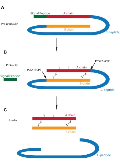
three disulphide bonds (Fu et al., 2013). Insulin is initially synthesized as the precursor
molecule preproinsulin, consisting of an A-chain connected to the B-chain via a C-chain
and a signal peptide located at the end of the B-chain peptide (Figure 1-1A).
Preproinsulin is trafficked through the rough endoplasmic reticulum where the signal
peptide is cleaved and the remaining proinsulin is folded into its three dimensional
conformation (Figure 1-1B). Proinsulin is then loaded into immature granules and the
conversion of proinsulin to insulin occurs in these granules. The immature granules are
acidified as the prohormone processing enzymes prohormone convertase 2 (PCSK2)
and prohormone convertase 1/3 (PCSK1/3) have optimal enzymatic activity at a pH of
5.0 to 5.5. Proinsulin is cleaved at two sites by PCSK2 and PCSK1/3 releasing the C-
chain that is also known as c-peptide. Cleavage by the prohormone processing enzymes
results in the exposure of the C-terminal basic amino acids on the insulin molecule.
These residues are then cleaved by carboxypeptidase E (CPE) (Figure 1-1C) (Rhodes,
2004a;; Rhodes et al., 2005). As the granule matures, the high concentration of insulin
causes the insulin monomers to crystalize into hexamers associated with two zinc ions
(Bilous and Donnelly, 2010;; Fu et al., 2013). When viewed by electron microscopy
mature insulin granules often appear as crystalline dense core granules surrounded by a
Figure 1-1
A
B
C
Figure 1-1. Processing of the Insulin Molecule. (A) Preproinsulin is synthesized
clear halo. These insulin granules are ready to be released by exocytosis from the β-cell
upon stimulation (Bilous and Donnelly, 2010).
1.1. 4 Insulin Secretion
Glucose is one of the critical stimulants of insulin secretion. When human blood glucose
levels are higher than 5mmol/L (90 mg/dL), β-cells begin to release insulin. To study β
cell functionality, in vitro differentiated β cells should express the appropriate genes in
order to sense increased glucose concentrations as well as respond with insulin
secretion.
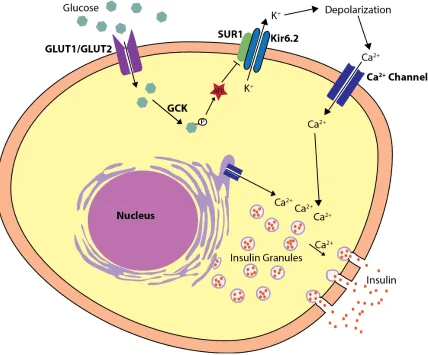
Glucose enters the β-cell through glucose transporters such as glucose transporter 1
(GLUT1 encoded by SLC2A1) or glucose transporter 2 (GLUT2 encoded by SLC2A2)
(De Vos et al., 1995;; McCulloch et al., 2011), where it is then phosphorylated by
glucokinase (GCK). The glucose is then metabolized by glycolysis, causing an increase
in the ratio of ATP relative to ADP. This causes the ATP sensitive potassium (KATP)
channels which are normally open to close (Henquin, 2000). The KATP channel is an
octamer composed of tetramers of the two subunits: the sulfonylurea receptor (SUR1 or
ABCC8) and an inwardly rectifying K+ channel (Kir6.2 or KCNJ11) (Koster et al., 2005).
The closing of the KATP channel results in the depolarization of the cell surface
membrane and the opening of the voltage gated calcium channels allowing calcium
influx into the cytoplasm. The increased cytoplasmic calcium stimulates the exocytosis of
insulin granules from the cell (Greeley et al., 2010). In addition to stimulating insulin
release in β-cells, high glucose levels also induce proinsulin biosynthesis. Within 20
minutes of glucose stimulation, the increase in insulin biosynthesis can be observed.
Figure 1-2
Figure 1-2. Insulin Secretion. Glucose enters the β cell via either GLUT1 or
reached. This can to 30 to 40 fold compared to basal levels (Alarcón et al., 2002;;
Rhodes, 2004b). In contrast, when unstimulated, β-cells are constantly releasing a basal
level of insulin. The glucose that undergoes oxidative glycolysis is low enough that the
ATP to ADP ratio does not change significantly and the KATP channels remain open
(Henquin, 2009).
When β cells are stimulated in vivo or in vitro, insulin release is biphasic. During the first
phase, large amounts of insulin are rapidly released from a ‘readily releasable pool’ of
insulin granules. These granules are located near the cell surface membrane and can
quickly undergo exocytosis. The first phase of insulin release lasts a few minutes from
the time of stimulation. With prolonged stimulation, during the second phase of
secretion, there is a decelerated rate of insulin secretion and the amount plateaus or
gradually increases lasting as long at the stimulus is applied (Bilous and Donnelly, 2010;;
Henquin et al., 2006;; Lacy et al., 1972). Insulin released during the second phase comes
from a reserved pool of insulin granules that can account for up to 90%-95% of the total
granules present within the β-cells. These granules are located further away and need to
be modified and transported to the cell surface membrane in order to undergo
exocytosis (Rorsman et al., 2000;; Seino et al., 2009). In addition to high glucose levels,
insulin secretion can also be stimulated by glucagon, gastric inhibitory peptides,
epinephrine, and amino acids such as arginine (Cleaver and Melton, 2004).
Insulin functions to lower blood glucose levels by stimulating a 10 to 40-fold increase in
glucose uptake by cells throughout the body with the liver, skeletal muscles, cardiac
muscles, central nervous system and adipose tissue being important sites. In the liver
generate ATP or is stored in the form of glycogen and tryiglycerides. In the adipose
tissues, glucose is stored in the form of triglycerides (Saltiel, 2001).
Full functionality of β cells requires a combination of correct insulin synthesis and
development of the appropriate mechanisms to respond to insulin secretion. During in
vitro differentiation of PSCs, functionality of β-like cells is determined by insulin secretion
in response to various stimuli, the most important being glucose. Furthermore, insulin
granule morphology is often examined as an indicator
1.2 Diabetes Mellitus
Diabetes Mellitus is a disease that characterized by the dysregulation of insulin secretion
and/or responsiveness in the body. Patients with this disease typically display
abnormally high levels of circulating glucose (Rhodes et al., 2005;; Saltiel, 2001). While
type I and type II diabetes mellitus are the most common forms of diabetes and have
been immensely studied (reviewed in Atkinson et al., 2014;; Kahn et al., 2014;; Lin and
Sun, 2010;; Nolan et al., 2011;; Olokoba et al., 2012;; Tuomi et al., 2014;; Van Belle et al.,
2011), this thesis will focus upon the less studied form of diabetes mellitus which results
from monogenetic mutations in GATA6.
1.2.1. Characteristics and Types of Diabetes Mellitus
In humans, physiological levels of blood glucose prior to meals are typically 3.9 mmol/L
to 7.2 mmol/L (or 70 to 130 mg/dL) and two hours after a meal should not be higher than
10 mmol/L (or 180 mg/dL). Blood glucose levels in the body are regulated by the release
of the antagonistic hormones from α-cells and β-cells. During regular homeostasis, when
glycogen to glucose, which is then released into the blood stream. When blood glucose
levels are elevated, β-cells respond by releasing insulin causing the uptake, utilization
and storage of glucose. Furthermore, insulin also subdues the hepatic production of
glucose resulting in the lowering of blood glucose levels (Rhodes et al., 2005;; Saltiel,
2001).
Diabetes Mellitus can be categorized into four main categories, type I diabetes, type II
diabetes, gestational diabetes and diabetes as a result of other causes. Other causes of
diabetes include monogenic mutations, diseases of the exocrine pancreas and drug or
chemical induced diabetes (American Diabetes Association, 2016).
1.2.2 Type I and Type II Diabetes Mellitus
Type I and Type II diabetes are the most common forms of diabetes. In 2014, an
estimated 22 million people in the United States were diagnosed with diabetes.
Approximately 5% of those diagnosed with diabetes have Type I diabetes while 90%-
95% of those diagnosed have Type II diabetes (Centers for Disease Control Prevention,
2015). Type I diabetes is an autoimmune disease that causes the selective destruction
and loss of β-cells. An absence of c-peptide occurs as the disease further progresses
and eventually there is complete loss of β-cells within the islets of Langerhans. Type I
diabetes is treated by rigorously monitoring glucose levels and daily insulin injections.
(American Diabetes Association, 2016;; Murphy et al., 2008). Type II diabetes is a
complex metabolic syndrome with multiple causes that are still being elucidated. Risk
factors for the development of type II diabetes include aging, obesity, a lack of exercise
and a genetic predisposition. Management of type II diabetes initially includes regulating
type II diabetes continued to be immensely studied (Atkinson, 2014, 2012;; Kahn et al.,
2014;; Lin and Sun, 2010;; Nolan et al., 2011;; Olokoba et al., 2012;; Röder et al., 2016;;
Todd, 2010;; Tuomi et al., 2014;; Van Belle et al., 2011), this thesis will focus upon
investigating monogenic diabetes, a rarer and less studied form of diabetes.
1.2.3 Monogenic Diabetes
Patients with monogenic diabetes have a mutation in a single gene involved in β-cell
development or functionality. These mutations can be inherited as dominant or
recessive, or arise spontaneously. Monogenic diabetes occurs approximately in 1 to 2 in
100,000 live births (Anik et al., 2015;; Greeley et al., 2010) which accounts for 1%-4% of
diabetes that are diagnosed in children (Rubio-Cabezas et al., 2014). Monogenic
diabetes can be subcategorized into neonatal diabetes and maturity onset diabetes of
the young (MODY) (American Diabetes Association, 2016).
A total of 13 types of MODY have been described in the literature. The four most
common types of MODYs result from mutations in either HNF1α or MODY3 (30% to
50% of the cases), GCK or MODY2 (20% to 50% of the cases, HNF1β or MODY5 (5%
of the cases) or HNF4α or MODY1 (5% of the cases) (Kim, 2015;; Steck and Winter,
2011). Patients with MODYs typically do not have the presence of pancreatic or islet
autoantibodies, have low insulin requirements and detectable c-peptide levels (Murphy
et al., 2008;; Rubio-Cabezas et al., 2014).
A distinguishing feature of patients with neonatal monogenic diabetes is that the majority
of these patients are diagnosed with diabetes prior to 6 months of age (Edghill et al.,
subcategories: those that have transient neonatal diabetes mellitus which resolves after
a few weeks to months and those who have permanent neonatal diabetes mellitus
(Steck and Winter, 2011). In addition to mutations that can result in non functional β
cells, permanent neonatal diabetes can also be a result of pancreatic agenesis.
Pancreatic agenesis is a rare congenital disease caused by heterozygous mutations in
PDX1 (Schwitzgebel et al., 2003;; Stoffers et al., 1997), PTF1A (Sellick et al., 2004;;
Weedon et al., 2014), GATA4 (Amato et al., 2010;; Shaw-Smith et al., 2014) or most
commonly GATA6 (Bonnefond et al., 2012;; Chao et al., 2015;; De Franco et al., 2013;;
Lango Allen et al., 2012;; Nakao et al., 2013;; Stanescu et al., 2014;; Suzuki et al., 2014;;
Yorifuji et al., 2012).
1.3 GATA6
Human GATA6 heterozygous mutations are the most common cause of pancreatic
agenesis. Due to discrepancies in the phenotype of currently available mouse models of
a heterozygous loss of GATA6 and human patient phenotypes, this thesis focuses upon
addressing this issue by development of an in vitro human based model system to study
a loss of GATA6.
1.3.1 Characteristics of GATA6
GATA6 is a part of the six member GATA family of transcription factors that share a
conserved DNA binding motif. GATA family members recognize and bind to the
consensus sequence A/T-GATA-A/G. While GATA1, GATA2 and GATA3 are primarily
expressed in the hematopoietic cell linages, GATA4, GATA5 and GATA6 are found in
endodermal derived tissues, the heart and gonads (Molkentin, 2000;; Viger et al., 2008).
Figure 1-3
GATA6
Figure 1-3. GATA6 protein structure. A schematic representation of GATA6
protein structure with the alternative transcriptional start site indicated.
domains followed by a DNA binding domain and a nuclear localization signal (Figure 1-
3). GATA6 has two transcriptional start sites, both located in the second exon resulting
in the production of two isoforms of GATA6, one which is 595 amino acids in length and
the other 449 amino acids long (Brewer et al., 1999). In mice, GATA6 is expressed in the
primitive streak, heart, lung, intestine, gonads, adrenal and pancreatic tissues
(Koutsourakis et al., 1999;; Liu et al., 2002;; Sartori et al., 2014).
1.3.2 The Function of GATA6
GATA6 is a transcription factor that is critically important for normal human pancreatic
development. Heterozygous mutations in GATA6 are the most common cause of
pancreatic agenesis (Amato et al., 2010;; Shaw-Smith et al., 2014). Patients with a
heterozygous mutation in GATA6 have been found with a range of phenotypes from no
pancreatic defects to adult onset diabetes and pancreatic agenesis (De Franco et. al.,
2013). The majority of GATA6 mutations in patients with pancreatic agenesis are de
novo mutations. However, in rare cases where the mutation has been inherited, not all
patients with the same mutation display the same phenotype suggesting incomplete
penetrance. There have been studies of pancreatic agenesis patients and family
members with identical GATA6 mutations who have adult onset diabetes or even no
abnormalities of the pancreas (Bonnefond et al., 2012;; De Franco et al., 2013;; Yu et al.,
2014). In addition to the pancreatic defects, GATA6 heterozygous patients often have a
combination of other additional defects including gut abnormalities, intrauterine growth
retardation and congenital heart defects (Chao et al., 2015).
In murine studies, mice with heterozygous loss of GATA6 are fertile and phenotypically
homozygous GATA6 null alleles are embryonic lethal by embryonic day 7.5 (Morrisey et
al., 1998). Tetraploid complementation studies have shown that GATA6 is essential for
extra embryonic endoderm formation and thus explains the embryonic lethality of
GATA6 null mice However, it has also been shown that GATA6 null ES cells can form
definitive endoderm and contribute to the primitive gut tube (Koutsourakis et al., 1999;;
Zhao et al., 2005). These studies were based upon a global loss of GATA6 within the
mouse. As human patients with a heterozygous loss of GATA6 have phenotypes of the
pancreas, mouse models with a pancreatic specific loss of GATA6 are beneficial in
gaining an understanding of the function of GATA6 in the pancreas.
During the development of the mouse pancreas, GATA6 is found to be expressed in the
pancreatic epithelium and eventually becomes restricted to the endocrine pancreas and
ductal cells (Decker 2006). Mice with a homozygous conditional loss of GATA6 in the
pancreas during embryonic development driven by either the PDX1 or the PTF1a
promoter were born normal and had a normal life span. Furthermore, the loss of GATA6
in these mice did not affect glucose tolerance test or insulin levels when examined as
adults (Carrasco et al., 2012;; Martinelli et al., 2013;; Xuan et al., 2012). Additionally, a
conditional loss of GATA6 in adult β-cells leads to increased endoplasmic reticulum
stress and minor β-cell death but does not have any effect on β-cell mass or glucose
homeostasis (Sartori et al., 2014). This suggest that GATA6 is not required in the
endocrine compartment in mice during the later stages of development and in the
pancreas of adult mice, GATA6 plays a non essential role in β-cell functionality.
However, GATA6 is required in the exocrine pancreas for the maintenance and
lost resulting in the majority of the pancreas being replaced by fat (Martinelli et al.,
2013).
In an alternative experimental system to study GATA family function in pancreas
development, GATA6 was fused to an engrailed dominant negative repressor and driven
by the PDX1 promoter. Expression of the GATA6-engrailed dominant negative protein
lead to the majority of embryos having either the complete absence of a pancreas or the
presence of a partial pancreas with disrupted morphology. Utilizing this more severe
method to inhibit targets of GATA6 further demonstrates the importance of GATA family
members in pancreatic development (Decker et al., 2006).
1.3.3 Functional Redundancy of GATA6
GATA4 and GATA6 have been demonstrated to have functional redundancy in the
development of the heart (Zhao et al., 2008), gonads (Padua et al., 2015, 2014), adrenal
glands (Tevosian et al., 2015) and intestine(Walker et al., 2014) in the mouse and in liver
and pancreatic development in the zebra fish (Holtzinger and Evans, 2005). A
conditional loss of both GATA4 and GATA6 during pancreatic epithelium development
leads to mice being born with growth retardation and hyperglycemia. These mice died
shortly after birth and upon examination were found to completely lack a pancreas
suggesting functional redundancy between GATA4 and GATA6 (Carrasco et al., 2012;;
Xuan et al., 2012). This functional redundancy was further studied by substituting
GATA6 cDNA in place of the GATA4 coding sequence. GATA6 was able to compensate
for a loss of GATA4 during early development replacing the function of GATA4 and
allowing the formation of the extra embryonic endoderm. Additionally, at embryonic day
suggesting that at this stage GATA6 could compensate for a lack of GATA4. However,
by embryonic day 12.5 the embryos displayed severe cardiac malformations.
Furthermore, examination of the embryos revealed that there was agenesis of the liver
and ventral pancreas indicating a necessity for GATA6 in the maturation of these organs
(Borok et al., 2015).
In human patients a heterozygous loss of GATA6 results in more severe phenotypes in
comparison to murine models. Additionally, in humans, there is incomplete penetrance
of GATA6 mutations as patients have a range of phenotypes from adult onset diabetes
to pancreatic agenesis with other organ deficiencies. Haploinsufficiency of GATA6 may
be occurring in human patients while this is not the case in murine models. Additionally,
GATA4 and GATA6 may have greater functional redundancy in the mouse as compared
to humans and thus in mouse models, a heterozygous loss of GATA6 can be
compensated for with GATA4. To study a loss of GATA6 in a human based system we
chose to use the differentiation of pluripotent stem cells.
1.4 Pluripotent Stem Cells
Pluripotent stem cells (PSC) are cells that have the ability to indefinitely self renew in an
undifferentiated state and have the potential to be differentiated into any cell type in the
body. Embryonic stem (ES) cells can be derived from the inner cell mass of the
blastocyst while induced pluripotent stem (iPS) cells are generated by introduction of
pluripotency factors. PSCs have the potential to give rise to unlimited supplies of
functional human cells that can be used in the study of developmental biology, disease
mechanisms, drug discovery and therapeutics (Girlovanu et al., 2015;; Irion et al., 2008;;
for differentiation of human PSCs through an endoderm progenitor cell intermediate to β-
like cells. We then utilize these cells as a model system to study human monogenetic
diabetes and pancreas agenesis caused by heterozygous mutations in GATA6.
1.4.1 Mouse Embryonic Stem Cells
Pluripotent cells were first studied in vitro through the use of mouse teratocarcinoma
stem cells or also known as embryonal carcinoma cells (ECCs) (Strickland, 1981). ECCs
are formed by either grafting a 11 to 12-day old fetal germinal ridge or a 1 to 6-day old
fertilized egg into the testes of adult of mice. The grafted cells then spontaneously
proliferate and can be isolated as ECCs (Stevens, 1970). ECCs are pluripotent and
single cells can be transplanted in vivo to form teratomas (Kleinsmith and Pierce, Jr.,
1964). Teratomas are benign tumors that grow in vivo and contain tissues derived from
all three germ layers, the ectoderm, mesoderm and endoderm. Due to these properties,
teratoma formation is often used to prove the pluripotency of a cell (Zhang et al., 2012).
In vitro, ECCs can also be differentiated to multiple cell lineages by formation of
embryoid bodies or through use of chemical induction (Strickland, 1981). However,
ECCs often have multiple chromosomal rearrangements, have an abnormal karyotype
and in some lines have presence of only one X sex chromosome (Evans, 1981). Based
on the pluripotency of ECCs it was hypothesized that ECCs were derived from a
population of embryonic pluripotent stem cells and if these cells would be experimentally
advantageous if they could be isolated (Evans, 1981).
Murine embryonic stem (ES) cells were first isolated in 1981 by Martin Evans, Matthew
Kaufman and independently in the same year by Gail Martin. In their experiments,