University of Pennsylvania
ScholarlyCommons
Publicly Accessible Penn Dissertations
1-1-2014
Cellular and Circuit Level Responses to Neural
Stem Cell Transplantation in the Rodent Cortex
Tanya Weerakkody
University of Pennsylvania, [email protected]
Follow this and additional works at:
http://repository.upenn.edu/edissertations
Part of the
Neuroscience and Neurobiology Commons
This paper is posted at ScholarlyCommons.http://repository.upenn.edu/edissertations/1495
For more information, please [email protected].
Recommended Citation
Weerakkody, Tanya, "Cellular and Circuit Level Responses to Neural Stem Cell Transplantation in the Rodent Cortex" (2014).Publicly Accessible Penn Dissertations. 1495.
Cellular and Circuit Level Responses to Neural Stem Cell Transplantation
in the Rodent Cortex
Abstract
Neural stem cell (NSC) transplantation is a promising strategy for the treatment of neurological disease and
injury. NSC transplants have been documented to exert both neurotrophic and immunomodulatory effects in
pathological contexts, but grafted cells frequently remain undifferentiated. The specific interactions between
undifferentiated NSCs and the normal host microenvironment are not well understood. To investigate the
functional impact of undifferentiated NSCs on host activity, a clonal NSC line (C17.2) was utilized. Network
dynamics were monitored post-transplant in acute slices of somatosensory cortex using voltage sensitive dye
imaging. Single and repetitive callosal stimuli elicited activity that originated in deep layers, propagated
vertically along cortical columns, and spread horizontally across superficial layers. Very high levels of C17.2
engraftment (>25%) interfered with parameters of cortical function, including the amplitude, spatial extent,
velocity, and integration of evoked potentials. These levels also raised the current threshold required to
activate cortical microcircuitry by ten-fold. Conversely, moderate levels of engraftment (<15%) preserved
network properties and induced only subtle changes in facilitation during repetitive stimulation. A binning
analysis of cortical activity showed that deep cortical layers were more susceptible to the presence of ectopic
NSCs than superficial layers. Pharmacological blockade of GABA-A signaling indicated that inhibition was
not the predominant cause of circuit dampening in these layers. Instead, highly engrafted cortices showed a
marked depletion in host neurons and associated neuronal metabolites. Microglial activation preceded
neuronal loss in the transplanted brain and deactivation with doxycycline exerted a neuroprotective effect.
Analysis of C17.2-conditioned supernatants showed they secrete a number of proinflammatory cytokines and
chemokines. However, these factors did not induce direct toxicity, but rather enhanced microglial-mediated
neuronal apoptosis in vitro via tumor necrosis factor alpha-dependent signaling. Primary NSCs from the
postnatal subventricular zone showed similar effects on microglial-mediated cytotoxicity. Together, these
results suggest that undifferentiated NSCs possess an inherent capacity to modulate microglial functions
which can affect neuronal survivability and activity in the host brain.
Degree Type
Dissertation
Degree Name
Doctor of Philosophy (PhD)
Graduate Group
Neuroscience
First Advisor
John H. Wolfe
Keywords
Circuit, Cortex, Microglia, Neural Stem Cell, Transplantation
Subject Categories
Neuroscience and Neurobiology
CELLULAR AND CIRCUIT LEVEL RESPONSES TO NEURAL STEM CELL
TRANSPLANTATION IN THE RODENT CORTEX
Tanya Nayomi Weerakkody
A DISSERTATION
in
Neuroscience
Presented to the Faculties of the University of Pennsylvania
in
Partial Fulfillment of the Requirements for the
Degree of Doctor of Philosophy
2014
Supervisor of Dissertation
____
John H. Wolfe, Professor of Pathology and Medical Genetics
Graduate Group Chairperson
____
Joshua I. Gold, Professor of Neuroscience
Dissertation Committee:
Jonathan Raper, Professor of Neuroscience Diego Contreras, Professor of Neuroscience
CELLULAR AND CIRCUIT LEVEL RESPONSES TO NEURAL STEM CELL
TRANSPLANTATION IN THE RODENT CORTEX
COPYRIGHT
2014
iii
ACKNOWLEDGMENTS
I would not be here today were it not for the love and encouragement of my parents Maya
and Sunil Weerakkody who have taught me to persevere in the face of adversity. Their endless
support has made the completion of this work possible. I would also like to thank my younger
brother Sean Weerakkody whose determination and fierce work ethic has been an inspiration to
me these past years. I also thank Reid Compton and Bill Higgins whose expert advice during my
undergraduate years at the University of Maryland advanced me towards pursuing a joint career
in teaching and biomedical research. I am very grateful to Parakrama Perera who graciously offered me my first opportunity to experience laboratory research at the National Institutes of
Health.
Next, I would like to thank the exemplary faculty, students, and administrative staff of the
Neuroscience Graduate Group for their unyielding support throughout my time at the University of
Pennsylvania. I feel incredibly fortunate to have been a member of such a talented and
well-rounded community. In particular, I thank my advisor John Wolfe for granting me the opportunity
to join his research team, even as a naïve summer intern. He has consistently encouraged me to
follow novel and collaborative lines of research and remained patient despite the many twists and
turns my project has taken me. He has further challenged me to think critically and creatively,
strive for excellence, and engage in an honest scientific pursuit, regardless of the answer. I would
also like to thank Jon Raper, Judy Grinspan, Tom Curran, Diego Contreras, and Stewart
Anderson for serving as members of my thesis committee and helping me become a more
rigorous scientist. Their thoughtful advice has been instrumental to the evolution and completion
of my dissertation research.
I am extremely grateful to Doug Coulter for his willingness to undertake an ambitious
collaboration and for his professional guidance. He has provided me with the tools to approach a
future career in systems neuroscience research. I also thank all the members of the Coulter Lab
who were so generous with their time. In particular, I thank Cuiyong Yue and Hajime Takano for
iv
without their help. I also thank Shanti Frausto whose positive attitude and professional advice
have been invaluable to me. I acknowledge David Meaney and his former graduate student
Tapan Patel in the Department of Bioengineering for their significant contribution to this body of
work. I have greatly appreciated Tapan’s superb programming skills and more importantly, his
friendship.
In addition, I would like to thank Harish Poptani and Manoj Verma in the Department of
Radiology for their expert assistance with in vivo proton magnetic resonance spectroscopy of the
brain. I also thank Margie Maronksi and Marc Dichter for providing the dissociated cortical
neurons that were critical for this research and Emilia Moscato and Rita Balice-Gordon for
providing primary glial cultures. I thank the Vector Core, Immunology Core, and Flow Cytometry
and Sorting Facility at the University of Pennsylvania for their technical assistance. I also thank
the Pathology Core and the NAPcore at the Children’s Hospital of Philadelphia for their frequent
support.
It is with the deepest sentiment and regard I thank my colleagues and dearest friends in
Philadelphia who have played a significant role in both my personal and professional
development. My lab members have been a surrogate family to me throughout these years. I
have learned much from their openness to teach and help others. I thank Erlinda Cabacungan,
Ara Polesky, and Michael Parente for making every day in the Wolfe lab stimulating and fun. I
thank Trena Clarke for providing an enormous amount of technical assistance and endless
encouragement. Her positive outlook has been an inspiration to me. I thank Faez Siddiqi for his
unrelenting support and professional input, both of which have been invaluable. I am particularly
indebted to Hayley Anderson who has been my cheerleader and confidant these past years. Her
tireless help has been instrumental to the development of this project. On a personal and equally
important note, I thank Tagan Griffin who has been with me during every step in this process. He
has been a remarkable friend and companion to me throughout the years.
Finally, I would like to acknowledge the funding that directly supported my research: the
v
ABSTRACT
CELLULAR AND CIRCUIT LEVEL RESPONSES TO NEURAL STEM CELL
TRANSPLANTATION IN THE RODENT CORTEX
Tanya Nayomi Weerakkody
John H. Wolfe
Neural stem cell (NSC) transplantation is a promising strategy for the treatment of neurological
disease and injury. NSC transplants have been documented to exert both neurotrophic and
immunomodulatory effects in pathological contexts, but grafted cells frequently remain
undifferentiated. The specific interactions between undifferentiated NSCs and the normal host
microenvironment are not well understood. To investigate the functional impact of undifferentiated
NSCs on host activity, a clonal NSC line (C17.2) was utilized. Network dynamics were monitored
post-transplant in acute slices of somatosensory cortex using voltage sensitive dye imaging.
Single and repetitive callosal stimuli elicited activity that originated in deep layers, propagated
vertically along cortical columns, and spread horizontally across superficial layers. Very high
levels of C17.2 engraftment (>25%) interfered with parameters of cortical function, including the
amplitude, spatial extent, velocity, and integration of evoked potentials. These levels also raised
the current threshold required to activate cortical microcircuitry by ten-fold. Conversely, moderate
levels of engraftment (<15%) preserved network properties and induced only subtle changes in
facilitation during repetitive stimulation. A binning analysis of cortical activity showed that deep
cortical layers were more susceptible to the presence of ectopic NSCs than superficial layers.
Pharmacological blockade of GABAA signaling indicated that inhibition was not the predominant
cause of circuit dampening in these layers. Instead, highly engrafted cortices showed a marked
depletion in host neurons and associated neuronal metabolites. Microglial activation preceded
neuronal loss in the transplanted brain and deactivation with doxycycline exerted a
neuroprotective effect. Analysis of C17.2-conditioned supernatants showed they secrete a
vi
direct toxicity, but rather enhanced microglial-mediated neuronal apoptosis in vitro via tumor
necrosis factor alpha (TNFα)-dependent signaling. Primary NSCs from the postnatal
subventricular zone showed similar effects on microglial-mediated cytotoxicity. Together, these
results suggest that undifferentiated NSCs possess an inherent capacity to modulate microglial
vii
TABLE OF CONTENTS
Acknowledgements ...iii
Abstract ... v
Table of Contents ...vii
List of Figures ... ix
CHAPTER 1: General Introduction ... 1
I. Neural Stem Cells ... 2
II. NSC Transplantation in CNS Disorders ... 8
III. Modulation of NSC Fate by Immune Cells ... 15
IV. Effects of NSCs on Host Microenvironment ... 17
IV. Rationale for Dissertation Studies ... 21
CHAPTER 2: Undifferentiated Neural Stem Cells Reduce Cortical Network Excitability in a Dose-Dependent Manner ... 22
I. Introduction ... 23
II. Results ... 24
III. Discussion ... 44
IV. Materials and Methods ... 48
CHAPTER 3: Undifferentiated Neural Stem Cells Induce Microglia-Mediated Cytotoxicity ... 54
I. Introduction ... 55
II. Results ... 56
III. Discussion ... 72
viii
CHAPTER 4: Conclusions and Future Directions ... 82
I. Modulation of Host Cellular and Circuit Functions by NSCs... 83
II. Implications for NSC Therapy in the CNS... 87
III. Future Directions ... 88
REFERENCES ... 92
ix
LIST OF FIGURES
CHAPTER 2
Figure 2.1 ... 25
Figure 2.2 ... 26
Figure 2.3 ... 28
Figure 2.4 ... 29
Figure 2.5 ... 31
Figure 2.6 ... 32
Figure 2.7 ... 34
Figure 2.8 ... 36
Figure 2.9 ... 39
Figure 2.10 ... 42
CHAPTER 3 Figure 3.1 ... 57
Figure 3.2 ... 58
Figure 3.3 ... 60
Figure 3.4 ... 61
Figure 3.5 ... 62
Figure 3.6 ... 65
Figure 3.7 ... 66
Figure 3.8 ... 68
Figure 3.9 ... 70
1
CHAPTER 1
2
I. Neural Stem Cells
For over 100 years, the adult mammalian brain was thought to remain structurally
constant after birth, with no further addition of neurons (Gross, 2000). This central dogma
stemmed from the histological and anatomic studies of Koelliker, His, and Cajal. In his seminal
work (Ramón y Cajal, 1913), Cajal commented “Once the development was ended, the founts of
growth and regeneration of the axons and dendrites dried up irrevocably. In the adult centers, the
nerve paths are something fixed, ended, and immutable. Everything may die, nothing may be
regenerated. It is for the science of the future to change, if possible, this harsh decree.” Up to the
mid1980s, autoradiographic analysis of S-phase nuclei suggested that neurogenesis was limited
to prenatal and early postnatal stages (Rakic, 1985; Eckenhoff and Rakic, 1988). Several
developments eventually led to the general acceptance of adult-generated neurons. In vivo
fate-mapping using BrdU (5-bromo-3'-deoxyuridine) revealed that neurons constitute a large
proportion of adult-born cells (Gratzner, 1982; Kuhn et al., 1996). The trophic and mitogenic
actions of growth factors, including fibroblast growth factors (FGF) and epidermal growth factor
(EGF) family proteins, further permitted the long-term maintenance and subsequent study of
neural cells in vitro (Morrison et al., 1987; Nurcombe et al., 1993). In the early 1990s, neural cells
were isolated from the adult brain that exhibited colony-forming activity, self-renewal, and
multipotency (Reynolds and Weiss, 1992, 1996). These bona fide neural stem cells (NSCs) were
also identified in other regions of the mammalian nervous system (Turner and Cepko, 1987;
Morshead et al., 1994; Palmer et al., 1995, 1997; Weiss et al., 1996; Gage, 2000). Advances in
the derivation and culture of NSCs led to the idea that these cells could be used to restore normal
function in the diseased or damaged brain. Numerous transplantation experiments have been
undertaken to test the therapeutic potential of NSCs. The beneficial effects of transplanted NSCs
were first demonstrated in a mouse model of lysosomal storage disease (Snyder et al., 1995).
Upon neonatal injection, clonally-derived progenitors exhibited widespread engraftment and
ameliorated neuropathological lesions. Different sources of NSCs have since been identified for
3
Identification of NSCs in the embryonic VZ
Multipotent NSCs reside in the germinal neuroepithelium or ventricular zone (VZ) of the
embryonic brain (Frederiksen and McKay, 1988; Anderson, 1989; Temple, 1989; Cattaneo and
McKay, 1990; Kilpatrick and Bartlett, 1993; Davis and Temple, 1994). Early in development,
neuroepithelial cells in the VZ undergo expansion through symmetric division. At the onset of
cortical neurogenesis, they elongate and convert into radial glial (RG) cells (Noctor et al., 2001).
RG cells divide asymmetrically to generate neurons directly or indirectly through intermediate
progenitors cells (IPCs) (Noctor et al., 2004). Newly generated neurons migrate along radial glial
fibers past earlier-generated cells to reside in successively more superficial positions (Kriegstein
et al., 2006). Postmitotic progeny ultimately coalesce into six distinct cortical layers that have
specific projections patterns. Sister excitatory neurons are also radially aligned in ontogenic
columns (Price and Thurlow, 1988; Kornack and Rakic, 1995) . Subsequent studies have
demonstrated that clonally related neurons within a column electrically couple (Yu et al., 2012)
and preferentially form synaptic connections (Yu et al., 2009).
The fate of germinal cells in the developing cortex is temporally regulated. Heterochronic
transplantation studies have examined the laminar fate of early and late cortical progenitor cells
(McConnell and Kaznowski, 1991; Frantz and McConnell, 1996). Early cortical progenitors, which
give rise to deep layer neurons, can retain this fate even after being transplanted into an older
host (McConnell and Kaznowski, 1991). Late cortical progenitors grafted into the cerebral cortex
of a younger host environment do not acquire an earlier fate. Instead, these cells are restricted to
producing upper layer neurons (Frantz and McConnell, 1996). Even in culture, individual
progenitor cells sequentially generate distinct neuronal subtypes in the same order as they occur
in vivo, suggesting that this phenomenon is cell-autonomous (Shen et al., 2006). The separate
timing of neurogenesis and gliogenesis in the developing cortex is also well established
(Takahashi et al., 1995). Isolated multipotent NSCs in vitro recapitulates the normal order of cell
generation: neurons followed by glia (Qian et al., 2000). Gliogenic cues, transcription factor
sequences, DNA methylation changes, and chromatin modifications may underlie this switch from
4
Despite their intrinsic capacity to generate multiple neural lineages, embryonic or fetal NSCs raise
ethical concerns that might preclude their use in therapies.
Immortalized cell lines
Immortalized lines offer a number of advantages for transplantation studies
(Martínez-Serrano, 1997; Jandial et al., 2008). They are self-renewing and can be expanded to large
numbers in vitro. Immortalized cells are also genetically homogenous because they are isolated
from single clone. Due to easy handling of these lines, stable expression of therapeutic genes
can be achieved readily.
Although spontaneously occurring NSC lines exist, specifically those isolated from
neuronal and glial brain tumors, the vast majority have been generated through genetic
manipulation. The introduction of viral oncogenes is an effective approach, presumably because
viruses have evolved to enhance host cell proliferation. The SV40 large T antigen has been used
extensively for the immortalization of neural progenitors. It maintains host cell replication by
inhibiting tumor-suppressor genes p53 and retinoblastoma protein. The mutated form of the large
T antigen, tsA58, is particularly favorable because it is inactivated at physiologic temperature
(Ray et al., 1992). Thus, NSCs expressing tsA58 can be propagated in vitro, but their proliferative
drive is removed following transplantation. Two well-characterized, tsA58-carrying lines, HiB5 and
RN33B, derive from the embryonic rat hippocampus and medullary raphe, respectively
(Frederiksen et al., 1988; Renfranz et al., 1991; Whittemore and White, 1993). Retroviral transfer
of v-myc is another strategy for generating NSC lines. V-myc was identified as the transforming
component of the retrovirus MC29 (Alitalo et al., 1983). Retroviruses encoding this oncogene
were introduced into primary cultures of dissociated postnatal mouse cerebellum to produce the
C17.2 line (Ryder et al., 1990; Snyder et al., 1992). Progenitors from the adult rat hippocampus
have also been rendered immortal using v-myc (Hoshimaru et al., 1996). The adenoviral early
region (E1A) is another viral oncogene capable of inducing sustained replication in neural cells.
5
Genetic perpetuation of human progenitors has been achieved using the retroviral
transfer of v-myc. Flax et al. (1998) isolated primary cells from the periventricular region of a
fifteen week human embryo and subsequently generated an immortalized line that retained
multipotency in vitro. Additional studies have demonstrated that human lines can be generated
from the diencephalic and telencephalic tissue (Villa et al., 2000). Immortalized human NSC lines
have also been generated with telomerase. Telomeres are TTAGGG repeats flanking the ends of
chromosomes that protect against degradation, fusion, and recombination. Reduction of telomere
length after replication contributes to cellular senescence. Telomerase is a cellular
ribonucleoprotein reverse transcriptase that increases telomere length and in effect, prolongs cell
cycling. Telomerase activity has been conferred upon fetal SVZ cells via introduction of the
human TERT gene. Transduced cells could be expanded indefinitely and were found to maintain
a normal diploid karyotype (Bai et al., 2004).
One major drawback of immortalized lines is the possibility of cell transformation
(Martínez-Serrano, 1997; Jandial et al., 2008). Transformed cells show independence from
mitogenic factors, unresponsiveness to antigrowth signals, resistance to apoptosis, and
independence from substrate anchorage. In addition, it is unclear whether immortalization itself
alters the developmental potential of transplanted NSCs. However, this remains primarily a
hypothetical case as no bona fide transformation events have been fully documented in the
literature after immortalized NSC transplantation.
Adult NSCs
Altman and Das provided the first evidence of adult neurogenesis in the subventricular
zone and hippocampal dentate gyrus (DG) of rodents (Altman, 1962; Altman and Das, 1965).
Bromodeoxyuridene (BrdU) labeling of dividing cells in the adult rodent forebrain confirmed these
early reports (Kuhn et al., 1996, 1997; Kempermann et al., 1997) and showed that adult
hippocampal neurogenesis generalized to the human brain (Eriksson et al., 1998). Fate-mapping
approaches revealed that a subpopulation of astrocytes gave rise to all adult-born neurons
6
counterparts, these neurons displayed spiking activity and were functionally active (Song et al.,
2002; van Praag et al., 2002).Although these newborn cells have been shown to synaptically
integrate into existing circuits, their contribution to higher order brain functions remains elusive.
Several studies have suggested that newborn neurons in the DG may be essential for making
fine discriminations between highly similar environments (Clelland et al., 2009; Creer et al., 2010;
Sahay et al., 2011; Gu et al., 2012; Nakashiba et al., 2012).
Much attention has been focused on methods to harvest and propagate NSCs from the
adult brain for therapeutic use. Cells have been successfully isolated from the striatum as well as
the subgranular zone (SGZ) of the hippocampus of the adult mouse brain and expanded in vitro
using mitogenic factors (Reynolds and Weiss, 1992; Palmer et al., 1995, 1997). Advanced sorting
techniques have been applied to further purify adult progenitors from the SGZ and SVZ (Roy et
al., 2000; Pastrana et al., 2009). Progenitor proliferation also occurs in nonneurogenic regions of
the adult brain (i.e. cortex and optic nerve), but these cells maintain properties of precursors or
become glia. However, progenitors isolated from these nonneurogenic regions can be directed
towards a neuronal fate in the presence of retinoic acid and forskolin (Palmer et al., 1999). These
studies demonstrate that cell-extrinsic factors are critical for NSC differentiation.
Transplantation experiments further support the importance of a neurogenic niche. For
example, DG precursors deposited into the SVZ follow the developmental trajectory of
endogenous SVZ precursors and differentiate into olfactory neurons (Suhonen et al., 1996).
Moreover, adult NSCs ectopically grafted in nonneurogenic regions generate predominantly
oligodendrocytes and astrocytes (Seidenfaden et al., 2006). Interestingly, NSCs from a
nonneurogenic region, such as the spinal cord, adopt mature neuronal phenotypes when
transplanted into the DG (Shihabuddin et al., 2000). These data support the idea that external
cues from the local microenvironment promote neuronal specification of NSCs. More specifically,
resident astrocytes (Song et al., 2002), endothelial cells (Shen et al., 2004), microglia (Sierra et
al., 2010), and vasculature (Palmer et al., 2000) in the SGZ and SVZ regulate site-specific NSC
behavior and contribute to neurogenesis. It is not well understood how these cellular constituents
7
Embryonic and induced pluripotent stem cells
Two major hurdles limit the use of primary adult NSCs in cell-based therapies (Elkabetz
et al., 2008). First, adult NSCs show an increased gliogenic bias after long-term culturing.
Second, in vitro expanded NSCs cannot recapitulate the endogenous diversity of neuronal
subtypes. Embryonic stem cell (ESC) lines derived from human blastocysts represent an
alternative to primary adult NSCs (Thomson et al., 1998). One notable advancement is the
isolation of neural rosettes (Tropepe et al., 2001). ESC-derived neural rosettes are radially
organized columnar epithelial cells capable of differentiating into region-specific glial and
neuronal cell types (Perrier et al., 2004; Li et al., 2005). They are comparable to in vivo neural
precursors which emerge at the neural plate after tube closure (Jessell, 2000) and provide
improved access to therapeutically relevant neuronal types upon transplantation (Koch et al.,
2009). However, human embryonic stem research is ethically and politically controversial
because it involves the destruction of human embryos.
Takahashi and Yamanaka devised a method to dedifferentiate mouse somatic cells into
pluripotent cells that avoids the problems specific to ESCs (Takahashi and Yamanaka, 2006).
They were able to circumvent the rigid bounds of cell fate and lineage commitment using four
transcription factors, Oct 3/4, Sox2, c-Myc, and Klf4. Several groups replicated and refined these
methods to generate induced pluripotent stem cells (iPSCs) of human origin (Meissner et al.,
2007; Okita et al., 2007; Takahashi et al., 2007). IPSCs can be guided towards an NSC fate using
a number of different culturing protocols (Chambers et al., 2009). This technology promises
patient-derived tissue for future transplantation, thereby minimizing the possibility of immune
rejection. However, there are several practical concerns introduced by the selective pressures
inherent to the reprogramming procedure. These issues include chromosomal aberrations
(Mayshar et al., 2010), somatic mutations (Gore et al., 2011), abnormal DNA methylation (Lister
et al., 2011), and copy number variations (Hussein et al., 2011). The pluripotency of these cell
types and their extended derivation times increase their potential for tumorigenicity (Ben-David
and Benvenisty, 2011). There is also concern that iPSC-derived NSCs may generate tumors due
8
multipotent, lineage-restricted cells could lower the potential risk of teratoma formation. More
recently, human fibroblasts have been directly converted into induced neural stem cells (iNSCs),
bypassing a pluripotent state (Han et al., 2012; Ring et al., 2012; Thier et al., 2012).
Another outstanding issue is the generation of mature neural progeny from iPSC-derived
transplants. Over the past decade, researchers have demonstrated that the morphogenic
gradients present in neurodevelopment may be applied in vitro to drive the differentiation of
specific neuronal subtypes from iPSCs (Ma et al., 2011; Shi et al., 2012; Liu et al., 2013; Qu et
al., 2014). However, these precisely regulated developmental processes do not tolerate
significant in vivo deviation, making the replacement of adult neurons by ectopic NSCs a
considerable challenge. For example, in the absence of developmental guidance cues, deriving
spinal motor neurons that can receive the appropriate regulatory input and are able to extend
their axons long distances to reinnervate muscles is a significant experimental hurdle. Apart from
this issue, both transplanted ESC and iPSC-derived cells may have an intrinsic specification that
may limit the cell types that can be produced in vivo (Gaspard et al., 2008; Espuny-Camacho et
al., 2013). ESC and iPSC lines may also vary in their neurogenic capacity as revealed by
genomic and functional analyses (Wu et al., 2007; Hu et al., 2010; Kim et al., 2010, 2011)
II. NSC Transplantation in CNS Disorders
The feasibility of different NSC-based strategies has been tested in a number of
preclinical models of human disease. Many of these therapeutic platforms have raised issues
related to the restricted differentiation potential of transplanted precursors.
Lysosomal storage disease
Lysosomal storage diseases (LSDs) represent a group of more than fifty metabolic
disorders typically inherited in an autosomal recessive manner (Shihabuddin and Cheng, 2011).
LSDs are caused by mutations in one or more catabolic enzymes involved in the degradation of
macromolecules within the lysosome. Impaired lysosomal activity leads to progressive
9
replacement therapy has been effective at correcting visceral disease for some LSDs, such as
Gaucher, Fabry, Pompe, mucopolysaccharidosis (MPS) type I, II, and VII. Treating CNS
pathology associated with many LSDs remains a major technical challenge due to limited
blood-brain barrier permeability. Moreover, the disseminated lesions characteristic of LSDs necessitate
widespread delivery of therapeutic products.
An effective stem cell-based therapy for LSDs requires cells to migrate throughout brain
parenchyma and secrete appropriate levels of lysosomal enzyme (Desnick and Schuchman,
2002). Transplanted NSCs can produce deficient enzyme for uptake by adjacent diseased host
cells, contributing to a larger area of clearance and neuroprotection. This phenomenon of
cross-correction is mediated by mannose 6-phosphate receptor-mediated endocytosis. NSCs can be
further engineered ex vivo to maximize expression and secretion of lysosomal enzymes that are
deficient in the host brain. Preclinical studies must establish the levels of enzyme activity required
for therapeutic effect.
Several mouse models of LSDs with neuropathic disease have been treated using NSC
transplants. The first successful demonstration came from studies in MPS VII rodents using the
NSC line C17.2 (Snyder et al., 1995) . Neonatal intraventricular transplantation resulted in the
widespread engraftment of donor cells, β-glucuronidase secretion, and cross-correction of
neighboring neurons and glia. Similar findings were reported using a human NSC line (HB1.F3) in
this mouse model (Meng et al., 2003). Cell-type specific markers and morphological features
suggested that some grafted cells in this study differentiated into astrocytes, however, no
evidence of mature neuronal differentiation was reported. In a Tay-Sachs rodent model,
C17.2-mediated gene transfer resulted in broad distribution of β-hexosaminidase, at levels potentially
sufficient to correct pathology (Lacorazza et al., 1996). Benefits of cell-based therapies have also
been reported in rodent models of Krabbe disease (Taylor et al., 2006), metachromatic
leukodystrophy (Klein et al., 2006), and Niemann-Pick type A (Shihabuddin et al., 2004). In the
majority of these studies, neonatal transplants result in the arrest of disease progression.
However, most affected patients are not diagnosed until their symptomology is clinically evident.
10
Sandhoff disease mice restored β-hexosaminidase activity, reduced storage levels, and
decreased inflammation (Jeyakumar et al., 2009). Although both neurons and glia were present
in C17.2 grafts, the vast majority of grafted cells persisted as undifferentiated cells.
Parkinson’s disease
Parkinson’s disease (PD) is a neurodegenerative disorder associated with a progressive
loss of dopaminergic neurons in the substantia nigra (SN) (Gaillard and Jaber, 2011). Symptoms
include rigidity, hypokinesia, tremor, and postural instability. Current treatments for PD such as
L-dihydroxyphenylalanine (L-dopa), dopamine agonists, enzyme inhibitors, and deep brain
stimulation fail to counteract disease progression. Cell-based approaches aim to provide
long-term clinical benefit by restoring physiological levels of dopamine in the striatum.
Open-label clinical trials have demonstrated that intrastriatal grafting of human fetal
mesencephalic tissue, rich in dopaminergic neuroblasts, can provide some relief (Lindvall et al.,
1992; Kordower et al., 1995; Piccini et al., 1999). The resulting neuronal progeny can reinnervate
the denervated striatum and restore striatal dopamine release. Contradictory data obtained from
two double-blind placebo trials reported no significant transplant effects (Freed et al., 2001;
Olanow et al., 2003). A considerable issue in these particular studies was the occurrence of
post-operative graft-induced dyskinesia, or involuntary muscle movements. This adverse event was
thought to arise from unregulated dopamine release by grafted cells. In fact, fetal tissue obtained
from mice lacking the dopamine active transporter (DAT), which regulates dopamine transmission
via reuptake, exacerbates L-dopa-induced dyskinesias in grafted rodents (Vinuela et al., 2008).
Recent work has addressed the long-term survival of fetal-derived grafts in PD patients
(Kordower et al., 2008; Li et al., 2008; Mendez et al., 2008). In one study, dopaminergic neurons
derived from transplants survived without signs of degeneration for up to 14 years(Mendez et al.,
2008). Conflicting reports cite that a small proportion of transplanted cells acquired host
pathology (Kordower et al., 2008; Li et al., 2008). However, the progression of pathology in
11
The substantial variability in patient outcomes may partly stem from the poor
standardization of transplanted material. Post-mortem analysis of PD patients reveals that
transplanted cells yielded a proportion of serotonergic neurons (Mendez et al., 2008). Grafts of
fetal ventral mesencephalon also contain a mixture of SN and ventral tegmental area (VTA)
dopaminergic neuronal subtypes. Only the SN subtype retains the capacity to innervate the
striatum and induce substantial benefit in PD (Mendez et al., 2005; Grealish et al., 2010). In fact,
when fetal neurons from the olfactory bulb are transplanted into the SN, few fibers extend to the
striatum, despite a significant number of dopaminergic neurons being present in the transplant
(Gaillard et al., 2009). These issues and the limited fetal mesencephalic tissue available for
transplantation may be resolved by generating dopaminergic neuroblasts from stem cells.
Dopaminergic neuroblasts have been derived from human ESCs using a variety of
methods, including stromal feeder layers, growth factors, morphogens, and transcription factors
(Kawasaki et al., 2000; Bjorklund et al., 2002; Kim et al., 2002; Takagi et al., 2005; Roy et al.,
2006; Rodríguez-Gómez et al., 2007; Cho et al., 2008; Sanchez-Pernaute et al., 2008; Kriks et
al., 2011). ESC-derived neurons can survive in rodent models of PD and mediate functional
recovery (Roy et al., 2006; Cho et al., 2008). IPSCs from PD patients have also been
differentiated into dopaminergic neurons (Hargus et al., 2010). IPSC-derived grafts in the adult rat
striatum did not show signs of neurodegeneration. However, few dopaminergic neurons within
the graft projected to appropriate subcortical targets. A significant therapeutic concern related to
the use of ESC and iPSC-derived transplants is the presence of undifferentiated tumor-forming
cells (Roy et al., 2006). Cell purification with FACS prior to transplantation may alleviate these
concerns (Ganat et al., 2012). There is a current need for strategies that will promote the proper
differentiation of grafted cells into SN dopaminergic neurons.
Epilepsy
Epileptic seizures may develop as result of brain insult, developmental malformation, and
genetic mutation, or may be idiopathic in nature (Sebe and Baraban, 2011). These surges of
12
brain. Approximately one third of affected individuals are unresponsive to existing medications,
which primarily target the GABAergic system. Although surgical resection may be option for a
minority of these patients, it can lead to further neurological impairment. Thus, refractory epilepsy
remains a large clinical problem. Cell-based approaches are currently being considered as an
alternative treatment (Alvarez Dolado and Broccoli, 2011; Sebe and Baraban, 2011; Tyson and
Anderson, 2014). GABAergic grafts may directly replace dysfunctional or lost interneurons or
indirectly modulate a hyperactive excitatory network.
Several studies over the past decade have elucidated the origin of cortical GABAergic
interneurons (Lavdas et al., 1999; Sussel et al., 1999; Xu et al., 2004; Butt et al., 2005) . Located
in a restricted region of the ventral telencephalon known as the medial ganglionic eminence
(MGE), these precursors migrate tangentially over long distances to populate the cortex (Marín
and Rubenstein, 2001). Grafting studies have further demonstrated the remarkable migratory
capacity of MGE cells in an adult host (Wichterle et al., 1999). Accordingly, MGE-derived
interneuron precursors may be especially suited for use in the epileptic brain.
In a proof-of-principle study, MGE precursors ectopically grafted into the normal postnatal
brain (P3-P4) gave rise to neurons that migrated extensively throughout the cortex, striatum, and
hippocampus (Alvarez-Dolado et al., 2006). Over seventy percent of grafted cells adopted a
GABAergic interneuron phenotype, based on electrophysiological measures, and a subset
expressed subtype specific markers (i.e. somatostatin, neuropeptide Y, parvalbumin, and
calretinin). More importantly, patch-clamp recordings of IPSCs in host pyramidal neurons
revealed that GABAergic-mediated inhibition was elevated in engrafted regions. Thus,
MGE-derived grafts could increase the inhibitory tone of local circuits via a synaptic mechanism. MGE
transplants also show intrinsic anti-epileptogenic activity. Cells transplanted into the cortex of
neonatal Kv1.1 mutant mice, which lack the voltage-gated K+ channel, Kv1.1, significantly
reduced the frequency and duration of spontaneous electrographic seizures (Baraban et al.,
2009). This approach can also be used to ameliorate seizure activity and attenuate behavioral
13
amygdala of pilocaprine-treated rodents (Hunt et al., 2013). Nearly sixty percent of grafted cells
exhibited electrophysiological phenotypes consistent with those of mature interneurons.
The limited number of cells that can be harvested from the MGE for therapeutic use is a
potential concern. MGE cells can be expanded in vitro as neurospheres, but the interaction of
MGE cells with mitogens in culture may alter their neurogenic potential. Consequently, MGE
neurospheres injected into a kainic acid model of epilepsy generated largely astrocytes and a
small proportion of GABAergic cells after transplantation into the adult hippocampus (Waldau et
al., 2010). Interestingly, grafted cells still restrained spontaneous recurrent motor seizures,
suggesting that these cells acted through a non-synaptic mechanism. Another strategy to obtain
sufficient cell numbers for transplantation is the directed differentiation of ESC and iPSC cells into
MGE like progenitors. Several studies have demonstrated that such cell types, which mimic
endogenous human neural development, can be derived in vitro (Maisano et al., 2012; Maroof et
al., 2013; Nicholas et al., 2013).
Stroke
Cell death following ischemic stroke can contribute to sensory, motor, and cognitive
impairments (Kokaia and Lindvall, 2012). Though endogenous, surviving neurons may contribute
to the restoration of disrupted circuitries, the plasticity of these mature cells is limited, particularly
in the aged brain. Cell-based approaches are of particular interest as a means to restore function
via neuronal replacement.
Human fetal-derived NSCs have been transplanted into the stroke-damaged rodent
cortex (Kelly et al., 2004) and striatum (Darsalia et al., 2007). Intracortically transplanted
neurospheres survived robustly in ischemic brains for at least four weeks post-transplant. Human
cells exhibited directed, long-distance migration towards the lesion in ischemic animals. Migrating
cells expressed the neuroblast marker doublecortin (DCX). Cells at the lesion border
predominantly expressed the immature neuronal marker βIII-tubulin and a small percentage
expressed glial fibrillary acidic protein (GFAP). Human NSCs in the lesioned striatum also
14
grafted cells displayed mature neuronal morphology and expressed subtype-specific markers,
such as calbindin and parvalbumin. However, the majority of cells remained undifferentiated and
expressed nestin, a marker of neural stem cells. Immunohistochemistry also revealed a very low
percentage of proliferating cells within the graft core. Altering the timing of transplantation and
number of input cells did not significantly increase the extent of neuronal differentiation (Darsalia
et al., 2011). Despite their lack of complete neuronal maturation, fetal grafts may promote
dendritic plasticity in stroke-lesioned and contralateral cortices (Andres et al., 2011). Transplanted
rats exhibited enhanced corticocortical, corticostriatal, corticothalamic, and corticospinal axonal
rewiring from the intact hemisphere, which correlated with functional recovery.
Fetal-derived NSCs are currently being used in clinical trial in stroke patients. Clinical use
is limited to the human NSC line CTX0E03, which was derived from fetal cortex following
conditional immortalization (Pollock et al., 2006). Previously, intrastriatal transplantation of
CTX0E03 in a rat model of stroke significantly improved sensorimotor function and gross motor
asymmetry. Grafted cells expressed the neuron-specific cytoskeletal protein neurofilament (NF).
None of the queried NF-reactive cells had elaborated dendritic arborizations characteristic of
striatal neurons. Thus, it is unlikely these cells act by neuronal replacement.
NSCs for neuronal replacement may also be obtained from human ESCs. Human
ESC-derived NSCs migrated towards the ischemic lesion following intrastriatal delivery in a rat stroke
model (Daadi et al., 2008). Treated rats improved in the independent use of their impaired
forelimb two months post-grafting. Nearly sixty percent of grafted cells differentiated into β
III-tubulin-positive neurons, half of which were GABAergic and only two percent of which were
glutamatergic. Further analysis revealed the presence of voltage-gated sodium currents and
excitatory postsynaptic currents in a subset of grafted cells, suggesting that they were functionally
active (Daadi et al., 2009). In another study, seven percent of mouse ES-derived NSCs
differentiated into mature glial cells and thirty percent differentiated into mature neurons that were
capable of firing action potentials (Bühnemann et al., 2006). While ES-derived NSCs can give rise
to functional neurons, it is unclear whether these cells integrate into host circuits to restore
15
More recently, human iPSC-derived NSCs were transplanted into the stroke-injured rat
brain (Oki et al., 2012; Tornero et al., 2013). Long-term expandable neuroepithelial-like stem cells
(lt-NES) generated from human iPS cells promoted the recovery of forepaw movements at one
week post-transplant (Oki et al., 2012). The rapid recovery period suggests that improvement was
most likely not due to neuronal replacement. However, a proportion of lt-NES cells were capable
of differentiating into morphologically mature neurons that received synaptic inputs and sent
projections to the proper target area. Nearly twenty-five percent of iPSC derived progenitors
grafted into the ischemic cortex generated electrophysiologically mature neurons, a subset of
which adopted cortical phenotypes (Tornero et al., 2013). Although transplanted groups exhibited
bilateral recovery in a motor task, this improvement occurred at an early time point and was likely
not due to the reconstruction of circuitry.
III. Modulation of NSC Fate by Immune Cells
The limited survival and differentiation of grafted NSCs may be a consequence of innate
and adaptive immune mechanisms that accompany virtually every injury or disease state.
Immune signaling in the CNS
Innate immunity in the CNS is stimulated by tissue injury, infection, and degenerative
disease (Lampron et al., 2013). Molecular hallmarks of these pathological processes, coined
pathogen-associated molecular patterns (PAMPs) and danger-associated molecular patterns
(DAMPs), are recognized by pattern recognition receptors (PRRs). PRRs are subdivided into
three major receptor families: Toll-like receptors (TLRs), Nod-like receptors (NLRs), and
RIG1-like receptors (RLRs). Although PRR functions are best characterized in resident microglia, the
innate immune cells of the CNS, astrocytes also express a subset of these receptors. PRR
signaling in glia converges on the nuclear factor κB (NFκB) and p38 mitogen-activated protein
kinase (p38 MAPK) pathways to induce the production of cytokines (IL-1β, IL-6, TNFα, IL-12,
IFNγ, TGFβ, etc.), chemokines (MIP-1α, MCP-1, RANTES, etc.), reactive oxygen species, and
16
immune signaling has time- and context-dependent proinflammatory and anti-inflammatory
components.
Cytokine release also triggers the adaptive immune response, which involves the
activation and recruitment of peripheral immune cells to sites of injury or infection (Ransohoff and
Brown, 2012). Cytokines act locally to activate vasculature and in circulation to activate
lymphocytes. Circulating lymphocytes bind to activated endothelium and extravasate into brain
parenchyma. Once present, lymphocytes can eliminate pathogens through antigen recognition,
T-cell mediated cytolysis and B-T-cell mediated antibody production. The brain has mechanisms in
place to resist an adaptive immune response. Relative to other tissues of the body, peripheral
lymphocyte recruitment is attenuated by the astrocytic blood-brain barrier (BBB). The brain also
tightly regulates T-cell mediated adaptive immune recognition. This is achieved in part by
constitutive expression of Fas ligand (FasL) by astrocytes and neurons, which triggers
FasL-mediated apoptosis of activated T-cells (Niederkorn, 2006). The CNS parenchymal environment
is also anti-inflammatory, containing elevated concentrations of anti-inflammatory cytokines
(i.e.TGF-β and IL-10) and gangliosides that are toxic to T-cells. Still, a sufficiently large immune
stimulus can degrade BBB function and permit a full-scale adaptive response (Ransohoff and
Brown, 2012) .
Immunogenicity of grafted NSCs
Neural stem cells, surprisingly, express numerous immune receptors and ligands,
including TLRs, cytokine and chemokine receptors, major histocompatibility complex type I and
type II (MHCI and II) molecules, and several cell adhesion molecules utilized for extravasation by
lymphocytes (Pluchino and Cossetti, 2013). Consequently, signaling mechanisms thought to
pertain only to cells of the immune system can potentially influence the survival and differentiation
of transplanted NSCs. These mechanisms may be significantly altered or even enhanced by
pathologic processes. Allogeneic NSCs in the healthy brain can evade immune surveillance
through the downregulation of MHC antigens (Hori et al., 2003). However, proinflammatory
17
particular, exposure to IFNγ or TNFα can upregulate surface expression of MHC and
costimulatory markers that subsequently trigger an adaptive immune response (Imitola et al.,
2004; Mammolenti, 2004). IFNγ or TNFα can also enhance the expression of toll-like receptors
(TLR2, TLR4) in NSCs and affect how these cells respond to TLR2 and TLR4 agonists (Covacu,
2009). The immunogenicity of NSCs may also be elevated by long-term culturing or cycling in
vivo (Fainstein et al., 2013). Considerable efforts are underway to improve graft survival through
suppression of the adaptive response (Pearl et al., 2011). However, innate immune signaling also
plays a prominent role in graft survival (Chen et al., 2011). In some circumstances, the actions of
the innate immune system may not be sufficient to eliminate allografts. Instead, innate responses
may suppress the neuronal differentiation of transplanted progenitors (Ideguchi et al., 2008; Gomi
et al., 2011). Consistent with these data, numerous studies have reported the detrimental effect of
inflammation on hippocampal neurogenesis (Carpentier and Palmer, 2009).
IV. Effects of NSCs on Host Microenvironment
The limited terminal differentiation of transplanted NSCs within host tissue suggests that
they may exert therapeutic effects that are alternative to cell replacement.
Neurotrophic support
Grafted NSCs can enhance endogenous repair and protective mechanisms via
neurotrophin signaling (Martino and Pluchino, 2006). Neurotrophins are a family of proteins
central to a number of CNS functions, including differentiation, neuronal survival, synaptogenesis,
and activity-dependent synaptic plasticity (Lu et al., 2005). Four neurotrophins have been
identified in the mammalian brain: nerve growth factor (NGF) (Levi-Montalcini, 1987), brain
derived neurotrophic factor (BDNF) (Barde et al., 1982) , neurotrophin 3 (NT3) (Maisonpierre et
al., 1990), and neurotrophin 4/5 (NT4/5) (Hallböök et al., 1991) . These molecules act by binding
two distinct categories of transmembrane receptors: the low-affinity p75 neurotrophin receptor
(p75NTR) and the high-affinity Trk family of receptor tyrosine kinases (TrkA, TrkB, TrkC) (Meakin
18
factors similarly possess neurotrophic activities. These include ciliary neurotrophic factor
(CNTF)(Ip and Yancopoulos, 1996) , glial cell line–derived neurotrophic factor (GDNF)(Lindsay
and Yancopoulos, 1996), insulin-like growth factor (IGF)(Doré et al., 1997), and basic fibroblast
growth factor (bFGF)(Walicke et al., 1986).
The increased bioavailability of neurotrophic factors correlates with functional recovery in
several disease models. NSC-secreted factors, such as CNTF and leukemia inhibitory factor
(LIF), are known to enhance mature oligodendrocyte survival in demyelinating CNS diseases
(Butzkueven et al., 2002; Linker et al., 2002). Transplanted cells releasing platelet-derived growth
factor (PDGF)-AA and FGF-2 induce oligodendrocyte precursor cell (OPC) proliferation and
remyelination in a rodent model of multiple sclerosis (MS) (Einstein et al., 2009). Similarly,
transplantation of iPSC-derived neural precursors promotes the survival, differentiation, and
remyelination capacity of endogenous precursors via LIF secretion (Laterza et al., 2013). Grafted
NSCs also influence the injury environment in the spinal cord; GDNF, NGF, and BDNF released
by an immortalized NSC line (C17.2) promote substantial outgrowth of host axons after spinal
cord injury (SCI) (Lu et al., 2003). Human iPSC-derived neurospheres similarly secrete BDNF
and NGF and support improved motor function post-SCI (Nori et al., 2011).
NSCs also confer neuroprotection via growth factors in neurodegenerative diseases.
Transplanted C17.2 cells intrinsically express GDNF and rescue dysfunctional host neurons in
Parkinsonian mice (Ourednik et al., 2002). Moreover, human progenitor cells engineered to
constitutively secrete GDNF can preserve the viability of neurons in experimental models of
Parkinson’s disease (Behrstock et al., 2005) and amyotrophic lateral sclerosis (ALS) (Suzuki et
al., 2007). Transplantation of NSCs into the lumbar spinal cord of ALS rodents further postpones
disease onset and progression, an effect that is correlated to increased levels of VEGF and IGF-1
(Corti et al., 2007). In models of stroke, VEGF also appears to be required for NSC-induced
recovery (Horie et al., 2011). In particular, human fetal-derived neurospheres secreting VEGF
enhanced peri-infarct vascular regeneration, an effect that was abolished following selective
19
dendritic sprouting, axonal plasticity, and axonal transport in vitro and may contribute to cortical
rewiring in stroke (Andres et al., 2011).
Transplanted NSCs may also exert neuroprotective antioxidant effects towards
surrounding host neurons. Accumulating evidence suggests that oxidative stress, which results
from the upregulated production of reactive oxygen species (ROS), may contribute to the
pathogenesis and progression of several neurodegenerative diseases (Andersen, 2004; Barnham
et al., 2004). Although mammalian cells have evolved repair and resistance mechanisms to
counteract oxidative stress, the activities of several antioxidant molecules (i.e. SOD2, CAT, and
GPx) are reduced in the brain. NSCs appear to be more resistant to oxidative insult than their
postmitotic progeny. At steady state, primary and immortalized NSCs have a lower content of
intracellular ROS and higher basal levels of key antioxidant enzymes UCP2 and GPx (Madhavan
et al., 2006). Following exposure to the mitochondrial toxin 3-nitropropionic acid, NSCs can
upregulate UCP2, GPx, and SOD2 to recover from initial deterioration (Madhavan et al., 2006).
Furthermore, NSCs can promote an effective antioxidant response in surrounding neuronal cells
when co-cultured with primary neurons or grafted into the striatum (Madhavan et al., 2008). In
particular, NSCs interfere with the production of free radicals by upregulating neuronal SOD2
expression, which is mediated by upstream VEGF and CNTF signaling.
Although future therapies may take advantage of the neuroprotective properties of
undifferentiated NSCs, such strategies will require an in-depth understanding of how trophic
factors signal and how these specific mechanisms are regulated. The biological actions of
neurotrophins depend on the form of the neurotrophin (i.e. pro- versus mature) and the class of
receptor that is activated. Pro-neurotrophins are functionally active and preferentially bind p75NTR
to mediate apoptosis whereas mature neurotrophins preferentially bind Trk to initiate survival (Lee
et al., 2001; Lu et al., 2005). One could imagine that pro- and mature neurotrophins exert
diametrically opposing effects on neuronal survival through their preferred cognate receptors.
However, evidence suggests each receptor can engage in both survival-promoting and
death-promoting signaling pathways (Kalb, 2005). Therapeutic manipulation of neurotrophin signaling
20
Immune modulation
Undifferentiated NSCs can also modify the inflammatory environment. NSCs isolated
from the neonatal rat SVZ can attenuate pathology in acute experimental allergic
encephalomyelitis (EAE) (Einstein et al., 2003). Acute EAE is a model for disseminated brain
inflammation associated with CNS injury and insult (i.e. traumatic, ischemic, and
immune-mediated). Neurological symptoms in acute EAE result from the breakdown of the blood-brain
barrier. The extravasation of T cells and peripheral monocytes/macrophages into the CNS gives
rise to widespread perivascular lesions. Transplanted NSCs downregulate the expression of
intercellular adhesion molecule-1 (ICAM-1) and the leukocyte integrin LFA-1, limiting the
transendothelial migration of leukocytes (Einstein et al., 2003). NSCs in this model can further
inhibit the proliferation of infiltrating T-lymphocytes.
The immunomodulatory effect of NSCs has also been reported in a mouse model of
chronic CNS inflammation, relapsing-remitting experimental autoimmune encephalomyelitis
(R-EAE), which mimics aspects of human MS (Pluchino et al., 2005). Unlike acute EAE, this model
exhibits significant immune-mediated demyelination, leading to chronic neurologic disability in
affected mice. NSCs harvested from the adult mouse SVZ and intravenously injected into R-EAE
mice can home to and persist in perivascular regions of the CNS. Grafted NSCs induce the
programmed death of infiltrating pro-inflammatory T-cells, reducing the extent of demyelination
and axonal loss. This effect has been attributed to binding of extrinsic death receptor and intrinsic
mitochondrial-mediated pathways. In fact, inhibition of death receptor ligands (i.e. FasL, Apo3L or
TRAIL) significantly reduces the NSC-mediated pro-apoptotic effect in vitro (Pluchino et al.,
2005).
Intravenous delivery of adult NSCs acutely after ischemic stroke can protect the brain
from delayed injury by altering the immune landscape. In particular, NSCs downregulate the
expression of proinflammatory cytokines (i.e.TNFα, IFNγ, IL-1β, and IL-6) and significantly reduce
numbers of activated microglia and infiltrating leukocytes in peri-ischemic regions (Bacigaluppi et
21
NSC transplantation in the subacute phase of spinal cord injury also induces an
immunomodulatory effect (Cusimano et al., 2012). NSCs occupying perivascular regions
establish contacts via junctional coupling with macrophages. These NSCs reduce the proportion
of infiltrating proinflammatory macrophages, and have profound effects on their cytokine profile.
Specifically, transplanted NSCs enhance levels of neuroprotective Nos2 in macrophages invading
the lesioned cord environment.
V. Rationale for Dissertation Studies
The in vivo replacement of degenerating neurons in the diseased brain with exogenous
NSCs remains an elusive goal. The adult brain largely lacks the extrinsic factors that regulate
differentiation developmentally. NSC transplants in this non-permissive environment generate few
cells that appropriately mature and integrate into host tissue. Accumulating evidence suggests
that ectopic NSCs exert remarkable trophic and immunomodulatory effects in a number of CNS
diseases while retaining predominantly undifferentiated features. In addition, NSCs express a
number of costimulatory and possibly antigenic components that may elicit an immune response
even in the absence of host pathology. The therapeutic potential of undifferentiated NSCs relies
on an improved understanding of their effects on the normal host brain. To address this issue,
research was undertaken with the following discrete goals. First, to identify alterations to neural
network activity induced by different densities of NSC engraftment in the mature rodent cortex.
Second, to investigate the interactions of undifferentiated NSCs with resident microglia and
neurons in vivo as well as in a well-controlled co-culture system. Completion of these goals will
22
CHAPTER 2
Undifferentiated Neural Stem Cells Reduce Cortical Network Excitability in a Dose-Dependent Manner
23
I. Introduction
Neural stem cells (NSCs) are promising candidates to treat a number of
neurodegenerative diseases, as reviewed in (Lindvall and Kokaia, 2010). Such neurological
disorders have been refractory to therapy due to their ubiquitous pathology. NSCs possess an
inherent ability to self-renew and migrate to multifocal lesions, circumventing limitations of other
gene delivery vehicles (Müller et al., 2006). However, primary NSC transplants, as well as NSCs
derived from embryonic stem cells (ESC) and induced pluripotent stem cells (iPSC) generate a
high proportion of cells that do not show evidence of neuronal differentiation or synaptic
integration (Roy et al., 2006; Wernig et al., 2008; Koch et al., 2009; Miura et al., 2009; Jeon et al.,
2012; Chaubey and Wolfe, 2013). It is important to understand whether undifferentiated or
non-integrating donor cells influence host circuit activity and if these cells cause unintended
neurological impairment.
Neurophysiological data from previous transplantation studies exclusively characterized
single-cell dynamics and did not assess the emergent properties of neuronal ensembles (Englund
et al., 2002; Wernig et al., 2004; Alvarez-Dolado et al., 2006; Koch et al., 2009; Oki et al., 2012).
The neocortex, which largely mediates cognitive processes, is composed of interacting laminar
and columnar circuits (Petersen, 2007). Due to its stereotypic connectivity, the cortex is an
amenable system to define host circuit properties and identify abnormalities induced by
exogenous cells. Voltage sensitive dye (VSD) imaging directly measures the spatiotemporal
dynamics of neural networks, including the functional connectivity of the neurons involved, with
high temporal resolution (Cohen and Salzberg, 1978; Contreras and Llinas, 2001; Carlson and
Coulter, 2008). Furthermore, since VSD signals reflect membrane depolarization, subthreshold
synaptic connections between functionally related areas that are difficult to detect with
conventional electrophysiology can be visualized.
In this study, we used VSD imaging to test the functional impact of physiologically
immature, non-integrating donor cells in the cerebral cortex. For donor NSCs, we selected the
well established clonal line C17.2 (Ryder et al., 1990) that is refractory to differentiation in the
24
2013) and immortalized NSC transplants(Lundberg et al., 2002; Demeter et al., 2004) that show
limited distribution, C17.2 cells yield high density, titratable levels of engraftment. This system
provides an ideal, testable model to evaluate the limits of physiological tolerance of host circuits
to donor cells, without confounding contributions from ectopic neurons and glia. Here, we provide
the first direct evidence that exogenous NSCs can disrupt neural network activity. While moderate
NSC levels largely preserved physiological function, high levels severely dampened cortical
activity through a mechanism not requiring GABAergic neurotransmission. Furthermore, our study
revealed that there was a significant dose-dependent depletion of host cells within engrafted
regions. We demonstrate that non-integrating NSCs can induce differential network alterations as
a function of engraftment level, which puts a premium on methods used to derive donor cells as
well as appropriate controls for engraftment effects.
II. Results
Distribution and Differentiation of Grafted NSCs
To evaluate the functional impact of exogenous NSCs on host cortical networks in vivo,
we used the immortalized NSC line C17.2 in an established murine transplantation model
(Snyder et al., 1995) (Fig. 2.1a). C17.2 cells are amenable to expansion and genetic
manipulation in vitro, and able to migrate and survive long term in vivo (Snyder et al., 1995; Flax
et al., 1998; Park et al., 2002; Riess et al., 2002; Teng et al., 2002) compared to primary-derived
cells (Magnitsky et al., 2008; Chaubey and Wolfe, 2013). The NSCs were modified to
constitutively express GFP and injected intraventricularly into the neonatal (P0-P2) mouse brain
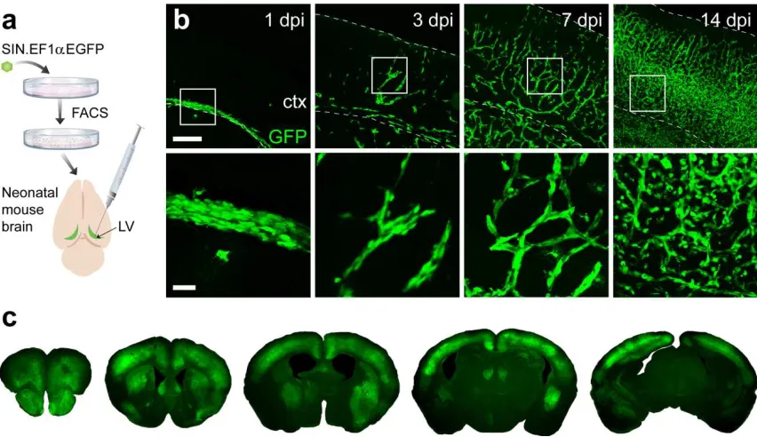
(Snyder et al., 1995). At 1 day post-injection (DPI), donor NSCs occupied periventricular regions
(Fig. 2.1b), at 3 DPI we observed chains of migrating NSCs, and by 14 DPI in vivo expansion
resulted in robust cortical engraftment throughout the neuroaxis (Fig. 2.1c). To phenotype donor
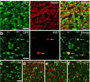
cells, we performed immunofluorescence analysis 2 months post-transplant (Fig. 2.2), which
25
Figure 2.1. Engrafted NSCs migrate and proliferate extensively during first two postnatal
weeks. (a) Schematic illustration of intraventricular NSC transplantation in neonatal rodent brain.
(b) Trajectory of transplanted NSCs during first two postnatal weeks. Lower panels are magnified
view (4x) of boxed region in upper panels. (c) Representative coronal sections along rostrocaudal
axis feature stable cortical grafts at 8 wks post-transplant. (Scale bars in b: 250 µm, Upper; 50
26
Figure 2.2. Exogenous NSCs show limited differentiation potential in vivo. (a) Cortical grafts
are immunopositive for nestin, a marker of undifferentiated NSCs, at 8 wks post-transplant. (b)
GFP-labeled cells were largely quiescent, with only a small percentage continuing to proliferate,
as indicated by Ki67 immunonoreactivity. (c-f) Exogenous NSCs show no evidence of
differentiation into mature neural lineages, as suggested by absence of DCX, βIII-tubulin, NeuN,
27
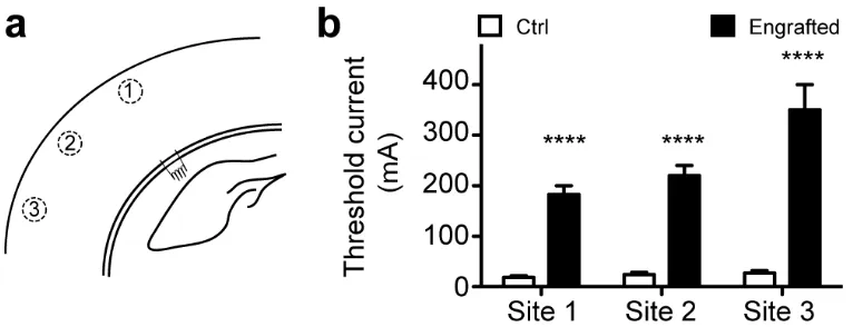
Dose-Dependent Effects on Amplitude of Cortical Activation
We previously found that stable engraftment of ectopic NSCs caused no gross behavioral
abnormalities (Snyder et al., 1995). However, it is unclear whether high density of engraftment in
some areas could disrupt existing neural networks. To investigate whether cortical dynamics were
influenced by engraftment density, NSC levels were titrated in vivo using three different input
doses (80,000, 40,000, and 8,000 cells/ventricle). We quantified engraftment using
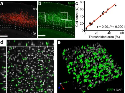
two-dimensional confocal projections of each slice and expressed values as percent GFP-positive
area normalized to total cortical area (Fig. 2.3a). Automated cell counts on an independent set of
slices validated this measurement method. Graft area measurements strongly correlated to cell
counts (Pearson’s correlation r=0.99 p<0.0001), and thus served as a metric for NSC engraftment
level (Figs. 2.3b-e).
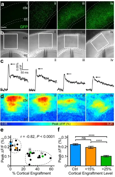
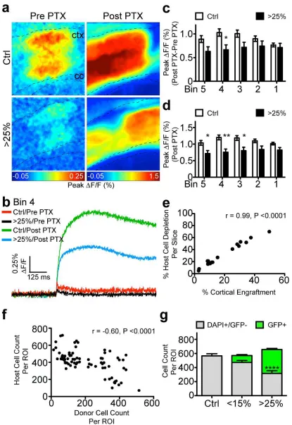
Optical recordings were made in acute slices ofsomatosensory cortex at 2 months
post-transplant in response to a single callosal stimulation (Fig. 2.4a,b). We observed a progressive
reduction in peak signal amplitude (∆F/F0) with increased cortical engraftment, suggesting that
exogenous NSCs can modulate network excitability (Fig. 2.4c). To determine the locus of
dampened cortical activity, we generated color-coded maps depicting maximum ∆F/F0 for
individual pixels across all movie frames (Fig. 2.4d). We observed a strong negative correlation
between engraftment level and corresponding peak ∆F/F0 values (Pearson’s correlation r=-0.82
p<0.0001) (Fig. 2.4e). K-means clustering of maximum ∆F/F0 values partitioned the slices into
three engraftment densities: control, moderate, and high (Fig. 2.4f). We expressed engraftment
as percent GFP-positive area normalized to total cortical area. Whereas high levels (> 25%)
caused marked reductions in the amplitude of activation (0.10±0.01% vs.0.22±0.01%, p<0.0001),
moderate levels (<15%) did not alter this network property (0.19±0.01% vs. 0.22±0.01%, p>0.05).
In accordance with these results, we found that highly engrafted slices had elevated thresholds of
cortical activation, as determined by local field recordings (Fig. 2.5). Furthermore, the injection
procedure itself did not significantly perturb host physiology (Figs. 2.6a,b). Collectively, these
28
Figure 2.3. Exogenous NSCs exhibit robust levels of engraftment in cortex. (a) Maximum
intensity projection showing thresholded GFP+ graft at 8 wks (red mask represents all pixel
intensities ≥ 2 SD above mean background intensity). (b) Automated counts performed on 5
randomly selected cortical ROIs (white boxes) validate graft area measurements. (c) Correlation
plot with linear fit comparing quantitation methods from a and b (n=16 slices). (d) Representative
optical plane from engrafted ROI in E showing colocalization of GFP and DAPI fluorescence. (e)
3-D reconstruction of engrafted ROI in E rendered from confocal z-stack. (Scale bars in a and b:
29
Figure 2.4. NSC engraftment reduces the amplitude of cortical activation in a