DESIGN AND CHARACTERIZATION OF CLARITHROMYCIN BEADS USING A BIODEGRADABLE CATIONIC POLYMER
5
0
0
Full text
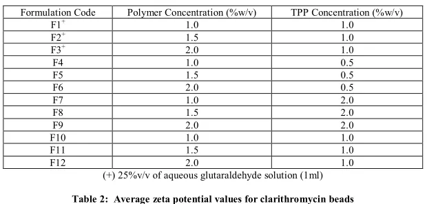
(2) P Jeyaprabha et al. Int. Res. J. Pharm. 2013, 4 (3) Surface Charge Measurements The surface charge on control and chitosan beads containing clarithromycin were determined by Zeta sizer (NANO ZS, ZEN 3600, MALVERN Company) in de ionized water and in S.G.F pH 1.2 and in S.I.F pH 7.4. Determination of Drug Load Clarithromycin content in beads were determined by digestion method. Drug loaded beads (150mg) were pulverized and incubated in HCl buffer pH 1.2. The suspension was sonicated and vibrated in a water bath for 150 min. Then, it was cooled to room temperature. The supernatant was assayed spectrophotometrically for clarithromycin content at 210 nm. Supernatant from empty beads (without drug) was taken as a blank. All the samples were analyzed in triplicate.. Equilibrium Swelling Studies A known weight (100mg) of clarithromycin beads (prepared with 2% chitosan) from formulations F3, F6, F9 and F12 were placed in various solutions like deionized water, enzyme-free simulated gastric fluid (HCl / NaCl solution; pH 1.2) and enzyme-free simulated intestinal fluid ((KH2PO4) NaOH buffer; pH 7.4), allowed to swell for 4 hours at 37 ± 0.50C.The beads were periodically removed, blotted with filter paper and their change in weight was measured until equilibrium swelling was attained. Swelling ratio (SR) was calculated for swollen beads using the formula, SR= (Wc – Wo) / Wo Where Wo is the initial weight of dry beads and Wc is the weight of the swollen beads after equilibrium swelling. The experiment was repeated three times and the average value ± S.D was taken as SR.. Table 1: Formulation variables for clarithromycin beads Formulation Code F1+ F2+ F3+ F4 F5 F6 F7 F8 F9 F10 F11 F12. Polymer Concentration (%w/v) TPP Concentration (%w/v) 1.0 1.0 1.5 1.0 2.0 1.0 1.0 0.5 1.5 0.5 2.0 0.5 1.0 2.0 1.5 2.0 2.0 2.0 1.0 1.0 1.5 1.0 2.0 1.0 (+) 25%v/v of aqueous glutaraldehyde solution (1ml). Table 2: Average zeta potential values for clarithromycin beads Formulation Code Zeta potential (mV) 8.37±0.21 A B 5.72±0.12 C 16.24±0.86 11.36±0.07 D A= chitosan beads without drug; B= chitosan beads with drug; C= crosslinked beads without drug; D= crosslinked beads with drug Table 3: Determination of equilibrium swelling ratio of clarithromycin beads Formulation Code+ F3 F6 F9 F12. Deionized water S.G.F pH 1.2 0.75±0.11 1.26±0.15 0.360±0.054 0.56±0.21 0.04±0.01 0.18±0.02 0.811±0.014 2.85±0.28 (+) 2%w/v of chitosan solution. S.I.F pH7.4 0.45±0.18 0.47±0.18 0.16±0.04 0.91±0.20. Table 4: In vitro release kinetics of clarithromycin from chitosan beads Formulation Code F1 F2 F3 F4 F5 F6 F7 F8 F9 F10 F11 F12. Zero order 0.9836 0.9656 0.9706 0.9938 0.9648 0.9604 0.9821 0.9648 0.9804 0.9745 0.9685 0.9643. Co.efficient of variation ‘r’ values First order 0.9886 0.9919 0.9925 0.9735 0.9685 0.9808 0.9278 0.9696 0.9855 0.9696 0.9927 0.9886. Higuchi equation 0.9975 0.9965 0.9940 0.9899 0.9954 0.9937 0.8629 0.9953 0.9949 0.9961 0.9953 0.9956. Page 178.
(3) P Jeyaprabha et al. Int. Res. J. Pharm. 2013, 4 (3). Figure: 1 FTIR spectral analysis of drug and excipients physical mixture 1.6 1.4. Average particle size (mm). 1.2 1 0.8 0.6 0.4 0.2 0 F1. F2. F3. F4. F5. F6. F7. F8. F9. F10. F11. F12. Formulation code. Figure: 2 Average particle size of clarithromycin beads prepared using chitosan. Bars represent mean ±S.D. (n=3). Figure: 3(a) SEM photographs of clarithromycin beads prepared with 2% TPP. Figure: 3(b) SEM photographs of clarithromycin beads prepared with aqueous glutaraldehyde solution. Page 179.
(4) P Jeyaprabha et al. Int. Res. J. Pharm. 2013, 4 (3) 40 35. Percentage loading. 30 25 20 15 10 5 0 F1. F2. F3. F4. F5. F6. F7. F8. F9. F10. F11. F12. Formulation code. Figure: 4 Percentage of clarithromycin loaded in chitosan beads. Bars represent mean±S.D. (n=3) 120. 100. Percent drug release. 80 F1 F2. 60. F3 F4. 40. F5 F6. 20. 0 0. 1. 2. 3. 4. 5. 6. 7. 8. 9. -20 Time (h). Figure: 5 Percentage of clarithromycin released from formulations F1 to f6 as a function of time at 370C in S.G.F (pH 1.2). Bars represent mean ±S.D. (n=3). 120. 100. Percent drug release. 80 F7 F8. 60. F9 F10. 40. F11 F12. 20. 0 0. 1. 2. 3. 4. 5. 6. 7. 8. 9. -20 Time (h). Figure: 6 Percentage of clarithromycin released from formulations F7 to F12 as a function of time at 370C in S.G.F (pH 1.2). Bars represent mean ±S.D. (n=3). Page 180.
(5) P Jeyaprabha et al. Int. Res. J. Pharm. 2013, 4 (3) In Vitro Release Study The release rate of clarithromycin from chitosan beads were studied using USP dissolution testing apparatus I (basket method). The dissolution test was performed using 900 ml HCl buffer pH 1.2 at 37± 0.50C with an agitated speed of 100 rpm. About 5 ml of sample was withdrawn at appropriate time intervals, filtered through 0.45mm membrane filter and the absorbance was measured spectrophotometrically at 210 nm. The volume withdrawn was replaced with fresh medium and adjusted to 900ml every time. All the batches were studied in triplicate. RESULTS The physical mixture of drug and polymer was characterized by FTIR spectral analysis for any significant interaction between the drug and the excipients used during the formulation. From the results, it was evident that there was no interference in the functional groups and the principle peaks of clarithromycin were found to be unaltered in the drug-polymer physical mixture, indicating they were chemically compatible, which was shown in Figure 1. Clarithromycin beads were prepared by ionotropic gelation method using chitosan as a polymer which were shown in Table 1. The average particle size of clarithromycin beads ranges between 0.87 ± 0.020mm and 1.394 ±0.025mm were shown in Figure 2. The particle size was greater for batches prepared with high concentration of TPP (2%) but showed irregular beads [Figure 3(a)]. Formulations (F1, F2, and F3) those prepared with aqueous glutaraldehyde solution showed uniform spherical particles Figure 3(b). Formulations (F4, F5, and F6) showed flat irregular particles (upon drying). The surface charge for drug loaded and unloaded beads were measured using zeta sizer in deionized water, S.G.F pH 1.2 and in S.I.F pH 7.4. The average zeta potential for drug loaded and unloaded beads were +16.24mV and +8.37mV respectively which were shown in Table 2. Figure: 4 showed that the drug content was increased up to 37.83 % upon addition of aqueous glutaraldehyde solution. A low drug entrapment was observed when the concentration of TPP was kept low (0.5%) but the high TPP concentration (2%) does not showed much deviation in drug content of the clarithromycin beads. Swelling ratio was conducted up to 4 hours in S.G.F pH 1.2, S.I.F pH 7.4 and in deionized water. The results indicated that the degree of swelling was influenced greatly by pH of the medium, greater swelling was observed at lower pH value (S.G.F) than in S.I.F and in de ionized water. The study also reveals that the concentration of polymer have significant role in determining the swelling character of bead. Formulation F3 showed reduced swelling than F9 and F12 but F6 could not maintain the shape and formed gel due to low TPP concentration which was shown in Table 3. The in vitro drug release was studied using S.G.F pH 1.2 at 37 ± 0.50C shown in Figure 5 and 6. Formulations prepared with low TPP concentrations (F4, F5 and F6) released almost 98.9% within 7 hours whereas F2 showed 96.2% at 8 hours. Drug release was faster for formulations F10, F11, F12 compared to F1, F2 and F3. Therefore from the above study it was understood that those batches (F1, F2, and F3) prepared with glutaraldehyde were able to sustain the drug release for longer time than those prepared without glutaraldehyde. The release data were fitted to models representing zero order,. first order and Higuchi’s square root of time. The linear regression analyses were summarized in Table 4. DISCUSSIONS In conclusion, clarithromycin beads prepared with cationic polymer has the possibility of sustaining the drug release over the period of at least 8 hours to increase the local concentration of drug in the stomach. The coefficient of determination of ‘r’ values for different formulations indicated that the drug release from clarithromycin beads followed a first order diffusion controlled mechanism of drug release. ACKNOWLEDGEMENTS The authors would like to acknowledge Dr. Sasval (Birla Institute of Technology, Ranchi, India) and Dr. Padmavathi (Perundurai Medical College, Perundurai, India) for their timely help and support during this work. REFERENCES 1. Warren, J.R, Marshall, B. Unified curved bacilli on gastric epithelium in active chronic gastritis. Lancet 1983; 321: 1273-1275. http://dx.doi.org/ 10.1016/S0140-6736(83)92719-8 2. Megraud, F., Lamouliatte, H., Helicobacter pylori and duodenal ulcers. Dig.Dis.Sci. 1992; 37: 769-772. http://dx.doi.org/10.1007/BF01296437 PMid:1563322 3. Forman, D., et al H.pylori and gastric cancer. Lancet 1994; 343: 243-244 http://dx.doi.org/10.1016/S0140-6736(94)91034-0 4. Peterson, W.L., Helicobacter pylori and peptic ulcer disease. N.Engl. med. 1991; 324: 1043-1048. 5. Suleymanlar, I., et al Response to triple treatment with omeprazole, amoxicillin and clarithromycin for Helicobacter pylori infections in continuous ambulatory peritoneal dialysis patients. Adv.Perit.Dial .1999; 15:79-81 PMid:10682076 6. Vakil, N., Cutler, A., Ten-day triple therapy with Ranitidine, Bismuth citrate, Amoxicillin and clarithromycin in eradicating Helicobacter pylori. Am. J. Gastroenterol. 1999; 94(5): 1197-1199. http://dx.doi.org /10.1111/j.1572-0241.1999.01065.x PMid:10235192 7. Ito, M., Ban,A., Ishihara, M., Anti-ulcer effects of chitin and chitosan, healthy foods, in rats.Jpn. J. Pharmacol. 2000; 82: 218-225. http:// dx.doi.org/10.1254/jjp.82.218 PMid:10887952 8. Lehr, C.M., Bouwstra, J.A., Schacht, E.H., Junginger, H.E., In vitro evaluation of mucoadhesive properties of chitosan and some other natural polymer. Int. J. Pharm. 1992; 78: 43-48. http://dx.doi.org/ 10.1016/0378-5173(92)90353-4 9. Needleman, I.G., Smales, F.C., In vitro assessment of bio adhesion for periodontal and buccal drug delivery. 1995; 16: 617-624. 10. Rillosi, M., Buckton, G., Modelling mucoadhesive by use of surface energy terms obtained from the lewis acid-lewis base approach. II. Studies on anionic, cationic and un ionisable polymers. Pharm. Res. 1995; 12: 669-675 http://dx.doi.org/10.1023/A:1016299223369 PMid:7479551 11. He, P., Davis, S.S., Illum, L., In vitro evaluation of mucoadhesive properties of chitosan microspheres. Int. J. Pharm. 1998; 166:75-88. http://dx.doi.org/10.1016/S0378-5173(98)00027-1 12. Shimoda, J., Onishi, H., Machida, Y., Bioadhesive characteristics of chitosan microspheres to the mucosa of rat small chitosan-intestine. Drug Dev. Ind. Pharm.2001; 27: 567-576. http://dx.doi.org/10.1081/ DDC-100105182 PMid:11548864 13. Kockisch, S., Rees, G.D., Young, S.A., et al polymeric microspheres for drug delivery to the oral cavity: An in vitro evaluation of mucoadhesive potential. J. Pharm.sci. 2003; 92: 1614-1623 http://dx.doi.org/10.1002 /jps.10423 PMid:12884248 14. V.R. Singla., A.K. Sindla., et al chitosan microspheres as a potential carrier for drugs. Int. J. Pharm. 2004; 274: 1-33. http://dx.doi.org /10.1016/j.ijpharm.2003.12.026 PMid:15072779 15. Calvo, P., et al Novel hydrophilic chitosan-polyethylene oxide nanoparticles as protein carriers. J. Appl. Sci. 1997; 63: 125-132 http://dx.doi.org/10.1002/(SICI)1097-4628(19970103)63:1<125::AIDAPP13>3.0.CO;2-4 Cite this article as: P Jeyaprabha, T Sudhamani. Design and characterization of Clarithromycin beads using a biodegradable cationic polymer. Int. Res. J. Pharm. 2013; 4(3):177-181. Source of support: Nil, Conflict of interest: None Declared. Page 181.
(6)
Figure
Related documents
Present study focuses on different aspects of habitat ecology, adaptability to artificial habitat, floral biol- ogy, reproductive biology and seed biology of Paris
Bycatch of the Lake Pontchartrain Basin inshore shrimp fishery Bycatch of the Lake Pontchartrain Basin inshore shrimp fishery and its effect on two sea catfish species:
Iranian Women Writers, Islamic Feminism, Iranian Nationalism, Gender, Literary Subterfuge, Censorship... Introduction:
In the United States, a significant proportion of child- hood asthma may be attributable to modifiable risk factors including acute viral respiratory infections, antibiotic use,
On the other hand, when we located the targets outside the PlanetLab network, we observed a number of location underestimations.. However, this is the global IP
The action plan at the end of this report identifies where improvements are needed to meet the requirements of the Health Act 2007 (Care and Welfare of Residents in Designated
The proposed techniques begin by detecting the hand, tracking the hands movements and analysing the variations in the hand locations (i.e. Motion detection), and
