Congenital Salivary Gland Anlage Tumor of the Nasopharynx
Erik G. Cohen, MD*; Michael Yoder, MD‡; Rebecca M. Thomas, MD‡; Denise Salerno, MD, FAAP§; and Glenn Isaacson, MD, FACS, FAAP*
ABSTRACT. Objective. Nasal and upper respiratory tract obstruction in the neonatal period can result from a variety of conditions, and may present with variable symptoms. In the absence of dysmorphic features or other abnormalities, causes of nasal obstruction may be difficult to differentiate on initial examination. We re-port an unexpected and potentially life-threatening con-dition arising during the work-up of this common neo-natal complaint.
Design. Case report with literature review.
Results. A male neonate presented with complaints of nasal obstruction and feeding difficulties. A common diagnostic approach to neonatal nasal obstruction was performed, resulting in an unexpected and potentially life-threatening, albeit curative, result. Cannulation of the nasal cavity to rule out choanal atresia resulted in a burst of bleeding from the nose and mouth. A finger sweep of the oropharynx produced a dislodged mass lesion. Pathology revealed a salivary gland anlage tumor of the nasopharynx.
Conclusions. The diagnosis of a nasopharyngeal mass lesion should be considered in neonates with nasal ob-structive symptoms. It is wise to place an index finger in the oropharynx when passing catheters to rule out cho-anal atresia to feel a dislodged mass lesion before it can become an airway foreign body. Should passage of nasal catheters result in bleeding and/or respiratory distress, the possibility of a displaced mass lesion must be con-sidered immediately to institute prompt intervention. Pediatrics2003;112:e66 –e69. URL: http://www.pediatrics. org/cgi/content/full/112/1/e66;nasopharyngeal tumor, sal-ivary gland tumor, congenital tumor, nasal obstruction.
ABBREVIATIONS. SGAT, salivary gland anlage tumor; EMA, epithelial membrane antigen; CT, computed tomography.
N
asal and upper respiratory tract obstructionin the neonatal period can result from a va-riety of congenital malformations, inflamma-tory conditions, hamartomas, and tumors. Obligate nose breathing in neonates makes nasal obstruction potentially life-threatening. Respiratory difficulties may be present at birth, or develop over the first few weeks or months of life. Neonates may present with
symptoms ranging from nasal discharge to feeding difficulties to severe respiratory distress and hyp-oxia.
The causes of nasal obstruction in the neonatal period are varied. These include bony abnormalities, cystic lesions, meningoencephalocele, glioma, nasal mucosal edema, and several infectious causes. Naso-pharyngeal neoplasms less commonly cause nasal obstruction in this age group, but may occur with similar, nonspecific presenting features. In the ab-sence of dysmorphic features or other abnormalities, causes of nasal obstruction may difficult to differen-tiate on initial examination.
Salivary gland anlage tumor (SGAT), also referred to as congenital pleomorphic adenoma, is a benign congenital tumor of the nasopharynx, which may produce nasal obstruction and other associated, non-specific symptoms. We report a case of SGAT caus-ing nasal obstruction and feedcaus-ing difficulties in the neonatal period. A common diagnostic approach to neonatal nasal obstruction was performed, resulting in an unexpected and potentially life-threatening, albeit curative, result. The differential diagnosis and clinical approach to managing neonates with symp-toms of nasal obstruction is discussed.
METHODS
Medical records and pathology reports were retrospectively reviewed. The English language literature was reviewed using a PubMed-based search (www.ncbi.nlm.nih.gov).
A frozen section of the tumor was performed and the diagnosis was made on the routine hematoxylin and eosin-stained perma-nent sections. Immunohistochemistry was performed for confir-mation of the diagnosis, using 5-m sections from paraffin-em-bedded tissue blocks. The staining was performed by the standard avidin-biotin-conjugate technique using antibodies against AE1/ AE3 (Zymed, South San Francisco, CA), vimentin (DAKO, Carpin-teria, CA), S-100 (DAKO), epithelial membrane antigen (EMA; DAKO) and smooth muscle actin (DAKO).
RESULTS Case Report
A full-term boy was born by normal spontaneous vaginal delivery. His mother had had no prenatal care. No difficulties were noted immediately after delivery. He weighed 2835 g (6.2 lbs), and APGAR scores were 8 and 9 at 1 minute and 5 minutes, respectively. He presented to the outpatient pediat-rics clinic with his mother on day of life 4 for weight follow-up. The mother reported breastfeeding the child without difficulty. Physical examination re-vealed nasal congestion. He again presented for eval-uation on day of life 11. His mother was concerned about his nasal congestion, and complained that he From the *Department of Otolaryngology-Head and Neck Surgery, Temple
University School of Medicine, Philadelphia, Pennsylvania; ‡Department of Pathology and Laboratory Medicine, Temple University Hospital, Philadel-phia, Pennsylvania; §Department of Pediatrics, Temple University Chil-dren’s Medical Center, Philadelphia, Pennsylvania.
Received for publication Nov 5, 2002; accepted Mar 4, 2003.
Reprint requests to (G.I.) Department of Otolaryngology, Temple Univer-sity School of Medicine, 3400 N Broad St, Kresge West Building, Suite 102, Philadelphia, PA 19140. E-mail: glenn@ent.temple.edu
had been spitting up his feedings since day of life 5. He had no history of fever or ill contacts.
During this outpatient visit with his pediatrician (D.S.), pediatric otolaryngology consultation was ob-tained in the outpatient pediatric clinic. On initial examination, the patient was found to have normal external ears and craniofacial structure. Anterior rhi-noscopy was within normal limits. The oral cavity and oropharynx were without clefts or other lesions. The neck had no palpable masses or lymphadenop-athy. There was no respiratory distress or stridor noted at rest; however, the patient was mouth breathing.
A flexible suction catheter was then inserted into the nose to rule out choanal atresia. Its passage was anticipated with a finger in the oropharynx. Mild resistance was encountered in the posterior nasal cavity, followed by a burst of bleeding from the nose and mouth. A finger sweep of the pharynx produced a 1.5-cm spherical mass. Bleeding slowed and then stopped spontaneously after several minutes. Imme-diate resolution of mouth-breathing and improved feeding was seen. The mass was then sent to pathol-ogy for frozen section analysis, as well as routine pathologic examination.
An unenhanced computed tomography (CT) scan
of the head and neck was then obtained. There were no defects in the skull base, nor was any residual mass seen in the nasopharynx.
The patient has done well since the mass was removed. A follow-up CT scan of the head revealed no evidence of recurrence or abnormality at 20 months of age.
Pathology
The tumor was a single, firm, roughly spherical mass of tan-yellow tissue with a smooth surface. It measured 1.5 cm in greatest dimension. The cut sur-face was also firm and tan-yellow. Microscopically, the surface was of thin, nonkeratinizing squamous epithelium; the underlying stroma was composed of spindled cells of variable cellularity (Fig 1A). Embed-ded in this stroma were squamous islands and duct-like structures, which often blended in with the stroma (Fig 1, B and C). Areas of dense cellularity were occasionally seen (Fig 1D). Mitoses were present, but not prominent. There was no necrosis and no significant pleomorphism. On immunohisto-chemical studies, the epithelial component was pos-itive for broad-spectrum keratin (AE1/AE3) and EMA. The stromal component was positive for vi-mentin and smooth muscle actin, with scattered
in-Fig 1. Histopathology of SGAT. A, Surface squamous epithelium with underlying epithelial and stromal elements (magnification 200⫻, hematoxylin and eosin stain). B, The underlying stroma with squamous islands (magnification 200⫻, hematoxylin and eosin stain). C, Loosely cellular stroma with small duct-like structures (magnification 100⫻, hematoxylin and eosin stain). D, Densely cellular area with epithelial and spindled cells (magnification 100⫻, hematoxylin and eosin stain).
http://www.pediatrics.org/cgi/content/full/112/1/ at Viet Nam:AAP Sponsored on August 30, 2020 e66 e67
dividual cells positive for S-100 protein. The stromal cells also showed patchy staining for AE1/AE3 and EMA.
DISCUSSION
The term, “salivary gland anlage tumor” was in-troduced in a report by Dehner et al,1in 1994. They
reported 9 cases involving lesions of the nasophar-ynx with similar histologic appearance presenting in the neonatal period. One mass was expelled during resuscitation during an episode of respiratory dis-tress, although most cases presented with respiratory and/or feeding difficulties. Several other cases have been reported in the English language literature.2– 4
The first reported case, described as a congenital pleomorphic adenoma, was reported by Har-El et al5
in 1985. An earlier report of a squamous proliferative lesion presented in strikingly similar fashion to the current case report, and may represent the same type of lesion.6
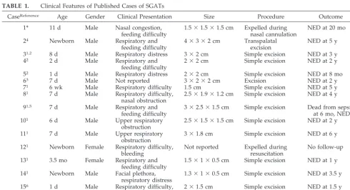
Clinical features of the published cases of SGAT are summarized in Table 1. Thirteen of 15 cases (87%) have been reported in males. The large majority of cases have presented within the first 2 weeks of life. SGATs have been reported as smooth or nodular, midline nasopharyngeal masses. A narrow pedicle may be attached to the nasopharyngeal mucosa1,5or
to the mucosa of the posterior nasal septum.1,2,4No
recurrences have been reported after simple excision with 6 months to 6 years follow-up.1– 6
The pathologic and immunohistochemistry fea-tures of the SGAT, as described by Dehner et al,1
were constant between cases in that report and sim-ilar to the case presented here. The histology of the SGAT suggests that of a normally developing
sali-vary gland, with invaginating surface epithelium into mesenchymal tissue, suggesting that SGAT may be a hamartoma, rather than a neoplasm.1
Symptoms of nasal obstruction from SGAT are similar to nasal obstruction from other causes, and may include sleeping difficulty, feeding problems, episodes of respiratory distress and/or cyanosis, and nasal discharge. Neonates with complete nasal ob-struction may have severe respiratory distress which is relieved by crying7; however, neonates with even
partial nasal obstruction may be symptomatic.8
Re-tractions, stridor, a weak or breathy cry, or respira-tory distress not relieved or worse with crying may indicate laryngotracheal abnormalities.
The cause of neonatal nasal obstruction may some-times be revealed by careful history and physical examination. Maternal history including medication use, substance abuse, and infectious diseases should be elicited. Family history of congenital or childhood abnormalities should be elicited as well.
A child’s general appearance may reveal evidence of a congenital abnormality. Neonates with bilateral choanal atresia may have severe respiratory distress and ⬃75% have other features of the CHARGE as-sociation.9Patients with unilateral atresia have
asso-ciated abnormalities less frequently, but usually present later in life.9,10Patients with nasal pyriform
aperture stenosis may have a single, medial upper incisor, and may have midfacial abnormalities and forms of holoprosencephaly.11Abnormalities such as
mandibulofacial dysostosis (eg, Treacher Collins syndrome and Crouzon disease), coronal craniosyn-ostosis, and midfacial and nasal hypoplasia associ-ated with fetal alcohol syndrome may be apparent on
TABLE 1. Clinical Features of Published Cases of SGATs
CaseReference Age Gender Clinical Presentation Size Procedure Outcome
1* 11 d Male Nasal congestion,
feeding difficulty
1.5⫻1.5⫻1.5 cm Expelled during nasal cannulation
NED at 20 mo
24 Newborn Male Respiratory and
feeding difficulty
4⫻3⫻2 cm Transpalatal excision
NED at 5 y
31,2 8 d Male Respiratory distress 3⫻2 cm Simple excision NED at 3 y
42 2 d Male Respiratory and
feeding difficulty
2⫻2 cm Simple excision NED at 2 y
52 1 d Male Respiratory distress 2⫻2 cm Simple excision NED at 8 mo
63 7 d Male Not reported 3⫻2⫻2 cm Excision NED at 2 y
71 6 wk Male Respiratory difficulty 1.5 cm Simple excision NED at 5 y
81 7 d Male Respiratory difficulty,
nasal obstruction
2.5⫻1.9⫻1.2 cm Simple excision NED at 4 y
91,5 7 d Male Respiratory and
feeding difficulty
3⫻2.5⫻1.5 cm Simple excision Dead from sepsis at 6 mo, NED
101 6 d Male Upper respiratory
obstruction
2.5⫻1.5⫻1.5 cm Simple excision NED at 2 y
111 7 d Male Upper respiratory
obstruction
3⫻1.8 cm Simple excision NED at 6 y
121 Newborn Female Respiratory difficulty,
bleeding
Not reported Expelled during resuscitation
No follow-up
131 3.5 mo Female Respiratory and
feeding difficulty
1.5⫻1⫻0.5 cm Simple excision NED at 1 y
141 Newborn Male Facial plethora,
respiratory distress
1.3⫻1⫻0.5 cm Simple excision NED at 3.5 y
156 1 d Male Respiratory difficulty,
bleeding after nasal cannulation
2⫻1.5 cm Simple excision NED at 1.5 y
initial inspection, and can cause nasal obstruc-tion.12,13
Anterior rhinoscopy may reveal nasal pyriform aperture stenosis or traumatic septal deviation, which may occur in as many as 1% of newborns.14
Nasolacrimal duct cysts can also be seen, and may be associated with epiphora.15 Lack of response of the
nasal mucosa to decongestion may indicate rhinitis medicamentosum with chronic use of topical decon-gestants in the child or vasoactive medications by the mother. Mucopurulent rhinorrhea may indicate con-genital syphilis (“snuffles”), whereas Chlamydial in-fection may cause also cause rhinorrhea and obstruc-tion.10
Neonates with gliomas or skull base defects of the anterior neuropore, eg, meningocele, encephalocele, or meningoencephalocele may present with nonspe-cific symptoms of nasal obstruction,16and may have
a normal anterior rhinoscopy. Neonates with hamar-tomas (eg, SGAT), benign neoplastic lesions (eg, hemangiomas), or rarely, malignant lesions (eg, rhabdomyosarcoma) involving the nasopharynx, may also have nonspecific symptoms of nasal ob-struction,12,16 and frequently have normal anterior
rhinoscopy.
Passage of a soft, flexible suction catheter through the nose, at least 32 mm, can be performed to exclude choanal atresia,16 as was performed in this case.
However, the inability to pass a catheter is not diag-nostic of choanal atresia.8
Fiber-optic endoscopy after topical decongestion can be useful in visualizing lesions of the posterior nasal cavity and nasopharynx. Radiographic imag-ing with CT scan can offer valuable information re-garding bony anatomy of the skull base and poste-rior nasal cavity if the diagnosis is unclear. Magnetic resonance imaging can be used selectively to evalu-ate connections of intranasal lesions with intracranial contents, and other intracranial or soft tissue abnor-malities before diagnostic or therapeutic procedures. Endoscopy under general anesthesia may be per-formed if the diagnosis remains uncertain.
In this case, SGAT was not suspected because it is a relatively uncommon cause of nasal obstruction. A flexible catheter was passed to evaluate patency of the nasal cavity. It is wise to place an index finger in the oropharynx when passing catheters. If the cath-eter is palpated in the nasopharynx, choanal atresia is definitively ruled-out (ie, the catheter is not coiled in the nose). In addition, masses or foreign bodies will be felt before they can fall into the airway.
SGATs are attached only by a thin, delicate vascu-lar pedicle which may be easily torn, even with
gen-tle probing. These lesions are of sufficient size to cause complete upper airway obstruction. Should passage of nasal catheters result in bleeding and/or respiratory distress, the possibility of a displaced mass lesion acting as an airway foreign body must be considered immediately to institute prompt inter-vention.
CONCLUSIONS
Nasal obstruction in the neonatal period may be caused by a wide variety of disorders. SGAT is a recently described hamartoma of the nasopharynx, which presents with symptoms that are difficult to differentiate from other causes of nasal obstruction. Potential for dislodgement of this mass from its delicate pedicle with resultant airway obstruction should be considered when assessing the patency of the posterior choanae.
REFERENCES
1. Dehner LP, Valbuena L, Perez-Atayde A, Reddick RL, Askin FB, Rosai J. Salivary gland anlage tumor (“congenital pleomorphic adenoma”): a clinicopathologic, immunohistochemical and ultrastructural study of nine cases.Am J Surg Pathol. 1994;18:25–36
2. Boccon-Gibod LA, Grangeponte MC, Boucheron S, Josset PP, Roger G, Berthier-Falissard ML. Salivary gland anlage tumor of the nasopharynx: a clinicopathologic and immunohistochemical study of three cases.
Pediatr Pathol Lab Med. 1996;16:973–983
3. Michal M, Sokol L, Mukensnabl P. Salivary gland anlage tumor. A case with widespread necrosis and large cyst formation.Pathology. 1996;28: 128 –130
4. Bondeson L, Andreasson L, Olsson M, Rausing A. Salivary gland anlage tumor: cytologic features in a case examined by fine-needle aspiration.
Diagn Cytopathol. 1997;160:518 –521
5. Har-El G, Zirkin HY, Tovi F, Sidi J. Congenital pleomorphic adenoma of the nasopharynx (report of a case).J Laryngol Otol. 1985;99:1281–1287 6. Stillwater L, Fee WE. Squamous cell proliferative lesion of the
naso-pharynx in a newborn.Otolaryngol Head Neck Surg. 1980;88:240 –247 7. Fearon B, Dickson J. Bilateral choanal atresia in the newborn: plan of
action.Laryngoscope. 1968;78:1487–1498
8. Shott SR, Myer III CM, Willis R, Cotton RT. Nasal obstruction in the neonate.Rhinology. 1989;27:91–96
9. Duncan NO, Miller RH, Catlin FI. Choanal atresia and associated anomalies: the CHARGE association.Int J Pediatr Otolaryngol. 1988;15: 129 –135
10. Coates H. Nasal obstruction in the neonate and infant.Clin Pediatr. 1992;31:25–28
11. Van Den Abbeele T, Triglia JM, Francois M, Narcy P. Congenital nasal pyriform aperture stenosis: diagnosis and management of 20 cases.Ann Otol Rhinol Laryngol. 2001;110:70 –75
12. Bluestone CD, Stool SE, Kenna MA.Pediatric Otolaryngology.3rd ed. Philadelphia, PA: W B Saunders Co; 1996
13. Usowicz AG, Golabi M, Curry C. Upper airway obstruction in infants with fetal alcohol syndrome.Am J Dis Child. 1986;140:1039 –1041 14. Jaffee BF. Classification and management of anomalies of the nose.
Otolaryngol Clin NA. 1981;14:989 –1004
15. Lusk RP, Muntz HM. Nasal obstruction in the neonate secondary to nasolacrimal duct cysts.Int J Pediatr Otolaryngol. 1987;13:315–322 16. Myer III CM, Cotton RT. Nasal obstruction in the pediatric patient.
Pediatrics. 1983;72:766 –777
http://www.pediatrics.org/cgi/content/full/112/1/ at Viet Nam:AAP Sponsored on August 30, 2020 e66 e69
DOI: 10.1542/peds.112.1.e66
2003;112;e66
Pediatrics
Isaacson
Erik G. Cohen, Michael Yoder, Rebecca M. Thomas, Denise Salerno and Glenn
Congenital Salivary Gland Anlage Tumor of the Nasopharynx
Services
Updated Information &
http://pediatrics.aappublications.org/content/112/1/e66
including high resolution figures, can be found at:
References
http://pediatrics.aappublications.org/content/112/1/e66#BIBL
This article cites 15 articles, 1 of which you can access for free at:
Subspecialty Collections
sub
http://www.aappublications.org/cgi/collection/hematology:oncology_ Hematology/Oncology
following collection(s):
This article, along with others on similar topics, appears in the
Permissions & Licensing
http://www.aappublications.org/site/misc/Permissions.xhtml
in its entirety can be found online at:
Information about reproducing this article in parts (figures, tables) or
Reprints
http://www.aappublications.org/site/misc/reprints.xhtml
DOI: 10.1542/peds.112.1.e66
2003;112;e66
Pediatrics
Isaacson
Erik G. Cohen, Michael Yoder, Rebecca M. Thomas, Denise Salerno and Glenn
Congenital Salivary Gland Anlage Tumor of the Nasopharynx
http://pediatrics.aappublications.org/content/112/1/e66
located on the World Wide Web at:
The online version of this article, along with updated information and services, is
by the American Academy of Pediatrics. All rights reserved. Print ISSN: 1073-0397.
the American Academy of Pediatrics, 345 Park Avenue, Itasca, Illinois, 60143. Copyright © 2003 has been published continuously since 1948. Pediatrics is owned, published, and trademarked by Pediatrics is the official journal of the American Academy of Pediatrics. A monthly publication, it
at Viet Nam:AAP Sponsored on August 30, 2020 www.aappublications.org/news