R E V I E W
Transforaminal Lumbar Interbody Fusion For
Lumbar Degenerative Disease: Patient Selection
And Perspectives
This article was published in the following Dove Press journal: Orthopedic Research and Reviews
Bekir Yavuz Uçar
Çağri Özcan
Ömer Polat Tayfun Aman
University of Health Sciences, Umraniye Education and Research Hospital, Department of Orthopaedics and Traumatology, Istanbul, Turkey
Abstract: Most adults will experience low back pain during their lifetime, with most of these instances resolving or improving without sequelae in a few weeks. For the small number of patients with severe, recalcitrant pain, lumbar fusion may be required, particularly when concomitant leg pain or deformity is present. Lumbar interbody fusion surgery is the usual treatment for degenerative lumbar disease, but it requires a long recovery period. Many surgical techniques have been described in the literature for spondylolisthesis. The main objective is to create interbody fusion, decompression of normal structures and a stable vertebrae. TLIF surgical techniques has a long learning curve. Comorbidities of the patient may make surgery more difficult. Methods such as transforaminal lumbar interbody fusion (TLIF), posterior lumbar interbody fusion, anterior lumbar interbody fusion and lateral lumbar interbody fusion are also available for interbody fusion in the literatüre. The aim of this review is to show which patients are more suitable for TLIF surgery and to discuss the advantages and disadvantages of TLIF surgery over other techniques.
Keywords:low back pain, transforaminal lumbar interbody fusion, lumbar spinal stenosis, degenerative spine disease, lumbar fusion, lumbar spondylosis
Introduction
Most adults will encounter low back pain amid their lifetime, with the greater part of these situations settling or improving without sequelae. For the modest number of patients with severe, persistent pain, lumbar fusion might be required, especially when attendant leg pain or deformity is existing.1
Nowadays, transforaminal lumbar interbody fusion (TLIF) is a commonly performed operation used to treat degenerative lumbar diseases. A variety of fusion techniques applied where necessary are available. TLIF is a spinal fusion procedure that fuses the anterior and posterior sections of the spine through a posterior approach which was developed by Harms.2,3Also, TLIF is an alternative technique that is used to archive disc resection, neural decompression and circumferential arthrodesis in the lumbar spine. The purpose of the present study was to investigate the patient selection and the perspectives of TLIF for lumbar degenerative diseases.
Patient Selection For TLIF
Despite the fact that the etiology of low back pain stays multifactorial, degenerative changes in the intervertebral discs of the spine have been emphatically connected Correspondence: Çağri Özcan
University of Health Sciences, Umraniye Education and Research Hospital, Department of Orthopaedics and Traumatology, Elmalikent Mh 34764 Umraniye, Istanbul, Turkey Email cagriozcann@gmail.com
Orthopedic Research and Reviews
Dove
press
open access to scientific and medical research
Open Access Full Text Article
Orthopedic Research and Reviews downloaded from https://www.dovepress.com/ by 118.70.13.36 on 26-Aug-2020
with the start of pain. Learning of the pathophysiology of the disc degeneration can help in the decision of treatment and to improve tissue engineering for biological restora-tion of degenerated discs. Intervertebral disc degenerarestora-tion includes a derangement in anabolic and catabolic activity within the annulus fibrosus and nucleus pulposus cells.4 As age increases, the water content of the intervertebral disc decreases andfissures may occur in the nucleus pul-posus, potentially extending to annulus fibrosus, and the onset of this process, called chondrosis intervertebralis, can mark the onset of degenerative destruction of the intervertebral disc, end plates, and vertebral bodies.5 There are many factors affecting the extracellular environ-ment around intervertebral disc cells, including nutrient levels, mechanical loading, and chemical factors. The change in disc cellular viability and activity affects the ability of annulus fibrosus and nucleus pulposus cells to produce extracellular matrix components and maintain tissue health.4
Patients with lumbar disc disease often present with numerous symptoms including pain, radicular symptoms, and weakness. Flexion often aggravates the symptoms, whereas extension prolongs them. When examining patients with predicted lumbar degenerative disc disease, it is important to exclude other possible etiologies known for pain. Abdominal pathologies such as aortic aneur-ysms, pancreatic disease, and kidney stones should be excluded.5
Although there are some classifications of disc degen-eration classified as radiological, the most frequently used methods are described by Mimura et al and Pathria et al.
Mimura et al described the rating-system I–IV with changes in disk height, osteophytes, and end plate sclerosis on radiographs anterior/posterior and lateral.6 (Table 1).
Pathria et al described the rating-system 1–4 with joint space narrowing, sclerosis, and hypertrophy on oblique radiographs7(Table 2).
Also, macroscopic and microscopic classification of disk degeneration can be made. Macroscopic disk degen-eration can be graded A–D according to the criteria devel-oped by Nachemson8(Table 3).
For the purpose of microscopic classification, the spe-cimens are arranged in four groups A–D (Table 4) accord-ing to thefindings of reactive chondrocytes, necrosis, and
fissures; a modified version of the general criteria put forth by Vernon-Roberts.9
The basic surgical principle is to provide stability and fusion in adult lumbar deformity. We can list the indica-tions of TLIF under the basic neurological spine surgery principle.10
Indications
● Grade 1 and 2 spondylolisthesis (degenerative or lytic) with mechanical lumbar pain or radicular syndromes,
● Reduced high-grade spondylolisthesis,
● Central canal stenosis,
Table 1Radiographic System For Grading Disk Degeneration On Antero-Posterior And Lateral Radiographs
Lumbar Radiographs In Anterio-Posterior And Lateral Position
Grade Of Disk Degeneration Disk Height Changes (% Of Adjacent Disc) Osteophytes Formationa End Plate Sclerosis
Normal 0 = 100% 0 0 points 0: None
I 0–1 1 > 75% 1 1–4 points 1: Either end plate
II 2–3 2 > 50% 2 5–8 points 2: Both end plates
III 4–6 3 > 25% 3 9–12 points
IV 7–10 4 < 25% 4 13–16 points
a
Sum of points on eight edges <3 mm 1 pt, >3 mm 2 pt. Data used from Mimura et al.6
Table 2Grading Of Facet Joint Disease On Oblique Radiographs
Lumbar Radiographs In Oblique Position
Grad Of Facet Degeneration
Changes Of The Facet Joint
0 No changes
1 Joint space narrowing
2 Narrowing plus sclerosis or hypertrophy 3 Severe osteoarthritis with beginning
narrowing, sclerosis, and osteophytes 4 Advanced osteoarthritis with hypertrophy,
narrowing, sclerosis, and osteophytes
Modified data used from Pathria et al.7
Orthopedic Research and Reviews downloaded from https://www.dovepress.com/ by 118.70.13.36 on 26-Aug-2020
● Lateral recess syndrome,
● Facet joint disease,
● Severe discogenic back pain,
● Lumbar segmental instability,
● Recurrent disc herniation,
● Postlaminectomy instability,
● Treatment of pseudarthrosis,
● Failed lumbar fusion with other techniques.
Contraindications
● High-grade spondylolisthesis,
● Severe osteoporosis,
● Presence of active infection,
● Malignities,
● Traumatic instability,
● Other diseases that prevent surgery.
Surgical Technique Of TLIF
The patient is anesthetized under general anesthesia. One gram of cefazolin is administered prophylactically.26 Baseline values were evaluated after neuromonitoring was started. Afterward, the patient is placed in the prone posi-tion and the basal values recorded again. After proper sterilization, the patient is prepared for surgery. The preop planned level is determined by C-armfluoroscopy. A stan-dard midline incision is made. With dissection, the para-vertebral muscles are carefully retracted. The dissection is completed so as to reveal transverse processes. Polyaxial pedicle screws are placed on both sides to the specified levels. Specially prepared retractors for distraction are placed on the screw heads. Distraction is performed with special retractor. In this way, instead of performing lami-nectomy, we have an adequatefield of view and application with the inferior facet joint resection of the upper segment of the facet joint. We prefer partial facetectomy through pathologic side determined with preoperative examination, MRI image, and intraoperative neuromonitoring (neural integrity monitor, NIM) values. The main priorities include preoperative examination, MRIfindings, and complaints of the patient. If the patient’s complaints are unilateral, then unilateral decompression is performed. If there are com-plaints in both lower extremities, we decide to perform on the laminectomy and ligamentumflavum excision with the lower NIM values. According to the change in NIM values, we decide to perform laminectomy on the other side. We try not to decompress the posterior elements completely to achieve fusion. The bleeding control is preferably per-formed with the help of a bipolar cautery. A gentle retrac-tion is made with the dura retractor. Thus, root and dura decompression is performed. The disc level is reached between the root and the dura, and the disc content is carefully excised. Upper and lower end plates are excised with curette. The disc and the end plate residues are com-pletely removed by washing thoroughly with water. The Table 3Classification Of Macroscopic Patho-Anatomic Changes
Associated With Disk Degeneration According To Nachemson
Degenerative Disk Disease Assessed By Macroscopic Inspection
Grade Of Degeneration
A Disks without changes visible to the naked eye. In these cases, a gelatinous shiny nucleus pulposus was seen; it was easily delimited from the annulusfibrosus, which was free from macroscopic ruptures from the annulusfibrosus
B Disks that showed macroscopic changes in the nucleus pulposus. The nucleus was somewhat morefibrous, but could be clearly distinguished from the annulus, which was intact
C Specimens that showed macroscopic changes in both the nucleus pulposus and the annulusfibrosus. The nucleus in these discs was morefibrotic but still soft. The boundary between nucleus and and annulus was no longer so distinct, but could be seen. Changes in the annulusfibrosus consisted of isolatedfissures D Specimens that showed more severe macroscopic changes. The
disk in this group exhibitedfissure formation and cavities in both the nucleus and the annulus. Marginal osteophytes were often found in adjoining vertebrae
Table 4 Intervertebral Disk Degeneration Classified According To A Modified Version Of The Microscopic Criteria Of Vernon-Roberts
Intervertebral Disc Degeneration Assessed By Microscopic Examination
Grading Of Degeneration
Reactive Chondrocytes
“Brutkapseln”
Fissures, Clefts, Splints
Areas Of Necrosis Damage Of Annular layers
A Few Isolated,flat Isolated, small 0–1 ring
B Moderate Ample,flat Several, focal 1–2 rings
C Ample Numerous deep Multiple, partly confluent 2–3 rings
D A lot Numerous very deep Great, diffuse extended 3–4 rings
Orthopedic Research and Reviews downloaded from https://www.dovepress.com/ by 118.70.13.36 on 26-Aug-2020
resulting grafts are placed in the disc space and the TLIF cage is placed appropriately. Retractors are removed after NIM control. Compression and fixation are provided with the help of rods. After manipulation, NIM control is per-formed again. After the control, a Hemovac drain is placed under the skin and the layer above the drain skin is closed (Figures 1–4).
The patient is mobilized on the first postoperative day. We do not recommend any brace or orthosis to our patients. Clinical studies have shown that using brace
after TLIF surgery is not effective.27 Hemovac drain is kept until 50 cc per day and is removed when less than 50 cc. We do not use any antibiotics other than prophy-lactic antibiotics.26 After the removal of Hemovac drain, the patient is discharged and the wound is found to be clean. The patient is followed up in the outpatient clinic at 15-day intervals. For the first 6 weeks, walking pro-gram is applied to the patient. After 6 weeks, the phy-sical therapy program is started and ap/lateral direct radiographies are taken. We do not perform CT for our patients in the first year unless needed. In this phy-siotherapy program, the patient is intended to perform Figure 1Preoperative CT images.
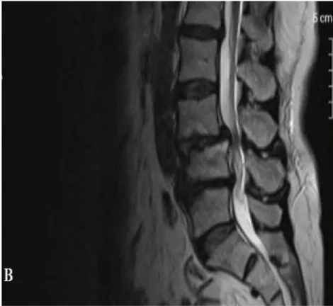
Figure 2Preoperative MR images.
Figure 3Preoperative AP view.
Figure 4Preoperative lateral radiographs view.
Orthopedic Research and Reviews downloaded from https://www.dovepress.com/ by 118.70.13.36 on 26-Aug-2020
range-of-motion exercises and strengthening exercises. Physical therapy is recommended until 3 months. Until this time, contact sports are not allowed. Full activity and return to work are allowed after 6 months. The patient is followed up for up to 1 year in 3-month periods. At the end of the first year, 6 months follow-up is done. After 2 years, the patient is taken into the annual follow-up program (Figures 5 and 6).
Complications Of TLIF Surgery
TLIF surgical techniques have a long learning curve. Comorbidities of the patient may make surgery more diffi -cult. However, successful TLIF operation causes the patient to have no further complaints.11,12The wound problem can be seen because a wide approach is applied for TLIF surgery. In a study by Potter et al, in the series of 100 cases, 5% wound site problem was seen. However, none of these patients required a secondary surgical debridement. In the same study, four patients had gastrointestinal disor-ders and were treated with postoperative nasogastric tube and follow-up.13Postoperative transient radiculopathy pain can be seen postoperatively that may be most commonly affected by L5 nerve root.13Dural rupture may occur during surgery. Especially in the case of TLIF after revision sur-gery, dural tear rate is higher in the literature.18360 degree fusion is aimed in TLIF surgery, but pseudarthrosis can be seen.19 Major complications such as vascular injury, ALL rupture, bowel injury, neurological injury, pulmonary embo-lism, deep vein thrombosis can be seen.13,19
What Is The Difference Between
TLIF And Other Interbody
Techniques?
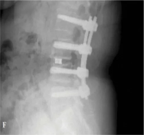
Many surgical techniques for spondylolisthesis have been described in the literature. Their basic application is simi-lar. The main objective is to create interbody fusion, decompression of normal structures, and a stable verteb-rae. Methods such as TLIF, posterior lumbar interbody fusion (PLIF), anterior lumbar interbody fusion (ALIF), and lateral lumbar interbody fusion (LLIF) are also avail-able for interbody fusion. The advantages of these proce-dures in each other should be discussed in the literature.15 TLIF surgery has been shown to be more appropriate in order to eliminate the potential concerns of PLIF sur-gery such as epidural adhesion, root damage, and extent of neural retraction.14 We reduce these risks by opening the Neural foramen one-sided. In a meta-analysis study, the complication rates in TLIF and PLIF literature were com-pared, and the complication rate in TLIF surgery was 50% lower than PLIF.14These complications are not only asso-ciated with surgery-related infection complications but include nerve injury, dura-tear, implant failure, and other complications. In the same study, TLIF surgery was found to be superior to PLIF in terms of complication rate and blood loss operation time. In addition, surgical approach has a relatively easier access to posterior structures Figure 6Postoperative lateral radiographic view of the patient who underwent
TLIF for L3 disc space.
Figure 5Postoperative AP view of the patient who underwent TLIF for L3 disc space.
Orthopedic Research and Reviews downloaded from https://www.dovepress.com/ by 118.70.13.36 on 26-Aug-2020
including lamina, ligamentum flavum, and facet joints. This method also provides better stabilization than PLIF by maintaining posterior structures.20
In ALIF technique, a fusion is obtained by avoiding the spinal canal. But compared to other methods, this technique may be very difficult and the morbidity rate is high.16In the systemic meta-analysis performed by Phan et al, the clinical and radiological results of ALIF and TLIF were compared. No difference was found between fusion rates and clinical results.21In patients who underwent ALIF, lumbar lordosis was found to be better than the other group, but longer duration of hospitalization and major complication rate were higher. In this method, the morbidity rate is high.16
In Teng et al's study, a meta-analysis comparing ALIF, PLIF, LLIF and TLIF was conducted. From the results obtained, ALIF is the most common technique, and the best improvement in the ODI scale in the literature was found in patients receiving TLIF compared to other groups.15 LLIF is the least studied method in the literature.15 The LLIF technique is usually suitable for T12-L1 vs L4-L5 interbody disc space. This technique is not suitable for the L5/S1 level, due to the location of the iliac crest that obstructs lateral access.17TLIF surgery can easily be applied to all levels of interbody disc space.16In addition, the LLIF technique is not a suitable method for patients with retroperitoneal surgery or retroperitoneal abscess or abnormal vessel placement.
In the literature, there are many studies comparing minimally invasive (MI) surgery with open TLIF surgery. In the study performed by Samuel et al, it was shown that patients had similar clinical benefits in both surgical techniques.28 In Kulkarni et al's study, there was no sig-nificant difference in postop outcomes between both groups. There is a significant difference in the duration of hospital blood loss in MI TLIF.29
The learning curve MI TLIF is difficult. In this process, the surgeon is exposed to higher radiation. This showed that more complications were seen in patients during learning. According to our interpretation, MI TLIF learn-ing curve is difficult for the surgeon to apply. To achieve the desired results at this stage is more difficult.
Why Apply TLIF In Recurrent Disc
Surgery?
In degenerative lumbar disc herniation treated with micro-discectomy without fusion, recurrence rates are high in the literature. The rate of recurrence was 27%, especially in
patients with more than 6 mm annular defects.22 Achieving a stable spine after surgery will minimize recur-rence rates. So a lot of fusion technique has been devel-oped. In a study by Barth et al, 84 patients showed that the rate of recurrent herniation after radical discectomy was 12.5% and 10% in the cases with limited discectomy.23 High recurrence rates may be seen, especially because of deterioration of stabilization after radical discectomy.24 After discectomy, fusion application creates a more stable spine. This fusion also helps to protect the lumbar lordosis in the advanced period.
In a study by Nei, Interbody fusion was performed, and non-performed discectomies were compared. Severe pain in long-term results was observed in patients unable to perform interbody fusion. Clinical results were shown to be better in patients with performed interbody fusion.25
We recommend the use of interbody fusion, especially in elderly patients with degenerated spine, as they provide better stability.
Disclosure
The authors report no conflicts of interest in this work.
References
1. Bydon M, De la Garza-ramos R, Macki M, et al. Lumbar fusion versus nonoperative management for treatment of discogenic low back pain: a systematic review and meta-analysis of randomized controlled trials.J Spinal Disord Tech. 2014;27(5):297–304. doi:10.1097/BSD.0000000 000000072
2. Harms J, Jeszenszky D. The unilateral transforaminal approach for posterior lumbar interbody fusion.Orthop Traumatol.1998;6:88–99. 3. Saglam N, Dogan S, Ozcan C, Turkmen I. Comparison of four
differ-ent posteriorfixation techniques for the treatment of thoracolumbar junction fractures.World Neurosurg.2019;123:e773–e780. doi:10.10 16/j.wneu.2018.12.030
4. Jakoi AM, Pannu G, D’Oro A. The clinical correlations between diabetes, cigarettes smoking and obesity on intervertebral degenerative disc disease of the lumbar spine.Asian Spine J.2017;11(3):337–347. doi:10.4184/asj.2017.11.3.337
5. Taher F, Essig D, Lebl DR. Lumbar degenerative disc disease: current and future concepts of diagnosis and management. Adv Orthop.
2012;9:70–75.
6. Mimura M, Panjabi MM, Oxland TR, et al. Disc degeneration affects the multidirectional flexibility of the lumbar spine. Spine (Phila Pa 1976).1994;19(12):1371–1380. doi:10.1097/00007632-199406000-00 011
7. Pathria M, Sartoris DJ, Resnick D. Osteoarthritis of the facet joints accuracy of oblique radiographic assessment. Radiology. 1987;164 (1):227–230. doi:10.1148/radiology.164.1.3588910
8. Nachemson A. Lumbar intradiscal pressure. Experimental studies on post-mortem material. Acta Orthop Scand Suppl. 1960;43:1–104. doi:10.3109/ort.1960.31.suppl-43.01
9. Vernon-Roberts B.Pathology of Intervertebral Discs and Apophyseal Joints. The Lumbar Spine and Back Pain. London: Churchill-Livingstone;1987:37–55.
Orthopedic Research and Reviews downloaded from https://www.dovepress.com/ by 118.70.13.36 on 26-Aug-2020
10. Özkara E, ArslantaşA. Transforaminal lumbar interbody. Füzyon-TLIF Türk Nörosirürji Dergisi.2009;3:223–226.
11. Çaçan MA, Uçar BY. What every spine surgeon should know about transforaminal lumbar interbody fusion surgery for herniated discs.
Int Orthop.2019;43(4):883–889. doi:10.1007/s00264-018-4251-x 12. Deutsch H, Musacchio MJ Jr. Minimally invasive transforaminal
lumbar interbody fusion with unilateral pedicle screw fixation.
Neurosurg Focus.2006;20(3):E10. doi:10.3171/foc.2006.20.3.11 13. Potter BK, Freedman BA, Verwiebe EG, et al. Transforaminal lumbar
interbody fusion: clinical and radiographic results and complications in 100 consecutive patients.J Spinal Disord Tech.2005;18(4):337– 346. doi:10.1097/01.bsd.0000166642.69189.45
14. De Kunder SL, van Kuijk SMJ, Rijkers K, et al. Transforaminal lumbar interbody fusion (TLIF) versus posterior lumbar interbody fusion (PLIF)in lumbar spondylolisthesis: a systematic review and meta-analysis. Spine J. 2017;17(11):1712–1721. doi:10.1016/j. spinee.2017.06.018
15. Teng I, Han J, Phan K, Mobbs R. A meta analysis comparing ALIF, PLIF, TLIF and LLIF.J Clin Neurosci.2017;44:11–17. doi:10.1016/j. jocn.2017.06.013
16. Mobbs RJ, Phan K, Malham G, Seex K, Rao PJ. Lumbar interbody fusion: techniques, indications and comparison of interbody fusion options including PLIF, TLIF, MI-TLIF, OLIF/ ATP,LLIF and A LIF.
J Spine Surg. 2015;1(1):2–18. doi:10.3978/j.issn.2414-469X.2015. 10.05
17. Joseph JR, Smith BW, La Marca F, Park P. Comparison of compli-cation rates of invasive transforaminal lumbar interbody fusion and lateral lumbar interbody fusion: a systematic review of the litera-ture.Neurosurg Focus.2015;39(4). doi:10.3171/2015.7.FOCUS15 278
18. Takahashi T, Hanakita J, Ohtake Y, et al. Current status of lumbar interbody fusion for degenerative spondylolisthesis.Neurol Med Chir (Tokyo).2016;56(8):476–484. doi:10.2176/nmc.ra.2015-0350 19. Krüger MT, Naseri Y, Hohenhaus M Impact of morbid obesity (BMI
> 40 kg/m2) on complication rate and outcome following minimally invasive transforaminal lumbar interbody fusion (MIS TLIF).Clin Neurol Neurosurg. 2019;178:82–85. doi:10.1016/j.clineuro.2019.02. 004
20. Audat Z, Moutasem O, Yousef K, et al. Comparison of clinical and radiological results of posterolateral fusion, posterior lumbar inter-body fusion and transforaminal lumbar interinter-body fusion techniques in the treatment of degenerative lumbar spine. Singapore Med J.
2012;53:183.
21. Phan K, Thayaparan GK, Mobbs RJ. Anterior lumbar interbody fusion versus transforaminal lumbar interbody fusion–systematic review and meta-analysis. Br J Neurosurg. 2015;29(5):705–711. doi:10.3109/02688697.2015.1036838
22. Carragee EJ, Han MY, Suen PW, Kim D. Clinical outcomes after lumbar discectomy for sciatica: the effects of fragment type and annular competence.J Bone Joint Surg Am.2003;85-A(1):102–108. doi:10.2106/00004623-200301000-00016
23. Barth M, Weiss C, Thomé C. Two-year outcome after lumbar micro-discectomy versus microscopic sequestrectomy: part 1: evaluation of clinical outcome. Spine (Phila Pa 1976). 2008;33(3):265–272. doi:10.1097/BRS.0b013e318162018c
24. Watters WC, Bono CM. An evidence-based clinical guideline for the diagnosis and treatment of degenerative lumbar spondylolisthesis.
Spine J.2009;9(7):609–614. doi:10.1016/j.spinee.2009.03.016 25. Eie N. Comparison of the results in patients operated upon for
ruptured lumbar discs with and without spinal fusion. Acta Neurochir (Wien).1978;41(1–3):107–113. doi:10.1007/BF01809141 26. Anderson PA, Savage JW, Vaccaro AR, et al. Prevention of surgical
site infection in spine surgery. Neurosurgery. 2017;80(3S):S114– S123. doi:10.1093/neuros/nyw066
27. Yao YC, Lin HH, Chang MC. Bracing following transforaminal lumbar interbody fusion is not necessary for patients with degenera-tive lumbar spine disease: a prospecdegenera-tive, randomized trial.Clin Spine Surg.2018;31(9):E441–E445. doi:10.1097/BSD.0000000000000697 28. Terman SW, Yee TJ, Lau D, et al. Minimally invasive versus open transforaminal lumbar interbody fusion: comparison of clinical out-comes among obese patients.J Neurosurg Spine. 2014;20(6):644– 652. doi:10.3171/2014.2.SPINE13794
29. Kulkarni AG, Bohra H, Dhruv A, et al. Minimal invasive transfor-aminal lumbar interbody fusion versus open transfortransfor-aminal lumbar interbody fusion.Indian J Orthop.2016;50(5):464–472. doi:10.4103/ 0019-5413.189607
Orthopedic Research and Reviews
Dove
press
Publish your work in this journal
Orthopedic Research and Reviews is an international, peer-reviewed, open access journal that focusing on the patho-physiology of the musculoskeletal system, trauma, surgery and other corrective interven-tions to restore mobility and function. Advances in new technologies, materials, techniques and pharmacological agents are particularly
welcome. The manuscript management system is completely online and includes a very quick and fair peer-review system, which is all easy to use. Visit http://www.dovepress.com/testimonials.php to read real quotes from published authors.
Submit your manuscript here:https://www.dovepress.com/orthopedic-research-and-reviews-journal
Orthopedic Research and Reviews downloaded from https://www.dovepress.com/ by 118.70.13.36 on 26-Aug-2020