Abstract
Malaria is a disease that causes enormous human morbidity and mortality. One feature of mature Plasmodium falciparum-infected erythrocytes leading to the development of severe malaria is thought to be cytoadherence and blockage of the microvasculature. Therefore, an understanding of mechanisms that mediate parasite adhesion leading to malaria pathology is needed to yield new treatments for malaria. However, to date, cytoadherence-associated pathology is still under debate. Is cytoadherence needed to develop severe malaria? This review will discuss the available information on associations of cytoadherence with the development of severe malaria.
Keywords: cerebral malaria, cytoadherence, endothelium, malaria
Introduction
Malaria is a serious burden, particularly to low and middle-income countries, and a major contributor to morbidity and mortality. The aetiological agents of malaria to humans are recognised as 6 distinct protozoan species of Plasmodium: Plasmodium falciparum (1),
Plasmodium vivax (2), Plasmodium malariae (3), 2 species of Plasmodium ovale (P. ovale curtisi
and P. ovale wallikeri) (4), and Plasmodium knowlesi (5), which was recently recognised as the 6th human Plasmodium after cross infection from long-tailed Macaca monkey to humans was reported in Malaysia (6–9). P. falciparum has often been seen as the most clinically significant infection due to an association with mortality and the intensity of infection in some regions of sub-Saharan Africa, but P. vivax has a wider geographical distribution, and its categorisation as benign has been challenged (10). What is clear is that an episode of P. falciparum malaria in a non- or semi-immune host can lead to severe malaria (SM) if untreated, with a high risk of death. However, recently a study from Papua New Guinea and Malaysian Borneo extended the pattern of severe disease by showing a strong association of P. vivax (11) and P. knowlesi
(8) infection, respectively, to SM and death. Studies of the pathology of SM were started in the late of 19th century by 2 Italian pathologists
Marchiafava and Bignami (12), where they found, post-mortem, the presence of higher parasite load in a comatose malignant blood fever patient compared with in those with benign fever. They saw high parasite levels and parasite pigment predominantly retained in the tissue microvessels compared with in the peripheral circulation, as well as the existence of necrosis and alterations in the endothelium of the cerebral vessels. This discovery and subsequent observations have led to a suggestion that the preferential accumulation of parasitised red blood cell (pRBC) in tissues might be linked to the disease and its severity. The manifestations of SM are highly variable and are determined by factors from both the human host and the parasite. The most common clinical features of SM are high fever, respiratory distress, vascular obstructions, metabolic disturbances (e.g., acidosis), multi-organ dysfunction (e.g., renal failure), severe anaemia, and cerebral malaria (CM); these features differ among areas of varying transmission intensity and between adults and children. This creates problems in comparing studies as the clinical definitions can vary and the impact of cytoadherence on these variable clinical outcomes is difficult to define. Some aspects of SM occur because the parasite has developed mechanisms to escape the host immune system, which we will discuss later, so one of the features of CM (an important subset of severe cases) is that it is more common
AlisterG Craig1, Mohd Fadzli Mustaffa Khairul1,2,
Pradeep R Patil1,3
1 Department of Molecular and Biochemical Parasitology, Liverpool School of Tropical Medicine, Pembroke Place, L3 5QA Liverpool, United Kingdom
2 Institute for Research in Molecular Medicine, Health Campus, Universiti Sains Malaysia, 16150 Kubang Kerian, Kelantan, Malaysia
3 Department of Pharmaceutical and Applied Chemistry, University of Siena, Vai Aldo Moro-2, 53100 Siena, Italy
Submitted: 30 Jun 2011
in semi-immune children in sub-Saharan Africa (13).
It is still unclear how infection with P. vivax
and P. knowlesi lead to SM, and it is possible that research on understanding P. falciparum– derived SM may help us to understand and predict how P. vivax and P. knowlesi act. Once thought to be unique to P. falciparum, the ability of the mature pRBC to undergo a range of adhesive interactions (cytoadhesion), such as the binding of pRBC with endothelial cells (sequestration) and the interaction of pRBC with non-infected RBC (rosetting) and with other pRBC (auto-agglutination), is now thought to be shared with other species. One of the big questions in
P. falciparum research is, “Is parasite adhesive behaviour linked to SM?” This question can now be extended to P. vivax and P. knowlesi with the discovery of adhesion to endothelial receptors by RBC infected with these species (8,14,15), although the timing and extent of cytoadherence in these 2 species differs from that of P. falciparum, with the latter exhibiting earlier (from 15 hours post-invasion) and more pronounced sequestration, whereas only the schizont stages of P. vivax and
P. knowlesi show this phenotype.
One molecule identified on the surface of
P. falciparum pRBC, known as P. falciparum
erythrocyte membrane protein-1 (PfEMP-1) encoded by var genes, has been correlated with P. falciparum cytoadherence (16–18). It has been thought that the antigenic switching between different PfEMP-1s constitutes an important virulence factor by facilitating the parasite’s escape from the host’s immune response, thus establishing chronic infection (19,20). Considering the potential harmful effect of P. falciparum cytoadherence to the host, early treatment or even prophylaxis would be highly desirable in preventing cytoadhesion and progression of disease. Unfortunately, falciparum malaria has become increasingly refractory to chloroquine (21,22), the cheapest and most widely available antimalarial, and this emergence of drug resistance in Southeast Asia and Africa was closely associated with the increased incidence of SM (23) . The World Health Organization advises all countries experiencing antimalarial drug resistance (including monotherapies such as chloroquine, amodiaquine, or sulfadoxine-pyrimethamine) to use combination therapies, preferably those containing artemisinin derivatives (artemisinin-based combination therapies, ACTs).
Recent clinical trials in Asia and Africa using ACTs showed improved recovery of SM patients
(24,25), but mortality reported shortly after hospital admission (within 48 hours) was still high despite the administration of highly effective anti-parasitic drugs. This finding is consistent with our recent data showing that after exposure to drugs, killed pRBC were still able to cytoadhere (26), which has led us to suggest that this persistent mortality may be due to the effects of adherent pRBC in the microvasculature. Is there any way of reducing this mortality? Perhaps adjunct therapies that can block and reverse the pathogenic effect of pRBC adhesion will lighten the disease burden. However, before embarking on this course, what evidence is there that cytoadherence is involved in SM?
pRBC Cytoadhesion
Why and how does parasite cytoadherence– related morbidity take place? Several hypotheses associated with the binding of pRBC in the microvasculature have been proposed and reviewed elsewhere, such as i) changes of the RBC and pRBC rigidity (27–29), ii) pro-inflammatory induction of the adhesion-receptor expression (30,31), iii) binding of pRBC to specific adhesion receptors on endothelial cells (32), iv) endothelial activation (33–35), and v) malaria toxins (36), with various levels of evidence to support them. However, there are also more recent discoveries such as the relevance of platelets and microparticles as well as the role for the coagulation cascade in mediating pRBC binding to endothelial cells (37,38).
A major question is how P. falciparum has adapted to bind in the microvasculature to such an extent that mature pRBC are rarely seen in the peripheral circulation, unlike other human-invading malaria parasite species. An important difference in P. falciparum is the modification to the surface of the host erythrocytes to become rigid and inflexible by exporting specific proteins to the RBC membrane during the intra-erythrocytic stages. This reduction of flexibility of RBC makes their circulation through the microvasculature difficult and favours pRBC adhesion to endothelial cells (39).
been demonstrated in in vitro studies. Knobs are distortions on the surface of P. falciparum
pRBC caused by deposition of knob-associated His-rich protein (KAHRP) at the cytoplasmic side of the pRBC membrane (41); these knobs contain several other proteins including PfEMP-1 as well as ring-infected and mature parasite–infected erythrocyte surface antigens (42). It is generally accepted that PfEMP-1 is largely responsible for pRBC adhesion in P. falciparum, and various associations between var gene expression and complicated or uncomplicated disease have been reported. However, are knobs essential to establish an interaction in the microvasculature? Some other Plasmodium species such as
P. brasilianum, P. vivax, and P. malariae also have knob-like structures but do not always exhibit cytoadherence properties, suggesting that these membrane modifications are not identical to those seen with P. falciparum (14,43). Biggs et al. (44) demonstrated that knobless P. falciparum
could bind to host receptors, although later work (45) showed that a KAHRP knockout line could not bind under more physiological flow conditions.
What Factors Mediate Adhesion of the
pRBC in Host Microvasculature?
Several receptors on endothelial cells have been shown to support interactions with pRBC, including thrombospondin; CD36; immunoglobulin superfamily cell adhesion molecules, e.g., intercellular adhesion molecule 1 (ICAM-1), vascular cell adhesion molecule, (VCAM), platelet endothelial cell adhesion molecule, and neural cell adhesion molecule; selectins, e.g., P-selectin and E-selectin; integrin αvβ3; globular C1q receptor; and glycoaminoglycans, e.g., chondroitin sulphate A (CSA) and heparin sulphate (35). With such a diverse collection of host receptors, how might one investigate associations between disease and specific adhesion phenotypes? It has been reported that ICAM-1 and CD36 are the most commonly used adhesion receptors by patient isolates, except in placental malaria (46), and correlations with severe and uncomplicated disease have been suggested (47), which has often been a starting point for clinical studies. It has been proposed that synergism (or at least co-operation) between these two receptors makes the binding of pRBC stronger (48,49). Therefore, it is not unusual that clinical studies examining the association between receptor usage and disease have concentrated on these two proteins, as indicated in Table 1 (31,45,46,50–58).
In addition to ICAM-1 and CD36, CSA is also one of the most common and successfully studied adhesion receptors. CSA provides the clearest example of an interaction of pRBC with an adhesion receptor in causing disease; however, this example of adhesion-related pathology does not come from endothelial cytoadherence but rather adhesion of pRBC to CSA in the placenta of pregnant women through a set of semi-conserved PfEMP-1 proteins (59–61). The restricted variation in this important facet of malaria pathology provides one of the most hopeful cases for the development of a disease-specific vaccine for malaria (62). In the case of placental malaria, the association of a specific var gene (var2csa) with adhesion and disease has been possible, but this has been much harder to define in other syndromes of SM, such as CM.
Another factor that has been postulated to have an association with host-mediated cytoadherence is the role of host pro-inflammatory cytokines. These cytokines have long been implicated in the pathogenesis of SM (63), where changes in cytokine plasma levels have paralleled the rise of temperature during fever paroxysms in SM (64), and an increase of pro-inflammatory cytokines, especially tumour necrosis factor (TNF), in CM in children especially from Africa and its correlation with mortality (65–68) have been observed. Nevertheless, how are these pro-inflammatory cytokines regulated and how might they mediate parasite adhesion and SM, or is this just a general effect? Other studies (33,69) have challenged the correlation of cytokines, especially TNF, towards malaria disease severity and claimed it is quite poor at predicting SM. It is thought that pro-inflammatory cytokines are central to the pathophysiology of systemic disease caused by infectious and non-infectious agents, and cytokines such as TNF and interleukin (IL)-10 have been proposed to have a protective role to clear the infections and to avoid inappropriate host responses that might lead to cell destruction and be harmful to the host. In the case of malaria and SM, high levels of pro-inflammatory cytokines TNF, IL-1, IL-6, IL-12, and interferon (IFN)-γ have been observed in patients with malaria, and low levels of IL-10 and tumour growth factor (TGF)-β have been correlated with fatal outcome (70). It is thought that these cytokines are produced by activated macrophages, dendritic cells, and, potentially, endothelial cells during the host response to pRBC and schizont rupture (36,71).
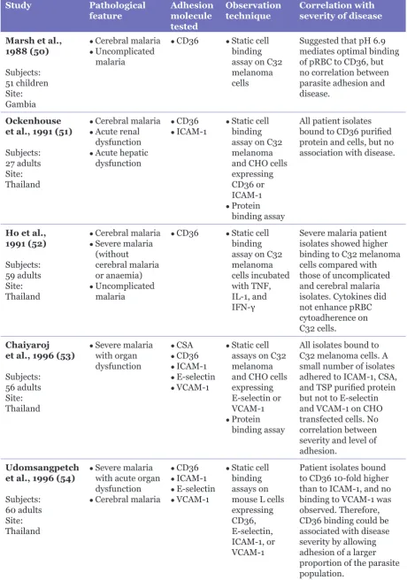
Table 1: Clinical studies on cytoadherence-related pathology in malaria
Study Pathological
feature Adhesion molecule
tested
Observation
technique Correlation with severity of disease
Marsh et al., 1988 (50)
Subjects: 51 children Site: Gambia
● Cerebral malaria ● Uncomplicated
malaria
● CD36 ● Static cell
binding assay on C32 melanoma cells
Suggested that pH 6.9 mediates optimal binding of pRBC to CD36, but no correlation between parasite adhesion and disease.
Ockenhouse et al., 1991 (51)
Subjects: 27 adults Site: Thailand
● Cerebral malaria ● Acute renal
dysfunction
● Acute hepatic
dysfunction
● CD36 ● ICAM-1
● Static cell
binding assay on C32 melanoma and CHO cells expressing CD36 or ICAM-1
● Protein
binding assay
All patient isolates bound to CD36 purified protein and cells, but no association with disease.
Ho et al., 1991 (52)
Subjects: 59 adults Site: Thailand
● Cerebral malaria ● Severe malaria
(without
cerebral malaria or anaemia)
● Uncomplicated
malaria
● CD36 ● Static cell
binding assay on C32 melanoma cells incubated with TNF, IL-1, and IFN-γ
Severe malaria patient isolates showed higher binding to C32 melanoma cells compared with those of uncomplicated and cerebral malaria isolates. Cytokines did not enhance pRBC cytoadherence on C32 cells.
Chaiyaroj et al., 1996 (53)
Subjects: 56 adults Site: Thailand
● Severe malaria
with organ dysfunction
● CSA ● CD36 ● ICAM-1 ● E-selectin ● VCAM-1
● Static cell
assays on C32 melanoma and CHO cells expressing E-selectin or VCAM-1
● Protein
binding assay
All isolates bound to C32 melanoma cells. A small number of isolates adhered to ICAM-1, CSA, and TSP purified protein but not to E-selectin and VCAM-1 on CHO transfected cells. No correlation between severity and level of adhesion.
Udomsangpetch et al., 1996 (54)
Subjects: 60 adults Site: Thailand
● Severe malaria
with acute organ dysfunction
● Cerebral malaria
● CD36 ● ICAM-1 ● E-selectin ● VCAM-1
● Static cell
binding assays on mouse L cells expressing CD36, E-selectin, ICAM-1, or VCAM-1
Study Pathological
feature Adhesion molecule
tested
Observation
technique Correlation with severity of disease
Newbold et al., 1997 (55)
Subjects: 150 children Site: Kenya
● Cerebral malaria ● Severe anaemia ● Non-severe
malaria
● CD36 ● ICAM-1 ● VCAM-1 ● E-selectin
● Static protein
binding assay CD36 was quantitatively the major receptor for all isolates, but some isolates bound strongly to ICAM-1, less to VCAM-1, and none to E-selectin.
Binding to ICAM-1 was associated with disease, but not cerebral malaria.
Rogerson et al., 1999 (56)
Subjects: 158 children Site: Malawi
● Severe malaria ● Cerebral malaria ● Severe anaemia
● CD36 ● ICAM-1 ● CSA ● TM
● Static protein
binding assay Varied cytoadherence profiles from patient isolates; all isolates bound to CD36, and severe anaemia isolates had low binding to ICAM-1. No correlation with severe disease.
Heddini et al., 2001 (57)
Subjects: 111 children Site: Kenya
● Severe malaria
without cerebral malaria or severe anaemia
● Cerebral malaria ● Severe anaemia ● Uncomplicated
malaria
● Control
● PECAM ● CD36 ● TSP ● ICAM-1
● Static cell
binding assays on mouse L cells expressing PECAM-1 and CHO cells expressing CD36 or ICAM-1
● Protein
binding assays using FACS technique
Rosetting associated with blood group A and heparin-type receptors (e.g., heparin sulphate) were prone to severe malaria. Binding to multiple receptors promoted pRBC sequestration in the severe malaria group.
Cojean et al., 2008 (47)
Subjects: 22 adults Site: France
● Uncomplicated
malaria
● Cerebral malaria ● Severe malaria
(without cerebral malaria)
● ICAM-1 ● CD36
● Static cell
binding assays on CHO cells expressing CD36 or ICAM-1
Binding of isolates from severe malaria showed no significant difference compared with uncomplicated malaria pRBC.
Chilongola et al., 2009 (46)
Subjects: 155 children Site: Tanzania
● Uncomplicated
and dendritic cells probably through direct or indirect interaction of PfEMP-1 via CD36, and it is known that CD36 is expressed on the surfaces of macrophages and dendritic cells. The biological role of these interactions is not clear. For example, there is conflicting data about the effect of pRBC adhesion on dendritic cells: some studies showed that decreased activation was associated with cytoadherence (74), while others showed that this adhesion process was not required for the modulation of dendritic cell activity (75). These findings in the literature are variable due to the use of different host species and parasite strains, at pre-erythrocyte or blood stages of infection (76). How might cytokines support pRBC binding to microvascular endothelium? One thought is that it is through endothelial activation. For example, TNF is known to act by increasing the expression of host adhesion molecules. TNF binding to TNF receptor type 2 induces recruitment of signal transduction that activates effector molecules and transcription factors, leading to a strong increase in the expression of ICAM-1, VCAM, and E-selectin. The involvement of TNF in the upregulation of adhesion molecules has been clearly reported in different in vitro and in vivo studies (49,77,78). However, does TNF induction alone do enough to exacerbate SM? If pathology of malaria was the result of a high level of TNF, patients with SM could be treated using
TNF specific antibody. However, a trial using a monoclonal antibody against TNF did not show any protection and, in fact, worsened neurological sequelae in patients (68).
The pattern of pathology in malaria is variable, and the profound cytokine-mediated changes and tissue oedema seen in other infections are not characteristic of this disease, although some signs of these pathologies are available. Thus, it seems that malaria pathology can be linked to a pro-inflammatory response, but this is not enough to explain the disease. This is consistent with recent studies that have shown that in children, TNF level was a poor discriminator of severity of disease, whereas proteins associated with endothelial activation (e.g., angiopoietin-1, angiopoietin-2, von Willebrand factor [vWF], soluble ICAM-1) were relatively good markers (69).
Endothelial activation, in response to inflammatory mediators, collectively increases the expression of adhesion molecules, including E-selectin, ICAM-1, and VCAM-1, on the cell surface through the activation of nuclear factor κB signalling transduction. Increases of P-selectin on endothelial cells following activation have also been reported (79). P-selectin is different from other adhesion molecules as it is stored in endothelial cell specific storage vesicles called Weibel–Palade bodies, together with other molecules such as vWF. How Weibel–Palade
Study Pathological
feature Adhesion molecule
tested
Observation
technique Correlation with severity of disease
Ochola et al., 2011 (32)
Subjects: 101 children Site: Kenya
● Cerebral malaria
● Severe anaemia
● Uncomplicated malaria
● CD36
● ICAM-1
● Static and flow protein binding assays
High pRBC binding to CD36 was associated with uncomplicated malaria. High ICAM-1 binding under flow correlated with cerebral malaria.
Mayor et al., 2011 (58)
Subjects: 46 children Site: Mozambique
● Severe malaria
(with cerebral malaria, severe anaemia, respiratory distress, prostration)
● Uncomplicated
malaria
● CD36 ● ICAM-1 ● gC1qR
● Static protein
binding assays
Higher levels of adhesion to gC1qR in isolates from children with multiple seizures.
bodies are activated in malaria infection is still unknown; the parasite has a protein that can cause basophils to release histamine, which is known as P. falciparum translationally controlled tumour protein (80), but some reports have also suggested that activated platelets and fibrin might mediate the release of P-selectin and vWF (81). vWF recently has been found to be a good prognostic marker for SM in children, and it has been thought that vWF might mediates pRBC binding on endothelial cell via ultra-large vWF multimers by producing a bridge via platelets (81–85).
Scientists have speculated that the febrile temperature seen as a part of malaria infection might enhance cytoadherence. We know that a fever is due, in part, to the increase in TNF seen during infection, but does temperature elevation help pRBC to bind to endothelial cells? Udomsangpetch et al. (86) showed that PfEMP-1 expression was accelerated by febrile temperature and increased cytoadherence. However, this is contrary to other findings where febrile temperature affected intra-erythrocyte growth, and upregulation of PfEMP-1 was not seen (87). Recently, Pattanapanyasat et al. (88) showed that febrile temperature induced and enriched expression of phosphatidylserine on the pRBC membrane surface. Several studies (89–92) have reported that phosphatidylserine promoted pRBC binding to CD36 and thrombospondin. Febrile temperature can also lead to endothelial cell disruption (93). In a clinical trial (94), the use of antipyretic intravenous ibuprofen was able to control fever but delayed parasite clearance. This finding suggests that ibuprofen and fever reduction does not act to reduce cytoadherence as might be expected from previous work, and there is some evidence that fever temperatures might act in the opposite way to reduce endothelial cell binding (Craig, unpublished observations). There is also evidence showing that febrile temperatures may increase pRBC rigidity (95), and this finding might cause at least some of the reduced RBC deformity, leading to the vascular flow obstruction seen in SM; however, there is a need for further studies to confirm this. In the absence of a consistent association with inflammation and malaria pathology as well as the observation of preferential pRBC accumulation in microvessels in SM, researchers have turned to cytoadherence, and many clinical studies (31,45,46,50–58) have attempted to correlate adhesion with disease (Table 1), particularly with CM where cerebral pRBC sequestration is an invariant feature of the disease.
How Might Cytoadherence Cause SM?
As mentioned earlier, complex interactions, including the host inflammatory response and endothelial activation, may contribute to SM, but how does cytoadherence itself modulate the severity of the disease?
When P. falciparum infects an RBC, the parasite expresses proteins that are transported to the RBC membrane, causing changes in rigidity and shape of the infected RBC. This may lead to difficulties in RBC flow through the microvasculature, and studies in Thailand (39) and Bangladesh (96) have shown that increased rigidity and reduced flow through blood vessels were associated with severe disease. Other studies on the retinal vasculature (97–99) have shown that micro-haemorrhages and vessel changes, thought to reflect blockage, were highly predictive of CM.
The hypothesis is that adhesion of pRBC in the deep vasculature leads to organ dysfunction. What evidence do we have to support this? As stated earlier, MacPherson et al. (40) showed that there was preferential pRBC accumulation in the brains of people dying of CM compared with in non-CM. One way that this might be taking place is that in some malaria infections, there is higher recruitment of pRBC to cerebral vessels due to the increased levels of receptors such as ICAM-1 (77) and the presence of parasites that are able to bind efficiently to these receptors (32). This clearly oversimplifies the potential mechanisms contributing to the preferential recruitment of pRBC in the brain, and there are likely to be several pathways by which this can be achieved. The role of the infecting parasite variant should not be ignored in this equation, and data from the analysis of pRBC in post-mortem tissues have shown the enrichment of specific PfEMP-1 variant types in the brains of children dying of CM (100).
Trans-endothelial electrical resistance experiments showed that when pRBC adhered to human brain microvascular endothelial cells, the integrity of the human blood–brain barrier (BBB) reduced 3-fold, causing increased permeability (105). The leakage of BBB leads to serum protein penetrating into the central nervous system (106). This influx of foreign substances activates the microglial cells that release pro-inflammatory cytokines, damaging astrocytes and glial cells that are crucial for BBB maintenance (107). It has also been suggested that interaction of serum protein with TGF-β receptors TGFBR1 and TGFBR2 could result in astrocyte dysfunction, followed by seizures and neuronal death (108).
The binding of pRBC to brain endothelial cells has also been reported to induce endothelial cell apoptosis (109,110). Pino et al. (109) have demonstrated pRBC modulation of the expression of endothelial cell genes such as TNF superfamily genes (Fas, Fas L, and DR-g) and apoptosis-related genes (Bad, Bax, Caspase-3, SARP2, DFF45/ ICAD, IFN-γ Receptor 2, Bcl-w, Bik, and iNOS). Toure et al. (110) subsequently showed for the first time that clinical isolates could sometimes induce endothelial cell apoptosis, and Herbert et al. (111) showed that the presence of apoptotic cells might upregulate the expression of cellular adhesion molecules, resulting in hyperadhesiveness, leading to a greater accumulation of pRBC and subsequent endothelial cell apoptosis.
How Might We Alleviate the Symptoms
of SM by Targeting Cytoadherence?
As described above, cytoadherence, which we believe may lead to some aspects of disease severity, is a process where mature pRBC erythrocytic stages escape from splenic clearance by binding to endothelial cells and promoting parasite growth in a relatively hypoxic environment. Therefore, can we use this information to devise treatments to prevent death or neurological sequelae?
Antiparasite drugs will still be the main treatment of choice to reduce mortality in patients with malaria; preferentially, these should kill the malaria parasite in early stages (in terms of the erythrocytic cycle) as destruction of non-adhesive ring stages will prevent the next wave of pRBC from sequestering. Therefore, artemisinin is a good choice as it kills ring-stage parasites; this might explain the reduced mortality seen in the field studies from South East Asia (24) and Africa (25) that compared artemisinin and quinine (which only kills mature pRBC). However, even with this welcome progress, there is still over 50% of the mortality recorded during first
48 hours after hospital admission that is largely unaffected by the use of ACTs. This may be because the pRBC has already sequestered to the endothelium. Therefore, there is a need for adjunct therapies to support the critically ill patients, to be used in combination with antimalarials such as artemisinin to remove the sequestered pRBC mass or reduce its effects on the host, while the standard drugs kill the parasite effectively.
To date, several compounds have been explored and screened for their potential to improve SM. N-acetylcysteine (NAC) is an antioxidant drug that is widely used in humans for the treatment of paracetamol overdose and has been shown to be able to reverse almost 72% of pRBC binding to CD36 (112). In addition, it also reduced the rigidity of pRBC (113). A pilot clinical trial study in Thailand (114) showed that NAC was able to normalise serum lactate (an indicator of SM) significantly in SM patients. NAC is thought to inhibit TNF release, thereby reducing cytoadherence. It is also a potent scavenger of free oxygen radicals, which are produced in response to TNF, and can mediate some toxic effects. However, despite these encouraging features, NAC has recently been shown to antagonise the action of artesunate (115), and clinical trials have been disappointing (116), with no reduction in TNF release.
Levamisole is an alkaline phosphatase inhibitor that is used as an antihelminthic drug. Using levamisole for treatment of endothelial cells in vitro showed that it was able to reduce the binding of P. falciparum pRBC through dephosphorylated ectodomain of CD36 (117,118). A clinical trial of Levamisole in combination with artesunate is currently underway, and so far, treatment with Levamisole has been shown to be safe and to cause the release of mature pRBC into the peripheral circulation (117). Epigalloyl-catechin-gallate, a naturally occurring polyphenol compound from green tea, was identified as being able to inhibit pRBC binding to ICAM-1 by 50% at micromolar concentrations (119) and has been postulated to synergise the effect of artemisimin on malaria by lowering the IC50 from 14 nM to 8.4 nM (120), but unfortunately, this compound does not appear to be able to reverse adhesion. This highlights the need to test potential anti-cytoadherence agents for inhibition and reversal.
follows on from a study (121) in SM patients showing low NO production and low plasma arginine. In normal conditions, NO mediates host resistance to a wide variety of infectious microorganisms, and some in vitro studies have shown that it possessed antiparasitic effects by killing pRBC, as well as an anti-adhesion effect. NO is also a potent inhibitor of TNF production and other pro-inflammatory cytokines implicated in malaria immunopathology (122). Therefore, L-arginine is a good candidate to be used as an adjunct therapy for SM by improving endothelial function. Clinical studies measuring reperfusion parameters have been encouraging (123), and further work is needed in this area to provide better understanding of adhesion-related pathology in malaria and to conduct more clinical trials.
Erythropoietin (EPO) is a hormone produced by the kidney that modulates the survival of developing erythroid precursors and the production of new erythrocytes in the bone marrow. In SM patients, low EPO has been detected and correlated with severe anaemia. Injection of high doses of EPO in mice infected with P. berghei showed a significant reduction of pro-inflammatory cytokines TNF and IFN-γ (124) and an increased survival rate when used in combination with artesunate (125). Preliminary clinical trials of EPO in combination with quinine in CM children in Mali showed that it was safe and did not show any side effects (126).
As mentioned before, apoptosis is postulated to be one way in which cytoadherence can cause disease. Therefore, the use of anti-apoptotic agents should be advantageous. Fasudil is a Rho kinase inhibitor and widely used in humans for cardio- and neurovascular diseases. An in vitro study using clinical parasite isolates showed that fasudil has the potential to inhibit apoptosis mediated by P. falciparum pRBC adhesion to endothelial cells but showed no effect on reversing or inhibiting pRBC cytoadherence (127). It appears to be a promising adjunctive therapeutic approach for reducing neurological sequelae by reversing endothelial permeability through reducing NFκB activation and endothelial apoptosis (102). The use of statins to control blood cholesterol level has also been shown to be able to restore endothelial damage caused by pRBC cytoadherence (128). Atorvastatin appears to improve endothelial function by increasing NO production, protecting endothelial barrier integrity, reducing oxidative stress, and inhibiting inflammatory responses (129) through activated anti-apoptotic Akt cascade. There is also evidence that statins decrease ICAM-1 expression in
stimulated endothelial cell and monocytes (130), but as yet, there is no evidence to show that they are able to reverse established pRBC adhesion.
Further work is needed in this area, such as a better understanding of adhesion-related pathology in malaria and more clinical trials. The latter are complicated by the need to record mortality as an outcome, making the number of patients that need to be recruited relatively large. This means that we need to have better measures of clinical success if this development is to be viable.
What Can We Do for the Future?
The use of adjunct treatments to reverse adhesion of sequestered pRBC is a rational approach to reduce disease severity, but the release of large amount of pRBC into the circulation could be damaging. Can the spleen deal with removing the released pRBC or might it lead to side effects such as splenic dysfunction? Better animal models would help to address this and identify and test lead compounds. Efforts are underway to develop humanised animal models and transgenic parasites (containing PfEMP–1 adhesion domains) that could provide a resource to study the pathophysiology of SM in humans. If we are to preserve some of the advantages gained using ACTs, then the design of new antiparasite drugs should incorporate the ability to kill ring stages as well as mature pRBC and gametocytes.
The need for rapid acting adjunct treatments is critical in order to reduce mortality in SM cases. Anti-adhesion therapies form a part of this portfolio, but we need to understand the biology of this interaction and have better tools to test potential therapies prior to clinical trials.
Authors’ Contributions
Conception and design: MFMK, PRP
Drafting and final approval of the article: AGC, MFMK
Critical revision of the article: AGC
Correspondence
Professor Dr Alister G Craig BSc Genetics (Edinburgh) PhD Molecular Biology (Leicester) Liverpool School of Tropical Medicine Pembroke Place
L3 5QA Liverpool, United Kingdom Tel: +44 151 705 3161
Fax: +44 151 705 3171
References
1. Perlmann P, Troye-Blomberg M. Malaria blood-stage infection and its control by the immune system. Folia Biol (Praha). 2000;46(6):210–218.
2. Sharma A, Khanduri U. How benign is benign tertian malaria? J Vector Borne Dis. 2009;46(2):141–44.
3. Siswantoro H, Russell B, Ratcliff A, Prasetyorini B, Chalfein F, Marfurt J, et al. In vivo and in vitro efficacy of chloroquine against Plasmodium malariae
and P. ovale in Papua, Indonesia. Antimicrob Agents Chemother. 2011;55(1):197–202.
4. Sutherland CJ, Tanomsing N, Nolder D, Oguike M, Jennison C, Pukrittayakamee S, et al. Two nonrecombining sympatric forms of the human malaria parasite Plasmodium ovale occur globally.
J Infect Dis. 2010;201(10):1544–1550.
5. Sabbatani S, Fiorino S, Manfredi R. The emerging of the fifth malaria parasite (Plasmodium knowlesi): A public health concern? Braz J Infect Dis. 2010;14(3):299–309.
6. Cox-Singh J. Malaria: What can apes teach humans?
Future Microbiol. 2010;5(8):1157–1160.
7. Cox-Singh J, Davis TM, Lee KS, Shamsul SS, Matusop A, Ratnam S, et al. Plasmodium knowlesi malaria in humans is widely distributed and potentially life threatening. Clin Infect Dis. 2008;46(2):165–171.
8. Cox-Singh J, Hiu J, Lucas SB, Divis PC, Zulkarnaen M, Chandran P, et al. Severe malaria - A case of fatal
Plasmodium knowlesi infection with post-mortem findings: A case report. Malar J. 2010;9:10.
9. Cox-Singh J, Singh B. Knowlesi malaria: Newly emergent and of public health importance? Trends Parasitol. 2008;24(9):406–410.
10. Anstey NM, Russell B, Yeo TW, Price RN. The pathophysiology of vivax malaria. Trends Parasitol.
2009;25(5):220–227.
11. Genton B, D’Acremont V, Rare L, Baea K, Reeder JC, Alpers MP, et al. Plasmodium vivax and mixed infections are associated with severe malaria in children: A prospective cohort study from Papua New Guinea. PLoS Med. 2008;5(6):e127.
12. Marchiafava E, Bignami A, Mannaberg J. On
summer-autumn malaria fevers. London (GB): The
New Sydenham Society; 1894.
13. Dubos F, Dauriac A, El Mansouf L, Courouble C, Aurel M, Martinot A. Imported malaria in children: Incidence and risk factors for severity. Diagn Microbiol Infect Dis. 2010;66(2):169–174.
14. Carvalho BO, Lopes SC, Nogueira PA, Orlandi PP, Bargieri DY, Blanco YC, et al. On the cytoadhesion of Plasmodium vivax-infected erythrocytes. J Infect Dis. 2010;202(4):638–647.
15. Fatih FA, Siner A, Ahmed A, Woon LC, Craig AG, Singh B, et al. Cytoadherence and virulence - The case of Plasmodium knowlesi malaria. Malar J. 2012;11(1):33.
16. Pasternak ND, Dzikowski R. PfEMP1: An antigen that plays a key role in the pathogenicity and immune evasion of the malaria parasite
Plasmodium falciparum. Int J Biochem Cell Biol. 2009;41(7):1463–1466.
17. Scherf A, Lopez-Rubio JJ, Riviere L. Antigenic variation in Plasmodium falciparum. Annu Rev Microbiol. 2008;62:445–470.
18. Flick K, Chen Q. var genes, PfEMP1 and the human host. Mol Biochem Parasitol. 2004;134(1):3–9.
19. Newbold CI, Craig AG, Kyes S, Berendt AR, Snow RW, Peshu N, et al. PfEMP1, polymorphism and pathogenesis. Ann Trop Med Parasitol. 1997;91(5):551–557.
20. Newbold C, Craig A, Kyes S, Rowe A, Fernandez-Reyes D, Fagan T. Cytoadherence, pathogenesis and the infected red cell surface in Plasmodium falciparum. Int J Parasitol. 1999;29(6):927–937.
21. Krishna S, White NJ. Pharmacokinetics of quinine, chloroquine and amodiaquine. Clinical implications.
Clin Pharmacokinet. 1996;30(4):263–299.
22. Zucker JR, Lackritz EM, Ruebush TK 2nd, Hightower AW, Adungosi JE, Were JB, et al. Childhood mortality during and after hospitalization in western Kenya: Effect of malaria treatment regimens. Am J Trop Med Hyg. 1996;55(6):655–660.
23. Giha HA, Elbashir MI, A-Elbasit IE, A-Elqadir TM, ElGhazali GE, Mackinnon MJ, et al. Drug resistance-virulence relationship in Plasmodium falciparum
causing severe malaria in an area of seasonal and unstable transmission. Acta Trop. 2006;97(2): 181–187.
24. South East Asian Quinine Artesunate Malaria Trial (SEAQUAMAT) group. Artesunate versus quinine for treatment of severe falciparum malaria: A randomised trial. Lancet. 2005;366(9487):717–725.
25. Dondorp AM, Fanello CI, Hendriksen ICE, Gomes E, Seni A, Chhaganlal KD, et al. Artesunate versus quinine in the treatment of severe falciparum malaria in African children (AQUAMAT): An open-label, randomised trial. Lancet. 2010;376(9753): 1647–1657.
26. Hughes KR, Biagini GA, Craig AG. Continued cytoadherence of Plasmodium falciparum infected red blood cells after antimalarial treatment. Mol Biochem Parasitol. 2010;169(2):71–78.
27. Dondorp AM, Nyanoti M, Kager PA, Mithwani S, Vreeken J, Marsh K. The role of reduced red cell deformability in the pathogenesis of severe falciparum malaria and its restoration by blood transfusion.
Trans R Soc Trop Med Hyg. 2002;96(3):282–286.
28. Dondorp AM, Kager PA, Vreeken J, White NJ. Abnormal blood flow and red blood cell deformability in severe malaria. Parasitol Today. 2000;16(6): 228–232.
30. Armah H, Wired EK, Dodoo AK, Adjei AA, Tettey Y, Gyasi R. Cytokines and adhesion molecules expression in the brain in human cerebral malaria. Int J Environ Res Public Health. 2005;2(1):123–131.
31. Armah H, Dodoo AK, Wiredu EK, Stiles JK, Adjei AA, Gyasi RK, et al. High-level cerebellar expression of cytokines and adhesion molecules in fatal, paediatric, cerebral malaria. Ann Trop Med Parasitol. 2005;99(7):629–647.
32. Ochola LB, Siddondo BR, Ocholla H, Nkya S, Kimani EN, Williams TN, et al. Specific receptor usage in
Plasmodium falciparum cytoadherence is associated with disease outcome. PLoS One. 2011;6(3):e14741.
33. Conroy AL, Phiri H, Hawkes M, Glover S, Mallewa M, Seydel KB, et al. Endothelium-based biomarkers are associated with cerebral malaria in Malawian children: A retrospective case-control study. PLoS One. 2010;5(12):e15291.
34. Garcia F, Cebrian M, Dgedge M, Casademont J, Bedini JL, Neves O, et al. Endothelial cell activation in muscle biopsy samples is related to clinical severity in human cerebral malaria. J Infect Dis. 1999;179(2):475–483.
35. Chakravorty SJ, Hughes KR, Craig AG. Host response to cytoadherence in Plasmodium falciparum.
Biochem Soc Trans. 2008;36(Pt 2):221–228.
36. Schofield L, Novakovic S, Gerold P, Schwarz RT, McConville MJ, Tachado SD. Glycosylphosphatidylinositol toxin of Plasmodium
up-regulates intercellular adhesion molecule-1, vascular cell adhesion molecule-1, and E-selectin expression in vascular endothelial cells and increases leukocyte and parasite cytoadherence via tyrosine kinase-dependent signal transduction. J Immunol. 1996;156(5):1886–1896.
37. Francischetti IM. Does activation of the blood coagulation cascade have a role in malaria pathogenesis? Trends Parasitol. 2008;24(6): 258–263.
38. Francischetti IM, Seydel KB, Monteiro RQ, Whitten RO, Erexson CR, Noronha AL, et al. Plasmodium falciparum-infected erythrocytes induce tissue factor expression in endothelial cells and support the assembly of multimolecular coagulation complexes.
J Thromb Haemost. 2007;5(1):155–165.
39. Dondorp AM, Pongponratn E, White NJ. Reduced microcirculatory flow in severe falciparum malaria: Pathophysiology and electron-microscopic pathology.
Acta Trop. 2004;89(3):309–317.
40. MacPherson GG, Warrell MJ, White NJ, Looareesuwan S, Warrell DA. Human cerebral malaria. A quantitative ultrastructural analysis of parasitized erythrocyte sequestration. Am J Pathol. 1985;119(3):385–401.
41. Howard RJ, Handunnetti SM, Hasler T, Gilladoga A, de Aguiar JC, Pasloske BL, et al. Surface molecules on Plasmodium falciparum-infected erythrocytes involved in adherence. Am J Trop Med Hyg. 1990;
43(2 Pt 2):15–29.
42. Craig A, Scherf A. Molecules on the surface of the
Plasmodium falciparum infected erythrocyte and their role in malaria pathogenesis and immune evasion. Mol Biochem Parasitol. 2001;115(2): 129–143.
43. Moreno-Perez DA, Mongui A, Soler LN, Sanchez-Ladino M, Patarroyo MA. Identifying and characterizing a member of the RhopH1/Clag family in Plasmodium vivax. Gene. 2011;481(1):17–23.
44. Biggs BA, Culvenor JG, Ng JS, Kemp DJ, Brown GV. Plasmodium falciparum: Cytoadherence of a knobless clone. Exp Parasitol. 1989;69(2):189–197.
45. Rug M, Prescott SW, Fernandez KM, Cooke BM, Cowman AF. The role of KAHRP domains in knob formation and cytoadherence of
P. falciparum-infected human erythrocytes. Blood. 2006;108(1):370–378.
46. Chilongola J, Balthazary S, Mpina M, Mhando M, Mbugi E. CD36 deficiency protects against malarial anaemia in children by reducing Plasmodium falciparum-infected red blood cell adherence to vascular endothelium. Trop Med Int Health. 2009;14(7):810–816.
47. Cojean S, Jafari-Guemouri S, Le Bras J, Durand R. Cytoadherence characteristics to endothelial receptors ICAM-1 and CD36 of Plasmodium falciparum
populations from severe and uncomplicated malaria cases. Parasite. 2008;15(2):163–169.
48. Yipp BG, Anand S, Schollaardt T, Patel KD, Looareesuwan S, Ho M. Synergism of multiple adhesion molecules in mediating cytoadherence of
Plasmodium falciparum-infected erythrocytes to microvascular endothelial cells under flow. Blood. 2000;96(6):2292–2298.
49. Gray C, McCormick C, Turner G, Craig A. ICAM-1 can play a major role in mediating P. falciparum adhesion to endothelium under flow. Mol Biochem Parasitol. 2003;128(2):187–193.
50. Marsh K, Marsh VM, Brown J, Whittle HC, Greenwood BM. Plasmodium falciparum: The behavior of clinical isolates in an in vitro model of infected red blood cell sequestration. Exp Parasitol. 1988;65(2):202–208.
51. Ockenhouse CF, Ho M, Tandon NN, Van Seventer GA, Shaw S, White NJ, et al. Molecular basis of sequestration in severe and uncomplicated
Plasmodium falciparum malaria: differential
adhesion of infected erythrocytes to CD36 and ICAM-1. J Infect Dis. 1991;164(1):163–169.
52. Ho M, Singh B, Looareesuwan S, Davis TME, Bunnag D, White NJ. Clinical correlates of in vitro
Plasmodium falciparum cytoadherence. Infect
Immun. 1991;59(3):873–878.
53. Chaiyaroj SC, Angkasekwinai P, Buranakiti A, Looareesuwan S, Rogerson SJ, Brown GV. Cytoadherence characteristics of Plasmodium falciparum isolates from Thailand: Evidence for chondroitin sulfate a as a cytoadherence receptor.
54. Udomsangpetch R, Taylor BJ, Looareesuwan S, White NJ, Elliott JF, Ho M. Receptor specificity of clinical
Plasmodium falciparum isolates: Nonadherence
to cell-bound E-selectin and vascular cell adhesion molecule-1. Blood. 1996;88(7):2754–2760.
55. Newbold C, Warn P, Black G, Berendt A, Craig A, Snow B, et al. Receptor-specific adhesion and clinical disease in Plasmodium falciparum. Am J Trop Med Hyg. 1997;57(4):389–398.
56. Rogerson SJ, Tembenu R, Dobano C, Plitt S, Taylor TE, Molyneux ME. Cytoadherence characteristics of
Plasmodium falciparum-infected erythrocytes from Malawian children with severe and uncomplicated malaria. Am J Trop Med Hyg. 1999;61(3):467–472.
57. Heddini A, Pettersson F, Kai O, Shafi J, Obiero J, Chen Q, et al. Fresh isolates from children with severe
Plasmodium falciparum malaria bind to multiple receptors. Infect Immun. 2001;69(9):5849–5856.
58. Mayor A, Hafiz A, Bassat Q, Rovira-Vallbona E, Sanz S, Machevo S, et al. Association of severe malaria outcomes with platelet-mediated clumping and adhesion to a novel host receptor. PLoS One. 2011;6(4):e19422.
59. Fried M, Duffy PE. Adherence of Plasmodium falciparum to chondroitin sulfate A in the human placenta. Science. 1996;272(5267):1502–1504.
60. Salanti A, Resende M, Ditlev SB, Pinto VV, Dahlback M, Andersen G, et al. Several domains from VAR2CSA can induce Plasmodium falciparum adhesion-blocking antibodies. Malar J. 2010;9(1):11.
61. Salanti A, Staalsoe T, Lavstsen T, Jensen AT, Sowa MP, Arnot DE, et al. Selective upregulation of a single distinctly structured var gene in chondroitin sulphate A-adhering Plasmodium falciparum involved in pregnancy-associated malaria. Mol Microbiol. 2003;49(1):179–191.
62. Schofield L. Rational approaches to developing an anti-disease vaccine against malaria. Microbes Infect. 2007;9(6):784–791.
63. Karunaweera ND, Grau GE, Gamage P, Carter R, Mendis KN. Dynamics of fever and serum levels of tumor necrosis factor are closely associated during clinical paroxysms in Plasmodium vivax malaria.
Proc Natl Acad Sci U S A. 1992;89(8):3200–3203.
64. Brown H, Turner G, Rogerson S, Tembo M, Mwenechanya J, Molyneux M, et al. Cytokine expression in the brain in human cerebral malaria.
J Infect Dis. 1999;180(5):1742–1746.
65. Grau GE, Taylor TE, Molyneux ME, Wirima JJ, Vassalli P, Hommel M, et al. Tumor necrosis factor and disease severity in children with falciparum malaria. N Engl J Med. 1989;320(24):1586–1591.
66. Grau GE, Bieler G, Pointaire P, De Kossodo S, Tacchini-Cotier F, Vassalli P, et al. Significance of cytokine production and adhesion molecules in malarial immunopathology. Immunol Lett. 1990;25(1–3):189–194.
67. Kwiatkowski D, Molyneux ME, Stephens S, Curtis N, Klein N, Pointaire P, et al. Anti-TNF therapy inhibits fever in cerebral malaria. Q J Med. 1993;86(2): 91–98.
68. Van Hensbroek MB, Palmer A, Onyiorah E, Schneider G, Jaffar S, Dolan G, et al. The effect of a monoclonal antibody to tumor necrosis factor on survival from childhood cerebral malaria. J Infect Dis. 1996;174(5):1091–1097.
69. Erdman LK, Dhabangi A, Musoke C, Conroy AL, Hawkes M, Higgins S, et al. Combinations of host biomarkers predict mortality among Ugandan children with severe malaria: A retrospective case-control study. PLoS One. 2011;6(2):e17440.
70. Day NP, Hien TT, Schollaardt T, Loc PP, Chuong LV, Chau TT, et al. The prognostic and pathophysiologic role of pro- and antiinflammatory cytokines in severe malaria. J Infect Dis. 1999;180(4):1288–1297.
71. Richards AL. Tumour necrosis factor and associated cytokines in the host’s response to malaria. Int J Parasitol. 1997;27(10):1251–1263.
72. Serghides L, Smith TG, Patel SN, Kain KC. CD36 and malaria: friends or foes? Trends Parasitol. 2003;19(10):461–469.
73. Patel SN, Serghides L, Smith TG, Febbraio M, Silverstein RL, Kurtz TW, et al. CD36 mediates the phagocytosis of Plasmodium falciparum-infected erythrocytes by rodent macrophages. J Infect Dis. 2004;189(2):204–213.
74. Urban BC, Ferguson DJ, Pain A, Willcox N, Plebanski M, Austyn JM, et al. Plasmodium falciparum-infected erythrocytes modulate the maturation of dendritic cells. Nature. 1999;400(6739):73–77.
75. Elliott SR, Spurck TP, Dodin JM, Maier AG, Voss TS, Yosaatmadja F, et al. Inhibition of dendritic cell maturation by malaria is dose dependent and does not require Plasmodium falciparum erythrocyte membrane protein 1. Infect Immun. 2007;75(7): 3621–3632.
76. Wykes MN, Liu XQ, Beattie L, Stanisic DI, Stacey KJ, Smyth MJ, et al. Plasmodium strain determines dendritic cell function essential for survival from malaria. PLoS Pathog. 2007;3(7):e96.
77. Turner GD, Morrison H, Jones M, Davis TM, Looareesuwan S, Buley ID, et al. An immunohistochemical study of the pathology of fatal malaria. Evidence for widespread endothelial activation and a potential role for intercellular adhesion molecule-1 in cerebral sequestration. Am J Pathol. 1994;145(5):1057–1069.
78. Turner GD, Ly VC, Nguyen TH, Tran TH, Nguyen HP, Bethell D, et al. Systemic endothelial activation occurs in both mild and severe malaria. Correlating dermal microvascular endothelial cell phenotype and soluble cell adhesion molecules with disease severity. Am J Pathol. 1998;152(6):1477–1487.
79. Van Mourik JA, Romani de Wit T, Voorberg J. Biogenesis and exocytosis of Weibel-Palade bodies.
Histochem Cell Biol. 2002;117(2):113–122.
80. Beghdadi W, Porcherie A, Schneider BS, Dubayle D, Peronet R, Huerre M, et al. Inhibition of histamine-mediated signaling confers significant protection against severe malaria in mouse models of disease.
81. Bridges DJ, Bunn J, van Mourik JA, Grau G, Preston RJ, Molyneux M, et al. Rapid activation of endothelial cells enables Plasmodium falciparum adhesion to platelet-decorated von Willebrand factor strings.
Blood. 2010;115(7):1472–1474.
82. Hollestelle MJ, Donkor C, Mantey EA, Chakravorty SJ, Craig A, Akoto AO, et al. von Willebrand factor propeptide in malaria: Evidence of acute endothelial cell activation. Br J Haematol. 2006;133(5): 562–569.
83. Lowenberg EC, Charunwatthana P, Cohen S, van den Born BJ, Meijers JC, Yunus EB, et al. Severe malaria is associated with a deficiency of von Willebrand factor cleaving protease, ADAMTS13. Thromb Haemost. 2010;103(1):181–187.
84. Larkin D, de Laat B, Jenkins PV, Bunn J, Craig AG, Terraube V, et al. Severe Plasmodium falciparum
malaria is associated with circulating ultra-large von Willebrand multimers and ADAMTS13 inhibition.
PLoS Pathog. 2009;5(3):e1000349.
85. Tripathi K, Kumar R, Bharti K, Kumar P, Shrivastav R, Sundar S, et al. Adenosine deaminase activity in sera of patients with visceral leishmaniasis in India.
Clin Chim Acta. 2008;388(1–2):135–138.
86. Udomsangpetch R, Pipitaporn B, Silamut K, Pinches R, Kyes S, Looareesuwan S, et al. Febrile temperatures induce cytoadherence of ring-stage Plasmodium falciparum-infected erythrocytes. Proc Natl Acad Sci U S A. 2002;99(18):11825–11829.
87. Oakley MS, Kumar S, Anantharaman V, Zheng H, Mahajan B, Haynes JD, et al. Molecular factors and biochemical pathways induced by febrile temperature in intraerythrocytic Plasmodium falciparum
parasites. Infect Immun. 2007;75(4):2012–2025.
88. Pattanapanyasat K, Sratongno P, Chimma P, Chitjamnongchai S, Polsrila K, Chotivanich K. Febrile temperature but not proinflammatory cytokines promotes phosphatidylserine expression on Plasmodium falciparum malaria-infected red blood cells during parasite maturation. Cytometry A. 2010;77(6):515–523.
89. Sherman IW, Prudhomme J. Phosphatidylserine expression on the surface of malaria-parasitized erythrocytes. Parasitol Today. 1996;12(3):122; author reply 122.
90. Sherman IW, Eda S, Winograd E. Cytoadherence and sequestration in Plasmodium falciparum: Defining the ties that bind. Microbes Infect. 2003;5(10): 897–909.
91. Eda S, Sherman IW. Cytoadherence of malaria-infected red blood cells involves exposure of phosphatidylserine. Cell Physiol Biochem. 2002;12(5–6):373–384.
92. Ho M, White NJ. Molecular mechanisms of cytoadherence in malaria. Am J Physiol. 1999;
276(6 Pt 1):C1231–C1242.
93. Riedel W, Maulik G. Fever: An integrated response of the central nervous system to oxidative stress. Mol Cell Biochem. 1999;196(1–2):125–132.
94. Krudsood S, Tangpukdee N, Wilairatana P, Pothipak N, Duangdee C, Warrell DA, et al. Intravenous ibuprofen (IV-ibuprofen) controls fever effectively in adults with acute uncomplicated Plasmodium falciparum malaria but prolongs parasitemia. Am J Trop Med Hyg. 2010;83(1):51–55.
95. Marinkovic M, Diez-Silva M, Pantic I, Fredberg JJ, Suresh S, Butler JP. Febrile temperature leads to significant stiffening of Plasmodium falciparum
parasitized erythrocytes. Am J Physiol Cell Physiol. 2009;296(1):C59–C64.
96. Dondorp AM, Ince C, Charunwatthana P, Hanson J, van Kuijen A, Faiz MA, et al. Direct in vivo assessment of microcirculatory dysfunction in sever falciparum malaria. J Infect Dis. 2008;197(1):79–84.
97. Maude RJ, Beare NA, Abu Sayeed A, Chang CC, Charunwatthana P, Faiz MA, et al. The spectrum of retinopathy in adults with Plasmodium falciparum malaria. Trans R Soc Trop Med Hyg. 2009;103(7):665–671.
98. Medana IM, Chan-Ling T, Hunt NH. Reactive changes of retinal microglia during fatal murine cerebral malaria: Effects of dexamethasone and experimental permeabilization of the blood-brain barrier. Am J Pathol. 2000;156(3):1055–1065.
99. Chang-Ling T, Neill AL, Hunt NH. Early microvascular changes in murine cerebral malaria detected in retinal wholemounts. Am J Pathol. 1992;140(5):1121–1130.
100. Janes JH, Wang CP, Levin-Edens E, Vigan-Womas I, Guillotte M, Melcher M, et al. Investigating the host binding signature on the Plasmodium falciparum PfEMP1 protein family. PLoS Pathog. 2011;7(5):e1002032.
101. Adams S, Brown H, Turner G. Breaking down the blood-brain barrier: Signaling a path to cerebral malaria? Trends Parasitol. 2002;18(8):360–366.
102. Taoufiq Z, Gay F, Balvanyos J, Ciceron L, Tefit M, Lechat P, et al. Rho kinase inhibition in severe malaria: Thwarting parasite-induced collateral damage to endothelia. J Infect Dis. 2008;197(7):1062–1073.
103. Tripathi AK, Sullivan DJ, Stins MF. Plasmodium
falciparum-infected erythrocytes increase
intercellular adhesion molecule 1 expression on brain endothelium through NF-kappaB. Infect Immun. 2006;74(6):3262–3270.
104. Jenkins N, Wu Y, Chakravorty S, Kai O, Marsh K, Craig A. Plasmodium falciparum intercellular adhesion molecule-1-based cytoadherence-related signaling in human endothelial cells. J Infect Dis. 2007;196(2):321–327.
105. Wassmer SC, Combes V, Candal FJ, Juhan-Vague I, Grau GE. Platelets potentiate brain endothelial alterations induced by Plasmodium falciparum.
Infect Immun. 2006;74(1):645–653.
107. Medana IM, Turner GD. Human cerebral malaria and the blood-brain barrier. Int J Parasitol. 2006;36(5):555–568.
108. Rao A, Kumar MK, Joseph T, Bulusu G. Cerebral malaria: Insights from host-parasite protein-protein interactions. Malar J. 2010;9:155.
109. Pino P, Vouldoukis I, Kolb JP, Mahmoudi N, Desportes-Livage I, Bricaire F, et al. Plasmodium falciparum–infected erythrocyte adhesion induces caspase activation and apoptosis in human endothelial cells. J Infect Dis. 2003;187(8):1283–1290.
110. Toure FS, Ouwe-Missi-Oukem-Boyer O, Bisvigou U, Moussa O, Rogier C, Pino P, et al. Apoptosis: A potential triggering mechanism of neurological manifestation in Plasmodium falciparum malaria.
Parasite Immunol. 2008;30(1):47–51.
111. Hebert MJ, Gullans SR, Mackenzie HS, Brady HR. Apoptosis of endothelial cells is associated with paracrine induction of adhesion molecules: Evidence for an interleukin-1beta-dependent paracrine loop.
Am J Pathol. 1998;152(2):523–532.
112. Treeprasertsuk S, Krudsood S, Tosukhowong T, Maek-A-Nantawat W, Vannaphan S, Saengnetswang T, et al. N-acetylcysteine in severe falciparum malaria in Thailand. Southeast Asian J Trop Med Public Health. 2003;34(1):37–42.
113. Nuchsongsin F, Chotivanich K, Charunwatthana P, Omodeo-Sale F, Taramelli D, Day NP, et al. Effects of malaria heme products on red blood cell deformability.
Am J Trop Med Hyg. 2007;77(4):617–622.
114. Watt G, Jongsakul K, Ruangvirayuth R. A pilot study of N-acetylcysteine as adjunctive therapy for severe malaria. QJM. 2002;95(5):285–290.
115. Arreesrisom P, Dondorp AM, Looareesuwan S, Udomsangpetch R. Suppressive effects of the anti-oxidant N-acetylcysteine on the anti-malarial activity of artesunate. Parasitol Int. 2007;56(3):221–226.
116. Charunwatthana P, Abul Faiz M, Ruangveerayut R, Maude RJ, Rahman MR, Roberts LJ 2nd, et al. N-acetylcysteine as adjunctive treatment in severe malaria: Arandomized, double-blinded placebo-controlled clinical trial. Crit Care Med. 2009;37(2):516–22.
117. Dondorp AM, Silamut K, Charunwatthana P, Chuasuwanchai S, Ruangveerayut R, Krintratun S, et al. Levamisole inhibits sequestration of infected red blood cells in patients with falciparum malaria.
J Infect Dis. 2007;196(3):460–466.
118. Ho M, Hoang HL, Lee KM, Liu N, MacRae T, Montes L, et al. Ectophosphorylation of CD36 regulates cytoadherence of Plasmodium falciparum to microvascular endothelium under flow conditions.
Infect Immun. 2005;73(12):8179–8187.
119. Dormeyer M, Adams Y, Kramer B, Chakravorty S, Tse MT, Pegoraro S, et al. Rational design of anticytoadherence inhibitors for Plasmodium falciparum based on the crystal structure of human intercellular adhesion molecule 1. Antimicrob Agents Chemother. 2006;50(2):724–730.
120. Sannella AR, Messori L, Casini A, Francesco Vincieri F, Bilia AR, Majori G, et al. Antimalarial properties of green tea. Biochem Biophys Res Commun. 2007;353(1):177–181.
121. Lopansri BK, Anstey NM, Weinberg JB, Stoddard GJ, Hobbs MR, Levesque MC, et al. Low plasma arginine concentrations in children with cerebral malaria and decreased nitric oxide production. Lancet. 2003;361(9358):676–678.
122. John CC, Kutamba E, Mugarura K, Opoka RO. Adjunctive therapy for cerebral malaria and other severe forms of Plasmodium falciparum malaria.
Expert Rev Anti Infect Ther. 2010;8(9):997–1008.
123. Yeo TW, Rooslamiati I, Gitawati R, Tjitra E, Lampah DA, Kenangalem E, et al. Pharmacokinetics of L-arginine in adults with moderately severe malaria.
Antimicrob Agents Chemother. 2008;52(12): 4381–4387.
124. Kaiser K, Texier A, Ferrandiz J, Buguet A, Meiller A, Latour C, et al. Recombinant human erythropoietin prevents the death of mice during cerebral malaria.
J Infect Dis. 2006;193(7):987–995.
125. Bienvenu AL, Ferrandiz J, Kaiser K, Latour C, Picot S. Artesunate-erythropoietin combination for murine cerebral malaria treatment. Acta Trop. 2008;106(2):104–108.
126. Picot S, Bienvenu AL, Konate S, Sissoko S, Barry A, Diarra E, et al. Safety of epoietin beta-quinine drug combination in children with cerebral malaria in Mali. Malar J. 2009;8:169.
127. Zang-Edou ES, Bisvigou U, Taoufiq Z, Lekoulou F, Lekana-Douki JB, Traore Y, et al. Inhibition of
Plasmodium falciparum field isolates-mediated endothelial cell apoptosis by Fasudil: Therapeutic implications for severe malaria. PLoS One. 2010;5(10):e13221.
128. Taoufiq Z, Pino P, N’dilimabaka N, Arrouss I, Assi S, Soubrier F, et al. Atorvastatin prevents Plasmodium falciparum cytoadherence and endothelial damage.
Malar J. 2011;10:52.
129. Laufs U. Beyond lipid-lowering: Effects of statins on endothelial nitric oxide. Eur J Clin Pharmacol. 2003;58(11):719–731.