R E V I E W
Open Access
Experimental and computational studies of
poly-L-lactic acid for cardiovascular
applications: recent progress
Raasti Naseem, Liguo Zhao
*, Yang Liu and Vadim V. Silberschmidt
Abstract
Stents are commonly used in medical procedures to alleviate the symptoms of coronary heart disease, a prevalent modern society disease. These structures are employed to maintain vessel patency and restore blood flow. Traditionally stents are made of metals such as stainless steel or cobalt chromium; however, these scaffolds have known disadvantages. An emergence of transient scaffolds is gaining popularity, with the structure engaged for a required period whilst healing of the diseased arterial wall occurs. Polymers dominate a medical device sector, with incorporation in sutures, scaffolds and screws. Thanks to their good mechanical and biological properties and their ability to degrade naturally. Polylactic acid is an extremely versatile polymer, with its properties easily tailored to applications. Its dominance in the stenting field increases continually, with the first polymer scaffold gaining FDA approval in 2016. Still some challenges with PLLA bioresorbable materials remain, especially with regard to understanding their mechanical response, assessment of its changes with degradation and comparison of their performance with that of metallic drug-eluting stent. Currently, there is still a lack of works on evaluating both the pre-degradation properties and degradation performance of these scaffolds. Additionally, there are no established material models incorporating non-linear viscoelastic behaviour of PLLA and its evolution with in-service
degradation. Assessing these features through experimental analysis accompanied by analytical and numerical studies will provide powerful tools for design and optimisation of these structures endorsing their broader use in stenting. This overview assesses the recent studies investigating mechanical and computational performance of poly(l-lactic) acid and its use in stenting applications.
Keywords:Biodegradable, Polymer scaffolds, Degradation, Mechanical properties, PLLA
Introduction
Atherosclerosis, a type of coronary heart disease (CHD), is a process, in which a build-up of fatty material (atheroma) occurs inside arterial walls (Fig. 1). This leads to narrow-ing of the artery, causnarrow-ing severe ailments (i.e., heart attack or angina) and a high rate of fatality (NHS 2014). CHD is a leading cause of mortality worldwide and contributes to the death of more than one in seven men and almost one in ten women in the United Kingdom, making it the UK’s biggest killer (British Heart Foundation 2015).
Coronary artery bypass grafting (CABG) or percutan-eous coronary intervention (PCI) are generally used to treat patients with a coronary artery disease. CABG is a
procedure, where a healthy artery section is removed from another site in the body and grafted in order to bypass the site of occlusion, ultimately creating a new path for con-tinuous blood flow (NIH 2012). In contrast, PCI is a non-surgical procedure, with the occluded artery section wid-ened through inflation of a balloon mounted onto a cath-eter (Fig. 2). Balloon angioplasty, pioneered by Andres Grüntzig in 1977, was the first procedure by which med-ical professionals were able to intervene non-surgmed-ically in order to establish coronary revascularization (Gruntzig 1978; Simard et al. 2014; Kočka et al. 2015). This method was found to be effective in initiating patency to a nar-rowed or blocked artery; however, severe life-threatening risks were also associated with it as a result of a high threat of restenosis (occurrence of vessel blockage trig-gered by arterial remodelling and intima hyperplasia) due * Correspondence:[email protected]
Wolfson School of Mechanical, Electrical and Manufacturing Engineering, Loughborough University, Loughborough LE11 3TU, UK
Fig. 1Atherosclerosis sequence (Widmer et al. 2016)
to coronary dissection and arterial recoil (Oberhauser et al. 2009; Serruys et al. 2012; Bourantas et al. 2013; Simard et al. 2014; Kočka et al. 2015).
The development of coronary stents began in the 1980s as a means to overcome the restenosis issues re-lated to balloon angioplasty (Hoffmann and Mintz 2000; Kočka et al. 2015). This scheme is based on the intro-duction of scaffolding of the artery with balloon inflated metallic stents averting early constrictive remodelling, stabilizing vascular dissections and eliminating arteral recoil. Bare metal stents (BMSs) were introduced to alle-viate problems seen with balloon angioplasty, e.g. acute vessel closure caused by dissection, elastic recoil, neoin-timal proliferation and a late occurrence of constrictive remodelling (Hara et al. 2006; O′Brien et al. 2011; Iqbal et al. 2014; Simard et al. 2014). Generally, the permanent structures were able to maintain a widened lumen and deemed an improvement on balloon angioplasty; how-ever, the occurrences of in-stent restenosis increased with time as a result of neointimal proliferation (Schatz et al. 1991). Although incidences of restenosis declined with BMS implants, vessel narrowing still occurred in 20–30% of stented lesions (Serruys et al. 1994). Bare metal stents also pose a risk to surgical revasculariaza-tion and impaired non-invasive imaging techniques, such as multi-slice computed tomography, used to image an implant in vivo (Agrawal et al. 1992; Serruys et al. 2009; Oberhauser et al. 2009; Nishio et al. 2012). Per-manent metallic stents also have potential to jail side branches, prevent expansive positive remodelling and eliminate reactive vasomotion (Ormiston et al. 2008). The risk of complication is even more critical due to fa-tigue loading, experienced by a stented artery and caused by a compliance disparity between the metal im-plant and the arterial wall (Nishio et al. 2012). In order to overcome these limitations, research was directed at new types of stents.
In 2002, the third generation of stents (see Table 1), drug eluting stent (DES), was introduced. These stents were based on permanent scaffolds, i.e., a metallic backbone with a polymeric drug-delivery coating (Kočka et al. 2015). The main objective of DES was to reduce a vascular in-flammatory response and cell proliferation in order to eliminate an excessive hyperplastic healing response ob-served in BMSs to diminish a chance of ISR occurance (Abizaid and Costa Jr., 2010; Simard et al. 2014; Verheye et al. 2014). Pharmacologic agents commonly used in
DESs are usually cytotoxic, antiproliferative and/or im-munosuppressive to reduce a growth of smooth muscle cells growth post coronary intervention (Simard et al. 2014). Studies showed that drug-eluting stents, in com-parison to BMSs, minimize smooth-muscle proliferation (neointimal hyperplasia), reduced restenosis and decreased the rates of target lesion revascularization by 50–70% (Ser-ruys et al. 2006; Kočka et al. 2015). Unfortunately, safety problems arose with the use of first-generation DESs par-ticularly, with respect to late stent thrombosis that delayed healing (Gada et al. 2013). This is associated with antipro-lifertive drugs used to coat the stent backbone, delaying endothelialisation of the implanted stent (Iakovou et al. 2005). Additionally, polymers used to load drugs onto the stent surface, could initiate an inflammatory response con-tributing to a prothrombotic environment (Kristensen and Grove 2007). To address these issues, second-generation DESs were developed, with thinner struts, enhanced bio-compatibility and biodegradable polymers for the stent backbone. This greatly improved the safety profile of DESs however, they were still permanent implants. A new gener-ation of stents, bioresorbable scaffolds, are under research and development, with a focus on full biodegradability.
Absorbable cardiovascular scaffolds deliver temporary support to vasculature; they are implanted with the intention of degradation and resorption by the body’s metabolic pathways, the key notion being the transient re-quirement for vessel scaffolding (Oberhauser et al. 2009). Scaffolds play a role in vascular restoration therapy, in-volving an endothelial function and vasomotion, where endothelium dysfunction was shown to be reversible (Celermajer 1997; Oberhauser et al. 2009). In comparison to permanent stenting (BMSs & DESs), bioresorbable stents enhance arterial recovery, allowing the vessel to react nor-mally to pulsatile flow, positively remodel and respond in a normal manner to biochemical factors released by the endothelium (Ormiston et al. 2007). Functional endothelial coverage can also contribute towards a diminished risk of stent thrombosis and meet a requirement for long-term use of antiplatelet therapy (Ormiston and Serruys 2009). Res-toration of mechano-transduction, adaptive shear stress, late luminal gain (as opposed to late luminal loss as seen with permanent stents) and late expansive remodelling are major enhancements achieved with BRSs (Serruys et al. 2012; Iqbal et al. 2014). The predominant aspect which dis-tinguishes bioabsorbable stents from BMSs and DESs, is a capacity to restore natural vascular response in the vessel. Together with a significant reduction in restenosis and late stent thrombosis as well as a potential for re-intervention at the injury site, reduction in dual antiplatelet therapy pre-scription and improved non-invasive imaging bioabsorbable stents are an extraordinary device, which could change the outcomes of PCI (Oberhauser et al. 2009). Bioresorbable stents also have higher flexibility in comparison to metal Table 1Stent evolution
1st Generation (1977) •Balloon Angioplasty
2nd Generation (1980's) •Bare metal stents
3rd Generation (2002) •Drug-eluting stents (permanent)
ones, enabling better tracking when implanting the device into complex anatomies (Bourantas et al. 2013). A compari-son of main types of stents is given in Table 2.
The intent of temporary scaffolding of the artery is to provide initial mechanical support, but with elastic re-coil, constrictive remodelling and endothelium dysfunc-tion being of concern. Alleviadysfunc-tion of these matters make permanent stenting redundant after the vessel has fully recovered (Oberhauser et al. 2009; Simard et al. 2014). It is therefore crucial, in order to achieve successful revas-cularization at the injury site, that there is a sufficient level of luminal support for the required time frame (Luo et al. 2014). This involves the adjustment of device performance (i.e., duration of radial scaffolding and struc-tural continuity), with the consideration of physiological behaviours (e.g. lumen stabilization and cellular matrix de-position). Bioabsorbable scaffolds are revolutionizing car-diovascular interventions; however their use in clinical procedures is still sparse due to the lack of experimental and computational analysis of their efficiency and suitabil-ity. This review assesses the current state of such studies performed in the past 15 years to contribute to accelerated introduction of implants into medical practice. It covers mechanical testing of PLLA material and scaffolds and ad-dresses factors, affecting behaviour of polymer. Computa-tional studies of PLLA scaffolds introduced in more recent years are also noted and discussed.
Review
Poly (L-lactic) acid used in bioresorbable stenting
Material
PLLA is the material currently used for bioresorbable stents available on the market, due to its high biocompatibility,
reasonable rates of biodegradation and adaptable physical and chemical nature (Li and Vert 2002). Naturally sourced lactic acid undergoes condensation polymerization in order to produce PLLA (Zhang 2015). Poly (L-lactide) is bio-degradable, biocompatible, non-toxic, semi-crystalline poly-ester, which forms a blend of amorphous and crystalline phases upon solidification; the proportion of these fractions is defined by the thermal and deformation history during processing within the PLLA structure (Quynh et al. 2007; Ormiston et al. 2008; Foin et al. 2014). Mechanically, it has a high tensile strength which makes it an ideal material for use in load-bearing applications (Weir et al. 2004), though raw polylactic acid, a glassy polymer, has a moderately low strength and poor elongation at break. Chemical modifica-tions, blending with other polymers and control of polymer chain orientation are amongst methods used to improve polymer’s mechanical properties and tailor its resorption rate (Grijpma et al. 2002; Pruitt and Chakravartula 2011; Foin et al. 2014). The radiolucent material is a clinically approved material, and illustrated to be safe in human cor-onary arteries (Tamai et al. 2000; Ormiston and Serruys 2009; Yang and Fernandez 2015). At room and physio-logical temperatures, PLLA is in a solid state as it is below the glass-transition temperature (Tg). Table 3 illustrates
typical properties of PLLA; however these can vary with parameters of manufacturing processes such as annealing temperature, extrusion method and blow moulding (Ormiston et al. 2008; Yang and Fernandez 2015). Variation of its elastic modulus with different manufacturing
pro-cesses, is presented in Table 4. T4
Mechanical testing of PLLA for use in cardiovascular interventions
Due to strict guidelines, which stent manufacturers must adhere to, there is an obligation for them to provide per-formance evidence for manufactured scaffolds. This, in turn, assists with further development of devices as well as a wider spread of device approval and clinical use. There are various techniques and guidelines, which can be employed to mechanical tests of stents, including three-point bending (ASTM F2606; ASTM 2014a), radial force testing (ASTM F3067; and a specific set of guidelines for testing of absorbable stents (ASTM F3067)(ASTM 2013).
Table 3PLLA properties (Röhm and Aldrich; Tamai et al. 2000; Hoffmann and Mintz 2000; Zhang 2015)
Property Value
Melting temperature–Tm(°C) 170–190
Transition temperature–Tg(°C) 50–65
Tensile strength (MPa) 50–70
Crystallinity (%) 37
Young’s modulus (GPa) 2.7–16
Elongation at break (%) 30–40
Table 2Comparison of BRS with other angioplasty techniques/ devices (Iqbal et al. 2014)
POBA BMS DES BRS
Acute occlusion − + + +
Acute recoil − + + +
Acute thrombosis − − − −
Sub-acute thrombosis ± − − −
Late thrombosis + − − ±
Very late thrombosis + ± −/ ± +/?
Neointimal hyperplasia − − + +
Constrictive remodelling − + + +
Adaptive (expansive) remodelling + − − +
Restoration of vasomotion + − − +
Late luminal enlargement + − − +
The last document detail securement of a vascular stent onto a delivery system, three-point bending of the whole device and a method to measure its intrinsic recoil. How-ever, these methods do not assess evolution of mechanical performance of stents with degradation. It is worthwhile to mention that testing of the bulk material may not pro-vide mechanical data suitable for devices due to changes caused by manufacturing and processing conditions when this bulk material is transformed into a tubular scaffold with fine details at a scale of 100μm. According to the lit-erature there are no guidelines for measurements of the mechanical performance of polymeric scaffolds with deg-radation and analysis of their local mechanical properties (i.e., for varying locations across the scaffold). It is also im-portant to note that in-vitro testing is not representative of the in-vivo environment, though it does allow for more comprehensive data for computational studies. In the sub-sequent sections, methods used by different research groups to characterize stent performance mechanically by taking into consideration the effect of manufacturing pro-cesses on material properties are reviewed. Experimental conditions in-vitro are also discussed, along with degrad-ation behaviour of PLLA.
Effect of processing on mechanical properties
Agrawal et al. (1992) performed one of the first studies investigating the mechanical properties of PLLA for use in vascular stents, specifically looking at the effects of processing. Elastic and shear moduli for PLLA fibres subjected to higher draw ratios were investigated. Stent cohorts were synthesized from PLLA monofilaments, each with varying draw ratios. The elastic modulus and UTS were found to increase with draw ratios of the monofilaments; however, this caused a decrease in the shear modulus and elongation at break. Such alterations in properties are related to an increased degree of longi-tudinal molecular orientation caused by drawing. Mono-filaments for each draw ratio were then split in three groups; control, annealing at 140 °C (constrained) and unconstrained annealing at the same temperature. Con-strained annealing, with the ends of the monofilaments fixed, gave enhanced elastic and shear moduli compared to the control group. The main outcome of this research
was the link between the mechanical properties of the PLLA monofilament and the draw ratio and thermal his-tory. Fambri et al. (1997) corroborated this research by assessing PLLA fibres produced by melt spinning. Pro-cesses such as extrusion and hot drawing contributed to the decline in molecular weight of a material. Gupta et al. (Gupta et al. 2007) presented a comprehensive over-view of PLLA fibres, including the variety of production methods and the correlation between their structure and properties. With regard to their use in medical applica-tions, e.g. stents, it was noted that tailoring the micro-structure enabled modification of mechanical properties of scaffolds. It was stated that due to the limitations of material properties of PLLA (weaker mechanical proper-ties compared with metals used in stents and more brittle nature) polymeric modifications, blending with other polymers and producing micro−/ nano-composites of PLLA could overcome these challenges (Madhavan Nampoothiri et al. 2010).
Grabow et al. (2005) assessed micro-specimens of PLLA to quantify the effect of laser machining and sterilization techniques on mechanical properties, with a comparison made between pure PLLA and PLLA with plasticizer. Dimensions of samples, (although not in the form of cylinder), were adapted to match the strut size of polymer scaffolds. Plasticizer incorporation signifi-cantly enhanced the elongation at break of the PLLA polymer, which would enable accumulation of higher maximum strain without strut rupture compared to pure PLLA; however the adverse effect was observed for creep behaviour of the material. Grabow et al. (2005) clearly demonstrated that processing conditions should not be disregarded when selecting a suitable material for stenting applications. Annealing is a secondary process, wherein the material is brought to a certain temperature, kept there for some time and then cooled down to room temperature. The main aims of for annealing include the reduction or removal of residual stresses and strains, di-mensional stabilization, reduction or elimination of de-fects and improvement of physical properties (LeGrand 2011). This process was found to have a significant effect on mechanical properties of PLLA samples where, gener-ally, a higher annealing temperature resulted in a polymer with higher crystallinity and a more ordered crystalline phase (Renouf-Glauser et al. 2005; Welch et al. 2008). This reiterates that material processing conditions affect signifi-cantly mechanical properties (Perego et al. 1996; Sarasua et al. 2005).
A review by Bergstrom and Hayman (2016) concluded that PLLA, like other bioresorbable polymers, demon-strated highly non-linear mechanical behaviour. This was attributable to strong effects of temperature, mo-lecular weight, momo-lecular orientation, crystallinity and physical ageing characteristics on properties, which Table 4Variation of PLLA elastic modulus with manufacturing
method
Reference Sample shape Manufacture
method
Elastic modulus (GPa)
Weir et al. (2004)
Dog bone Compression
moulding
0.62
Grabow et al. (2005)
Dog bone Laser cut 2.85
Leenslag et al. (1984)
Fibres Dry-spinning /
hot-drawing
could be tailored through material processing. The paper highlighted a link between the Young’s modulus and elongation to failure at room temperature for a range of PLLA materials with varying microstructure. So the pro-cessing history of the PLA material must be considered prior to extrusion into a scaffold (Fig. 3).
Experimental testing on virgin PLLA samples
Specimens for testing of materials for bioresorbable scaf-folds come in a variety of forms from fibre to dogbone and the whole scaffold. Thus, each set of data is specific to the used configuration and does not represent general PLLA behaviour. With complex scaffold geometries, it be-comes apparent that there is no simple way to test com-plex mechanical behaviour, especially in localized areas.
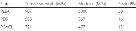
Zilberman et al. (2005) assessed behaviour of PLLA fibres and stent structures by subjecting them to uniaxial tensile testing. PLLA revealed high levels of tensile strength and modulus in comparison to other biodegradable polymers tested, polydioxanone (PDS) and poly(glycolide-co-epsilon-caprolactone) (PGACL) (Table 5). From Fig. 4, it is appar-ent that PLLA is the only polymer which exhibits yield.
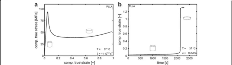
Engels et al. (2009) assessed the performance of amorphous PLLA materials under short- and long-term static loading conditions to investigate the time-dependent failure behaviour, especially the molecular and kinetic mechanisms which cause this time depend-ence. They found that premature failure seen in polylac-tides was due to the time-dependent behaviour of the materials, a common feature of polymer materials. The
work assessed compression behaviour of PLLA under a range of applied strain rates. As illustrated in Fig. 5a, PLLA initially behaved as a linear viscoelastic material, and changed to a non-linear response with increasing stress levels. A yield point of the material appeared at around 4% strain level with stress of 94 MPa. The initial response exhibited the trend for a typical brittle material, a dominant characteristic of PLLA, but strong softening (decreasing stress with increased strain) was followed by some hardening (upward trend of stress versus strain and large stretching before failure) (Meijer and Govaert 2005). Creep deformation of PLLA (Fig. 5b) is charac-terised by a sudden increase in strain to the point of final failure, which happened after some 30 min of the application of the stress equal to 70% of the yield point (Engels et al. 2009). An additional factor, which may affect the time-dependent performance of PLLA, is the application of dynamic load caused by blood pressure.
Bobel et al. (2015) recently characterized pre-degradation characteristics of PLLA used for cardiovascular application, as there remained a lack of data in this area. Performance of PLLA was strongly dependent on temperature and strain
Fig. 3Overview of the summarizing link between Young’s modulus and material microstructure (Agrawal et al. 1992; Perego et al. 1996; Wang et al. 1998; Middleton and Tipton 2000; Hermawan et al. 2010; Sin et al. 2012; Söntjens et al. 2012; Deroiné et al. 2014; Nerkar et al. 2014; Bergstrom and Hayman 2016)
Table 5Mechanical properties of bioresorbable fibres obtained in tension testing (Zilberman et al. 2005)
Fibre Tensile strength (MPa) Modulus (MPa) Strain (%)
PLLA 967 5000 50
PDS 583 367 161
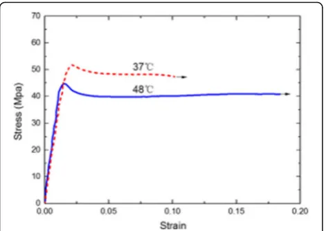
rate, and overall strain was revealed to contain a significant recoverable component. These factors were demonstrated at a displacement rate recommended for Absorb scaffolds (Abbott vascular, U.S.A.) [2.5 mm/min] (Fig. 6), which is the suggested inflation rate for scaffold deployment (Abbott Absorb and Absorb GT1, 2016). Work by Wang et al. (2017) presented uniaxial tensile tests on high molecular weight PLLA at two temperatures in order to correctly replicate mechanical behaviour of stent crimping (48 °C) and expansion (37 °C – body temperature) (Fig. 7). The obtained results demonstrated good plasticity of the material and a steady state of stress evolution in the material after yield stress was reached.
The testing schemes employed mostly two types of specimens- fibres and dogbone samples make of bulk material. Obviously, these specimens, generally, were not exposed to the same manufacturing/ processing condi-tions as a whole scaffold, questioning the suitability of obtained results to analysis of scaffolds. To overcome
these limitations, post-manufacture tests of scaffolds were performed.
Scaffold testing
Testing the macro properties of a scaffold is difficult to pursue due to their intricate geometry. At present, there is one set of ASTM guidelines for testing of absorbable stents (ASTM 2013) but they do not capture mechanics of the whole device before and during implantation and degradation. Eswaran et al. (2011) carried out a ring-test experiment, using Absorb scaffold, to quantify its mech-anical data. Geometry of the ring was non-uniform around the circumference; therefore, it could be as-sumed that, due to this, mechanical properties of the scaffold differed around the ring. The ring-tension ex-periments showed a breakage just below 3 N (Fig. 8a). To ensure the material tested was comparable to that of final stent, dog-bone samples used for tension testing were subjected to manufacturing processes that a scaf-fold tube would undergo. Figure 8b illustrates material anisotropy after yield and the flow resistance in the cir-cumferential direction continues to grow post yield; however, in the axial direction, there was a weaker Fig. 4Stress-strain curves for different bioresorbable fibres
(Zilberman et al. 2005)
Fig. 5(a) True stress versus strain (compressive) at constant strain rate; (b) evolution of compressive true strain with loading time at constant stress (Engels et al. 2009)
response with necking occurring. The displacement rate for testing is also shown to have an influence. Typical for PLLA, the material response was found to be strain-rate-dependent.
Oberhauser et al. (2009) investigated the Absorb scaffold by considering change in radial strength and molecular weight over time with incubation up to 90 days. It can be seen that Absorb stents had a radial strength comparable to that of common metallic stents (Fig. 9). This is despite weak bulk mechanical properties of polymers in compari-son to their metallic counterparts and due to post-extrusion manipulations with polymeric tubes increasing the circumferential orientation of crystallites in the semi-crystalline PLLA structure, resulting in higher resistance to radial loading (Oberhauser et al. 2009).
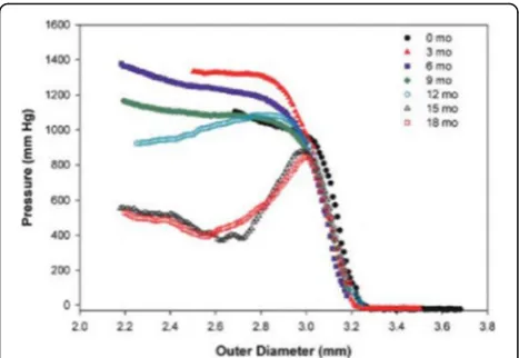
Additional data for Abbott Vascular stents (Fig. 10) show the evolution of pressure against the scaffolds’
outer diameter with degradation times, obtained by using a MSI RX550 Radial Force tester. The data show a noticeable change in behaviour with the onset of plastic deformation. For periods shorter than 12 months, the scaffold plastically deformed under larger compressive loads. However, it was still able to maintain radial sup-port to the artery beyond the yield point. For implant-ation longer than 12-months, a reduced resistance of stents to compressive load was found, indicating a lack of integral support to a vessel wall. This testing strategy is essential to observe maintenance of vessel patency is maintained during critical months of vessel healing (Kossuth et al. 2017).
Other features also affect mechanical properties of PLLA stents. Gomez-Lara et al. (2011) investigated the variation in curvature and angulation of the treated vessel post-deployment of BRSs. Absorb, in comparison to metal-lic stents, presented better conformability to vessel’s Fig. 7Stress–strain curves for PLLA at crimping and expansion
temperatures (Wang et al. 2017)
Fig. 8(a) Average force-displacement average curves from ring-tension experiment; (b) stress-strain data for uniaxial tension (1–circumferential-fast; 2 - circumferential-medium; 3–circumferential-slow; 4–longitudinal-fast; 5–longitudinal-medium; 6–longitudinal-slow) (Eswaran et al. 2011)
architecture and produced lower variations of these param-eters (Gomez-Lara et al. 2010). In 2011, the same authors demonstrated that BRS restored coronary configuration in comparison with metallic stenting which altered geometry permanently (Gomez-Lara et al. 2011). Foin et al. (2016) assessed overexpansion of Absorb scaffolds beyond their recommended diameter and concluded that this could result in strut breakage and a loss of central mechanical support. A similar work was conducted and corroborated by Ferdous et al. (2015).
A comprehensive overview of in-vitro tests assessing mechanics associated with the safety and efficacy of the implantable devices was presented by Schmidt et al. (2016). Scaffold expansion, elastic recoil, radial strength and flexibility/ bending stiffness were assessed for two
types of virgin bioabsorbable scaffolds and one metallic stent. As expected, the Absorb scaffold demonstrated lower crush resistance in comparison to a metallic stent; however, the collapse pressure for Absorb (172 kPa) was in the required range for radial strength of permanent stents (Fig. 11; Lanzer and Schmidt 2015). This data cor-roborates with that produced by Oberhauser et al. (2009), where strength of polymer scaffolds was compar-able to that of their metallic counterparts. Elastic recoil of the stents was assessed immediately after implant-ation and an hour after expansion in two vessel types, i.e., rigid (Fig. 9b) and mock vessels (Fig. 9c). Within the rigid vessel, the Absorb scaffold exhibited recoil compar-able to that of BIOTRONIK; however, it did not display the interesting behaviour of DESOLVE, whose diameter increased after an hour. The mock vessel did not affect the BIOTRONIK stent, but encouraged further recoil of the Absorb scaffold after an hour of implantation and significantly increased recoil of the DESolve scaffold. These results illustrate that it is crucial to correctly simu-late in-vivo conditions as they may impact a stents per-formance. With load, the Absorb scaffold did recoil further, as expected. The authors noted that good in-vitro performance would not always correlate to good perform-ance in-vivo, and poor in-vitro performperform-ance is indicative of potential complications. These obtained results showed the high performance of PLLA and, in particular, the Ab-sorb scaffold in comparison to metallic counterparts.
Mechanical performance of scaffolds with degradation
Obviously, in-vitro degradation of bioresorbable stents has a strong effect on their mechanical properties and
Fig. 11(a) Radial strength measured as a means of collapse pressure; acute and time dependent recoil after expansion in (b) a rigid vessel model and (c) in a mock vessel (Schmidt et al. 2016)
performance. Degradation is defined as the “ chain-scis-sion process, during which polymer chains are cleaved to form oligomers and, finally, to form monomers”, whereas erosion “designates the loss of material owing to mono-mers and oligomono-mers leaving the polymer” (Göpferich 1996). There are two methods of hydrolysis which PLLA can undergo: (1) chemical degradation via hydrolysis and (2) enzyme-catalysed hydrolysis; the former is the most prominent for artificial biodegradable polymers. Degrad-ation of polymers such as PLLA depends on parameters of the environment, only some of which are controllable; these include the type of chemical bond, pH level, molecular weight, polydispersity, crystallinity, morphology, ionic strength, temperature and water uptake (Göpferich 1996; Gong et al. 2007; Zhang 2015).
Degradation of PLLA fibres in vitro were investigated by Yuan et al. (2002) with fibres incubated in phosphate buffer solution (PBS) with pH 7.4 and at 37 C for 35– 45 weeks. The average molecular weight of fibres, pro-duced by melt spinning, dropped throughout degradation, with 40–60% reduction at the end of the incubation period and an increase in crystallinity. Fibres studied by the group were not investigated separately, instead, they were bundled together randomly which may have affected their biodegradation. PBS was changed fortnightly which is an acceptable time frame in accordance with ASTM standards. Prior to measurements of degraded samples, a bundle was removed from PBS, washed with water and dried in a vacuum oven at 48 C. Water absorbed by the fi-bres is a potential issue that could affect the results of this scheme. However, authors did not mention duration of this process. Additionally, drying in a vacuum oven is a good methodology to remove excess fluid from the fibres; however, a temperature of 48 C could affect the properties of the fibre, with potential to cause damage to the polymer. The group could have weighed fibres prior incubation in the vacuum oven in order to assess the water content in the material (Yuan et al. 2002).
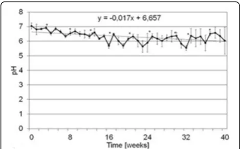
Mechanical, rheological, fatigue and degradation behav-iour of PLLA used in vascular implants was investigated by Bartkowiak-Jowsa et al. (2012). The authors highlighted the importance of adequate optimization of stent design. The study produced results (Fig. 12) confirming the need to change the incubation fluid at regular intervals. A saw-tooth character of the graph demonstrates that the incu-bation fluid required regular replenishment to maintain a pH of 7.4 ± 0.2. PLLA tested in this regime did not show a decrease of more than 10% in strength and deformabil-ity, ideal for providing sufficient support for an arterial wall. However, it was noted that the total degradation time for PLLA was 24 months, far beyond the time required for tissue regeneration. In order to combat this, the au-thors suggested the modification of PLLA through the addition of biodegradable filler, alginate fibres. These
fibres enhance liquid absorption at phase boundaries resulting in faster degradation, without affecting mechan-ical properties of PLLA. The study assessed the stability of elastic properties by testing their thermodynamic proper-ties. Assessing the storage-modulus values allowed the au-thors to determine the temperature range, in which the material demonstrated the desired elastic properties. All materials tested had glass-transition temperatures in the range of 56.1–68 C providing structural stability in the temperature range of biomedical applications.
Kimble and Bhattacharyya (2015) assessed mechanical properties of PLLA during in vitro degradation in com-parison to a co-polymer material (PLLA/PBS) by incu-bating dogbone specimens for 24 weeks in PBS at 37 °C. In comparison to the co-polymer tested, neat PLLA maintained integrity throughout 24 weeks, showing in-creasing creep resistance in the initial 8-week degrad-ation period. The initial modulus and tensile strength of PLLA are higher than those of a variety of co-polymer blends. The study demonstrated the necessity to obtain baseline properties of a material and that the co-polymeric blend in fact had lower mechanical properties and a higher, less stable degradation rate. The effect of fatigue on chemical and mechanical degradation of model stent sub units was analysed by Dreher et al. (2016) since this factor can impact greatly on a stent's performance and was studied insufficiently. Subunits composed of PLLA were exposed to fatigue and degrad-ation conditions simultaneously, amounting to one year of implantation within the body. Preliminary experi-ments, carried out by the authors, illustrated that 38 million fatigue cycles were concurrent with one year of implant of the scaffold within a pulsatile vessel. In com-parison to control samples tested under static conditions (i.e. without fatigue), the study indicated that fatigue loading, resulted in a significant increase of samples stiffness with a diminished force at break. Molecular
weight was also affected, with a greater decline seen at fatigue. The authors concluded that this illustrated a link between chemical degradation and mechanical loading.
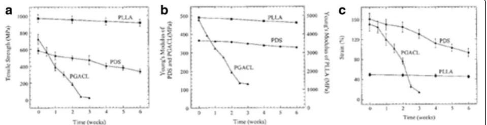
The effect of degradation on tensile mechanical prop-erties of PLLA fibres and expandable stents prepared from fibres was studied by Zilberman et al. (2005). The work discovered that PLLA, in contrast to other bioab-sorbable materials, underwent degradation at a slower rate, displaying no alterations in mechanical properties during 6 weeks (Fig. 13). It also confirmed results of the previous study by the same group, showing PLLA fibres exhibited good mechanical properties after 24 weeks of degradation incubation (Zilberman et al. 2004). Single fibres constructed into a stent structure for each polymer were subjected to radial compressive tests (Fig. 14) to obtain data for scaffold strength. PLLA showed consistent results over the 6-week period and confirmed its viability for use as an arter-ial support structure.
Dreher et al. (2015) focused on assessing the process of degradation of PLLA tubes in in-vivo tests as a model for coronary stent degradation. Their predominant aim was to estimate the effect of physiological factors, (pulsa-tile flow, body temperature), in testing of scaffolds. Their work demonstrated that PLLA tubes subjected to mock pulsatile flow displayed a statistically different degrad-ation pattern to tubes subjected to control conditions (inside the vessel but not subjected to pulsatile flow). The results indicated that to correctly investigate per-formance of polymeric scaffolds in-vivo, all constituents that stimulate degradation such as correct pulsatile flow, pressure, and temperature should be implemented in-vitro to obtain accurate results. After 6 months, it was seen that the scaffold in the loop set up presented a slower rate of molecular weight loss compared to the control samples. This was potentially due to oligomers, breakdown products from degradation, being removed with the flow system (as opposed to remaining within the control samples), creating localized degradation be-cause of their acidic nature. Crystallinity changes for
control samples were comparable; however, the loop samples showed that crystallinity was diminished signifi-cantly. This was accounted for by the group by relax-ation of polymer chain with certain stimuli such as frequency and temperature (Fig. 15; Dreher et al. 2015).
The effect of static and dynamic loading conditions on degradation of PLLA stent fibres was assessed by Hayman et al. (2014) over 15 months to ascertain whether dynamic conditions were required during in-vitro testing and it was found that it did affect output. In comparison to a static load, dynamic load seemed to accelerate degradation, especially at later time point; however an increase in static load also showed acce-lerated degradation (Hayman et al. 2014). Luo et al. (2014) assessed the degradation performance of high-molecular-weight PLLA scaffolds in a mock artery system. A plasma pump enabled simulation of systolic/ diastolic pressures comparable to those observed in the human circulatory system. The system also enabled in-corporation of fluid circulation to mimic blood flow. These data were compared to in-vivo results, where the scaffolds were implanted into animal models. There was a good correlation between the in-vivo and in-vitro results, and the slight discrepancies between the two conditions
Fig. 13Mechanical properties of 3 bioresorbable polymers subjected to degradation in phosphate buffer solution (PBS) at 37 °C:atensile strength;bYoung’s modulus;cstrain (Zilberman et al. 2005)
might be attributed to the biochemical environmental dif-ference (Luo et al. 2014).
From this review of degradation, manufacturing and processing studies for PLLA, it can be concluded that a significant number of factors affect mechanical proper-ties of PLLA scaffolds, from its initial post-implantation state and throughout degradation. Each PLLA material, depending on these conditions, can result in significantly altered mechanical properties and degradation profiles. Degradation performance of biodegradable polymers in-vitro showed that higher loads resulted in shorter deg-radation times (Athanasiou et al. 1998;,Middleton and Tipton 2000); therefore accurate simulation of mechan-ical conditions corresponding to real-life conditions (e.g., pulsatile flow and loads) is needed. Additionally, many studies appeared to use PBS as a fluid to incubate scaf-folds; however, the biochemical environment may also influence polymer degradation. Obtaining accurately quantified mechanical response data can then be used as input into a constitutive model for finite-element (FE) simulations (Bobel et al. 2016). This could diminish ex-perimental efforts and enable accurate prediction of scaffold performance throughout its lifecycle.
Computational studies for BRS performance with degradation
Numerical simulations, based on adequate mechanical models allow additional insights into a response of a physical system to specific loading and environmental conditions that can be used for making decisions from an engineering perspective (Babuška 1991). Computational
modelling of medical devices enables the users to obtain knowledge of their performance and precise locations of failure in a device, implement design optimization and reduce benchtop testing thus minimizing the device development time and cost (Eswaran et al. 2011; Bobel et al. 2015). The ability to accurately capture the behaviour of medical-device materials under physiological loads and during processing remains a challenge. There appears to be a lack of material models detailing the accurately the degradation process of bioabsorbable materials because of a wide range of factors affecting on a materials response, e.g., temperature, processing and extrusion procedure. Simulations run with FE analysis for complex loading con-ditions require precise experimental investigation into the modelled material in order to obtain fundamental geomet-ric and material parameters (Bobel et al. 2015). Through development of accurate validated computational models, high-quality predictions of device performance are pos-sible (Eswaran et al. 2011). Due to the complexity and lim-ited work carried out in the area of numerical simulations of PLLA BRSs, this section highlights the recent studies.
Material modelling of PLLA
devices are adequately captured by the model to provide accurate predictions. At low strains, the behaviour of this material is viscous whereas under high strain, it behaves as brittle elastic material (Bardenhagen et al. 1997). A non-linear mechanical response of PLLA is due to its low glass-transition temperature (Tg = 60 °C), sensitivity to molecular weight, degree of crystallinity and physical ageing time (Bergstrom and Hayman 2016). Classical linear viscoplas-tic/elastic models therefore cannot be used to describe PLLA behaviour as they do not capture its full anisotropic behaviour (Eswaran et al. 2011). Also typical elastic, hypere-lastic or viscoehypere-lastic models are not able to capture the ac-cumulation of plastic strain in a material, which is crucial for engagement of vascular scaffolds (Vieira et al. 2015).
Neo-Hookean or Mooney-Rivlin models, describing incompressible hyperelastic materials, could be used to predict material behaviour prior to any fissure of non-degraded PLLA. These models are useful as they can pre-dict monotonic behaviour of PLLA devices, also at specific degradation time points. Mechanical behaviour of PLLA, with large deformation and dynamic loads at different strain rates, is both viscous and elastoplastic. Also, hyster-esis behaviour was observed in biodegradable polyesters (Vieira et al. 2015). A handful of models at present ac-count for diverse behaviours of aliphatic polyester using varying combinations of hyperelastic, viscous and plastic models; however, they do not account for the degradation-related changes (Vieira et al. 2015). As scaffolds are sub-jected to loading above the elastic limit during processing/ manufacturing, it leads to an accumulation of plastic strain and contributes to device failure at these areas of high strain. Post-implantation scaffolds are also subjected to a cyclic stresses, which could cause evolution of significant plastic strain during their in-service life resulting in failure and laxity (Vieira et al. 2015). Hydrolysable bonds in PLLA make the material more susceptible to moisture, heat, light and mechanical stresses. The traditional degradation mechanism of biodegradable polymers is chemical degrad-ation via hydrolysis or enzyme-catalysed hydrolysis. Im-portantly, mechanical behaviour of the material evolves with time due to the hydrolytic chain scission (Pan 2015). Degradation results in a reduction in molecular weight of such polymers, there is also a reduction in its ductility, toughness and strength (Vieira et al. 2015). Some complex
material models based on a concept of networks, combin-ing elastic, slidcombin-ing and dissipative elements (based on Boltzmann’s superposition principle), are used to simulate this time dependent response; however, most models only simulate an initial mechanical response of the polymer and neglect changes in material properties during hydro-lytic degradation (Vieira et al. 2015).
Constitutive material models
Tailoring microstructural parameters for specific functions are depends on potential applications; these parameters can be controlled with resin formulation and processing conditions. Stress-strain, deformation and degradation re-sponses of a material is affected significantly by a type of, and changes in, applied boundary conditions, loads and environmental factors, the material is subjected to. Such multi-physics responses can be incorporated into relevant constitutive models to simulate material behaviour; how-ever, there remains doubts as to whether a whole-device response is accurately captured in simulations (Bergstrom and Hayman 2016). A stent technology and mechanical behaviour of metallic stents are relatively well established; however, due to the non-linear, viscoplastic and degrad-able behaviour of PLLA along with an altering mechanical response of the device in-vivo, the performance of BRSs with degradation is not as established (Bergstrom and Hayman 2016). The ideal material model for the Absorb PLLA scaffold should be able to capture the nature of the mechanical properties of the stent with evolving degrad-ation. As highlighted in Table 6, numerous materials modes were proposed in literature to capture complex re-sponses of biodegradable polymers - from their virgin state right through to full degradation (Soares et al. 2008, 2010a, b; Eswaran et al. 2011; Muliana and Rajagopal 2012; Khan and El-Sayed 2013; Vieira et al. 2015; Rosa et al. 2016). The past five years have seen significant ad-vancements in computational mechanics of bioresorbable scaffolds aimed at development of material models to cap-ably simulate adequately performance of PLLA stents.
Computational and mathematical modelling of BR scaffolds
Various groups of researchers concentrated on different aspects of simulation of properties and performance of
Table 6Different material model frameworks used for modelling of PLA (Soares et al. 2008; Eswaran et al. 2011; Söntjens et al. 2012; Khan and El-Sayed 2013; Hayman et al. 2014; Bergstrom and Hayman 2016)
Model Class Degradation modelled Visco-plastic effects Anisotropic effects Use in FE simulations
(Eswaran et al.2011) Viscoplastic x ✓ ✓ ✓
(Hayman et al.2014) Hyperelastic ✓ x x ✓
(Khan and El-Sayed2013) Linear viscoelastic ✓ x x ✓
(Soares et al.2008) Hyperelastic ✓ x x ✓
bioresorbable stents. Bobel et al. (2015) compiled a com-prehensive study of computational bench testing using Abaqus/Standard 6.12 (SIMULIA), to evaluate short-term mechanical performance of three different stent designs; however, with no consideration for polymeric degradation. A non-linear viscoelastic parallel network model was used to account for material performance; still, there was no account for materials plasticity. Whilst some more complex material representations are avail-able, the group discussed the advantage of this model, including its ease of use and access in Abaqus software, which captures PLLA behaviour for the testing method-ologies involved sufficiently well. An interesting notion is a “flexible” material model, where a range of elastic behaviours and properties of PLLA can be incorporated in it. Still, this model is only suitable for material behav-iour at so called “time zero” (i.e. immediately post de-ployment). A strong feature of this work is the use of real stent geometries and experimental testing to assess the radial strength, flexibility and longitudinal resistance (Fig. 16). Additionally, shorter sections of stents were used, 2 or 4 circumferential rings, to reduce the compu-tation time. There was a special focus on ensuring that the stents were exposed to the same real-life processes prior to implanting (e.g. expansion). Authors found that to meet the requirement of minimum collapse pressure of 0.04 MPa, the Young’s modulus of 4.1 GPa was re-quired for Absorb scaffolds. This was conflicting infor-mation based on an Abbott patent report, stating that PLLA has a modulus of 1.2–2.7 (Wang 2013). So, it would have been more accurate to accommodate this range of values in the employed material model. The ra-dial strength of the Absorb scaffold was reported to be comparable to that of metallic stents (Oberhauser et al. 2009), so long as they were deployed within their size limits, these experimental data contradict the computa-tional results of this work. It was suggested that to im-prove the radial performance of stents, the materials elastic modulus should be increased to 15 GPa. This, how-ever, would affect the compliance mismatch which is a goal for bioresorbable scaffolds (Brugaletta et al. 2012). According to this study, the wide variations in the poly-mer’s elastic modulus resulted in a corresponding range of
stent resistance to applied pressure, with weaker materials prone to collapse under lower pressure. Additionally, strut thickness was assessed computationally and it was found that as in the experiments, thicker struts could bear higher radial pressure than thinner struts. Finally, assessing a range of stent geometries, the mechanical performance of scaffolds was directly linked to their design and dimen-sions (Bobel et al. 2015). Schiavone and Zhao (2016) cor-roborated the factor emphasised in Bobel et al. (2015), that it is essential relevant biomechanical forces in the model are incorporated in computation performance studies in order to correctly simulate all steps of a scaffold lifecycle, from manufacturing to full degradation.
In another study Luo et al. (2014) conducted compre-hensive in-vitro and in-vivo analysis to validate their model by employing a user-defined constitutive model (USDFLD) in FE simulations. In this approach a user can manually define different field variables in order to ac-count for material properties at a certain time point. In-corporating varying degrees of degradation into the material model enabled assessment of evolution of stent properties with time. Numerical simulations were carried out using Abaqus, with C3D8I element used to mesh the geometric model. This incompatible-mode eight-node brick element was preferred to model stents with Abaqus as they are loaded predominantly in bending (Pham et al. 2014). Crimping and expansion tools were meshed with a surface element (SFM3D4R) and scaffold simulation car-ried out in four steps (Table 7). These steps were neces-sary to assess the forces the scaffold is exposed to prior to its implantation into an artery. A fatigue simulation was also conducted to ascertain the effect of pulsatile flow on the change in mechanical properties with time. In this simulation, the last effect was incorporated by application of pressure to the inner arterial walls. This stage was followed by simulations of the degradation process.
Figure 17a demonstrates the von Mises stress for crimp-ing to 1.5 mm (A) and expansion (B). Stresses were local-ized in the U-bend regions of the scaffold, common for polymeric stents (Schiavone et al. 2017). The stage of elastic recoil saw a stress decrease from 161 MPa to 102.4 MPa. In expansion, stress concentration was again found in the U-bend regions, with a high tensile stress of
172.3 MPa. The degradation process was assessed post-fatigue, thus ensuring the material state was as in-vivo, with a similar application of biomechanical forces. It was observed that degradation occurred at a greater rate in areas of previous higher strain, presenting spatially non-uniform degradation throughout the scaffold (Fig. 17b). Performing expansion on the incubated in-vitro scaffolds, the observed areas of breakage coincided with high-concentration areas obtained with FE. Thus, the in-vivo, in-vitro and FE results were consistent, implying suitabil-ity of the material model used.
Similar to Luo et al. (2014), Wang et al. (2017) pre-sented a combined experimental and computational study on the mechanical performance of PLLA coronary implants. To implement computation aspect, the Aba-qus/ Standard implicit solver was used to estimate radial strength and 3-point-bending performance of stents, with care taken to adhere to ASTM standards to ensure that simulations were as close to the testing protocol as possible. Model geometry closely mimicked that of Ab-sorb, and a mesh of hexahedral C3D8R elements was ap-plied. In comparison to Luo et al. (2014), the crimping and expansion simulations also incorporated the effect of temperature for each of these stages, i.e., crimping was at 48 °C and expansion at body temperature 37 °C. This was supported by testing pure PLLA at different temperatures and using the obtained data with respect-ive models. Results were acquiescent with those de-scribed by Luo et al. (2014); with localization of stress found in U-bends of the scaffolds. Simulations were reli-ant on the experimental data obtained by the group for crimping, expansion, radial strength and three-point bending. Table 8 presents the correlation between the
obtained in-vitro data and computational results. For crimping, the radial recoil ratio was not as critical as that observed for expansion. By over-expanding scaf-folds, apparently, recoil was reduced, indicative of plastic deformation in the material. These findings can be used to ensure a suitable recoil effect to prevent mechanical injury to the vessel wall due to excess expansion, result-ing in inflammation.
In comparison to Wang et al. (2017), Pauck and Reddy (2015) simulated the radial performance of PLLA stents within a remodelling artery, taking into consideration vari-ous geometries for the scaffolds. Material-characterization tests were conducted to obtain accurate data for high-molecular-weight PLLA and used in a standard aniso-tropic elastic-plastic material model. The data obtained from the material characterisation of PLLA was incorpo-rated into the material model, with all the scaffolds having these same material parameters. Three stent geometries were investigated: one mimicked a typical polymer scaffold (Absorb); a second was with a typical metal-stent design; a third addressed the shortfalls and weaknesses of the other two designs. C3D8RH elements were used in simulations as they were computationally inexpensive thanks to having only one integration point per element. Some hourglassing issues were found for C3D8R, how-ever, this was accounted for by enhancing hourglass con-trol in the simulations. It was concluded that design and geometry of scaffolds had an impact on the radial per-formance of stents. A geometry of the typical polymer scaffold was shown to provide the best performance in comparison to the other two. It was concluded that all Table 7Scaffold simulation steps (Luo et al. 2014)
Step Detail Description
1 Crimp Rigid cylindrical tube to simulate a crimp tool (3.5 mm➔1.5 mm)
2 Elastic recoil Removal of rigid cylindrical tube
3 Expansion Rigid cylindrical body denoting balloon catheter (➔3.2 mm inner radius)
4 Elastic recoil Removal of rigid cylindrical tube
Fig. 17(a) Results of crimping and expansion simulations and (b) degradation trend at different sites across the scaffold depending on stress (Luo et al. 2014)
Table 8Summary of results obtained in Wang et al. (2017)
Experiment Simulation
Recoil % Crimping
Outer diameter (mm) 1.41 n/a 10.87
Expansion
Inner diameter (mm) 3.10 4.7 4.19
3.7 ~ 2 2.92
3.92 ~ 2 1.81
stent geometries underperformed, with the polymer stent only just presenting adequate radial strength of 322 mmHg (magnitudes in excess of 300 mmHg are considered suffi-cient for vessel patency (Pauck and Reddy 2015)). This ra-dial strength value, however, was only achieved by a significant increase of the material-stiffness to 9 GPa. With depleting material stiffness values, the performance of all designs diminished. Additionally, improving thickness of scaffolds improved their performance; however, this, in turn, could instigate damaging physiological side effects.
Additionally a study by Schiavone et al. (2016) highlighted the importance of taking into account varia-tions of plaque morphology within the vessel as this can significantly impact the implantation and performance of a scaffold. The study investigated mechanics of stent-vessel interaction, comparing the performance of polymeric and metallic stents with consideration of eccentric and concentric shapes of the arterial plaque. It was shown that polymeric scaffolds generated lower stresses in the vessel wall, and recoiled further after expansion than their metallic counterparts. The study illustrated that eccentric plaque retained high stresses in different layers of the artery. Eccentricity caused uneven radial expansion, affecting the scaffold performance. Simulating performance of scaffolds in vessels with atherosclerotic lesions could enable assessment of mechanical behaviour of different stent designs with various plaque compositions, offering a best fit for a patient-specific plaque.
Conclusion
Due to the increasing prevalence of bioresorbable PLLA scaffolds, specifically stents in the medical-devices field, there is a need to understand and quantify the mechan-ical response of these scaffolds to in-service conditions prior to their implant together with associated changes in this response during their degradation. This know-ledge will allow engineers to enhance the development and design optimization of stents in order to improve their performance. Issues, arising from use of metal stents, can be relieved with the use of polymer scaffolds, presenting many benefits compared to permanent stent-ing, including restoration of vasomotion. Degradation profiles of polymer scaffolds are controlled by multiple factors including molecular weight, degree of crystallin-ity as well as loads applied to the structure. Experimental quantification of the non-linear mechanical response for the viscoelastic polymer enables validation of computa-tional (FE) models, with numerical simulations permit-ting predictions of in-vivo behaviour of scaffolds. This review highlighted recent progression in material models and FE simulations endorsed by experimental testing. Still, it is apparent that further experimental and compu-tational studies are required to understand specific
features of mechanical behaviour of PLLA scaffolds, in-cluding their performance prior to, and during, degrad-ation. Additionally, developing specific material models for each type of PLLA stent due to the different manu-facturing procedures and compositions is necessary be-cause of varying performance of different PLLA stents. Achieving this will enable improvement and enhance-ment of future generations of polymer scaffolds with im-proved prediction of their mechanical performance, allowing enhancement of medical procedures and accel-erated healing of patients.
Abbreviations
ASTM:American Society for testing and Materials; BMS: Bare metal stents; BRS: Bioresorbable scaffolds; CABG: Coronary artery bypass grafting; CHD: Coronary heart disease; DES: Drug eluting stents; FDA: Food and Drug administration; NHS: National Health Service; PCI: Percutaneous coronary intervention; PLLA: Poly-L-Lactic acid
Funding
RN is funded by the British Heart Foundation for her PhD research project (Grant number: FS/15/21/31424; Title: Towards controlling the mechanical performance of polymeric bioresorbable vascular scaffold during biodegradation).
Authors’contributions
RN carried out the literature review and wrote the paper; LGZ, YL and VVS edited the paper. All authors read and approved the final manuscript.
Authors’information
RN is a PhD student, LGZ is a professor in Solid Mechanics, YL is a Senior Lecturer of Healthcare Engineering and VVS is a professor in Mechanics of Materials. All work at Wolfson School of Mechanical, Electrical and Manufacturing Engineering, Loughborough University of the UK.
Consent for publication
All authors agree to publications.
Competing interests
The authors declare that they have no competing interests.
Publisher’s Note
Springer Nature remains neutral with regard to jurisdictional claims in published maps and institutional affiliations.
Received: 26 May 2017 Accepted: 10 July 2017
References
Abbott Absorb and Absorb GT1 - Bioresorbable Vascular Scaffold Systems. https:// www.vascular.abbott/int/products/coronary-intervention/absorb-bioresorbable-scaffold-dissolving-stent.html. Accessed 1 Oct 2016
Abizaid A, Costa JR Jr. (2010) New drug-eluting stents: an overview on biodegradable and polymer-free next-generation stent systems. Circ Cardiovasc Interv 8:384–393
Agrawal CM, Haas KF, Leopold DA, Clark HG (1992) Evaluation of poly(L-lactic acid) as a material for intravascular polymeric stents. Biomater 13:176–182 http://dx.doi.org/10.1016/0142-9612(92)90068-Y
ASTM (2014a) Standard guide for three-point bending of balloon expandable vascular stents and stent systems
ASTM (2013) Standard guide for testing absorbable stents
Athanasiou KA, Agrawal CM, Barber FA, Burkhart SS (1998) Orthopaedic applications for PLA-PGA biodegradable polymers. Arthroscopy 14:726–737 Babuška I (1991) Mathematical models and engineering decisions. In: Finite
Bardenhagen SG, Stout MG, Gray GT (1997) Three-dimensional, finite deformation, viscoplastic constitutive models for polymeric materials. Mech Mater 25:235–253 Bartkowiak-Jowsa M, Będziński R, Kozłowska A et al. (2012) Mechanical, rheological,
fatigue, and degradation behavior of PLLA, PGLA and PDGLA as materials for vascular implants. Meccanica 48:721–731. doi:10.1007/s11012-012-9626-2 Bergstrom JS, Hayman D (2016) An overview of mechanical properties and
material modeling of polylactide (PLA) for medical applications. Ann Biomed Eng 44:330–340. doi:10.1007/s10439-015-1455-8
Bobel AC, Lohfeld S, Shirazi RN, McHugh PE (2016) Experimental mechanical testing of poly (l-Lactide) (PLLA) to facilitate pre-degradation characteristics for application in cardiovascular stenting. Polym test 54:150–158 http://dx. doi.org/10.1016/j.polymertesting.2016.07.011
Bobel AC, Petisco S, Sarasua JR et al. (2015) Computational bench testing to evaluate the short-term mechanical performance of a polymeric stent. Cardiovasc Eng Technol 6:519–532
Bourantas CV, Onuma Y, Farooq V et al. (2013) Bioresorbable scaffolds: current knowledge, potentialities and limitations experienced during their first clinical applications. Int J Cardiol 167:11–21 http://dx.doi.org/10.1016/j.ijcard.2012.05.093 British Heart Foundation (2015) BHF Headline Statistics https://www.bhf.org.uk/
research/heart-statistics
Brugaletta S, Gogas BD, Garcia-Garcia HM et al. (2012) Vascular compliance changes of the coronary vessel wall after bioresorbable vascular scaffold implantation in the treated and adjacent segments. Circ J 76:1616–1623
Celermajer DS (1997) Endothelial dysfunction: does it matter? Is it reversible? J am Coll Cardiol 30:325–333 http://dx.doi.org/10.1016/S0735-1097(97)00189-7 Deroiné M, Le Duigou A, Corre Y-M et al. (2014) Accelerated ageing and lifetime prediction of poly(3-hydroxybutyrate-co- 3-hydroxyvalerate) in distilled water. Polym Test 39:70–78. doi:10.1016/j.polymertesting.2014.07.018
Dreher ML, Nagaraja S, Batchelor B (2016) Effects of fatigue on the chemical and mechanical degradation of model stent sub-units. J Mech Behav biomed mater 59:139–145 http://dx.doi.org/10.1016/j.jmbbm.2015.12.020 Dreher ML, Nagaraja S, Lu Q, et al. (2015) Degradation of PLLA tubes in a
pulsatile flow loop as a model for cardiovascular stent degradation Engels TAP, Söntjens SHM, Smit TH, Govaert LE (2009) Time-dependent failure of
amorphous polylactides in static loading conditions. doi:10.1007/s10856-009-3851-9 Eswaran SK, Kelley JA, Bergstrom JS, Giddings VL (2011) Material modeling of
Polylactide
Fambri L, Pegoretti A, Fenner R et al. (1997) Biodegradable fibres of poly(l-lactic acid) produced by melt spinning. Polymer (Guildf) 38:79–85. doi:10.1016/S0032-3861(96)00486-7
Ferdous J, Kolachalama VB, Kolandaivelu K, Shazly T (2015) Degree of bioresorbable vascular scaffold expansion modulates loss of essential function. Acta Biomater 26:195–204. doi:10.1016/j.actbio.2015.08.009
Foin N, Lee R, Mattesini A et al. (2016) Bioabsorbable vascular scaffold overexpansion: insights from in vitro post-expansion experiments. EuroIntervention 11:1389–1399. doi:10.4244/EIJY15M07_02
Foin N, Lee RD, Torii R et al. (2014) Impact of stent strut design in metallic stents and biodegradable scaffolds. Int J Cardiol 177:800–808 http://dx.doi.org/10. 1016/j.ijcard.2014.09.143
Fortier A, Gullapalli V, Mirshams RA (2014) Review of biomechanical studies of arteries and their effect on stent performance. IJC hear vessel 4:12–18 http://dx.doi.org/10.1016/j.ijchv.2014.04.007
Gada H, Kirtane AJ, Newman W et al. (2013) 5-year results of a randomized comparison of XIENCE V everolimus-eluting and TAXUS paclitaxel-eluting stents: final results from the SPIRIT III trial clinical evaluation of the XIENCE V everolimus eluting coronary stent system in the treatment of patient. JACC Cardiovasc Interv 6:1263–1266. doi:10.1016/j.jcin.2013.07.009
Gomez-Lara J, Brugaletta S, Farooq V et al. (2011) Angiographic geometric changes of the lumen arterial wall after bioresorbable vascular scaffolds and metallic platform stents at 1-year follow-up. JACC Cardiovasc Interv 4:789–799 http://dx.doi.org/10.1016/j.jcin.2011.04.009
Gomez-Lara J, Garcia-Garcia HM, Onuma Y et al. (2010) A comparison of the conformability of everolimus-eluting bioresorbable vascular scaffolds to metal platform coronary stents. JACC Cardiovasc Interv 3:1190–1198 http://dx.doi.org/10.1016/j.jcin.2010.07.016
Gong Y, Zhou Q, Gao C, Shen J (2007) In vitro and in vivo degradability and
cytocompatibility of poly(l-lactic acid) scaffold fabricated by a gelatin particle leaching method. Acta biomater 3:531–540 http://dx.doi.org/10.1016/j.actbio.2006.12.008 Göpferich A (1996) Mechanisms of polymer degradation and erosion. Polym
scaffolding hard tissue Eng 17:103–114. doi: http://dx.doi.org/10.1016/0142-9612(96)85755-3
Grabow N, Schlun M, Sternberg K et al. (2005) Mechanical roperties of laser cut poly(L-lactide) micro-specimens: implications for stent design, manufacture, and sterilization. J Biomech Eng 127:25–31. doi:10.1115/1.1835349 Grijpma DW, Altpeter H, Bevis MJ, Feijen J (2002) Improvement of the
mechanical properties of poly(D,L-lactide) by orientation. Polym Int 51: 845–851. doi:10.1002/pi.988
Gruntzig A (1978) Transluminal dilatation of coronary-artery stenosis. Lancet (London, England) 1, 263
Gupta B, Revagade N, Hilborn J (2007) Poly(lactic acid) fiber: an overview. Prog Polym Sci 32:455–482. doi:10.1016/j.progpolymsci.2007.01.005
Hara H, Nakamura M, Palmaz JC, Schwartz RS (2006) Role of stent design and coatings on restenosis and thrombosis. Drug-eluting stents an Innov Multidiscip drug Deliv Platf 58:377–386 http://dx.doi.org/10.1016/j.addr.2006.01.022 Hayman D, Bergerson C, Miller S et al. (2014) The effect of static and
dynamic loading on degradation of PLLA stent fibers. J Biomech Eng 6: 81006-81006–81006-81009
Hermawan H, Dubé D, Mantovani D (2010) Developments in metallic biodegradable stents☆. Acta Biomater 6:1693–1697. doi:10.1016/j.actbio.2009.10.006 Hoffmann R, Mintz GS (2000) Coronary in-stent restenosis - predictors, treatment
and prevention. Eur Heart J 21:1739–1749. doi:10.1053/euhj.2000.2153 Iakovou I, Schmidt T, Bonizzoni E et al. (2005) Incidence, predictors, and outcome
of thrombosis after successful implantation of drug-eluting stents. JAMA 293: 2126–2130. doi:10.1001/jama.293.17.2126
Iqbal J, Onuma Y, Ormiston J et al. (2014) Bioresorbable scaffolds: rationale, current status, challenges, and future. Eur Heart J 35:765–776. doi:10.1093/eurheartj/eht542 Khan KA, El-Sayed T (2013) A phenomenological constitutive model for the
nonlinear viscoelastic responses of biodegradable polymers. Acta Mech 224: 287–305. doi:10.1007/s00707-012-0760-7
Kimble LD, Bhattacharyya D (2015) In Vitro degradation effects on strength, stiffness, and creep of PLLA/PBS: a potential stent material. Int J Polym Mater Polym Biomater 64:299–310. doi:10.1080/00914037.2014.945203
Kočka V, Toušek P, Widimský P (2015) Absorb bioresorbable stents for the treatment of coronary artery disease. Expert Rev Med Devices 12:545–557. doi:10.1586/17434440.2015.1080119
Kossuth MB, Perkins LEL, Rapoza RJ (2017) Design principles of bioresorbable polymeric scaffolds. Interv Cardiol Clin 5:349–355 http://dx.doi.org/10.1016/j. iccl.2016.02.004
Kristensen SD, Grove E (2007) Stent thrombosis: Definitions, Mechanisms and Prevention http://www.escardio.org/Guidelines-&-Education/Journals-and-publications/ESC-journals-family/E-journal-of-Cardiology-Practice/Volume-5/ Stent-thrombosis-definitions-mechanisms-and-prevention-Title-Stent-thrombos Lanzer P, Schmidt W (2015) Instrumentation for coronary artery interventions. In:
Lanzer P (ed) PanVascular Medicine. Springer Berlin Heidelberg, Berlin, Heidelberg, pp 1979–2027
Leenslag JW, Gogolewski S, Pennings AJ (1984) Resorbable materials of poly(L-lactide). V. Influence of secondary structure on the mechanical properties and hydrolyzability of poly(L-lactide) fibers produced by a dry-spinning method. J Appl Polym Sci 29:2829–2842. doi:10.1002/app.1984. 070290913
LeGrand DG (2011) Annealing. In: In: Encyclopedia of Polymer Science and Technology Li S, Vert M (2002) Biodegradation of aliphatic polyesters. In: Degradable
Polymers. Springer Netherlands, Dordrecht, pp 71–131 Luo Q, Liu X, Li Z et al. (2014) Degradation model of bioabsorbable
cardiovascular stents. PLoS One 9:e110278. doi:10.1371/journal.pone.0110278 Madhavan Nampoothiri K, Nair NR, John RP (2010) An overview of the recent
developments in polylactide (PLA) research. Bioresour Technol 101:8493– 8501. doi:10.1016/j.biortech.2010.05.092
Meijer HEH, Govaert LE (2005) Mechanical performance of polymer systems: the relation between structure and properties. Prog Polym Sci 30:915–938. doi:10.1016/j.progpolymsci.2005.06.009
Middleton JC, Tipton AJ (2000) Synthetic biodegradable polymers as orthopedic devices. Biomaterials 21:2335–2346
Muliana A, Rajagopal KR (2012) Modeling the response of nonlinear viscoelastic biodegradable polymeric stents. Int J solids Struct 49:989–1000 http://dx.doi. org/10.1016/j.ijsolstr.2011.12.007
Nerkar M, Ramsay JA, Ramsay BA, Kontopoulou M (2014) Dramatic improvements in strain hardening and crystallization kinetics of PLA by simple reactive modification in the melt state. Macromol Mater Eng 299:1419–1424. doi:10.1002/mame.201400078