IJSRSET162643 | Received: 18 Nov-2016 | Accepted: 24 Nov-2016 | November-December-2016 [(2) 6: 149-154]
© 2016 IJSRSET | Volume 2 | Issue 6 | Print ISSN: 2395-1990 | Online ISSN : 2394-4099 Themed Section: Engineering and Technology
149
Enhancing Robustness of Embedded Medical Images with a 4
level Contourlet Transform
J. Samuel Manoharan, G. Jayaseelan, P. Muralidharan
Bharathiyar College of Engineering and Technology, Karaikal, UT, Thiruvettakudy, Puducherry, Tamil Nadu, India
ABSTRACT
Medical Images are an important class in the sense that they deal with real time conditions and are highly sensitive. Any slightest modification or manipulations done during their processing could degrade the quality of the image which could severely affect the efficiency of embedding techniques. Hence, Robustness and Fidelity are two important criteria that need to be adhered to in any data embedding technique. A multi resolution approximation technique using the Contourlet transform has been introduced in this paper and its effectiveness towards a wide range of aggressive image processing operations simulating the real time attacks has been found to make it a suitable transform to embed data into sensitiveness prone medical images. The results have been expressed in terms of well-known metrics like PSNR, Correlation Coefficient and the structural similarity index.
Keywords : Data Embedding, Multi resolution approximation, Robustness, Attacks, Image Quality.
I.
INTRODUCTION
Data Hiding is an age-old technique, which deals with concealing a data, which may be a audio, video or image, known as the watermark or payload, inside another data which may be a audio or video or image known as the Cover data. It might find various real time applications such as copyright protection, broadcast monitoring, tamper detection and covert communication [1]. Data embedding could be done in the spatial domain with pixel wise modifications or frequency domain using coefficient wise modifications with each having their own merits and demerits. Normally, a frequency domain technique is preferred if preserving of image quality is of concern. Three criteria govern the success of any embedding technique namely, robustness, perceptual imperceptibility and embedding capacity. Robustness denotes the capability of the embedded data to withstand intentional and unintentional attacks while perceptual invisibility denotes the degree of invisibility, of any modification made to the cover data, to the human eye. Embedding capacity measures the amount of payload that could be packed into the cover data without causing any degradation to the embedded data.
All three mutually exist and are interrelated and hence a proper balance is highly essential to preserve the efficiency of the embedding technique. A general embedding and extraction scheme is shown in figure 1. A recent trend is the utilization of data embedding techniques for storing medical data which may be a diagnosis report of the patient into a medical image belonging to the patient itself [2] to serve the purpose of tele based medical services, labeling and archival, data base storage for later retrieval and hospital patient record management systems.
Figure 1 illustrates a general data embedding and extraction scheme where the cover data of any of the format mentioned is embedded with the payload using a spatial or frequency domain algorithm to get the embedded data which is subjected to intentional or unintentional attacks in an attempt to illegally extract the hidden data or destroy it. The attacked data is subjected to extraction algorithm to get back the cover image and the hidden data. The extraction procedure may be blind (does not require the cover image) or non-blind (requires the cover image) for extraction. The extracted data are then tested for its robustness against the attacks and the performance established in terms of Peak signal to noise ratio (PSNR), Correlation coefficient and Structural similarity index (SSIM). Since, the image under experimentation is a medical image, its robustness and fidelity should be highly preserved.
II.
METHODS AND MATERIAL
A. Related Work
Since data hiding is an age old technique, techniques as early as 1960’s have been introduced [3]. Many techniques have been successfully introduced both in spatial and frequency domain addressing the embedding criteria. Since, our work is on a multi resolution based technique, the literary work is restricted to the frequency domain. A discrete cosine transform based technique [4] has been found to be used till date in hybrid combinations with other frequency domain transforms for its robustness towards a wide range of image processing operations. It has its unique energy compaction properties which makes it an useful transform in other applications too. The advent of multi resolution decomposition and analysis proved to be an important turning point as many transforms in use today are the derivatives of multi resolution approximation. Multi resolution approximation aids in analyzing the signal at various scales or degrees of resolution. In terms of embedding, it plays an active role by allowing the user to choose the ideal area of embedding according to the application. For example, the user could choose a low frequency are of the signal to be the embedding location if perceptual invisibility is not of much concern but robustness towards low pass filtering and compression attacks is of high priority. Similarly, if the embedded data needs to be highly imperceptible to the human visual system, then embedding in high
frequency sub bands is preferable. It is well known that the discrete wavelet transform [5] provides four decomposition sub bands at each level of approximation hence giving one out of four embedding locations to put the data into. A Ridgelet [6] based approach is much suitable if the cover data is characterized by a number of ridges like the fingerprint or palm print. If translational invariance towards rotation, scaling and translation attacks (RST) is preferred, a Singular value decomposition technique [7] in a hybrid combination with any of the frequency domain transforms could be utilized. Since, medical images are highly characterized with smooth edges and contours, wavelets would not be able to provide the much needed approximation as they are known as good approximates of edges with sharp discontinuities [8]. A recent derivative of the multi resolution approximation is the Contourlet transform [9], which is a Pyramidal filter bank structure, is found to be ideal for images with smooth contours and edges. It also increases the choice of embedding locations as a ‘k’ level decomposition generates 2k high sub bands. It also provides an ideal platform for multiple data embedding in each of the sub bands which have been experimented in this paper. The structure of the Contourlet transform is discussed in section III.
B. Contourlet Transform
The Contourlet Transform is a multiscale highly directional frequency domain transform that is capable of providing a good approximation for images with smooth edges or contours. It is composed of a Laplacian Pyramid (LP) followed by a Directional Filter Bank (DFB). For every ‘k’ level decomposition, it yields 2k
sub bands which means for a 3 level decomposition, 8 sub bands are generated and for a 4 level decomposition, 16 sub bands are generated and so on. The filter bank structure is shown in figure 2 and it is to be noted that in a 4 level decomposition, the first 8 sub bands are vertically oriented while the remaining are horizontally oriented. The sixteen sub bands generated offer a wide range of choice of embedding locations and energy level of each of the sub band has been taken as the criteria for embedding location selection. Since high energy levels are less prone to attacks, they offer an additional layer of robustness over the DCT.
depending upon the application. Each level generates a low pass version and a band pass version. Contourlet Transforms also find active utilization in image denoising techniques [10].
Figure 2. A k = 3 level Contourlet filter bank structure
C. Methodology
Since, robustness is the prime concern, the first and foremost objective would be to determine the embedding location, which would preserve the payload data, embedded into it. Since, high-energy areas are more robust towards attacks than their low energy counterparts, the energy of each of the 2k is computed and the one with the highest energy is designated to be the embedding location. The energy is computed as
∑{ } (1)
denote the coefficients in the sub band. If multiple
payloads are to be embedded, the sub bands in the decreasing order of energy levels are chosen. Usually, the payloads given for authentication and tamper detection are placed in low energy areas. This will favor identification of tampering even if a small change in an attempt to tamper has been made on the cover data. Prior to energy computation, the cover data and payload are decomposed into sub bands using a ‘k’ level Contourlet transform. The same ‘k’ level decomposition has to be carried out in the extraction phase at the destination. Once the labeling of embedding locations for each of the payload is accomplished, an embedding function inserts the payload into the pre designated embedding location. The insertion or embedding is just a modification or updating of existing cover data coefficients with the coefficients of the payload. Any embedding technique follows the following update equation given as
(2)
During the modification process, the payload is scaled with a strength factor which controls the degree of invisibility. During the extraction phase, the embedded image is now treated as the cover data and subjected to the same ‘k’ level Contourlet decomposition and a non-blind approach is utilized to get back the cover data and payload. The extracted payloads and cover data are tested for their quality. Figure 3 illustrates the process flow used in this technique.
Figure 3: Evaluation Methodology of the proposed technique
III.
RESULTS AND DISCUSSION
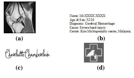
To begin with, a MRI knee image of dimension 512 x 512 has been taken as the cover data and the payloads multiple in nature consisting of the patient information, the doctor’s signature for copyright protection and hospital logo for tamper detection. Figure 4 depicts the cover data, doctor’s signature and the hospital logo.
(a) (b)
(c) (d)
Figure 4 : a. MR Knee Image (Cover Data) b. Sample EPR (Payload 1) – 1014 bits c. Doctor’s signature
(Payload 2)
The cover data are subjected to a 4 level decomposition using the Contourlet transform to generate 2k = 16 directional high frequency sub bands and its corresponding energy plot as shown in figure 5.
Figure 5. a 2k sub bands for a k level CT
Figure 5. b Energy plot of the sub bands
It is evident from the above plot that sub bands 4, 3 and 6 have high energies in the decreasing order and can be designated as the ideal embedding areas. The embedding is carried out using the principle of modification of coefficients of the cover image as per the update equation given by (2).The inverse transforms are computed to get back the embedded image as shown in figure 6.
Figure 6: a. Original Cover Image b. Embedded Image
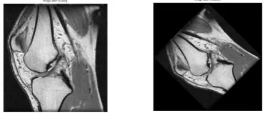
Figure 6 illustrates that there is no visual difference between the cover and the embedded image and a perfect correlation of 1is recorded. The embedded image is now subjected to a wide range of aggressive image processing operations simulating the real time attacks. The attacks that have been enforced upon the embedded image are filtering (low, high, median and wiener), noise exposure, scaling, rotation, blurring (horizontal) contrast enhancement. Figure 7 denotes the embedded image exposed to median filtering, wiener filtering, noise exposure and rotation attacks.
Figure 7: a. Embedded image scaled to 12% b. Embedded image rotated to 450
Figure 7 depicts the embedded image subjected to scaling of 12% and a 45 degree rotation attack. Scaling attack yielded a PSNR of 38.56dB while the rotation attack had a dip of PSNR down to 29dB with the correlation values of the payloads reaching as low as 0.4 indicating poor resilience towards RST attacks.
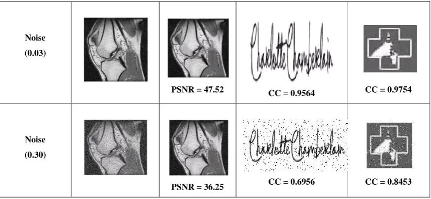
Table 1 : Response of embedded image and payloads towards Noise of varying intensities
Attack Embedded Image Extracted Cover
Image
Watermark1 Watermark2
Noise
(0.02)
PSNR = 49.52
Noise
(0.03)
PSNR = 47.52 CC = 0.9564 CC = 0.9754
Noise
(0.30)
PSNR = 36.25 CC = 0.6956 CC = 0.8453
Table 1 illustrates the recorded values in terms of PSNR and Correlation coefficient of the extracted cover image and the two payloads towards varying levels (variance) of noise intensities. It could be seen that at considerably peak noise levels of 0.03 a good value of PSNR of more than 45 dB is recorded with correlation values nearing 1. It undergoes a steep decrease for high variance levels of noise nearing 0.30. The embedded image is also subjected to filtering attacks like low pass (cut off frequency of 0.5), high pass, median (3 x 3 neighborhoods) and wiener filtering. The recorded values are tabulated in table 2.
Table 2: Response of Embedded Image towards filtering Attacks
Table 2 illustrates the superior nature of Contourlet transform over the other counterparts with respect to robustness towards filtering attacks. Each of the above recorded values have been tested with the same MR Knee image under three different techniques namely spatial domain using log average luminance [11], discrete cosine transform [4] and a 3 level discrete wavelet transform [5] with embedding of the patient record in high frequency sub band and the authentication payloads embedded into the mid frequency sub bands. Although, Contourlet transform
based watermarking have been done for natural images, EPR embedding into medical images with multiple payloads have not been reported so far up to our knowledge and this performance exhibited specifically towards robustness and fidelity indicate it be an ideal platform to be used for medical image data embedding applications. Future work could be focused on utilizing the Contourlet transform in a hybrid combination with certain translation invariant transforms like radon transform [12] to make the embedded image resilient to RST attacks to. This would however increase the complexity of the system. A future enhancement could be thought of by putting this idea into a real time implementation by arriving at a Contourlet filter bank structure and implementing in a FPGA.
IV.
REFERENCES
[1] Chu Shien Lu and Hong Yuan Mark Liao,
"Multipurpose Watermarking for Image
authentication and protection", IEEE Transactions on Image Processing, Vol. 10, No. 10, 2001.
[2] Rajendra Acharya et al, "Simultaneous storage of
patient information with medical images in the frequency domain", International Journal of Computer methods and programs in Biomedicine, Vol. 76, 2004.
[3] David Kahn, "The history of Steganography",
Proceedings of the First International Workshop on Information hiding, Lecture notes in computer science, Vol. 1174, 1996.
[4] Juan Hernandez, Martin Amado and Fernando
Perez Gonzalez, "DCT domain watermarking techniques for still images: detector performance Filtering
Attack
Spatial Domain (dB)
Frequency Domain DCT
(dB)
DWT (dB)
CT (dB) Low Pass 30.99 42.44 43.69 53.14
High Pass 33.69 41.22 44.91 49.88
Median 35.25 46.25 49.4 55.35
analysis and a new structure", IEEE transactions on Image Processing, Vol. 9, No. 1, 2000.
[5] G.S. El-Taweel et al, "Secure and Non-Blind
Watermarking Scheme for color Images based on DWT", International Journal on Graphics, Vision and Image Processing, Vol. 5, Issue 4, pp. 1-5, April 2005.
[6] Yuan Cheng Li, "An Image digital watermarking
method based on Ridgelet and KICA",
International Conference on Multimedia and Information Technology, 2008.
[7] Chin Chen Chang et al, "SVD based digital image
watermarking scheme", Pattern recognition
letters, Vol. 26, 2005.
[8] Kamstra et al, "Reversible data embedding into
images using wavelet techniques and sorting", IEEE transactions on Image processing, Vol. 14, 2005.
[9] Minh Do and Martin Vitterli, "The Contourlet
transform: An efficient directional multi
resolution image representation", IEEE
transactions on Image Processing, Vol. 14, No. 12, 2005.
[10] Sivakumar et al, "Image denoising using
Contourlet transform", International Conference on Computer and Electrical Engineering, 2009.
[11] Jamal Husein, "Spatial domain watermarking for
color images based on log average based luminance", International Journal of Computing, Vol. 2, Issue. 1, 2010.
[12] Dimitrios Simitotoulos et al, "Robust Image