ARTICLE
Rectal Bleeding in Infancy: Clinical, Allergological,
and Microbiological Examination
Taina Arvola, MD, PhDa,b, Tarja Ruuska, MD, PhDa,b, Jaakko Kera¨nen, MDc, Heikki Hyo¨ty, MD, PhDd,e, Seppo Salminen, MD, PhDf,
Erika Isolauri, MD, PhDg
aDepartment of Paediatrics,cCentre for Laboratory Medicine, Department of Pathology, andeCentre for Laboratory Medicine, Department of Clinical Microbiology, Tampere University Hospital, Tampere, Finland;bPaediatric Research Centre anddDepartment of Virology, University of Tampere, Tampere, Finland;fFunctional Foods Forum, University of Turku, Turku, Finland;gDepartment of Paediatrics, Turku University Central Hospital; Turku, Finland
The authors have indicated they have no financial relationships relevant to this article to disclose.
ABSTRACT
OBJECTIVE.Rectal bleeding is an alarming symptom and requires additional investi-gation. In infants it has been explained mainly by hypersensitivity. In addition to dietary antigens, intraluminal microbial agents challenge the immature gut mu-cosa. Although controlled in the mature gut, these antigens may induce inflam-mation in the developing gastrointestinal tract. The objectives of this study were to evaluate prospectively the clinical course of rectal bleeding and evaluate the impact of cow’s milk allergy and aberrant gut microbiota on the condition. Because withdrawal of cow’s milk antigens from the infants’ diet is used as a first treatment without evidence of its efficacy, we also aimed to asses the effect of a cow’s milk– elimination diet on the duration of rectal bleeding.
METHODS.The study involved 40 consecutive infants (mean age: 2.7 months) with visible rectal bleeding during a 2-year period at the Tampere University Hospital Department of Pediatrics. Most of the infants (68%) were fully breastfed. At enrollment the infants were randomly allocated to receive a cow’s milk– elimina-tion diet (n⫽19) or continue their previous diet (n⫽21) for 1 month. Findings of colonoscopy, fecal bacterial culture, fluorescence in situ hybridization of se-lected gut genera, specific detection of fecal enteroviruses, rotaviruses, and adeno-viruses, fecal electron microscopy for adeno-viruses, and mucosal electron microscopy for viruses were assessed. During each visit the severity of atopic eczema, if any, was assessed according to the SCORAD method. In evaluating the extent of sensitiza-tion, serum total immunoglobulin E (IgE) and specific IgE and skin-prick tests for cow’s milk, egg, and wheat were studied. Cow’s milk allergy was diagnosed by elimination and provocation testing. Five patients were hospitalized; all others were treated on an outpatient basis. The follow-up visits were scheduled 1 month later and at the age of 1 year. Sixty-four healthy reference infants were selected as controls according to the following criteria: age and timing of fecal sampling being identical to within 1 month.
www.pediatrics.org/cgi/doi/10.1542/ peds.2005-1069
doi:10.1542/peds.2005-1069
Key Words
rectal bleeding, cow’s milk allergy, elimination diet, microbes, colonoscopy
Abbreviations
FISH—fluorescence in situ hybridization IgE—immunoglobulin E
PCR—polymerase chain reaction Hb— hemoglobin
RESULTS.Altogether, 32 (80%) infants manifested bloody stools during follow-up (mean [range]: 2.1 [1–15] per day). The mean number of days with rectal bleeding on follow-up was 6. Typically, bloody stools occurred irreg-ularly, for which reason the mean time to the last oc-currence of rectal bleeding was 24 (range: 1– 85) days from admission. Atopic eczema at presentation or during follow-up was diagnosed in 38% of the infants. In-creased specific IgE concentrations or a positive skin-prick test were uncommon. The growth of the infants was normal on admission and during follow-up. Colonoscopy revealed typically focal mucosal erythema and aphthous ulcerations. The mucosa appeared normal in less than half of the patients. No anorectal fissures or colonic polyps were found. Light microscopy revealed that the overall architecture of the mucosa was well maintained. Acute inflammation or postinflammatory state and focal infiltration of eosinophils in the lamina propria were the most common abnormalities. A cow’s milk– elimination diet did not affect the duration of rec-tal bleeding. Cow’s milk allergy was diagnosed in 7 (18%) patients. Virus-particle aggregates were found in the microvillus layer of the colon epithelium in 8 cases. The surface epithelium of the virus-positive colon biopsy specimens regularly showed degenerative changes in the microvillus layer and epithelial cells. Electron micros-copy study of the colon biopsies disclosed virus particles (30 nm in diameter) on the surface of epithelial cells. Virus particles or RNA were present in feces in only a minority of the patients. All fecal cultures were negative forSalmonella,Shigella, andYersinia.Campylobacter jejuni was found in the feces of 1 patient, and fecal cultures were positive forClostridium difficilein 4 patients, Staph-ylococcus aureus in 8 patients, and yeast in 2 patients. Fluorescence in situ hybridization revealed that at the time of admission the total numbers of bacteria and the numbers of bifidobacteria and lactobacilli in feces were lower in the patients compared with controls. The fecal concentrations of microbes characterized in this study (Bacteroides, bifidobacteria, Clostridium, lactobacilli, and enterococci) did not differ significantly between the time of admission and the second visit in the patients or controls. At the age of 1 year, 7 patients still suffered from cow’s milk allergy, 5 of whom also suffered from multiple food allergies. Atopic eczema and histopatho-logically confirmed inflammation of the colonic mucosa at presentation were associated with persistence of cow’s milk allergy at the age of 1 year. No patients exhibited gastrointestinal complaints or visible blood in stools.
CONCLUSIONS.Rectal bleeding in infants is generally a be-nign and self-limiting disorder. Bloody stools occurred irregularly for only a few days during the following months. As in a previous report, most infants were ex-clusively breastfed. In the majority of the patients the cause of the condition remains unknown. An association
with viruses can be seen in some patients. The microbes that commonly lead to bloody diarrhea in older children and adults,Salmonella,Shigella, andYersinia, were absent in the present material. The low bifidobacterial numbers in fecal samples may indicate a significant aberrance that may provide a target for probiotic intervention to nor-malize gut microbiota. The gut microbiota overall seemed stable, because the numbers of major groups of microbiota tested did not change significantly between the time of admission and after 1 month. Cow’s milk allergy among these patients is more uncommon than previously believed. Cow’s milk challenge is thus essen-tial in infants who become symptom-free during a cow’s milk–free diet to reduce the number of false-positive cow’s milk–allergy diagnoses.
R
ECTAL BLEEDING INinfants is an alarming symptom and requires additional investigation. Among chil-dren of all ages, lower gastrointestinal bleeding has been the main symptom in 0.3% of patients admitted to an emergency department.1The condition comprisesheter-ogeneous manifestations such as allergic colitis, infective colitis, and the so-called ecchymotic colitis, characterized by ecchymotic hemorrhages at the mucosal surface of the colon.2,3Among older children rectal polyps are the
most common cause of rectal bleeding. The fact that in a significant proportion of cases investigations have not revealed an etiology for the bleeding3calls for new
an-gles on characterization of the disorder.
After birth the gut mucosa is challenged by a myriad of antigens, from viruses to commensal microbiota and dietary antigens. Although controlled in the mature gut, these antigens may induce inflammation in the devel-oping gastrointestinal tract. The purpose here was to evaluate prospectively the clinical course of rectal bleed-ing and evaluate the impact, if any, of cow’s milk allergy and aberrant gut microbiota on the condition. Because withdrawal of cow’s milk antigens from the infants’ diet is used as a first treatment4–6 without evidence of its
efficacy, we also assessed the effect of a cow’s milk– elimination diet on the duration of rectal bleeding.
METHODS
Patients
term. Five patients were hospitalized; all others were treated on an outpatient basis.
Controls
Sixty-four healthy reference infants were selected ac-cording to the following criteria: age and timing of fecal sampling being identical to within 1 month.
Ethics
The parents were informed verbally and in writing re-garding the nature and requirements of the study. Their written informed consent was obtained, and the study was approved by the Tampere University Hospital Ethics Committee.
Design
All patients received the same information, and the fol-low-up was conducted in a uniform manner. The infants were studied at enrollment at a mean age of 2.7 months (range: 3 weeks to 5.5 months); 27 (68%) were receiv-ing breast milk as the only source of milk (breastfed infants), 5 (12%) were receiving cow’s milk formula as the only source of milk (formula-fed infants), and 8 (20%) were receiving both breast milk and cow’s milk formula (mixed-fed infants). At enrollment, 2 breastfed and 2 formula-fed infants were fed also with solid foods. The follow-up visits were scheduled 1 month later (mean age: 3.7 months; range: 1.6 – 6.7 months) and at the age of 1 year (Fig 1). Two patients failed to attend the last examination, but on telephone inquiry their mothers reported them to be completely healthy.
Fecal samples from the controls were evaluated at a mean age of 3.6 months (range: 3 weeks to 9.2 months).
Clinical Evaluation
During each visit the patients were clinically examined by the same pediatricians (T.A. and T.R.), and blood (for analysis of blood and differential white blood cell count, sedimentation rate, and concentrations of C-reactive protein, serum albumin, sodium, and potassium) and fecal (for viral, fluorescence in situ hybridization [FISH], and␣-1 antitrypsin analysis) samples were obtained.
Evaluation of Endoscopy
Colonoscopy (with the small, flexible Olympus [Melville, NY] GIF-N30 endoscope) with multiple biop-sies was performed (by T.R. and T.A.) on 39 patients at enrollment without need of prior bowel preparation or anesthesia. In 1 case, the procedure was not undertaken after maternal refusal. All biopsies were taken under direct vision. Biopsy specimens for morphology were fixed in phosphate-buffered formalin and embedded in paraffin blocks by using standard methods. Paraffin sec-tions were stained routinely with hematoxylin and eosin and reviewed by a pathologist. Colonoscopy was sched-uled to be repeated after 1 month in patients with his-topathologically confirmed inflammation.
Evaluation of Hypersensitivity
During each visit the severity of atopic eczema, if any, was assessed according to the SCORAD method.7 In
evaluating the extent of sensitization, serum total im-munoglobulin E (IgE) (Phadebas IgE Prist; Pharmacia, Uppsala, Sweden) and specific IgE for milk, egg, and wheat (radioallergosorbent assay; Pharmacia) were studied at enrollment and at the age of 1 year. Skin-prick tests for cow’s milk, egg, and wheat allergies were stud-ied during the second visit 1 month after enrollment.
Cow’s milk allergy was diagnosed by elimination and provocation testing in 2 ways: (1) if rectal bleeding or atopic eczema disappeared during an elimination diet and reappeared at the second visit when cow’s milk was reintroduced to the diet of the infant or the lactating mother or (2) if an unambiguous adverse reaction to cow’s milk occurred between the second and the last visit in open cow’s milk challenge starting and controlled in the hospital as described previously.8Open cow’s milk
challenge was applied in 7 cases after cessation of breast-feeding because of symptoms suggestive of cow’s milk allergy. Placebo was not used in the challenge because placebo responses are rare in this age group.8,9
Evaluation of Dietary Intervention
To study the effect of dietary manipulation, the infants were randomly assigned at enrollment to start a cow’s milk– elimination diet or continue their previous diet. TABLE 1 Clinical Features of Infants With Rectal Bleeding Who Did Not Participate in the Study (Nⴝ4)
Gender Reason for Nonparticipation Outcome
Refused
4 d old F Exhaustion of the mother No bloody stools
Excluded
1.5 mo old M Suspicion of intussusception Salmonella typhimuriumin stools; no bloody stools after cefotaxime treatment
1 mo old M Suspicion of Hirschsprung disease No bloody stools; constant constipation; diagnosis of ultra–short-segment Hirschsprung disease 1.5 mo old F Risk of bone fracture during colonoscopy resulting
from osteogenesis imperfecta
Altogether, 19 infants went on an elimination diet and 21 continued their previous diet. The elimination diet comprised an amino acid– derived formula (Neocate; SHS Int, Liverpool, United Kingdom) in case supplemen-tary feedings were required (2 were formula-fed and 3 were mixed-fed). In breastfed infants (14 were exclu-sively breastfed and 3 were mixed-fed), instruction for the cow’s milk– elimination diet was given to the lactat-ing mother with the addition of 1000 mg of calcium gluconate daily. The elimination diet was recommended verbally and in writing in all cases. Thirteen breastfed, 3 formula-fed, and 5 mixed-fed infants continued their previous diet. The cow’s milk– elimination period was 1
month, after which time all infants and mothers re-turned to their previous diet.
The parents filled a daily symptom diary (bloody stools, fever, vomiting, abdominal pain, eczema, itching) and recorded stool frequency and consistency (solid, loose, watery) at home for 1 month between the first and second visits.
Fecal Bacterial and Viral Analysis
Fecal Bacterial Analysis
Fecal specimens were cooled immediately at 6 to 8°C after collection from diapers and within 24 hours frozen
FIGURE 1
at ⫺70°C until analysis. Fresh fecal samples were cul-tured for Salmonella, Shigella, Yersinia, Campylobacter, Clostridium difficile, Staphylococcus aureus, and yeast. Cy-closerine-cefoxitin fructose agar (CCFA; Oxoid, United Kingdom) was cultured forC difficileand Dixon agar for yeast. These cultures were also studied in fecal samples of the patients’ mothers.
Fecal Virus Analyses
Adenovirus and rotavirus antigens were assessed from fecal samples by using an enzyme-linked immunoassay (IDEIA; Dako Cytomation, Glostrup, Denmark) accord-ing to manufacturer instructions. Enterovirus and rhi-novirus RNA were detected by using a reverse-transcrip-tion polymerase chain reacreverse-transcrip-tion (PCR) method as described previously.10
Electron Microscopy of Fecal Specimens
Fecal samples from 34 patients were analyzed by trans-mission electron microscopy (model JEM 1200 EX; Jeol, College Station, TX). The samples were diluted with phosphate-buffered saline (pH 7.4) and placed on Form-var-coated grids, which then were negatively stained with 2% aqueous ammonium molybdate.
Electron Microscopy of Colon Biopsy Specimens
Small pieces from the large intestinal mucosa of 20 pa-tients were excised for a transmission electron micros-copy study. Details of the specimen-preparation proce-dure are described elsewhere.11
Fecal FISH
FISH of the fecal samples was performed as described previously.12In short, samples were suspended in
phos-phate-buffered saline and homogenized. Bacteria were fixed with paraformaldehyde and hybridized with Cy3
(a fluorophore) indocarbocyanine-labeled oligonucleo-tide probe. Probes included Bac303 for Bacteroides, Bif164 for bifidobacteria, His150 for clostridia of the Clostridium hystolyticumgroup, and Lab158 for lactobacilli and enterococci. Total bacterial counts were determined by staining with 4⬘,6-diamino-2-phenylindole (DAPI). The bacteria were washed and filtered on a 0.2-m polycarbonate filter. The filters were mounted on a slide and counted visually under an epifluorescence micro-scope using Cy3- and DAPI-specific filters.
Analysis of Fecal␣-1 Antitrypsin
Analysis of fecal ␣-1 antitrypsin was performed as de-scribed previously.13The results are given as milligrams
per gram dry weight of lyophilized feces.
Statistics
Data are given as means with range or medians with interquartile range. Analysis of variance was used in statistical comparisons.
RESULTS
Clinical Characteristics of Infants With Rectal Bleeding Twenty two (55%) of the patients were male. The mean (range) duration of bloody stools before admission was 10 (1–50) days, and mean (range) frequency was 2.7 (1–10) per day. In addition to bloody stools, watery or mucous stools, abdominal pain, and vomiting were com-mon (Table 2). The mean (range) duration of exclusive breastfeeding (not even 1 minute of exposure to solid foods or milk other than breast milk) was 2 (0 – 6) months, and that of total breastfeeding (last breastfeed-ing) was 8 (1–12) months.
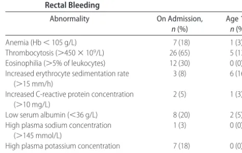
The laboratory abnormalities are presented in Table 3. Only 1 patient exhibited extensive blood loss and devel-oped anemia (hemoglobin [Hb]: 87 g/L) that required iron supplementation on admission. Blood transfusion with succeeding iron supplementation was given at the TABLE 2 Clinical Characteristics of Infants (Nⴝ40) With Rectal
Bleeding
Characteristic n(%)
Disease history from birth
Transient viral infection 14 (35)
Transient bacterial infection 5 (13)
Course of antimicrobial medication 6 (15)
On admission
Bloody stools 40 (100)
Mucous stools 29 (73)
Watery stools 15 (38)
Abdominal pain 23 (58)
Vomiting 7 (18)
Respiratory infection 9 (22)
Fever 3 (8)
During follow-up
Bloody stools 32 (80)
Watery stools 5 (13)
Abdominal pain 9 (23)
Vomiting 14 (35)
TABLE 3 The Frequency of Laboratory Abnormalities on Admission (Nⴝ40) and at 1 Year of Age (Nⴝ38) in Infants With Rectal Bleeding
Abnormality On Admission,
n(%)
Age 1 y, n(%)
Anemia (Hb⬍105 g/L) 7 (18) 1 (3)
Thrombocytosis (⬎450⫻109/L) 26 (65) 5 (13) Eosinophilia (⬎5% of leukocytes) 12 (30) 0 (0) Increased erythrocyte sedimentation rate
(⬎15 mm/h)
3 (8) 6 (16)
Increased C-reactive protein concentration (⬎10 mg/L)
2 (5) 1 (3)
Low serum albumin (⬍36 g/L) 8 (20) 2 (5)
High plasma sodium concentration (⬎145 mmol/L)
1 (3) 0 (0)
High plasma potassium concentration (⬎4.8 mmol/L)
age of 8 months (6 months after admission) to 1 infant who developed iron-deficiency anemia (Hb: 82 g/L; mean corpuscular volume: 59 fL; mean corpuscular Hb: 19 pg), the cause of which remained unexplained de-spite extensive examinations including gastroscopy and colonoscopy.
Altogether, 32 (80%) infants manifested bloody stools during follow-up (mean: 2.1 per day [range: 1–15 per day]). In 23 cases bloody stools continued longer than 2 weeks. The mean number of days with rectal bleeding on follow-up was 6. Typically, bloody stools occurred irregularly, for which reason the mean (range) time to the last occurrence of rectal bleeding was 24 (1– 85) days from admission. In addition, watery stools, abdominal pain, and vomiting were recorded during follow-up (Table 2).
A positive family history of atopy among first-degree relatives was found in most cases (Table 4). Atopic ec-zema at presentation or during follow-up was diagnosed in 38% of the infants. In these patients, the mean (range) SCORAD score was 7.4 (0.002–30.100) at en-rollment, 8.8 (0.002–27.800) after 1 month, and 5.5 (3.700 –14.700) at the age of 1 year. Total IgE was de-tectable (⬎5 kU/L) in 6 patients (range: 6 –19 kU/L) at presentation and 11 patients (range: 6 – 81 kU/L) at the age of 1 year. Increased specific IgE concentrations or a positive skin-prick test were uncommon (Table 4).
The growth of the infants was normal on admission and during follow-up when compared with Finnish growth charts for infants.
Concentration of Fecal␣-1 Antitrypsin
The median (interquartile range) concentration of fecal
␣-1 antitrypsin was 1.6 (1.1–2.6) mg/g on admission. After 1 month the concentration was 3.5 (2.7–5.0) mg/g. The difference is not statistically significant.
Characterization of Gut Mucosa
During colonoscopy, the cecum was reached in 8 (21%) patients, and the examination covered the mucosa from rectum to transverse colon for the rest of the patients. No anorectal fissures or colonic polyps were found. Macro-scopically, focal mucosal erythema and aphthous ulcer-ations were common (Table 5). The mucosa appeared normal in less than half of the patients.
Light microscopy revealed that the overall architec-ture of the mucosa was well maintained. Acute inflam-mation or postinflammatory state and focal infiltration of eosinophils in the lamina propria were the most com-mon abnormalities (Table 5). Only 2 patients manifested visible hemorrhage on the mucosa. In most cases, biopsy specimens evinced no diagnostic abnormalities.
In subsequent colonoscopic examination of 7 patients with histopathologically confirmed inflammation, le-sions were found to be healed in 4. Eosinophilia was still observed in 1 patient and inflammatory changes in 2 patients. In 6 patients with histopathologically con-firmed inflammation a pathologic review was not avail-able at the second visit, therefore bypassing the second colonoscopy.
Association of Cow’s Milk Antigens With Rectal Bleeding
Effect of Cow’s Milk Elimination
When evaluated in whole groups, a cow’s milk– elimi-nation diet did not affect the duration or severity of rectal bleeding during follow-up. The mean (range) number of days with rectal bleeding during follow-up was 5.6 (0 –22) in infants who were randomly assigned to a cow’s milk– elimination diet and 5.5 (0 –20) in those randomly assigned to continue their normal diet (P ⫽ .94). Also, the mean number of bloody stools per day during follow-up and the time to the last occurrence of rectal bleeding were comparable in both groups. How-ever, in patients who were later diagnosed to have cow’s milk allergy, random assignment to a cow’s milk– elimi-nation diet tended to shorten the duration of rectal bleeding as compared with those who were randomly assigned to continue their normal diet.
TABLE 4 Allergological Data on Infants (Nⴝ40) With Rectal Bleeding
n(%)
Family history of atopy/asthma 23 (58)
Atopic eczema 15 (38)
On admission 8 (20)
During follow-up 7 (18)
Positive RAST
On admission 0 (0)
At the age of 1 y
Cow’s milk 4 (10)
Egg 3 (8)
Wheat 1 (3)
Positive skin-prick test
Cow’s milk 2 (5)
Egg 1 (3)
Wheat 0 (0)
Diagnosis of cow’s milk allergy 7 (18)
On dietary intervention 2 (5)
On cow’s milk challenge 5 (13)
RAST indicates radioallergosorbent assay (positive indicatesⱖ0.4 kU/L).
TABLE 5 Colonoscopic Macroscopic and Microscopic Findings on Admission in Infants (Nⴝ39) With Rectal Bleeding
n(%)
Macroscopic
Normal mucosa 16 (41)
Focal erythema 20 (51)
Aphtous ulcerations 13 (33)
Microscopic
Normal 23 (59)
Inflammation 13 (33)
Eosinophilic infiltration 9 (23)
Outcome of Clinical Challenge
Cow’s milk allergy was confirmed by the reappearance of rectal bleeding and atopic eczema after reintroduction of cow’s milk to the diet of the infant in 1 case and to the lactating mother in another after a 1-month elimination period (Table 4). Between the second and third visits, open cow’s milk challenge elicited a positive reaction in 5 of 7 patients. In these 5 patients, the response to challenge was acute-onset urticaria after ingestion of 1 to 30 mL of cow’s milk. Altogether, the prevalence of cow’s milk allergy among the patients with rectal bleed-ing was 18%.
Association of Intraluminal Microbes With Rectal Bleeding All fecal cultures were negative for Salmonella,Shigella, andYersinia.Campylobacter jejuniwas found in the feces of 1 patient. Because of prolonged symptoms, the patient was treated with azithromycin for 3 days. Fecal cultures were positive for C difficile in 4 patients, S aureus in 8 patients, and yeast in 2 patients.
Also, 1 mother hadCampylobacterin the feces. How-ever, her infant had a negative fecal culture. Cultures were negative for Salmonella, Shigella, Yersinia, and C difficilein all mothers.S aureuswas found in the feces of 1 mother and yeast in 6 mothers.
FISH
At the time of admission the numbers of bifidobacteria and lactobacilli and total microbial counts in feces were lower in the patients with rectal bleeding compared with controls (Table 6). The fecal concentrations of microbes characterized in this study (Bacteroides, bifidobacteria, Clostridium, lactobacilli, and enterococci) did not differ significantly between the time of admission and the second visit in patients or controls (data not shown).
Detection of Viruses in Fecal Samples
Enterovirus RNA was present in the feces of 6 (15%) of 39 patients and 3 (6%) of 52 controls; the difference was not statistically significant (P⫽.13). Also, there was no difference in the presence of rhinovirus RNA in the feces of patients (9 of 39 [23%]) and controls (8 of 52 [15%]).
Neither rotavirus nor adenovirus antigens were not found in any of the patients.
Virus particles were detected by electron microscopy in 4 (12%) of 34 patients but in no controls. These patients were also PCR-positive for either enterovirus (1 of 4) or rhinovirus (3 of 4).
Electron Microscopy of Colon Biopsy Specimens
Virus-particle aggregates were found in the microvillus layer of the epithelium in 8 cases. In 7 of these, virus RNA was also detected in the PCR study. However, only 3 of them had virus particles detected with electron microscopy of fecal samples. The surface epithelium of the virus-positive colon biopsy specimens regularly showed degenerative changes in the microvillus layer and epithelial cells. Electron microscopy study of the colon biopsies disclosed virus particles (30 nm in diam-eter) on the surface of epithelial cells. Typically, numer-ous intercalated virus particles were located between microvilli (Fig 2). No virus particles were found in the specimens from the PCR-negative patients.
Clinical Outcome at the Age of 12 Months
At the age of 1 year, 7 patients still suffered from cow’s milk allergy, 5 of whom also suffered from multiple food allergies (including allergy to cereals). Atopic eczema and histopathologically confirmed inflammation of the colonic mucosa at presentation were associated with persistence of cow’s milk allergy at the age of 1 year. No patients exhibited gastrointestinal complaints or visible blood in stools. Laboratory results were generally normal (Table 3).
DISCUSSION
Our results demonstrate that rectal bleeding is a benign and self-limiting disorder in infants. Bloody stools
oc-FIGURE 2
Transmission electron micrograph of a colon biopsy specimen. Numerous virus particles located between microvilli are seen at the microvillus layer of the epithelium.
TABLE 6 Bacterial Counts as Determined by FISH in Fecal Samples From Patients With Rectal Bleeding (Nⴝ26) and Controls Patients (Nⴝ17)
Bacterial Genus Patients, Counts⫻109
Controls, Counts⫻109
P
Bifidobacterium 1.35 (0.68–2.10) 6.77 (3.20–10.33) .0004 Lactobacillus/Enterococcus 1.64 (0.63–2.65) 5.92 (2.60–9.24) .0039 Bacteroides 1.83 (0.45–3.22) 3.86 (0.59–7.13) .18 Clostridium 1.27 (0.58–1.96) 1.72 (0.69–2.76) .43 Total microbial counts 4.00 (2.57–5.42) 11.11 (6.49–15.74) .0007
curred irregularly for only a few days during the follow-ing months. As in a previous report,14most of the infants
were exclusively breastfed.
According to the literature, diarrhea, vomiting, ab-dominal pain, anorexia, and failure to thrive are the most common symptoms in addition to bloody stools.15
In the present study, loose and mucous stools, abdomi-nal pain, and vomiting were seen frequently. Diarrhea may have also led to dilution of intestinal contents and washing out of microbiota, as reflected by lower total microbial counts in the subjects with rectal bleeding. However, in our patients, growth was normal, perhaps because of the mild and transient nature of the disorder. Cow’s milk allergy has been considered to be the most common cause of bloody stools in infants.16 This has
been demonstrated in 33 newborn infants whose symp-toms disappeared after elimination of cow’s milk anti-gens from the infant’s or the lactating mother’s diet and recurred after an open milk challenge.17 However, the
diagnosis of cow’s milk allergy in some other reports on infantile rectal bleeding5,6was not based on elimination
and challenge testing, which is the only reliable means of diagnosing food allergy. In a study of 9 exclusively breastfed infants, rectal bleeding was suspected to be a result of cow’s milk allergy, because the symptoms dis-appeared during a maternal cow’s milk– elimination di-et.14 However, no challenge tests were performed to
confirm the suspicion. Indeed, histologic findings of in-creased numbers of eosinophils in the lamina propria together with negative stool bacterial culture have been considered to make food challenges unnecessary in the diagnosis of allergic proctocolitis.6,18In the present study,
cow’s milk allergy was diagnosed in only 18% of the patients. On the basis of our results, we strongly recom-mend a cow’s milk challenge in infants who become symptom-free during an elimination diet to reduce the number of false-positive cow’s milk–allergy diagnoses.
The elimination of cow’s milk antigens from the diet of infants with rectal bleeding or from the diet of the lactating mother is a commonly used and recommended practice,4,14,17although it seems effective for only some
patients.4 To our knowledge, our study is the first to
evaluate systematically the effect of a cow’s milk– elimi-nation diet on the duration and severity of rectal bleed-ing in infants. We showed that the number of days with rectal bleeding during follow-up was identical in infants who were randomly assigned to an elimination diet or to continue their previous diet. However, the elimination diet seemed to shorten the duration of rectal bleeding in infants who were diagnosed later as having cow’s milk allergy. We therefore suggest initiating a cow’s milk– elimination diet for a limited period of time in infants with rectal bleeding but stress the importance of a cow’s milk challenge because of spontaneously favorable out-come of the condition. However, it should be noticed that our study population may not represent a
homog-enous group. Exclusively breastfed, exclusively formula-fed, and mixed-fed infants with rectal bleeding may have various responses to an elimination diet and differ-ent outcome of the condition.
The intestinal microbiota have an important role in the etiology of many diseases. Evidence for microbial involvement has been described in case reports on young immunocompetent infants presenting with rectal bleeding and diarrhea and diagnosed as suffering from gastrointestinal infection caused by cytomegalovirus.19,20
Because cytomegalovirus infection is believed to be rare in healthy individuals, investigations for this microbe are not usually undertaken; it therefore remains unclear whether cytomegalovirus enterocolitis is truly rare or whether milder forms of the infection are not being recognized. The association of other viruses with rectal bleeding in infants has not been systematically studied. In the present study, electron microscopy examination of fecal and colon biopsy specimens revealed virus par-ticles in a few patients. Electron microscopy study may be an insensitive method to recognize virus infection, because it demands an abundance of virus particles in the samples. In the case of a sample with low virus concentration, virus-particle– enrichment procedures may increase the sensitivity of electron microscopy study, but these procedures were not used in our inves-tigation. In addition to electron microscopy, we used more sensitive virus-specific tests for the detection of enterovirus, rotavirus, and adenovirus, which represent the most common enteral viruses in this age group. Rhinovirus was analyzed as a control, because its pres-ence in feces is likely to represent swallowed respiratory viruses rather than a true intestinal pathogen (rhinovi-ruses do not replicate in the intestine). Rotavirus and adenovirus were not detected in any of the patients, and the highly sensitive enterovirus reverse-transcription PCR method demonstrated enterovirus RNA in only a minority of patients, with no difference compared with control subjects, suggesting that enterovirus could not be an important causative agent in infantile rectal bleeding. We had only 1 patient withC jejuni, and there was no evidence of mother-to-child transmission. The microbes that commonly lead to bloody diarrhea in older children and adults,Salmonella,Shigella, andYersinia, were absent from our patients. Altogether, 14 infants had fecal cul-tures positive for C difficile, S aureus, or yeast, the mi-crobes of which are regarded as part of the normal intestinal microbiota in infants.
The FISH technique used for enumeration of bacterial concentrations demonstrated that significantly lower numbers of bifidobacteria and lactobacilli were present in patients when compared with control subjects. In view of the well-characterized dominance of bifidobac-teria in breastfed infants,21representing the majority of
viable counts in feces of patients with rectal bleeding were lower compared with healthy controls. This sug-gests that there may have been a diluting effect by diarrhea on fecal microbiota and that the luminal micro-biota may also be affected by the condition. Bifidobac-teria and lactobacilli may prove to be biomarkers of such disturbances, providing a target for intervention by pro-biotic lactic acid bacteria or bifidobacteria. These micro-bial groups, especially bifidobacteria, are considered im-portant indicators of balanced succession of microbiota in breastfed infants.21 The health promoting effect of
breast milk has been linked to bifidogenic oligosaccha-rides, which promote a significant bifidobacteria micro-biota and provide the infant with improved colonization resistance against pathogens. Thus, the low bifidobacte-rial numbers may indicate a significant aberrance that may provide a target for probiotic intervention to nor-malize gut microbiota. The gut microbiota overall seemed stable, because the numbers of major groups of microbiota tested did not change significantly between the time of admission and after 1 month.
CONCLUSIONS
Rectal bleeding in infants is a benign and self-limiting disorder in which total numbers of bacteria in intestinal contents are decreased. Cow’s milk allergy among these patients is less common than previously believed, and a cow’s milk challenge is therefore essential in infants who become symptom-free during a cow’s milk– elimination diet. An association with microbes can be seen in some patients, but in the majority of cases, the cause of the condition remains unknown.
ACKNOWLEDGMENTS
The study was supported financially by the Medical Re-search Fund of Tampere University Hospital and the Academy of Finland.
We thank Immo Rantala, PhD, for collaboration and Mrs Leena Ripsaluoma, RN, Mrs Sirkka-Liisa Matikka, RN, Mrs Annette Tahvanainen, RN, and Mrs Raija Huk-kila for excellent assistance.
REFERENCES
1. Lawrence WW, Wright JL. Causes of rectal bleeding in chil-dren.Pediatr Rev.2001;22:394 –395
2. Balkan E, Kiris¸tiog˘lu I˙, Gu¨rpinar A, O¨ zel I˙, Sinmaz K, Dog˘ruyol H. Sigmoidoscopy in minor lower gastrointestinal bleeding. Arch Dis Child.1998;78:267–268
3. Dupont C, Badoual J, Le Luyer B, Le Bourgeois C, Barbet JP, Voyer M. Rectosigmoidoscopic findings during isolated rectal bleeding in the neonate. J Pediatr Gastroenterol Nutr.1987;6: 257–264
4. Machida HM, Catto Smith AG, Gall DG, Trevenen C, Scott RB. Allergic colitis in infancy: clinical and pathologic aspects.J Pe-diatr Gastroenterol Nutr.1994;19:22–26
5. Willetts IE, Dalzell M, Puntis JWL, Stringer MD. Cow’s milk enteropathy: surgical pitfalls. J Pediatr Surg. 1999;34:1486 – 1488
6. Kumar D, Repucci A, Wyatt-Ashmead J, Chelimsky G. Allergic colitis presenting in the first day of life: report of three cases. J Pediatr Gastroenterol Nutr.2000;31:195–197
7. European Task Force on Atopic Dermatitis. Severity scoring of atopic dermatitis: the SCORAD index.Dermatology.1993;186: 23–31
8. Isolauri E, Turjanmaa K. Combined skin prick and patch testing enhances identification of food allergy in infants with atopic dermatitis.J Allergy Clin Immunol.1996;97:9 –15
9. Bock SA, Atkins FM. Patterns of food hypersensitivity during sixteen years of double-blind, placebo-controlled food chal-lenges.J Pediatr.1990;117:561–567
10. Lo¨nnrot M, Sjo¨roos M, Salminen K, Maaronen M, Hyypia¨ T, Hyo¨ty H. Diagnosis of entero- and rhinovirus infections by RT-PCR and time-resolved fluorometry with lanthanide che-late labeled probes.J Med Virol.1999;59:378 –384
11. Isolauri E, Kaila M, Arvola T, et al. Diet during rotavirus enteritis affects jejunal permeability to macromolecules in suckling rats.Pediatr Res.1993;33:548 –553
12. Kallioma¨ki M, Kirjavainen P, Eerola E, Kero P, Salminen S, Isolauri E. Distinct patterns of neonatal gut microflora in in-fants developing or not developing atopy.J Allergy Clin Immu-nol.2001;107:129 –134
13. Arvola T, Moilanen E, Vuento R, Isolauri E. Weaning to hy-poallergenic formula improves gut barrier function in breast-fed infants with atopic eczema. J Pediatr Gastroenterol Nutr. 2004;38:92–96
14. Anveden-Hertzberg L, Finkel Y, Sanstedt B, Karpe B. Procto-colitis in exclusively breast-fed infants.Eur J Pediatr.1996;155: 464 – 467
15. Ojuawo A, St Louis D, Lindley KJ, Milla PJ. Non-infective colitis in infancy: evidence in favour of minor immunodefi-ciency in its pathogenesis.Arch Dis Child.1997;76:345–348 16. Vanderhoof JA, Murray ND, Kaufman SS, et al. Intolerance to
protein hydrolysate infant formulas: an underrecognized cause of gastrointestinal symptoms in infants. J Pediatr.1997;131: 741–744
17. Chouraqui JP, Barbier M, Joannard A, Rambaud P, Bost M. Cow’s milk protein-induced colitis of early infancy.Acta Endo-scopica.1994;24:461– 468
18. Kumagai H, Masuda T, Maisawa S, Chida S. Apoptotic epithe-lial cells in biopsy specimens from infants with streaked rectal bleeding.J Pediatr Gastroenterol Nutr.2001;32:428 – 433 19. Jonkhoff-Slok TW, Veenhoven RH, Graeff-Meeder ER, Bu¨ller
HA. An immunocompetent infant with cow’s milk allergy and cytomegalovirus colitis.Eur J Pediatr.1997;156:528 –529 20. Rongkavilit C, Bedard MP, Ang JY, Asmar BI, Tolia V. Severe
cytomegalovirus enterocolitis in an immunocompetent infant. Pediatr Infect Dis J.2004;23:579 –581
DOI: 10.1542/peds.2005-1069
2006;117;e760
Pediatrics
Erika Isolauri
Taina Arvola, Tarja Ruuska, Jaakko Keränen, Heikki Hyöty, Seppo Salminen and
Examination
Rectal Bleeding in Infancy: Clinical, Allergological, and Microbiological
Services
Updated Information &
http://pediatrics.aappublications.org/content/117/4/e760
including high resolution figures, can be found at:
References
http://pediatrics.aappublications.org/content/117/4/e760#BIBL
This article cites 21 articles, 3 of which you can access for free at:
Subspecialty Collections
http://www.aappublications.org/cgi/collection/gastroenterology_sub
Gastroenterology
sub
http://www.aappublications.org/cgi/collection/fetus:newborn_infant_
Fetus/Newborn Infant
following collection(s):
This article, along with others on similar topics, appears in the
Permissions & Licensing
http://www.aappublications.org/site/misc/Permissions.xhtml
in its entirety can be found online at:
Information about reproducing this article in parts (figures, tables) or
Reprints
http://www.aappublications.org/site/misc/reprints.xhtml
DOI: 10.1542/peds.2005-1069
2006;117;e760
Pediatrics
Erika Isolauri
Taina Arvola, Tarja Ruuska, Jaakko Keränen, Heikki Hyöty, Seppo Salminen and
Examination
Rectal Bleeding in Infancy: Clinical, Allergological, and Microbiological
http://pediatrics.aappublications.org/content/117/4/e760
located on the World Wide Web at:
The online version of this article, along with updated information and services, is
by the American Academy of Pediatrics. All rights reserved. Print ISSN: 1073-0397.