INTRODUCTION
There is a growing evidence that women with thrombo-philia are at increased risk, not only for pregnancy-related venous thromboembolism (VTE), but also for other vascular complications in pregnancy, such as fetal loss, pre-eclampsia (PE) and intrauterine growth restriction (IUGR) [1].
According to the definition, pregnancy loss (PL) is the spontaneous abortion of an embryo or fetus before the
20th week of pregnancy or when the fetus weighs <500 g or
measures <25 cm [2].
The F5 gene mutation 1691G>A (rs6025) results in an altered variant of factor V, namely Factor V Leiden, which cannot be easily cleaved by activated protein C (aPC). The 1691G>A mutation increases the risk of venous thrombosis (VT) up to 50-100 fold in adult homozygous [3]. In European populations, the frequency of F5 1691A allele is higher in Bosnians (6.0%) than in the Czechs (2.6%), Ukrainians (1.5%), and Croats (1.4%) [4]. Numerous studies indicated that the
F5 1691G>A is responsible for 3-42% of PL [5].
The F2 20210G>A (rs1799963) mutation, results in increased prothrombin levels and is considered as another risk factor for VT [6]. The heterozygotes have 2-5-fold increased risk of thrombosis which may also increase the risk for PL. The
Prevalence of
F5
1691G>A
,
F2
20210G>A,
and
MTHFR
677C>T polymorphisms in Bosnian women with
pregnancy loss
Emir Mahmutbegović1, Damir Marjanović2,3, Edin Medjedović4, Nevena Mahmutbegović5, Serkan Dogan2,
Amina Valjevac6, Ewa Czerska7, Anna Pawińska-Matecka7, Agnieszka Madlani8, Grażyna Adler8*
1Institution of Health Protection of Women and Motherhood Canton Sarajevo, Sarajevo, Bosnia and Herzegovina, 2International Burch
University, Sarajevo, Bosnia and Herzegovina, 3Institute for Anthropological Research, Zagreb, Croatia, 4Gynecology Clinic, Clinical Center
of University of Sarajevo, Sarajevo, Bosnia and Herzegovina, 5Neurology Clinic, Clinical Center of University of Sarajevo, Sarajevo, Bosnia
and Herzegovina, 6Laboratory for Molecular Medicine, Center for Genetics, Medical Faculty, University of Sarajevo, Sarajevo, Bosnia and
Herzegovina, 7Central Laboratory, Regional Hospital, Szczecin, Poland, 8Department of Gerontobiology, Pomeranian Medical University,
Szczecin, Poland
ABSTRACT
The relationship between genetic risk factors of thrombophilia and pregnancy loss (PL) is being discussed. The focus has been on F5 1691G>A,
F2 20210G>A, and MTHFR 677C>T polymorphisms that may predispose women to microthrombosis during the stages of embryo implan-tation and placenimplan-tation. Although, the frequencies of these polymorphisms were reported in different populations, such studies have not yet been performed in Bosnian population. In this study, we determined the prevalence of F5 G>A (rs6025), F2 G>A (rs1799963) and MTHFR
C>T (rs1801133) polymorphisms in Bosnian women. A total of 154 women with PL, mean age 33 (±5.4) years, were enrolled in the study. As a control group, 154 mothers [mean age 31.4 (±6.7) years] with at least one live-born child were included. We used real-time polymerase chain reaction (PCR) to determine the frequencies of F5 G>Aand F2 G>A genotypes, and PCR-restriction fragment length polymorphism (RFLP) for analyzing MTHFR C>T genotypes. The frequency of heterozygotes for F5 and F2 was significantly higher in women with venous thrombosis (VT) compared to women without VT (p = 0.047 and p = 0.001, respectively). There was no significant difference in the distribution of MTHFR
genotypes and alleles between these two groups. In addition, we observed no significant differences in the genotype and allele frequencies between the group with PL and control group, for all investigated polymorphisms. The allele frequencies for 1691A (F5), 20210A (F2), and 677T (MTHFR) reported in this study are consistent with the data obtained for other European countries, however, we were not able to confirm the association between the three polymorphisms and PL in Bosnian women.
KEY WORDS: Pregnancy loss; risk factors; polymorphisms; thrombophilia; F5; F2; MTHFR
DOI: http://dx.doi.org/10.17305/bjbms.2017.1954 Bosn J Basic Med Sci. xxxx;xx(x):1-6. © 2017 ABMSFBIH
*Corresponding author: Grażyna Adler, Department of Gerontobiology, Pomeranian Medical University, Żołnierska 48, 71-210 Szczecin, Poland. Phone: +48 91 48 00 958. E-mail: [email protected]
prevalence of F2 20210G>A in European Caucasians is from 1% to 8%, and from 1% to 12% in Mediterranean populations. Furthermore, in European Caucasians with VTE, the preva-lence of F2 20210G>A is from 3% to 17%, and in Mediterranean populations with VTE this figure is from 3% to 24% [7-9]. The
F2 20210A allele frequency in Bosnians is 6.0%, and is higher than in Poles and Ukrainians, 2.5% and 1.7%, respectively [4].
The 677C>T mutation (rs1801133) of the MTHFR gene leads to a defective methylene tetrahydrofolate reductase (MTHFR) enzyme that has only 50% of the normal activity and is a common cause of elevated levels of homocysteine, previously identified as a risk factor for VTE. The frequency of MTHFR 677T allele in Bosnians is 37.5% and is higher than those reported for the Czechs, Ukrainians, Norwegians, and Swedes (25.0%, 29.1%, 29.0%, and 25.0%, respectively), as well as for Austrians and the Dutch (each 29.3%) [4]. The American College of Obstetricians and Gynecologists recommends against screening for MTHFR variants to determine the pres-ence of thrombophilia in pregnant women [10]. On the other hand, the co-inheritance of the alleles linked to thrombophilia, such as the F5 1691A, F2 20210A, and MTHFR 677T, may increase the risk for PL.
The prevalence of the genetic risk factors for thrombo-philia is higher in Bosnian population compared to several European populations [4]. Therefore, the aim of our study was to investigate the prevalence of F5 G>A(rs6025), F2 G>A (rs1799963) and MTHFR C>T (rs1801133) polymorphisms in Bosnian women with miscarriages and to determine the cor-relation with the risk of PL.
MATERIALS AND METHODS
The medical history and buccal swabs were collected at the Institution of Health Protection of Women and Motherhood (Sarajevo, Bosnia and Herzegovina) from 2013 to 2015. The study was conducted according to the standards of the Declaration of Helsinki (1975, revised 2000), and the protocol was approved by the local Bioethical Committee (decision reference numbers 10-1285-03-14, KB-0012/46/15, and KB-0012/133/13). A total of 154 women with PL were enrolled in the study, and 121 (78.6%) of these women had at least one live born child. In this group, all pregnancies were anembryonic, and miscarriage took place between 6 and 28 weeks gestation. As a control group, 154 mothers without PL and with at least one live-born child were included. All 308 women were of Bosnian origin and came from the general population of Sarajevo (an estimated population of 369.534 inhabitants [http://fzs.ba/index.php/popis-stanovnistva/ popis-stanovnistva-2013/konacni-rezultati-popisa-2013/]). In our study ethnicity refers to linguistic, cultural, religious, and political aspects. Clinical and lab data were recorded for each
women in the StatView computer software version 5.0 (SAS Institute Inc. Cary, NC, USA).
Data on obstetric history, body weight, number of success-ful pregnancies, and history of VT are shown in Table 1.
DNA extraction and amplification
Genomic DNA from buccal swabs was extracted using QIAamp DNA Blood Mini Kit (Qiagen, Hilden, Germany). The extraction was performed according to the manufactur-er’s instructions. DNA samples were stored at +4°C for further analyses.
Following DNA extraction from buccal swabs, polymerase chain reaction (PCR) was performed. The F5 G>A and F2 G>A genotypes were determinedby real-time PCR on a Roche LightCycler® 2.0 (Roche, Bazylea, Switzerland) and using
Gene Proof PCR kits (Imogena, Brno, Czech Republic). The
MTHFR C>T genotypes were analyzed by PCR-restriction fragment length polymorphism (PCR-RFLP) using the follow-ing primers: forward 5’-CAAAGGCCACCCCGAAGC-3’ and reverse 5’-AGGACGGTGCGGTGAGAGTG-3’ (TIB MolBiol, Poznań, Poland). The PCR reactions were performed on a Hightech Thermocycler Cycler-Technology for Life (SensoQuest, Gottingen, Germany) according to previously described techniques by Schmitz et al. [11]. After digestion, the genotypes were determined by electrophoresis in 2-3% aga-rose gels, stained with DNA-star dye (Lonza, Inc., Rockland, ME, USA).
Statistical analysis
Statistical analysis was performed using IBM SPSS Statistics for Windows, Version 23.2. (IBM Corp, Armonk, NY). Deviation of Hardy–Weinberg equilibrium (HWE) was assessed by Student’s t-test for independent samples. Difference in genotype distribution between women with and without PL was determined using Fisher’s exact test. Odds ratio (OR) and 95% confidence interval (CI) were calculated. A value of p < 0.05 was considered statistically significant.
RESULTS
In our study group, 9.7% (n = 15) women with PL and 7.8% (n = 12) without PL had VT, but there was no statistically signif-icant difference between the two groups (χ2 = 0.688; p = 0.344).
A positive family history of VT was more frequent in the group of women with PL, 24.7% (n = 38) compared to the con-trol group, 16.2% (n = 25), and this difference was statistically significant (χ2 = 3.372; p = 0.045). Both groups were in HWE,
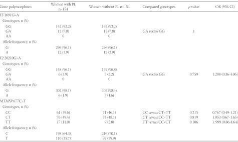
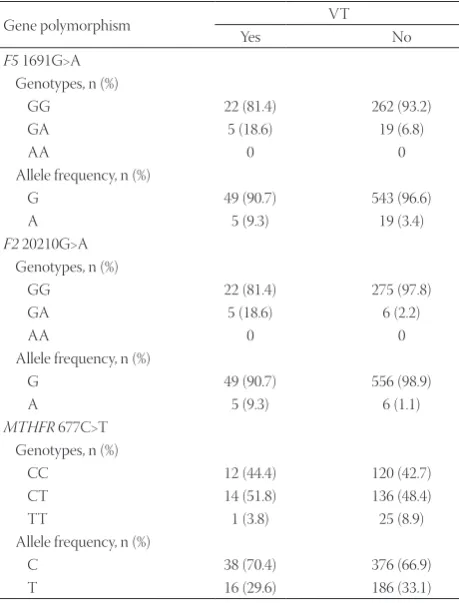
The distribution of genotypes and alleles in women with PL and control group is presented in Table 2. The distributions of genotypes and alleles in women with PL and control group in relation to the number of PL and the presence of VT are shown in Tables 3 and 4, respectively.
The F5 GA genotype was more frequent in women with multiple miscarriages (the borderline statistical significance,
p = 0.051, χ2 = 7.791). No statistically significant correlation
between the F2 and MTHFR genotype and allele distribution and the number of PL was observed.
Both F5 and F2 heterozygotes were significantly more fre-quent in women with VT compared to women without VT (p = 0.047 and 0.001, respectively). There was no statistically significant difference in the distribution of MTHFR genotypes and alleles between women with VT and those without VT.
DISCUSSION
Pregnancy is a physiological state linked to increased clotting, decreased anticoagulant activity, and decreased fibrinolysis [12].
Although the allele A of the F5 gene (1691A) is a risk fac-tor for thrombosis, the results on the association between
F5 A allele and PL are contradictory. In our study, the fre-quency of 1691A allele was 7.8% in both groups (i.e., women with and without PL), and the differences in the genotype and allele distributions between the two groups were not significant (p > 0.05). Similarly, Polish, German, and Turkish studies showed no significant differences in the frequency of
F5 1691A allele between women with PL and those without PL (p > 0.05). In the above-mentioned studies, the following frequencies of the 1691A allele were reported in women with PL versus women without PL: 3.2% versus 3.0% in the Polish study, 10.9% versus 7.4% in the German study, and 7.9% ver-sus 7.0% in the Turkish group [13-15]. In Serbian women with and without PL, the frequency of the 1691A allele was 4.0%
versus 1.5%, respectively (p = 0.05) [16]. Similarly, studies on Lebanese and Palestinian women with PL confirmed that the 1691A allele was significantly more frequent in PL group compared to controls (p < 0.001 and p < 0.0001, respec-tively) [6,17]. Surprisingly, in an Israeli study, the frequency of
TABLE 1. Characteristics of the study group
Characteristics Women with PL (mean±SD) Women without PL (mean±SD) p
Age 32.9±5.1 31.7±6.6 0.069
Weight 73.9±9.3 74.7±9.2 0.484
Number of pregnancies 1.4±1.1 1.2±0.8 0.066
VT, n (%) 15 (9.7) 12 (7.8) 0.344
VT in the family, n (%) 38 (24.7) 25 (16.2) 0.045
PL: Pregnancy loss; VT: Venous thrombosis; SD: Standard deviation
TABLE 2. Distribution of genotypes and alleles of F5, F2, and MTHFR polymorphisms in women with and without PL
Gene polymorphism Women with PLn=154 Women without PL n=154 Compared genotypes p value OR (95% CI)
F5 1691G>A Genotypes, n (%)
GG GA AA
142 (92.2) 12 (7.8)
0
142 (92.2) 12 (7.8)
0 GA versus GG 1 Allele frequency, n (%)
G
A 296 (96.1)12 (3.9) 296 (96.1)12 (3.9)
F2 20210G>A Genotypes, n (%)
GG GA AA
148 (96.1) 6 (3.9)
0
149 (96.8) 5 (3.2)
0 GA versus GG 0.759 1.208 (0.36-4.06) Allele frequency, n (%)
G
A 302 (98.1)6 (1.9) 303 (98.4)5 (1.6)
MTHFR 677C>T Genotypes, n (%)
CC CT TT
61 (39.6) 76 (49.4) 17 (11.0)
71 (46.1) 74 (48.1) 9 (5.8)
CC versus CT+TT CT versus CC+TT TT versus CC+CT
0.215 0.819 0.106
0.767 (0.49-1.21) 1.053 (0.67-1.65) 1. 999 (0.86-4.64) Allele frequency, n (%)
C
T 198 (64.3)110 (35.7) 216 (70.1)92 (29.9)
the mutated F5 allele was higher in women without PL com-pared to women with PL (p > 0.05) [18].
Rodger et al. [19] performed a meta-analysis on the rela-tionship between mothers, carriers of the F5 1691A and
F2 20210A alleles, and complications of pregnancy. They demonstrated no significant association between the mutated alleles and pregnancy complications, including PL [19]. In the present study, we also showed no significant differences in the frequency of F2 20210A allele and obstetric complica-tions between women with and without PL (p > 0.05); the fre-quency of the mutated allele was 1.9% and 1.6%, respectively.
Although Sehirali et al. [20] found the association between the F2 20210A allele and PL in Turkish women (p < 0.05) [20], these results should be interpreted with caution due to the small sample size; they included 55 women with PL and 49 controls.
Rey et al. [21] conducted a meta-analysis of 31 studies, published between 1975 and 2002. Their conclusion was that the F5 1691A allele is linked to early (OR: 2.01, 95% CI: 1.13 -3.58), late recurrent fetal loss (OR: 7.83, 95% CI: 2.83-21.67), and late non-recurrent fetal loss (OR: 3.26, 95% CI: 1.82-5.83). The
F2 20210A allele was linked to PL in the first trimester (OR: 2.32, 95% CI: 1.12-4.79), repeated miscarriages (OR: 2.56, 95% CI: 1.04-6.29), and late non-recurrent fetal loss (OR: 2.3, 95% CI: 1.09-4.87) [21].
In Spanish women with three or more unexplained PL, significant differences between the 677T allele of the MTHFR
gene and PL were not found [22].
In a Greek study, Sotiriadis et al. [23] reported that the
F5 1691G>A, F2 20210G>A, and MTHFR 677C>T polymor-phisms were not significantly associated with increased risk for PL (p = 1.00, p = 0.652, and p = 0.869, respectively) [23]. In another Greek study, the association between PL in the second trimester and primary PL with the F5 1691G>A and
F2 20210G>A was found (p = 0.03, p = 0.038, respectively), but not with the MTHFR 677C>T (p = 0.134) [24].
In the present study, the frequency of F5 1691A and
F2 20210A alleles was significantly higher in women with VT compared to women without VT, 9.3% versus 3.4% (χ2 = 4.739; p = 0.047) and 9.3% versus 1.1% (χ2 = 19.199, p = 0.001),
respec-tively. In 2006, Robertson et al. [25] reported a relationship between the risk of VTE and adverse pregnancy outcomes and thrombophilia in pregnancy. The authors concluded that the risk of VTE for F5 1691A homozygotes was 34.4 (95% CI: 9.86-120.05), while for the heterozygotes the risk was - 8.32 (95% CI: 5.44-12.70) [25].
In a meta-analysis of 63 studies, the authors found that the F5 1691G>A mutation was associated with a 4.5- and 8.6-fold increased risk of the first VTE during pregnancy (95% CI:
TABLE 3. Distribution of genotypes and alleles of F5 1691G>A, F2 20210G>A, and MTHFR 677C>T polymorphisms by the number of PL
Gene polymorphism Number of PL
1 2 3 4
F5 1691G>A Genotypes, n (%)
GG 98 (95.1) 32 (88.9) 11 (84.6) 1 (50.0) GA 5 (4.9) 4 (11.1) 2 (15.4) 1 (50.0)
AA 0 0 0 0
Allele frequency, n (%)
G 201 (97.6) 68 (94.4) 24 (92.3) 3 (75.0) A 5 (2.4) 4 (5.6) 2 (7.7) 1 (25.0)
F2 20210G>A Genotypes, n (%)
GG 100 (97.1) 34 (94.4) 12 (92.3) 2 (100.0) GA 3 (2.9) 2 (5.6) 1 (7.7) 0
AA 0 0 0 0
Allele frequency, n (%)
G 203 (98.5) 70 (97.2) 25 (96.1) 2 (100) A 3 (1.5) 2 (2.8) 1 (3.9) 0
MTHFR 677C>T Genotypes, n (%)
CC 42 (40.8) 15 (41.7) 4 (30.8) 0 CT 48 (46.6) 17 (47.2) 9 (69.2) 2 (100.0) TT 13 (12.6) 4 (11.1) 0 0 Allele frequency, n (%)
C 132 (64.1) 47 (65.3) 17 (65.4) 2 (50.0) T 74 (35.9) 25 (34.7) 9 (34.6) 2 (50.0)
χ2=7.791, p=0.051. PL: Pregnancy loss
TABLE 4. Distribution of genotypes and alleles of F5 1691G>A, F2 20210G>A, and MTHFR 677C>T polymorphisms in relation to the presence of VT
Gene polymorphism VT
Yes No
F5 1691G>A Genotypes, n (%)
GG 22 (81.4) 262 (93.2)
GA 5 (18.6) 19 (6.8)
AA 0 0
Allele frequency, n (%)
G 49 (90.7) 543 (96.6)
A 5 (9.3) 19 (3.4)
F2 20210G>A Genotypes, n (%)
GG 22 (81.4) 275 (97.8)
GA 5 (18.6) 6 (2.2)
AA 0 0
Allele frequency, n (%)
G 49 (90.7) 556 (98.9)
A 5 (9.3) 6 (1.1)
MTHFR 677C>T Genotypes, n (%)
CC 12 (44.4) 120 (42.7)
CT 14 (51.8) 136 (48.4)
TT 1 (3.8) 25 (8.9)
Allele frequency, n (%)
C 38 (70.4) 376 (66.9)
T 16 (29.6) 186 (33.1)
1.8-10.9; 95% CI: 5.9-12.6, respectively) [26]. Patients with VTE have a high risk of recurrent events. This risk is estimated to be 4.5%/year in the first 2 years after the initial event [27].
A Romanian retrospective study analyzed a possible asso-ciation between thrombosis and inherited thrombophilia in pregnant women. The risk of thrombosis in women with the
F5 1691G>A mutation was 2.66 times higher than in women without the mutation (OR = 2.66, 95% CI: 0.96-7.37, p = 0.059). The authors did not find any statistical association with the
MTHFR 677C>T polymorphism [28]. Furthermore, in another Romanian study, homozygous individuals for the F5 1691G>A had a 9-fold higher risk of thrombosis compared to individuals without the mutation (p = 0.015, 95% CI: 1.080-72.923) [29].
To our knowledge, this is the first report on the association between the F5 1691G>A, F2 20210G>A, and MTHFR 677C>T polymorphisms and risk of PL in Bosnian women. The allele frequencies reported in this study are consistent with the data obtained for other European countries. The main limitation of our study was the small sample size of women with three and four PL. We suggest further studies with larger sample size and equal samples of women with two, three, and four PLs.
CONCLUSION
In summary, we were not able to confirm the association between the F5 1691G>A, F2 20210G>A, and MTHFR 677C>T polymorphisms and PL in Bosnian women. The significant differences in the frequency of mutated alleles were found only between women with VT and those without VT.
ACKNOWLEDGMENTS
This work was funded in part by the Pomeranian Medical University, Szczecin, Poland (decision reference number WNoZ-307-01/S/13/2016).
DECLARATION OF INTERESTS
The authors declare no conflict of interests.
REFERENCES
[1] Walker ID. Thrombophilia in pregnancy. J Clin Pathol 2000;53(8):573-80.
https://doi.org/10.1136/jcp.53.8.573.
[2] WHO: Recommended definitions, terminology and format for sta-tistical tables related to the perinatal period and use of a new cer-tificate for cause of perinatal deaths. Modifications recommended by FIGO as amended October. Acta Obstet Gynecol Scand 1977;56(3):247-53.
[3] Rodeghiero F, Tosetto A. Activated protein C resistance and fac-tor V Leiden mutation are independent risk factors for venous thromboembolism. Ann Intern Med 1999;130(8):643-50.
https://doi.org/10.7326/0003-4819-130-8-199904200-00004.
[4] Adler G, Agnieszka G, Valjevac A, Czerska E, Kiseljakovic E, Salkic NN. Prevalence of genetic prothrombotic risk factors: 1691G > A FV, 20210G > A PT and 677C > T MTHFR mutations in the Bosnian population. Ann Hum Biol 2015;42(6):576-80.
https://doi.org/10.3109/03014460.2014.968618.
[5] Inbal A, Carp H. Defects in coagulation factor leading to recur-rent pregnancy loss. In: Carp H, editor. Recurrecur-rent Pregnancy Loss. Causes Controversies and Treatment. UK: Informa Healthcare; 2007. p. 127-39.
https://doi.org/10.3109/9780203931677.019.
[6] Finan RR, Tamim H, Ameen G, Sharida HE, Rashid M, Almawi WY. Prevalence of factor V G1691A (factor V-Leiden) and prothrombin G20210A gene mutations in a recurrent miscarriage population. Am J Hematol 2002;71(4):300-5.
https://doi.org/10.1002/ajh.10223.
[7] Franco RF, Reitsma PH. Genetic risk factors of venous thrombosis. Hum Genet 2001;109(4):369-84.
https://doi.org/10.1007/s004390100593.
[8] Poort SR, Rosendaal FR, Reitsma PH, Bertina RM. A common genetic variation in the 3’-untranslated region of the prothrombin gene is associated with elevated plasma prothrombin levels and an increase in venous thrombosis. Blood 1996;88(10):3698-703. [9] Jadaon MM. Epidemiology of prothrombin G20210A
muta-tion in the Mediterranean region. Mediterr J Hematol Infect Dis 2011;3(1):e2011054.
https://doi.org/10.4084/mjhid.2011.037.
[10] American College of Obstetricians and Gynecologists Women’s Health Care Physicians. ACOG Practice Bulletin No. 138: Inherited thrombophilias in pregnancy. Obstet Gynecol 2013;122(3):706-17. https://doi.org/10.1097/01.AOG.0000433981.36184.4e.
[11] Schmitz C, Lindpaintner K, Verhoef P, Gaziano JM, Buring J. Genetic polymorphism of methylenetetrahydrofolate reduc-tase and myocardial infarction. A case-control study. Circulation 1996;94(8):1812-4.
https://doi.org/10.1161/01.CIR.94.8.1812.
[12] Hellgren M. Hemostasis during normal pregnancy and puerpe-rium. Semin Thromb Hemost 2003;29(2):125-30.
https://doi.org/10.1055/s-2003-38897.
[13] Slezak R, Laczmanski L, Karpinski P, Reszczynska-Slezak D. The role of 1691G>A (Leiden) mutation in Factor V gene, 20210G>A in prothrombin gene and 677C>T in MTHFR gene in etiology of early pregnancy loss. [Article in Polish]. Ginekol Pol 2011;82(6):446-50. [14] Reznikoff-Etievan MF, Cayol V, Carbonne B, Robert A, Coulet F,
Milliez J. Factor V Leiden and G20210A prothrombin muta-tions are risk factors for very early recurrent miscarriage. BJOG 2001;108(12):1251-4.
https://doi.org/10.1016/S0306-5456(01)00298-4.
[15] Altintas A, Pasa S, Akdeniz N, Cil T, Yurt M, Ayyildiz O, et al. Factor V Leiden and G20210A prothrombin mutations in patients with recurrent pregnancy loss: Data from the southeast of Turkey. Ann Hematol 2007;86(10):727-31.
https://doi.org/10.1007/s00277-007-0327-1.
[16] Djurovic J, Stojkovic O, Todorovic J, Brajic A, Obradovic S, Stankovic S, et al. Genetics of suspected thrombophilia in Serbian females with infertility, including three cases, homozy-gous for FII 20210A or FV 1691A mutations. Hum Fertil (Camb) 2017;20(2):132-9.
https://doi.org/10.1080/14647273.2016.1255785.
[17] Hussein AS, Darwish H, Shelbayeh K. Association between factor V Leiden mutation and poor pregnancy outcomes among Palestinian women. Thromb Res 2010;126(2):e78-82.
https://doi.org/10.1016/j.thromres.2010.04.017
[18] Carp H, Salomon O, Seidman D, Dardik R, Rosenberg N, Inbal A. Prevalence of genetic markers for thrombophilia in recurrent preg-nancy loss. Hum Reprod 2002;17(6):1633-7.
https://doi.org/10.1093/humrep/17.6.1633.
cohort studies. PLoS Med 2010;7(6):e1000292. https://doi.org/10.1371/journal.pmed.1000292.
[20] Sehirali S, Inal MM, Yildirim Y, Balim Z, Kosova B, Karamizrak T, et al. Prothrombin G20210A mutation in cases with recurrent mis-carriage: A study of the mediterranean population. Arch Gynecol Obstet 2005;273(3):170-3.
https://doi.org/10.1007/s00404-005-0061-7.
[21] Rey E, Kahn SR, David M, Shrier I. Thrombophilic disorders and fetal loss: A meta-analysis. Lancet 2003;361(9361):901-8.
https://doi.org/10.1016/S0140-6736(03)12771-7.
[22] Creus M, Deulofeu R, Peñarrubia J, Carmona F, Balasch J. Plasma homocysteine and vitamin B12 serum levels, red blood cell folate concentrations, C677T methylenetetrahydrofolate reductase gene mutation and risk of recurrent miscarriage: A case-control study in Spain. Clin Chem Lab Med 2013;51(3):693-9.
https://doi.org/10.1515/cclm-2012-0452.
[23] Sotiriadis A, Vartholomatos G, Pavlou M, Kolaitis N, Dova L, Stefos T, et al. Combined thrombophilic mutations in women with unexplained recurrent miscarriage. Am J Reprod Immunol 2007;57(2):133-41.
https://doi.org/10.1111/j.1600-0897.2006.00454.x.
[24] Foka ZJ, Lambropoulos AF, Saravelos H, Karas GB, Karavida A, Agorastos T, et al. Factor V leiden and prothrombin G20210A
mutations, but not methylenetetrahydrofolate reductase C677T, are associated with recurrent miscarriages. Hum Reprod 2000;15(2):458-62.
https://doi.org/10.1093/humrep/15.2.458.
[25] Robertson L, Wu O, Langhorne P, Twaddle S, Clark P, Lowe GD, et al. Thrombophilia in pregnancy: A systematic review. Br J Haematol 2006;132(2):171-96.
https://doi.org/10.1111/j.1365-2141.2005.05847.x.
[26] Biron-Andreani C, Schved JF, Daures JP. Factor V Leiden muta-tion and pregnancy-related venous thromboembolism: What is the exact risk? Results from a meta-analysis. Thromb Haemost 2006;96(1):14-8.
https://doi.org/10.1160/th06-02-0086.
[27] Van Dongen CJ, Vink R, Hutten BA, Buller HR, Prins MH. The incidence of recurrent venous thromboembolism after treatment with vitamin K antagonists in relation to time since first event: A meta-analysis. Arch Intern Med 2003;163(11):1285-93.
https://doi.org/10.1001/archinte.163.22.2793-a.
[28] Coriu L, Ungureanu R, Talmaci R, Uscatescu V, Cirstoiu M, Coriu D, et al. Hereditary thrombophilia and thrombotic events in pregnancy: Single-center experience. J Med Life 2014;7(4):567-71. [29] Hotoleanu C, Popp R, Trifa A. Factor V Leiden, prothrombin