Transcriptome and Functional Analysis of Mating in the
Basidiomycete
Schizophyllum commune
Susann Erdmann,aDaniela Freihorst,aMarjatta Raudaskoski,bWolfgang Schmidt-Heck,cElke-Martina Jung,aDominik Senftleben,a and Erika Kothea
Institute of Microbiology—Microbial Phytopathology, Friedrich-Schiller University, Jena, Germanya
; Department of Biochemistry and Food Chemistry, Plant Physiology and Molecular Biology, University of Turku, Turku, Finlandb
; and Department of Molecular and Applied Microbiology, Leibniz Institute for Natural Product Research and Infection Biology—Hans Knöll Institute, Jena, Germanyc
In this study, we undertook a functional characterization and transcriptome analysis that enabled a comprehensive study of the
mating type loci of the mushroom
Schizophyllum commune
. Induced expression of both the
bar2
receptor and the
bap2(2)
pher-omone gene within 6 to 12 h after mates’ contact was demonstrated by quantitative real-time PCR. Similar temporal expression
patterns were confirmed for the allelic
bbr1
receptor and
bbp1
pheromone-encoding genes by Northern hybridization.
Interest-ingly, the fusion of clamp connections to the subterminal cell was delayed in mating interactions in which one of the compatible
partners expressed the
bar2
receptor with a truncated C terminus. This developmental delay allowed the visualization of a green
fluorescent protein (Gfp)-labeled truncated receptor at the cell periphery, consistent with a localization in the plasma membrane
of unfused pseudoclamps. This finding does not support hypotheses envisioning a receptor localization to the nuclear
mem-brane facilitating recognition between the two different nuclei present in each dikaryotic cell. Rather, Gfp fluorescence observed
in such pseudoclamps indicated a role of receptor-pheromone interaction in clamp fusion. Transcriptome changes associated
with mating interactions were analyzed in order to identify a role for pheromone-receptor interactions. We detected a total of 89
genes that were transcriptionally regulated in a mating type locus
A
-dependent manner, employing a cutoff of 5-fold changes in
transcript abundance. Upregulation in cell cycle-related genes and downregulation of genes involved in metabolism were seen
with this set of experiments. In contrast, mating type locus
B
-dependent transcriptome changes were observed in 208 genes, with
a specific impact on genes related to cell wall and membrane metabolism, stress response, and the redox status of the cell.
T
he filamentous fungus
Schizophyllum commune
is a widely
dis-tributed mushroom that lives saprophytically and can cause
white rot on wood. It has been extensively used to study mating
interactions for almost 100 years (47). In addition to studies
in-volving the mushroom
Coprinopsis cinerea
and the corn smut
Ustilago maydis
, the mating type genes have been extensively
an-alyzed for this tetrapolar basidiomycete (4, 29). The recent
publi-cation of the
S. commune
genome sequence enables global
expres-sion analyses (41). During the life cycle of this fungus, compatible
haploid monokaryons can mate to produce a fertile dikaryon with
two nuclei per cell, one derived from each mating partner. Under
appropriate environmental conditions, the dikaryon is able to
form the sexual reproductive fruiting bodies (40). While the
fu-sion of two monokaryotic hyphae (plasmogamy) is independent
of mating types, specific steps of sexual development are regulated
by the mating type genes encoded in the loci
A
and
B
(25, 47). For
each of these loci, two linked, multiallelic subloci (termed
␣
and

) have been determined by recombination analyses (27, 49, 51,
65). The
A
loci encode homeodomain transcription factors, while
the
B
loci code for multiallelic pheromone receptors and
phero-mones (55, 63, 66). Both the
A
and
B
pathways can be activated by
the combination of different allelic specificities in at least one
su-blocus derived from each of the two mates (46, 52, 53). Thus, for a
compatible mating interaction to occur, different
A
and
B
mating
types (
A
⫽
B
⫽
) in the partners are essential.
The migration of nuclei is triggered by the presence of different
B
mating types after the fusion of two compatible monokaryons
and occurs reciprocally from one mating partner throughout the
mycelium of the other (54). Subsequent nuclear pairing, as well as
the initiation of clamp cell development, is regulated by the
A
mating type. The formation of clamp cells at the septa of the
de-veloping dikaryon results in a regular distribution of the two
dif-ferent nuclei during mitosis within the dikaryon (4, 39). The last
step of clamp fusion is dependent on different
B
factors and
in-volves Ras signaling (31, 59). This sequence of events can be
ob-served in semicompatible mating interactions where a
hetero-karyon with an activated
B
locus (
B
onor
A
⫽
B
⫽
) undergoes
constant nuclear migration associated with irregular distribution
of nuclei (26, 38). This phenotype, termed flat, results in reduced
aerial mycelia, malformed hyphae, and no formation of clamps
(43, 48). The second type of semicompatible mating interaction
occurs with an activated
A
locus (
A
onor
A
⫽
B
⫽
) and is evident in
the formation of pseudoclamps and a die-off in the contact zone
(barrage phenotype) (38, 43, 44).
For the pheromone-receptor system encoded in the
B
locus of
S. commune
, the pheromone signaling is triggered by the
interac-tion of nonself-pheromones with G-protein-coupled receptors
(GPCRs) of class D (1, 20, 70). All pheromone receptors of
basid-iomycetes belong to the
Saccharomyces cerevisiae
Ste3-related
pheromone receptor family and bind
a
-factor-related lipopeptide
pheromones (12, 32, 67, 64). Multiple different cognate
phero-Received22 August 2011 Accepted22 December 2011
Published ahead of print30 December 2011
Address correspondence to Erika Kothe, [email protected].
Supplemental material for this article may be found athttp://ec.asm.org/.
Copyright © 2012, American Society for Microbiology. All Rights Reserved.
doi:10.1128/EC.05214-11
on September 8, 2020 by guest
http://ec.asm.org/
mones capable of activating a single receptor in a multistate model
of interaction have evolved in
S. commune
, most likely by
recom-bination between copies of the mating type locus (15, 20). The
function of pheromone signaling is thus linked to the processes of
nuclear migration, nuclear identity, and clamp fusion.
A model that links pheromone-receptor interactions to the
distance between nuclei in dikaryons had been proposed. Under
this model, productive interaction between non-self-pheromones
and their cognate receptor occurs only when the nuclei are in close
proximity to each other (60). Another hypothesis (5) proposed
that receptor localization to the nuclear envelope was necessary
for detection of intracellular pheromone(s). This would
necessi-tate an inverse orientation of the receptor within the membrane.
In order to investigate the role of the pheromone receptor and to
distinguish between the different models described above,
recep-tor localization has been of great interest for a number of years. A
plasma membrane localization was found for pheromone
recep-tors in single cells of the corn pathogen
U. maydis
(17). In
mush-room-forming homobasidiomycetes like
S. commune
or
C.
ci-nerea
, however, cellular localization of the pheromone receptor
has not yet been determined.
The role of pheromone perception in
S. cerevisiae
includes
both mate attraction and the formation of shmoo cells that show
growth directed toward the mating partner (33). In contrast,
at-traction and fusion of
S. commune
hyphae can be seen
indepen-dent of mating type. Directional growth toward a mate has been
observed at very short distances (54). In an earlier investigation on
the role of Ras in sexual development of
S. commune
, we
demon-strated that a deletion of a Ras-specific GTPase-activating protein,
⌬
gap1
, results in a lack of clamp fusion in
⌬
gap1
homoallelic
dikaryons (59). This uninucleate state is rescued by development
of a branch, which is then able to fuse with the clamp cell after
prolonged growth. We hypothesized that this situation in the
mu-tant represents two monokaryotic cells which show clear
attrac-tion toward each other. Thus, clamp fusion in dikaryons can be
viewed as a system in which mate attraction can be visualized at
very short distances.
The intracellular signaling via GPCRs has been shown to be
controlled by regulators of G-protein signaling (RGS) in other
studies of
S. commune
(6, 13). The RGS protein Thn1 negatively
regulates the activity of the G-protein
␣
subunit by acting as a
GTPase-activating protein (GAP) (42). A loss-of-function
muta-tion due to transposon integramuta-tion into
thn1
results in hyphae
with a characteristic corkscrew morphology which are unable to
accept nuclei of a compatible mating partner (13, 61). For
S.
cerevisiae
, RGS gene
SST2
was functionally linked to mating
inter-actions (7, 8).
In the study presented here, we show the localization of the
G-protein-coupled pheromone receptor protein in hyphal
fila-ments of
S. commune
. Furthermore, we report on the role of the
C-terminal region of the pheromone receptor Bar2 in mating
in-teractions. In combination with transcriptome analyses, a
com-prehensive study of the pheromone-receptor system for a
mush-room-forming basidiomycete is presented for the first time.
MATERIALS AND METHODS
Strains and culture conditions.TheS. communestrains 4-40, 23, 684, 1792-114-10, W21, W22, W22-thin, V153-21 (Bnull), 12-43, and 4-39
were obtained from the Jena Microbial Resource Center (JMRC) or the strain collection of University of Turku.S. communereceptor transfor-mants generated in this study (Vbar2f, Vbar2t, Vbar2tG1, Vbar2tG11) and strains relevant for microarray analysis are listed in Table 1.S.
com-munewas routinely grown at 28°C on minimal medium (MM) (50) or
complex yeast medium (CYM) (62) with 2% (wt/vol) glucose, with or without 1.8% (wt/vol) agar. Liquid cultures were shaken at 150 rpm. For RNA extraction, cultures were grown on a cellophane membrane placed on solid medium. TheS. communestrains investigated by Northern blot-ting or quantitative real-time PCR (RT-PCR) were cultured using a mod-ified sandwich method as described previously (67). Interacting mycelia were grown for 3 to 72 h at 28°C before harvest. For transcriptome anal-ysis, mycelia from a monokaryon or a mating interaction were transferred onto a cellophane membrane placed on fresh CYM plates and grown for 3 days.
Functional analysis and receptor localization. We generated the transformants that are expressing Vbar2f using aBnullstrain, which
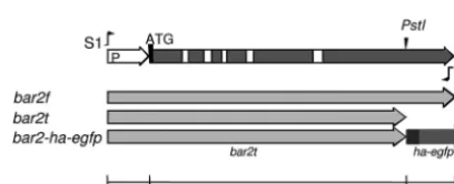
un-transformed does not expresses mating pheromones or receptors (14). For amplification of the pheromone receptor genebar2f(NCBI accession
numberX91168.4), we used primers S1 (5=
-GGATCCGCCCATTGTCC-3=) and S2 (5=-TCACACCGACGCGCGGT-3=) to produce an amplicon starting from approximately 300 bp upstream of ATG and extending to the last 17 bases ofbar2(Fig. 1). Comparison of the last 17 bases ofbar2
with those inbar1andbar3shows that they are identical, except for a different stop codon inbar3. The pheromone receptor genebar2fencodes a full-length protein of 636 amino acids (aa). In addition, we generated the strain Vbar2t, which was transformed with a mutated gene encoding a TABLE 1Schizophyllum communestrains and mating interactions
Strains and crosses Mating type, relevant genotype/description
H4-8 A4,6;B3,2
W21 A1,1;B1,1
W22 A4,6;B3,2
W22-thin A4,6;B3,2; thin (thin
a)
V153-21 A3,5;Bnull;trp1⫺
Vbar2f A3,5;Bnull::bar2 trp1
Vbar2t A3,5;Bnull::bar2⌬PstItrp1
Vbar2tG1 A3,5;Bnull::bar2⌬PstI-HA-Gfptrp1
Vbar2tG11 A3,5;Bnull::bar2⌬PstI-HA-Gfptrp1
12-43 A3,5;B2,3;ura1⫺; monokaryon (Mon1
a)
4-39 A1,1;B3,2; monokaryon (Mon2
a)
W22⫻12-43 Dikaryon (Dika)
W22⫻4-39 Semicompatible interaction:A⫽B⫽(Aon
a)
W21⫻4-39 Semicompatible interaction:A⫽B⫽(Bon
a)
Vbar2f⫻4-39 Dikaryon of receptor transformant with full-length
receptor (Dik-Vbar2fa)
Vbar2t⫻4-39 Dikaryon of receptor transformant with truncated
receptor (Dik-Vbar2ta)
aTerm for microarray analysis.
FIG 1Genebar2ofSchizophyllum commune. The primers S1 and S2 were used for amplification of full-length genebar2f(NCBI accession numberX91168.4) with promoter region (P). The genebar2twas truncated at the 3=PstI restric-tion site (see also NCBI accession numberX91168.2). The 3⫻HA-eGfp tag (ha-egfp) is indicated at the C terminus of thebar2tgene. The white squares indicate introns.
on September 8, 2020 by guest
http://ec.asm.org/
truncated receptor ofB␣2specificity with 518 aa in length; the sequence of
bar2tis truncated at a 3=PstI site (20) (NCBI accession numberX91168.2, GI 23954358). The truncatedbar2treceptors of strains Vbar2tG1 and Vbar2tG11 have been tagged with extended green fluorescent protein (eGfp) forin vivostudies and with hemagglutinin (3⫻HA) for receptor localization by indirect immunostaining. The HA-eGfp sequence (845
bp) was amplified with the primers 5=-GGC TGC AGG GAT ACC CGT
ATG ATG TTC CGG ATT ACG CTG GCT ACC CAT ACG ACG TCC CAG ACT ACG CTG GCT ACC CAT ACG ACG TCC CAG ACT ACG CTG GCG CAC CTG GAG CCA TGG TGA GCA AGG GCG AGG
AGC-3=and 5=-GTT GGA ATT CTG CAG TCG CGG CCG CTT TAC TTG-3=
using the vector pegfp (Clontech, The Netherlands) as template and li-gated into the PstI site at the C terminus ofbar2t. All transformed phero-mone receptor genes were expressed under their native promoter.
Fluorescence microscopy and immunostaining. For microscopic analysis, sterile glass coverslips were placed on an agar plate such that hyphae could attach to and grow onto the slides after 2 to 4 days of culti-vation. Mycelia ofS. communewere fixed for 90 min at room temperature with methanol and PME {50 mM PIPES [piperazine-N,N= -bis(2-ethane-sulfonic acid)], pH 6.7, 25 mM EGTA, pH 8.0, 5 mM MgSO4} plus 3.7% formaldehyde. Coverslips were then washed with PME and the cells were treated with 3% lysis enzyme (Trichoderma harzianum; L1412; Sigma, Germany) and 50% egg white. The cells were permeabilized with 0.3% Triton X-100 in phosphate-buffered saline (PBS; 137 mM NaCl, 2.68 mM KCl, 8.09 mM Na2HPO4, 1.76 mM KH2PO4, pH 7.4). Nonspecific bind-ing sites were blocked with 3% bovine serum albumin (BSA). The 1st antibody (mouse anti-HA 2367; Cell Signaling) was diluted 1:200 in PBS with 3% BSA, and coverslips were incubated overnight at 4°C. The fluo-rescein isothiocyanate (FITC)-labeled 2nd antibody (FITC-antimouse; F4018; Sigma) was diluted 1:100 in PBS and incubated for 60 min at 37°C. Following every incubation step, the coverslips were washed with PBS at least once. Nuclei were stained with DAPI (4=,6-diamidino-2= -phenylin-dole dihydrochloride; 0.1 to 1g/ml) added to the mounting medium (0.1 M Tris HCl, pH 8.0, 50% glycerol, and 1 mg/ml phenylenediamine). The mycelia were examined with a fluorescence microscope (Axioplan2) and confocal laser scanning microscope (LSM 5 Live) (Carl Zeiss Micro-Imaging, Jena, Germany). Images were processed with SPOT imaging software (Diagnostic Instruments, Sterling Heights, MI) and ZEN soft-ware (Carl Zeiss, Jena, Germany).
Quantitative real-time PCR and Northern hybridization.Mycelia were harvested, frozen in liquid nitrogen, and ground with mortar and pestle to a powder. Total RNA was isolated from not more than 100 mg of tissue powder using a commercial kit (RNeasy plant minikit; Qiagen, Hilden, Germany). Reverse transcription was performed using 1g of total RNA in a 20-l reaction mix (iScript cDNA synthesis kit; Bio-Rad, Munich, Germany). For each real-time PCR, we used 2l of cDNA as template in a 25-l reaction mix that additionally contained 12.5l 2⫻ reaction mix [1.25 U HotTaqDNA polymerase, 0.4 mM deoxynucleoside triphosphates, 40 mM Tris HCl, pH 8.55, 32 mM (NH4)2SO4, 0.02% Tween 20, and 4 mM MgCl2; peqGold hot start mix Y; Peqlab], 7.5l water (nuclease free), 1l of each primer (10 pmol/l), and 1l SYBR green I (using a 1:2,000 dilution of the stock reagent; Molecular Probes, Invitrogen) using a SmartCycler II thermocycler (Cepheid). The PCR program consisted of 5 steps (initial denaturation for 120 s at 94°C; 45 cycles of denaturation for 20 s at 94°C, annealing for 20 s at 50 or 55°C, and extension for 20 s at 72°C; and a final melting curve analysis at 60 to 95°C with 0.2°C/s). The SYBR green fluorescence was detected (excitation, 450 to 495 nm; emission, 510 to 527 nm) during both the extension phase of each cycle and the subsequent melting curve analysis. For amplification of the target genesbar2andbap2(2), as well as the reference genesact1and
tef1, the following oligomers were used: 5=-ATTACTCTTGGCGCCTCT
GTA-3=and 5=-AATGAGAGCGTCGACCATGACT-3=forbar2(yielding
a product of 138 bp), 5=-TTACTGATAGTCACAGATA-3=and 5=-ATGG
CGAACCGGAC-3=forbap2(2)(87 bp), 5=-GTCCGCCCTCGAGAAGA
GTTA-3=and 5=-TTGTACGTCGTCTCGTGGATA-3=foract1(141 bp),
and 5=-AGCTTGGCAAGGGTTCCTTCA-3=and 5=-AACTTCCAGAGG
GCGATATCA-3=fortef1(97 bp). The primers forbar2,act1, andtef1
span an intron to identify potential genomic DNA contamination. The genebap2(2)has a total length of only 87 bp and has no intron. Annealing was performed at 55°C for all genes exceptbap2(2), which was amplified at an annealing temperature of 50°C. The calculation of the gene-specific efficiencies was based on the slope of a standard curve generated from a series of different cDNA concentrations (0.04, 0.2, 1, 5, 25, 50, 100, and
200 ng cDNA/25-l PCR mixture). cDNA was derived from a mating
interaction between two compatible wild-type strains (12-43⫻4-39). The PCR efficiency forbar2was 1.83 (corresponding to 83% of exponen-tial expression), that forbap2(2)was 1.53 (53%), that foract1was 2.1 (110%), and that fortef1was 2.0 (100%).
Since the receptor and pheromone genes between different strains are not alike at the DNA sequence level but belong to the same family of gene products, the primer combinations were tested with templates of different
B␣specificities. The receptor and pheromone primers gave no amplifica-tion with specificities other thanB␣2(data not shown). The genesact1, coding for actin, andtef1, coding for translation elongation factor EF1␣, were suitable reference genes with stable expression levels under the tested conditions.
The expression of the target genes was determined relative to the ex-pression of reference genes, which were used for the normalization of expression between samples. We measured expression levels in mycelia isolated at a progression of different times during the mating interaction (after 3, 6, 9, 12, 24, 30, 48, 54, and 72 h; samplest3throught72). These times correspond to mycelia of different nuclear distributions. Samplet0 corresponds to a mix of two monokaryons which have been brought to-gether directly before sampling (t0⫽monokaryon, 0 h) and reflects the basal level of expression (control). For each time and individual mating interaction, the total RNAs of three independently grown mycelia have been isolated and transcribed into cDNA in duplicate, and each one was used as a template in real-time PCR, also measured in duplicate. The expression of the target genes was normalized and quantified relative to the expression of the reference genes and was also corrected for the calcu-lated efficiency (45, 68). Northern hybridization experiments and the la-beling of the receptor and pheromone cDNA fragments were performed as described previously (21).
Microarray-based transcriptome analysis.Quantification of tran-scriptome changes associated with mating interactions was performed in order to identify signaling components and targets of pheromone re-sponse. In addition, anS. communestrain showing the thin phenotype was investigated to resolve the role of Thn1 in mating. The microarrays (febit biomed GmbH, Heidelberg, Germany) used in the present study contain probes for all 13,181 predicted genes ofS. commune. It is based primarily upon the November 2008 version of theSchizophyllum commune(version 1.0) genome using strain H4-8 (A4,6;B3,2). The probes (oligonucleotides of ⬃50 bases in length) have been spotted on Geniom Biochips. The strains and mating interactions were investigated in microarray analysis in two biological or technical replicates (Table 1; see also Table S1 in the supple-mental material). Genes which show regulation (fold change,ⱖ2) be-tween the two monokaryotic strains 12-43 and 4-39 were eliminated from all other comparisons as strain-specific differences (Fig. 2).
A comparison of theAoncondition versus 4-39 (Mon2) yielded what were defined to beA-regulated genes. In contrast,B-regulated genes were defined by comparingBonversus Dik: this assumes that hyphae activated forB-dependent regulation (Bon) are more related to dikaryotic hyphae containing nuclei of both mating partners after nuclear migration. At the same time, no gene was included from the former set (A-regulated genes) so as not to involve cross-pathway signaling betweenAandB(Fig. 2).
Differentially expressed genes of the thin phenotype were up- or downregulated in both of the comparisons made: 12-43 (Mon1) versus W22-thin and 4-39 (Mon2) versus W22-thin (Fig. 2). To identify regu-lated genes influenced by the C-terminal region of Bar2, we defined two criteria: genes had to be up- or downregulated in both comparisons: Dik-Mating in the BasidiomyceteSchizophyllum commune
on September 8, 2020 by guest
http://ec.asm.org/
Vbar2t versus Dik and Dik-Vbar2t versus Dik-Vbar2f. In addition, these genes were unregulated in Dik-Vbar2f versus Dik (Fig. 2).
For each array, 1g of total RNA was labeled using the MessageAmp-biotin enhanced RNA kit from Ambion. Hybridization was performed automatically for 16 h at 45°C using RT-Analyzer (febit, Heidelberg, Ger-many). The biotin-labeled nucleic acid was detected using streptavidin-phycoerythrin (SAPE) in combination with consecutive signal enhance-ment (CSE). Feature recognition using the Cy3 filter set and signal calculation were automatically analyzed within milliseconds using the Ge-niom RT-Analyzer (febit, Heidelberg, Germany). For preprocessing, all data analyses of the febit microarrays were performed using the LIMMA (linear models for microarray data) packages of the Bioconductor soft-ware (18). Background correction was performed using the intensities of blank probes that consisted of only a single T nucleotide. The median background intensity was subtracted from the spot intensity. After con-verting any negative values to a low-positive value (8), signal intensities were log2transformed, and duplicate spots were averaged. The data ob-tained were processed using quantile normalization. To identify the genes with the greatest evidence of differential expression, a linear model fit was applied for each gene using LIMMA. Candidate genes were selected for
further analysis on the basis of their fold change (ⱖ5) in expression and theirPvalue (ⱕ0.05).
Microarray data accession number.The data discussed in this publi-cation have been deposited in NCBI’s Gene Expression Omnibus (9) and are accessible through GEO Series accession numberGSE26401(http: //www.ncbi.nlm.nih.gov/geo/query/acc.cgi?acc⫽GSE26401).
RESULTS
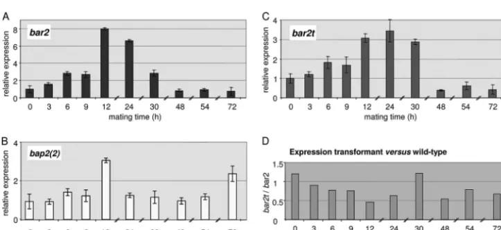
Expression of pheromone receptor and pheromone genes
dur-ing matdur-ing interactions.
In order to assess the expression of both
pheromone receptor and pheromone genes during the life cycle of
S. commune
, a specific pheromone receptor gene,
bar2
, and a
cor-responding self-pheromone gene,
bap2(2)
, were analyzed by
quantitative real-time PCR. As expected, expression levels for
both mating type-specific genes were low in monokaryons (
t
0),
and expression was specifically induced during mating
interac-tions. In a time series from 0 to 72 h (
t
0to
t
72) during a compatible
mating interaction, the expression of the receptor gene
bar2
in-creased gradually to a 3-fold higher level in the first 9 h and
reached a maximal change of 8-fold after 12 h (Fig. 3).
Subse-quently, the expression of
bar2
gradually decreased to a low level
comparable to monokaryotic expression levels. The expression of
the pheromone gene
bap2(2)
increased to a maximum of a 3-fold
increase over baseline after 12 h, decreasing shortly thereafter (Fig.
3). In contrast to the receptor, however, pheromone expression
increased steadily and then again up to 2.5-fold after 72 h. Overall,
the expression levels of receptor gene
bar2
and pheromone gene
bap2(2)
in wild type were extremely low, reaching the threshold
level only after 26 and 29 to 32 cycles, respectively, while the
ref-erence genes
act1
and
tef1
amplified with mean threshold cycles of
15 and 21, respectively.
The quantitative RT-PCR measurements of
bar2
and
bap2(2)
were in accordance with those from Northern blot
experiments determining both
bbr1
receptor and
bbp1(1)
pher-omone gene expression in a time series from 3 to 12 h (Fig. 4).
Induction of genes encoding receptors
bar1
and
bbr1
as well as
pheromones
bbp1(1)
,
bbp1(2)
, and
bbp1(3)
was obtained in
A
onand
B
onsemicompatible matings, although the signals in these
FIG 2Diagram of interactions screened to identify genes differentiallyregu-lated. The comparison of assumed different (green) and similar (red) interac-tions was used to identify genes regulated in transcriptome profiling.
FIG 3(A and B) Expression of the pheromone receptor genebar2(A) and the pheromone genebap2(2)(B) in a wild-type mating (12-43⫻4-39); (C) expression of a truncated version of pheromone receptor genebar2tin the transformant Vbar2t in a mating with the wild-type strain 4-39. The gene expression levels were determined by quantitative real-time PCR of compatible mating interactions over a time period of 72 h. The expression levels in monokaryons (t0) were normalized to 1, and all other mating times (t3tot72) are shown relative tot0. (D) Ratio ofbar2t/bar2expression, illustrating the effect of a C-terminal truncation of the pheromone receptor.
on September 8, 2020 by guest
http://ec.asm.org/
interactions were lower than those in a compatible mating
leading to dikaryon formation (Fig. 4).
The pheromone receptors of
S. commune
contain long
intra-cellular C termini and are considerably longer than those of the
S.
cerevisiae
Ste3 pheromone receptor. Thus, specific intracellular
binding sites for regulatory proteins could be present and might
also perform a function in the regulation of expression. This
prompted us to measure expression levels with quantitative
RT-PCR in the transformant Vbar2t, which carries only the one
trun-cated receptor gene (
bar2t
) but lacks all other
B
mating genes. The
expression level of
bar2t
was determined to be slightly higher than
the expression level of
bar2
in a wild-type strain at
t
0(Fig. 3D). The
induction of expression upon mating was lower, however. The
truncated gene also showed a slower increase in expression, which
prompted us to carefully reexamine its phenotype.
C-terminal truncation of the pheromone receptor affects
clamp fusion.
We examined the
B
nulltransformants containing
either the entire, full-length pheromone receptor gene
bar2f
or the
truncated version,
bar2t
, in order to investigate the potential
func-tions of the long intracellular C terminus of the receptor.
Integra-tion of either
bar2f
or
bar2t
reconstituted the wild-type phenotype
of mating competence in monokaryotic, vegetative mycelium.
Pheromone receptor transformants developed mycelia (substrate
and aerial) that grew in a normal fashion, but with a slightly
smaller colony diameter than wild-type strains (data not shown).
Pheromone receptor transformants mated with fully compatible
partners (A
⫽
B
⫽
) and showed the expected unilateral nuclear
mi-gration and formation of apparently clamped mycelium on the
side of the receptor transformant. Both versions of the receptor
recognized pheromones of all other
B
␣
specificities but the
self-specificity
B
␣
2
, and clamp structures were formed at every
sep-tum. In transformants carrying a truncated receptor gene, a delay
in development was seen, with these matings taking 1 to 2 days
longer before clamp cells became visible, correlating well with the
delayed upregulation of receptor and pheromone gene expression
measured by RT-PCR. Closer examination of all fully compatible
crosses (
A
⫽
B
⫽
) involving transformants carrying either
bar2f
or
bar2t
alleles revealed the formation of pseudoclamps with a
trapped nucleus in the unfused hook cells (Fig. 5). The tip cells of
mated receptor transformants contained two nuclei and hence
were dikaryotic. Subterminal cells often contained only one
nu-cleus, while the other remained trapped in the unfused
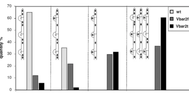
pseudoc-lamp (Fig. 5). For analysis, we classified four states of cpseudoc-lamp
fu-sion: (i) three successive clamps behind the tip cell are fused
clamps, (ii) only the first clamp (closest to the tip cell) is still
unfused, (iii) all three clamp connections are pseudoclamps, and
(iv) an alternate distribution of pseudoclamps and fused clamps
occurs (Fig. 6). Using this classification, it was observed that in
wild-type mating interactions, clamp fusion in tip cells was not
(yet) completed in 35% of the cases, while all subterminal cells had
only fused clamps. In transformants carrying either
bar2f
or
bar2t
,
however, all four patterns occurred, albeit with a higher rate of
clamps in the transformant with the full-length gene
bar2f
(Fig. 6).
Thus, the 118 aa missing from the C terminus in Vbar2t influence
the correct and timely development of dikaryotic hyphae.
Truncation of Bar2 affects fruit body development and spore
production.
Despite the formation of pseudoclamps, the receptor
transformants Vbar2f and Vbar2t were able to develop fruit
bod-FIG 4Northern hybridization of receptor and pheromone gene expression during compatible and semicompatible matings. (A) Expression of the Bbr1 receptor and the Bbp1 pheromone-encoding genes in a compatible mating, 4-40 (M1;A4,6;B1,1)⫻4-39 (M2;A1,1;B3,2), and the two respective monokaryons, as well as an incompatible cross between identical monokaryons (M1x). RNA extraction was performed after the indicated times (3, 6, 12 h). (B) Expression of the receptor genes encoding Bar1 and BbrI and also those encoding the pheromones Bbp1(1), Bbp1(2), and Bbp1(3). Lane 1, expression inAonsemicompatible mating between strains 23 (A4,6;B3,1)⫻684 (A2,6;B3,1); no signal was expected forbar1since neither of theBloci encodes thebar1receptor; lanes 2 and 3,Bon semicompatible matings between strains 1792-114-10 (A4,6;B3,6)⫻4-40 (A4,6;B1,1) and 43/26 (A4,6;B3,1)⫻4-40 (A4,6;B1,1), respectively; lane 4,Aon;Bonfully compatible mating between strains 4-40 (A4,6;B1,1)⫻4-39 (A1,1;B3,2); lane 5, strain 4-39 (A1,1;B3,2); no signal was expected in this control since no sequences encoding either the Bar1 and Bbr1 receptor or any of the three Bbp1 pheromones are present in strain 4-39; lane 6, strain 4-40 (A4,6;B1,1); lane 7, strain 23 (A4,6;B3,1). All strains were grown for 8 h after mating. Each well contains 20g of total RNA. Expression of glyceraldehyde-3-phosphate dehydrogenase (GPD) was monitored as a loading control.
Mating in the BasidiomyceteSchizophyllum commune
on September 8, 2020 by guest
http://ec.asm.org/
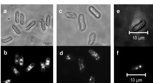
ies. However, fruit body development often stopped before
matu-rity. Lamellae were either absent or malformed, but production of
spores was recorded nevertheless (Fig. 7). The spores produced
from transformants carrying
bar2t
lacked nuclei in high
propor-tions (
⬎
70%). The majority of the spores of receptor
transfor-mant Vbar2t showed only mitochondrial DNA staining,
appear-ing as small spots distributed all over the cell (Fig. 8). However, a
small proportion of nucleated spores was formed in transformant
Vbar2t, and these were able to germinate and to develop into
monokaryotic mycelia. This is in contrast to the wild type, where
spores generally contain two nuclei per spore (Fig. 8) and
germi-nated at almost 100%. Transformants carrying
bar2f
showed an
intermediate phenotype, with anucleated spores and
wild-type-like spores being observed (Fig. 8).
Cellular localization of pheromone receptor.
We decided to
utilize the enhanced level of pseudoclamps for
in vivo
and
in situ
localization of receptor molecules. The prolonged presence of
un-fused clamp cells allowed us to examine the short-distance
attrac-tion between interacting cells. For this purpose, the receptor gene
bar2t
C-terminally fused to both Gfp and HA allowed us to
per-form both
in vivo
observations and immunofluorescence staining
of fixed cells, respectively. When this fusion construct was
trans-formed into the
B
nullrecipient strain, complementation was
ob-served for mating functions with all non-self-
B
␣
specificities, as
was the case for the untagged pheromone receptors. The hyphae
produced from the matings showed the expected occurrence of
pseudoclamps, with no apparent effect of the Gfp-HA tag on
re-ceptor function.
Expression of the Gfp-tagged receptor was induced by a mating
interaction with a compatible wild-type strain, and fluorescence
could be detected in pseudoclamps, at the cell periphery, and in
vesicles (Fig. 9). Thus,
in vivo
visualization confirmed the presence
of receptor molecules within pseudoclamps, while staining of the
nuclear envelope was never observed. To independently confirm
the results, the same transformants were used for
immunofluores-cence detection of the receptor. At the low expression levels
ob-served for the receptor gene, we reasoned that the signal
enhance-ment inherent in antibody detection would be of benefit to our
analyses. Localization of the receptor with predominant
occur-rence at the cell periphery was consistent with our observations of
membrane localization in pseudoclamps and clamp connections,
utilizing an FITC-labeled antibody specific for the HA tag (Fig. 9).
FIG 5Pseudoclamp formation in transformants carrying the truncated pheromone receptor genebar2tafter a compatible mating. (a) Pseudoclamp with a DAPI-stained nucleus trapped in the unfused clamp. Bar, 5m. (b) Bright-field micrograph of hyphae and DAPI staining of nuclei showing a nuclear pair in the tip cell and the two nuclei separated in the unfused clamp and subterminal cell (asterisk). The tip of the hypha is at the extreme right of the figure, and nuclei are indicated by an arrow. Bar, 20m.
FIG 6States of clamp fusion in a wild-type (wt) dikaryon (12-43⫻4-39) and in two pheromone receptor transformants carrying either the full-length genebar2f
(Vbar2f) or the truncated version,bar2t(Vbar2t), after interacting with a compatible mating partner (strain 4-39);n⫽100.
on September 8, 2020 by guest
http://ec.asm.org/
The fluorescence signal was especially intense at the septa, which
we interpret to be a result of cell membranes on both sides of a
single septum. For some pseudoclamps, detection of Gfp-tagged
receptor failed, presumably for those trapping a wild-type nucleus
not carrying Gfp.
Since the expression of pheromone receptor protein was
shown to be induced in compatible mating interactions,
fluores-cence analyses were performed predominantly on mycelia derived
from mated cultures. When monokaryotic, vegetative mycelium
of the tagged Gfp-receptor transformants was investigated, no
clear fluorescence signal was detectable for either Gfp or HA,
be-cause of the extremely low expression levels under these
condi-tions (data not shown). Mycelia of untagged wild-type strains
were used as negative controls in fluorescence microscopy.
FIG 7Fruiting bodies ofSchizophyllum communein the wild type (a and b), pheromone receptor transformant Vbar2t with the truncated receptor (c to e), and the pheromone receptor transformant Vbar2f with the full-length receptor gene (f and g). All strains have been mated with the compatible partner strain 4-39. While the wild type forms fruit bodies with ordinary pseudolamellae, the transformants show defects in fruit body development, resulting in fewer or absent pseudolamellae. Bars, 0.5 cm.
FIG 8Spores ofSchizophyllum communein the wild type (a and b) or pheromone receptor transformants encoding either the truncated receptor (Vbar2t) (c and d) or the full-length receptor (Vbar2f) (e and f). Spores were obtained from fruiting bodies generated from compatible crosses with strain 4-39. While almost all wild-type spores contained two nuclei (b), more than 70% of the spores from the truncated receptor transformants did not contain nuclei and only mitochondrial DNA was stained (d). The spores derived from outcrosses of the full-length receptor transformant Vbar2f showed a higher incidence of two nuclei than spores derived from outcrosses with the truncated version (f).
Mating in the BasidiomyceteSchizophyllum commune
on September 8, 2020 by guest
http://ec.asm.org/
Downstream signaling and target genes of pheromone
re-sponse.
In order to identify genes responding to pheromone
sig-naling at the transcriptional level, we used whole-genome
mi-croarrays hybridized with cDNA derived from different mating
interactions. These interactions included the heterokaryotic
A
on,
heterokaryotic
B
on, and dikaryotic (
A
onand
B
on) conditions.
Overall, 26% of the entire genome was determined to be
transcrip-tionally regulated (fold change,
ⱖ
2;
P
ⱕ
0.05) due to mating
in-teractions. This can be broken down into 974 genes (7% of the
genome) regulated by activation of the
A
pathway, 1,480 genes
(11% of the genome) regulated by the
B
pathway, and 1,016 genes
(8% of the genome) regulated by both
A
and
B
. The last type of
regulation was not analyzed further, since mushroom formation
would likely override or obscure any direct effects of combined
A
and
B
regulation. A higher threshold for transcriptional regulation
(change,
ⱖ
5-fold) yielded 89
A
-regulated genes (41 up, 48 down;
FIG 9(A) Micrographs of HA-Gfp-tagged pheromone receptor localization in dikaryoticS. communetransformants by immunostaining (Aa to Ad) andin vivo
(Ae and Af) in dikaryotic mycelium (Vbar2tG1 or Vbar2tG11 mated to 4-39); (B) differentiation of pseudoclamps showing strong receptor staining while others are nonfluorescent (top, bright field; middle, DAPI and calcofluor; bottom, Gfp). Bars, 5m.
on September 8, 2020 by guest
http://ec.asm.org/
see Tables S2 to S4 in the supplemental material) and 208
B
-reg-ulated genes (138 up, 70 down; see Tables S2, S5, and S6 in the
supplemental material), corresponding to 0.7% and 1.6% of
the genome, respectively. According to KOG (euKaryotic
Or-thologous Groups) classification, putative protein domains
and general functions for the obtained regulated genes were
classified (Fig. 10).
As is evident from this analysis, more genes were regulated by
activation via action of the
B
mating type genes than through the
A
-dependent pathways. We found that the
A
mating type genes,
which code for homeodomain transcription factors, primarily
ac-tivate genes involved in signal transduction, defense mechanisms,
transcription, and cell cycle control, while genes involved in
car-bohydrate metabolism are downregulated. Specifically, the
in-creased expression of genes coding for a splicing coactivator
sub-unit (protein identifier [ID] 234140) and the large subsub-unit of an
RNA polymerase II (ID 112761) hints to an enhancement of
tran-scriptional activity (Table 2).
In contrast to the
A
pathway, the response to
B
activation was
associated with more than 2-fold the number of transcriptionally
regulated genes. Most of the differentially expressed genes are
up-regulated and involved in information storage, metabolism, and
FIG 10Transcriptional regulation of genes associated with various mating interactions inS. commune. Functional groups of regulated genes (change,ⱖ5-fold;
Pⱕ0.05; green, upregulation; red, downregulation). KOG classification: cellular processes and signaling (M, cell wall/membrane/envelope biogenesis; N, cell motility; O, posttranslational modification, protein turnover, chaperones; T, signal transduction; U, intracellular trafficking, secretion, and vesicular transport; V, defense mechanisms; W, extracellular structures; Y, nuclear structure; Z, cytoskeleton), information storage and processing (A, RNA processing and modification; B, chromatin structure and dynamics; J, translation, ribosomal structure and biogenesis; K, transcription; L, replication, recombination, and repair), metabolism (C, energy production and conversion; D, cell cycle control, cell division, chromosome partitioning; E, amino acid transport and metabo-lism; F, nucleotide transport and metabometabo-lism; G, carbohydrate transport and metabometabo-lism; H, coenzyme transport and metabometabo-lism; I, lipid transport and metabolism; P, inorganic ion transport and metabolism; Q, secondary metabolites biosynthesis, transport and catabolism), and poorly characterized (R, general function prediction only; S, function unknown).
Mating in the BasidiomyceteSchizophyllum commune
on September 8, 2020 by guest
http://ec.asm.org/
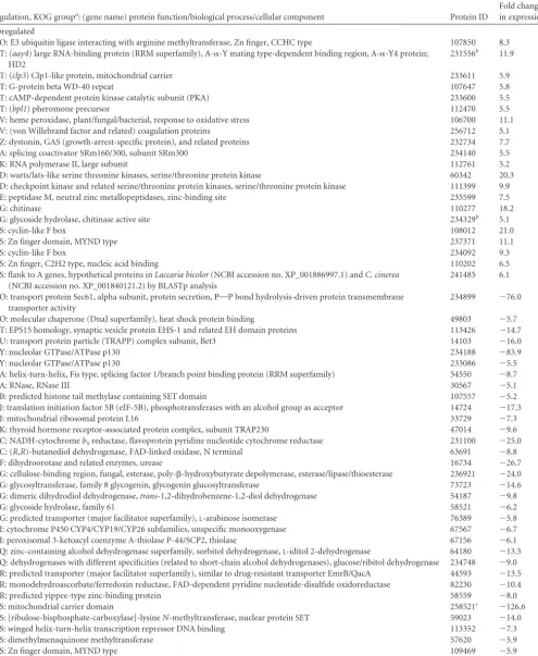
TABLE 2Identification of genes differentially expressed underA-regulated development ordered by KOG group
Regulation, KOG groupa: (gene name) protein function/biological process/cellular component Protein ID
Fold change in expression
Upregulated
O: E3 ubiquitin ligase interacting with arginine methyltransferase, Zn finger, CCHC type 107850 8.3
T: (aay4) large RNA-binding protein (RRM superfamily), A-␣-Y mating type-dependent binding region, A-␣-Y4 protein; HD2
231556b 11.9
T: (clp3) Clp1-like protein, mitochondrial carrier 233611 5.9
T: G-protein beta WD-40 repeat 107647 5.8
T: cAMP-dependent protein kinase catalytic subunit (PKA) 233600 5.5
T: (bpl1) pheromone precursor 112470 5.5
V: heme peroxidase, plant/fungal/bacterial, response to oxidative stress 106700 11.1
V: (von Willebrand factor and related) coagulation proteins 256712 5.1
Z: dystonin, GAS (growth-arrest-specific protein), and related proteins 232734 7.7
A: splicing coactivator SRm160/300, subunit SRm300 234140 5.5
K: RNA polymerase II, large subunit 112761 5.2
D: warts/lats-like serine threonine kinases, serine/threonine protein kinase 60342 20.3
D: checkpoint kinase and related serine/threonine protein kinases, serine/threonine protein kinase 111399 9.9
E: peptidase M, neutral zinc metallopeptidases, zinc-binding site 235599 7.5
G: chitinase 110277 18.2
G: glycoside hydrolase, chitinase active site 234329b 5.1
S: cyclin-like F box 108012 21.0
S: Zn finger domain, MYND type 237371 11.1
S: cyclin-like F box 234092 9.3
S: Zn finger, C2H2 type, nucleic acid binding 110202 6.5
S: flank to A genes, hypothetical proteins inLaccaria bicolor(NCBI accession no. XP_001886997.1) andC. cinerea
(NCBI accession no. XP_001840121.2) by BLASTp analysis
241485 6.1
O: transport protein Sec61, alpha subunit, protein secretion, POP bond hydrolysis-driven protein transmembrane transporter activity
234899 ⫺76.0
O: molecular chaperone (DnaJ superfamily), heat shock protein binding 49803 ⫺5.7
T: EPS15 homology, synaptic vesicle protein EHS-1 and related EH domain proteins 113426 ⫺14.7
U: transport protein particle (TRAPP) complex subunit, Bet3 14103 ⫺16.0
Y: nucleolar GTPase/ATPase p130 234188 ⫺83.9
Y: nucleolar GTPase/ATPase p130 233086 ⫺5.5
A: helix-turn-helix, Fis type, splicing factor 1/branch point binding protein (RRM superfamily) 54550 ⫺8.7
A: RNase, RNase III 30567 ⫺5.1
B: predicted histone tail methylase containing SET domain 107557 ⫺5.2
J: translation initiation factor 5B (eIF-5B), phosphotransferases with an alcohol group as acceptor 14724 ⫺17.3
J: mitochondrial ribosomal protein L16 33729 ⫺7.3
K: thyroid hormone receptor-associated protein complex, subunit TRAP230 47014 ⫺9.6
C: NADH-cytochromeb5reductase, flavoprotein pyridine nucleotide cytochrome reductase 231100 ⫺25.0
C: (R,R)-butanediol dehydrogenase, FAD-linked oxidase, N terminal 63691 ⫺8.8
F: dihydroorotase and related enzymes, urease 16734 ⫺26.7
G: cellulose-binding region, fungal, esterase, poly--hydroxybutyrate depolymerase, esterase/lipase/thioesterase 236921 ⫺24.0
G: glycosyltransferase, family 8 glycogenin, glycogenin glucosyltransferase 73723 ⫺14.6
G: dimeric dihydrodiol dehydrogenase,trans-1,2-dihydrobenzene-1,2-diol dehydrogenase 54187 ⫺9.8
G: glycoside hydrolase, family 61 58521 ⫺6.2
G: predicted transporter (major facilitator superfamily),L-arabinose isomerase 76389 ⫺5.8
I: cytochrome P450 CYP4/CYP19/CYP26 subfamilies, unspecific monooxygenase 67567 ⫺6.7
I: peroxisomal 3-ketoacyl coenzyme A-thiolase P-44/SCP2, thiolase 67156 ⫺6.1
Q: zinc-containing alcohol dehydrogenase superfamily, sorbitol dehydrogenase,L-iditol 2-dehydrogenase 64180 ⫺13.5
Q: dehydrogenases with different specificities (related to short-chain alcohol dehydrogenases), glucose/ribitol dehydrogenase 234748 ⫺9.0
R: predicted transporter (major facilitator superfamily), similar to drug-resistant transporter EmrB/QacA 44593 ⫺13.5
R: monodehydroascorbate/ferredoxin reductase, FAD-dependent pyridine nucleotide-disulfide oxidoreductase 82230 ⫺10.4
R: predicted yippee-type zinc-binding protein 58559 ⫺8.0
S: mitochondrial carrier domain 258521c ⫺126.6
S: [ribulose-bisphosphate-carboxylase]-lysineN-methyltransferase, nuclear protein SET 59023 ⫺14.0
S: winged helix-turn-helix transcription repressor DNA binding 113352 ⫺7.3
S: dimethylmenaquinone methyltransferase 57620 ⫺5.9
S: Zn finger domain, MYND type 109469 ⫺5.9
aKOG groups are defined in the legend to Fig. 10.
b
Additionally upregulated in thin phenotype. cAdditionally downregulated in thin phenotype.
on September 8, 2020 by guest
http://ec.asm.org/
signal transduction via 3
=
-5
=
-cyclic AMP (cAMP)
phosphodies-terase and small G proteins (for example,
S. cerevisiae
Pde1 [ID
105393]; GAP [ID 108327], and a signaling protein [ID 62504]).
Fewer downregulated genes were detected, and these were mostly
classified to either the metabolism of carbohydrates (IDs 31488,
55688, 13089, 70398, 108884, 13397, 110470, 109961, 236244),
transcription (IDs 61956, 35685, 65707, 114395, 256713), or
post-translational modification, protein turnover, and chaperones
(IDs 56996, 17256, 83759) (Table 3).
Intracellular modification of pheromone signaling.
To
ob-tain more information on cellular pathways involved in sexual
development, we investigated several mutant strains affected in
mating interactions. The influence of Thn1 was examined by the
expression profiling of a homokaryotic
S. commune
strain
express-ing a thin phenotype. The 114 regulated genes (72 up, 42 down)
(Fig. 10; see Tables S7 and S8 in the supplemental material)
showed high overlap with cellular responses, in accordance with
the known RGS function of Thn1 as a repressor of
G-protein-coupled signaling. Upregulated genes included those
influenc-ing processinfluenc-ing and posttranslational modification or genes
with a stress-related protein function. Proteins involved in
bio-genesis of the cell wall or membrane turnover were identified
among both the up- and downregulated genes, suggesting
re-programming of the cells via genetic means (Table 4; see also
Tables S7 and S8 in the supplemental material). Among the
upregulated genes, we also found several candidates for
chiti-nases (IDs 85084, 85210, 46134, 79630) and other glycoside
hydrolases (family 61, IDs 60863, 16233, 41145; family 5, ID
16928; others, ID 234329), suggesting functions associated
with a reorganization of the cell wall. The expression of
thn1
itself was highly downregulated in a loss-of-function
thn
mu-tant compared to a wild-type homokaryon (Table 4).
The truncation of the intracellular C terminus of the receptor
would be predicted to interfere with some intracellular
protein-protein interactions upon pheromone stimulation. Only 25 genes
with predicted function (46 in total) were altered in expression at
least 5-fold (Fig. 10; Table 5; see also Tables S9 and S10 in the
supplemental material). A strong effect on genes involved in
cel-lular processes and signaling was observed, with changes ranging
from 10- to 60-fold in magnitude. Of specific interest was the
finding that a small GTPase involved in nuclear protein import
(ID 43735) was downregulated more than 1,000-fold (Table 5).
DISCUSSION
The localization of the pheromone receptor protein in hyphae of
the fungus
S. commune
provides an important link between
pher-omone perception and the cellular responses to that signal (55). In
this study, the pheromone receptor was visualized at the cell
pe-riphery, consistent with a localization in the plasma membrane.
No evidence of nuclear membrane localization, as has been
pro-posed in some previous hypotheses, was found (5, 60). Another
prediction of these models was that pheromone receptors would
be expected to be localized in the plasma membrane close to the
encoding nucleus by virtue of localized expression and
incorpo-ration. We did not observe an increased occurrence of
fluores-cence label in areas close to one of the nuclei in the dikaryotic cells.
The involvement of pheromone activity in nuclear pairing could
not be verified by receptor localization
in vivo
and
in situ
.
Prior unsuccessful attempts to localize the pheromone
recep-tors in mushroom-forming basidiomycetes with Gfp labeling
were attributed to the low level of expression of the
B
genes, also
confirmed in this study. For these reasons, it seemed of vital
im-portance to determine the critical parameters necessary for high
receptor expression under natural conditions. This goal was
greatly facilitated by the induction of mating in many hyphal
com-partments at the same time through the setup consisting of a
top-to-top sandwich formed from two pregrown mycelia. This
experimental system allowed us to define the period of highest
expression to a time between 6 and 12 h in mating interactions of
compatible strains. To the best of our knowledge, this is the first
time that a pheromone receptor has been localized in the hyphae
of a mushroom-forming fungus.
Because the
B
genes regulate both nuclear migration and the
fusion of clamp cells, pheromone activity could be envisioned to
play a key role in the upregulation of pheromone expression in as
yet unfused clamps. It follows that pheromone receptors would be
expected to be localized to the plasma membrane of
pseudoc-lamps and to the hyphal cell close to the fusion site. However,
localization studies were hampered by both the low expression
level of the pheromone receptors and the transient nature of hook
cell formation, which is normally completed along with a fused
clamp connection within seconds (59). The unexpected finding
that clamp cell fusion was inhibited in mated mycelium of
recep-tor transformants carrying a C-terminally truncated receprecep-tor gene
enabled us to detect a fluorescence signal predominantly in
un-fused pseudoclamps. The Gfp-tagged receptor could be clearly
localized at the cell periphery in association with the plasma
mem-brane. In addition, septa of the clamped mycelia gave a strong
signal. Mislocalization of the tagged receptor seems unlikely
be-cause transformants of the
B
nullmutant integrating this construct
exhibited the normal self- versus non-self-recognition phenotype
characteristic of wild-type cells. Clamp connections were formed
more slowly than wild-type mating interactions, but this was also
observed in nontagged truncated and full-length receptor
trans-formants. While the transformants did contain mostly one copy of
the respective gene (but in one case the transformant contained up
to five copies of the respective gene), a difference in phenotype was
not connected to copy number, neither for number of
pseudoc-lamps formed or fruit body formation nor for formation of
anucleate spores.
In our receptor transformants of the
B
nullrecipient strain, the
distribution of nuclei in mated mycelia was disturbed, with one
nucleus being trapped in the unfused clamp cell and the other
being trapped in the main hyphal compartment. Thus, two
mo-nokaryotic compartments were formed, resulting from the failure
to achieve the dikaryotic condition after the initiation of mitosis
and clamp formation. The strong fluorescence visible in
pseudoc-lamps is an indication for mating competence associated with
higher levels of receptor within that cellular structure. A
fluores-cent signal would be visible only in those unfused pseudoclamps
trapping a transformed, Gfp-receptor-encoding nucleus and not
those trapping an untransformed nucleus. Given that we did not
observe fluorescence in every unfused pseudoclamp that had
trapped a nucleus, this observation supports the idea of
position-ing of either nucleus within the newly formed clamp. However,
we did not observe a regular, alternating distribution for the
nuclei going into the hook cells of
S. commune
, as has been
demonstrated for
C. cinerea
(24). Rather, nuclear distribution
is mixed in accordance with the possibility of nuclei randomly
Mating in the BasidiomyceteSchizophyllum commune
on September 8, 2020 by guest
http://ec.asm.org/
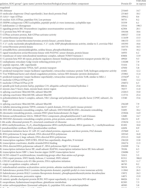
TABLE 3Identification of genes differentially expressed underB-regulated development ordered by KOG group
Regulation, KOG groupa: (gene name) protein function/biological process/cellular component Protein ID
Fold change in expression
Upregulated
M: chitinase 231665 14.7
O: molecular chaperone (DnaJ superfamily), heat shock protein DnaJ 231463 18.1
O: AAA⫹-type ATPase 109412 8.6
O: nuclear AAA ATPase, peptidase S16, Lon protease 58751 8.2
O: HSP90 cochaperone CPR7/cyclophilin, peptidyl-prolylcis-transisomerase, cyclophilin type 55359 6.1
T: indoleamine 2,3-dioxygenase 33516 198.5
T: signaling protein RIC-8/synembryn (regulates neurotransmitter secretion) 62504 20.1
T: G protein beta WD-40 repeat 104106 18.6
T: GTPase activator protein, Rab GTPase activator activity 108327 13.8
T: serine/threonine protein kinase 111639 13.5
T: casein kinase (serine/threonine/tyrosine protein kinase), protein kinase 106153 13.5
T: 3=,5=-cyclic nucleotide phosphodiesterase, 3=,5=-cyclic AMP phosphodiesterase activity, similar toS. cerevisiaePde1 105393 10.6
T: serine/threonine protein kinase 103735 10.5
T: armadillo/beta-catenin/plakoglobin, uridine kinase, phosphoribulokinase 71870 10.2
T: signal transduction serine/threonine kinase with PAS/PAC sensor domain, protein kinase 16935 6.2
U: translocase of outer mitochondrial membrane complex, subunit TOM37/metaxin 1 83604 13.9
U: G-protein beta WD-40 repeat, prolactin regulatory element-binding protein/protein transport protein SEC12p 49920 9.3
U: endoplasmic reticulum-Golgi vesicle-tethering protein p115 110288 7.9
U: peptide exporter, ABC superfamily 51416 7.4
U: vacuolar sorting protein VPS24, Snf7, protein transport 47850 5.8
V: predicted transporter (major facilitator superfamily), tetracycline resistance protein TetB, hydrogen antiporter activity 41682 12.2
V: Von Willebrand factor and related coagulation proteins, various SH3 domains (protein destination) 255861 11.4
V: predicted transporter (major facilitator superfamily), tetracycline resistance protein TetB, similar to Mfs1.1 233429b 5.2
Y: nucleolar GTPase/ATPase p130 51001 12.2
Y: nucleolar GTPase/ATPase p130 256072 6.3
Z: spindle pole body protein Sad1p, peptidase C19, ubiquitin carboxyl-terminal hydrolase 2 80616 25.6
Z: myosin class V heavy chain, myosin head, motor region 70377 11.0
A: splicing coactivator SRm160/300, subunit SRm300 232813 33.0
A: splicing coactivator SRm160/300, subunit SRm300 113731 25.4
A: polyadenylation factor I complex, subunit, Yth1 (cleavage and polyadenylation specific factor [CPSF] subunit), Zn finger
13540 15.6
A: splicing coactivator SRm160/300, subunit SRm300 256249 7.9
A: tuftelin-interacting protein TIP39, contains G-patch domain, D111/G patch (mouse protein) 36337 5.7
B: SWI-SNF chromatin remodeling complex, Snf5 subunit, SNF5/SMARCB1/INI1, chromatin remodeling 53495 18.2
B: chromatin remodeling protein, contains PHD (plant homeodomain) Zn finger 233026 14.9
B: histone acetyltransferase SAGA, TRRAP/TRA1 component, phosphatidylinositol 3 and 4 kinases 12680 14.7
B: SWI/SNF chromatin-remodeling complex protein, prion protein, aminoacyl-tRNA synthetase 106231 6.9
J: 60s acidic ribosomal protein P1, structural constituent of ribosome 233634 11.5
J: tRNA methyltransferase,N2,N2-dimethylguanosine tRNA methyltransferase, tRNA (guanine-N2-)-methyltransferase 81492 10.0
J: pseudouridylate synthase, tRNA pseudouridine synthase 42694 8.9
J: translation initiation factor 2C (eIF-2C) and related proteins, argonaute and dicer protein, PAZ domain 257069 6.1
K: RNA polymerase II, large subunit, DNA-directed RNA polymerase 105543 16.8
K: RNA polymerase I, large subunit, DNA-directed RNA polymerase 255115 14.8
K: transcription regulator XNP/ATRX, DEAD box superfamily, shugoshin, N terminal 256320 13.8
K: transcription coactivator, double-stranded RNA binding 104274 11.5
K: DNA-directed RNA polymerase subunit E=, RNA polymerase Rpb7, N terminal 234509 7.6
K: transcription initiation factor IIF, small subunit (RAP30); transcription initiation factor IIF, beta subunit 78184 6.5
K: transcription factor XBP-1, basic leucine zipper (bZIP) transcription factor 236086 5.7
K: GCN5-relatedN-acetyltransferase, transferring groups other than amino acyl groups 52412 5.5
L: DNA repair protein, SNF2 family, helicase, C terminal, SNF2 related 81511 306.0
L: CDC45 (cell division cycle 45)-like protein, DNA replication initiation 50273 11.7
L: origin recognition complex, subunit 4 46745 8.2
C: mitochondrial carnitine-acylcarnitine carrier protein, adenine nucleotide translocator 1 58334 17.4
D: halotolerance protein HAL3 (contains flavoprotein domain), peptidyl-prolylcis-transisomerase activity 113841 26.5
D: halotolerance protein HAL3 (contains flavoprotein domain), phosphopantothenoylcysteine decarboxylase 15072 24.3
D: Mis12, chromosome, pericentric region 14071 15.9
D: mitotic spindle checkpoint protein BUB3, WD repeat superfamily, G-protein beta WD-40 repeat 86026 5.5
E: oxoprolinase, hydantoinase/oxoprolinase, glutathione metabolism 82383 7.3
E: serine carboxypeptidases (lysosomal cathepsin A), peptidase S10, serine carboxypeptidase 40585 5.8
E: Xaa-Pro aminopeptidase, peptidase M24 58445 5.2
(Continued on following page)
on September 8, 2020 by guest
http://ec.asm.org/
TABLE 3(Continued)
Regulation, KOG groupa: (gene name) protein function/biological process/cellular component Protein ID
Fold change in expression
G: glucose-6-phosphate/phosphate and phosphoenolpyruvate/phosphate antiporter 36941 22.9
G: general substrate transporter, sugar transporter superfamily 66123 14.4
G: esterase, poly--hydroxybutyrate depolymerase, esterase/lipase/thioesterase, carbohydrate esterase family 1 protein 47380 12.1
G: inositol monophosphatase 47747 7.7
G: beta-1,6-N-acetylglucosaminyltransferase, contains WSC domain 110551c 7.5
G: glycoside hydrolase, family 43 232782 6.6
I: acyl coenzyme A:diacylglycerol acyltransferase (DGAT) 53861 21.1
I:S-adenosylmethionine-dependent methyltransferases, generic methyltransferase 14559 13.5
I: cytochrome P450 CYP4/CYP19/CYP26 subfamilies, E-class P450, group I, gamma-hexachlorocyclohexane degradation, ascorbate and aldarate metabolism
233430 11.6
I: cytochrome P450 CYP4/CYP19/CYP26 subfamilies, unspecific monooxygenase, tryptophan metabolism, fatty acid metabolism
76871 6.0
P: Ca2⫹/H⫹antiporter VCX1 and related proteins, sodium/calcium exchanger membrane region, calcium/proton
exchanger
27729 9.4
P: Ca2⫹transporting ATPase, potassium/sodium efflux P-type ATPase, fungal type 53464 5.4
Q: multidrug/pheromone exporter, ABC superfamily 113902 53.4
Q: multidrug/pheromone exporter, ABC superfamily 258386 5.0
R: Zn finger, C2H2 type, similar to vegetative cell wall protein gp1 precursor (hydroxyproline-rich glycoprotein 1) 113591 19.2
R: WD-40 repeat-containing protein 84316 9.0
R: Zn finger, cytochromecheme-binding site, electron transport activity 233610 5.7
S: UbiA prenyltransferase, prenyltransferase activity 238827 23.8
S: chloroperoxidase, peroxidase activity, electron transport 57566 21.7
S: cyclin-like F-box domain 111277 18.4
S: cyclin-like F box, Zn finger, C2H2 type 104797 8.7
S: glutathioneS-transferase, N terminal 111982 8.3
S: bucentaur or craniofacial development 112020 8.2
S: protein binding, BTB/POZ domain 236141 7.8
S: bacterial extracellular solute-binding protein, family 3, transporter activity 105400 7.7
S: predicted membrane protein 108226 7.7
S: Zn finger, MYND type 231200 7.1
S: target SNARE coiled-coil region 56068 6.5
S: cyclin-like F box, Zn finger, C2H2 type 234065 6.3
S: Zn finger, CCHC type 237097 6.0
S: notchless-like WD-40 repeat-containing protein, 2-acetyl-1-alkylglycerophosphocholine esterase, G-protein beta WD-40 repeat
37220 5.2
Downregulated
O: alkyl hydroperoxide reductase/peroxiredoxin 56996 ⫺20.1
O: E3 ubiquitin ligase interacting with arginine methyltransferase, Zn finger, CCHC type 83759 ⫺19.5
O: peptidase M28, transferrin receptor and related proteins containing the protease-associated (PA) domain 17256 ⫺11.3
T: serine/threonine protein kinase 108101 ⫺8.5
T: (brl3) fungal pheromone STE3 6-protein-coupled receptor, mating-type alpha-factor pheromone receptor activity 258344 ⫺5.6
T: (bbp2) pheromone precursor 12028 ⫺14.2
U: nuclear pore complex, Nup98 component (sc Nup145/Nup100/Nup116), ribosomal protein L9 N-terminal like 112737 ⫺5.9
U: clathrin adaptor complex, medium chain, adaptor complexes medium subunit family 61803 ⫺5.2
K: fungal transcriptional regulatory protein, N-terminal, fungus-specific transcription factor, DNA binding, zinc ion binding
65707 ⫺12.8
K: transcription factor activity, similar to Cu-dependent DNA-binding protein, copper fist DNA binding 61956 ⫺9.4
K: fungus-specific transcription factor, DNA binding, zinc ion binding 35685 ⫺7.3
K: peptidase M, neutral zinc metallopeptidases, zinc-binding site, selective LIM domain binding factor 114395 ⫺6.9
K: GATA-4/5/6 transcription factors, Zn finger, GATA type 256713 ⫺6.2
C: aldehyde dehydrogenase, tyrosine metabolism, glycolysis/gluconeogenesis 258124 ⫺7.7
C: kynurenine 3-monooxygenase and related flavoprotein monooxygenases, salicylate 1-monooxygenase 238637 ⫺6.5
G: gluconate transport-inducing protein 108884 ⫺25.9
G: predicted short-chain-type dehydrogenase, glucose/ribitol dehydrogenase 236244 ⫺20.5
G: predicted short-chain-type dehydrogenase, glucose/ribitol dehydrogenase 110470 ⫺10.0
G: general substrate transporter, permease of the major facilitator superfamily 55688 ⫺9.6
G: dTDP-glucose 4-6-dehydratase/UDP-glucuronic acid decarboxylase, erythromycin biosynthesis 13089 ⫺6.8
G: permease of the major facilitator superfamily 13397 ⫺6.0
G: glycoside hydrolase family 23 protein, candidate beta-glycosidase distantly related toN-acetylmuramidases 31488 ⫺5.4
G: alpha-amylase 70398 ⫺5.2
(Continued on following page) Mating in the BasidiomyceteSchizophyllum commune