ANALYSIS OF GENE REARRANGEMENTS AND PROTEIN
EXPRESSION OF THE TUMOUR SUPPRESSOR GENES,
RB^
AND p16, IN ACUTE MYELOID LEUKAEMIA: POSSIBLE
ROLES IN LEUKAEMOGENESIS
by
A.RAHMAN A.JAMAL
A thesis submitted for the degree of Doctor of Philosophy at the University of London
supervised by
Professor David C.Linch Department of Haematology
University College London Medical School London
ProQuest Number: 10017418
All rights reserved
INFORMATION TO ALL USERS
The quality of this reproduction is dependent upon the quality of the copy submitted.
In the unlikely event that the author did not send a complete manuscript and there are missing pages, these will be noted. Also, if material had to be removed,
a note will indicate the deletion.
uest.
ProQuest 10017418
Published by ProQuest LLC(2016). Copyright of the Dissertation is held by the Author.
All rights reserved.
This work is protected against unauthorized copying under Title 17, United States Code. Microform Edition © ProQuest LLC.
ProQuest LLC
789 East Eisenhower Parkway P.O. Box 1346
ACKNOWLEDGEMENTS
I would like to mention my co-sponsors, the Universiti Kebangsaan Malaysia (National University of Malaysia) and the Public Services Department of Malaysia, for the scholarship award.
I am indebted to Professor David 0. Linch for giving me the opportunity to undertake this Ph.D project and for his excellent supervision and moral support despite his hectic schedule.
I would also like to convey many special thanks to Dr.Rosemary E. Gale and Dr.Shaun Thomas for their wholehearted tutelage and for sharing their vast experience and scientific knowledge.
My appreciation as well to everyone in the department who have made my life as a scientist in Chenies Mews an unforgettable experience. Thank you for the top tips!
ABSTRACT
Carcinogenesis is multi-step process involving the activation of oncogenes and/or inactivation of tumour suppressor genes. One of these tumour suppressor genes is the human retinoblastoma susceptibility gene, RB1,
TABLE OF CONTENTS
Thesis title... 1
Acknowledgements... 2
Abstract... 3
Table of contents... 4
List of figures... 9
List of tables... 11
CHAPTER ONE GENERAL INTRODUCTION 1.1 Carcinogenesis... 12
1.2 Myelopoiesis and the molecular basis of leukaemia... 13
1.3 Tumour suppressor genes... 20
1.4 The human retinoblastoma susceptibility gene (RB1)... 22
1.4.1 The RB protein (pRB) and cell cycle control... 23
1.4.2 The role of pRB in differentiation... 31
1.4.3 The role of pRB in apoptosis... 32
1.4.4 The role of RB1 in tumour formation and progression 33
1.5 The role of cyclin dependent kinase inhibitors (CDKIs) in cell cycle regulation... 35
1.6 p75and p75genes... 37
1.6.1 p i 6 protein function in the cell cycle... 38
1.7 Oncogenic potential of GI checkpoint control...“... 40
1.8 Aims of study... 41
CHAPTER TWO MATERIALS AND METHODS 2.1 Patients and normal controls... 43
2.2 DMA extraction... 43
2.3 RNA extraction... 44
2.4 Preparation of protein lysates... 45
2.5 Southern blotting analysis... 45
2.7 Immunoprécipitation... 48
2.8 Cell culture... 49
2.9 Polymerase Chain Reaction... 50
2.10 Reverse Transcription-PCR... 51
2.11 Sequencing of PCR and RT-PCR products... 51
CHAPTER THREE ANALYSIS OF REARRANGEMENTS IN THE RB1 GENE IN ACUTE MYELOID LEUKAEMIA PATIENTS 3.1 Introduction... 52
3.2 Materials and Methods... 56
3.2.1 AML patients and normal controls... 56
3.2.2 Southern blotting analysis and Polymorphisms ... 57
3.2.3 Optimisation of conditions for RB1 Southerns... 59
3.2.2.1 Testing different RB1 cDNA fragments as probes and establishing the germline bands 59 3.2.2.2 Determining the amount of genomic DNA to be used in digest... 60
3.2.2.3 Establishing positive and negative controls 60 3.2.2.4 Comparing 2 different membranes... 62
3.2.2.5 [a-32p]dATP versus [a-32p]dCTP for labelling probes... 62
3.2.2.6 Comparing 3 different hybridisation protocols... 62
3.2.4 Testing the sensitivity of the Southern blotting technique... 64
3.2.5 Southern blot analysis of AML patients...64
3.3 Results... 64
3.3.1 Germline configuration in normal controls... 64
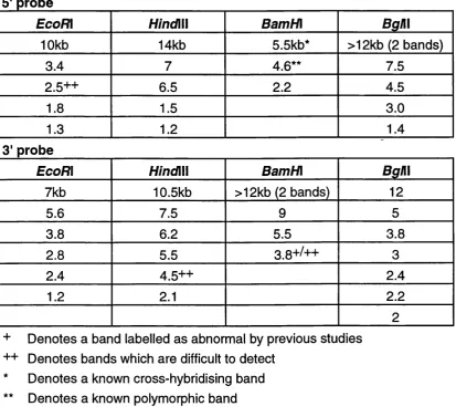
3.3.2 Polymorphisms in the RB1 gene... 67
3.3.3 Sensitivity of detection of bands by Southern blotting 69
3.3.4 Southern blotting results of 106 AML patients... 69
3.3.5.2 Southern blotting of samples using the RB1
promoter region as a probe... 75 3.3.5.3 RT-PCR of exons 1-9... 76 3.3.5.4 Southern blotting analysis using
3 different PCR probes... 76 3.4 D iscussion... 81
CHAPTER FOUR
ANALYSIS OF PROTEIN EXPRESSION OF THE RETINOBLASTOMA GENE IN PATIENTS WITH ACUTE MYELOID LEUKAEMIA
4.1 Introduction... 89 4.2 Materials and Methods... 92 4.2.1 The western blotting technique... 93 4.2.2 Showing the different phosphorylation states of pRB
during the cell cycle... 93 4.3 Results... 93 4.3.1 pRB expression in cell lines... 93 4.3.2 Protein loading and integrity in the AML samples 95 4.3.3 pRB expression in normal controls and AML patients 97 4.3.4 Complete remission and survival data of AML patients.... 100 4.4 Discussion... 100
CHAPTER FIVE
THE ANALYSIS OF DELETIONS AND REARRANGEMENTS IN THE p16
GENE USING MULTIPLEX POLYMERASE CHAIN REACTION AND QUANTITATIVE SOUTHERN BLOTTING TECHNIQUE
5.1 Intro d u ctio n ... I l l 5.2 Materials and M ethods... 117
5.2.1 Setting up the multiplex PCR... 117 5.2.1.1 PCR of exon 1 or 2 of the p 75 gene... 117 5.2.1.2 Multiplex PCR of exon 1 or 2 with an
5.2.1.3 Multiplex PCR of both exons 1 and 2 with an
internal control... 120
5.2.1.4 Evaluating the contribution of non-leukaemic cells in the PCR analysis of leukaemic samples... 122
5.2.2 Quantitative Southern blotting analysis... 124
5.2.2.1 Introduction... 124
5.2.2.2 Preparing the p 76 gene probe... 126
5.2.2.5 Southern blot analysis using different enzymes digests and the incorporation of an internal control... 126
5.2.2.4 Quantification of bands... 127
5.2.2.5 Optimisation of quantitative Southern blotting analysis... 127
5.2.2.6 Testing the sensitivity of quantitative Southern blotting... 131
5.2.2.7 Quantitative Southern blotting analysis of 18 normal controls... 131
5.2.2.B Quantitative Southern blotting analysis of 76 AML patients... 133
5.3 Results... 133
5.4 Discussion... 133
CHAPTER SIX ANALYSIS OF THE p 1 6 GENE FOR POINT MUTATIONS AND POLYMORPHISMS USING RT-PCR-SSCP TECHNIQUE 6.1 Introduction... 139
6.2 Materials and Methods... 142
6.2.1 RT-PCR... 142
6.2.2 SSCP analysis... 143
6.2.3 Sequencing of RT-PCR products... 143
6.3 Results... 143
6.3.1 Results of SSCP analysis using 7 different RT-PCRs 143
6.3.2 Results of sequencing of samples showing at least two abnormal SSCP patterns... 146
CHAPTER SEVEN
THE STATUS OF p16 PROTEIN EXPRESSION IN AML PATIENTS AND THE CORRELATION BETWEEN pRB AND p16 DATA
7.1 Introduction... 151
7.2 Materials and Methods... 153
7.2.1 p i 6 western blotting... 153
7.2.2 CDK4 western blotting... 153
7.2.3 Analysis of p i 6 protein expression in different cell lines and during differentiation... 154
7.2.4 The specificity of p i 6 by western blotting... 154
7.2.4.1 Peptide inhibition experiment... 154
7.2.4.2 In-vitro translation at p16 cDNA followed by immunoprécipitation... 156
7.2.4.3 Analysing the méthylation status of the 5' CpG island of p 76 gene in samples with reduced p i 6 protein expression... 156
7.3 Results... 157
7.3.1 p i 6 protein expression in AML patients... 157
7.3.2 p i 6 protein expression in cell lines... 157
7.3.3 pRB levels in AML patients with altered p i 6 expression...159
7.3.4 CDK4 protein levels in AML patients with altered p i 6 expression... 159
7.3.5 Méthylation status of the 5' CpG island in cases with reduced p i 6 expression... 163
7.4 Discussion... 163
CHAPTER EIGHT CONCLUSIONS... 168
REFERENCES... 174
ABSTRACTS AND SUBMISSIONS... 208
LIST OF FIGURES
No. Figure title Page
1.1 Schematic diagram of myelopoiesis... 14 1.2 Oncogenes known to be activated in leukaemia and their
roles in signal transduction pathways... 16 1.3 Two-hit mutation of the RB1 gene in retinoblastoma 24 1.4 Cell cycle progression and the phosphorylation of pRB by
cyclins and cyclin dependent kinases... 25 1.5 The binding of pRB to E2F represses the transcription of
genes involved in cell proliferation... 30 1.6 p i 6 inhibits CDK4/6 to prevent the phosphorylation of pRB
and progression of cells from Gi into the 8 phase... 39
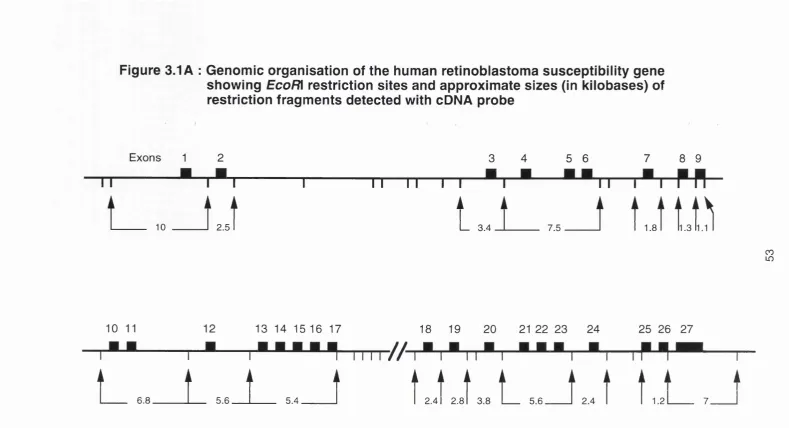
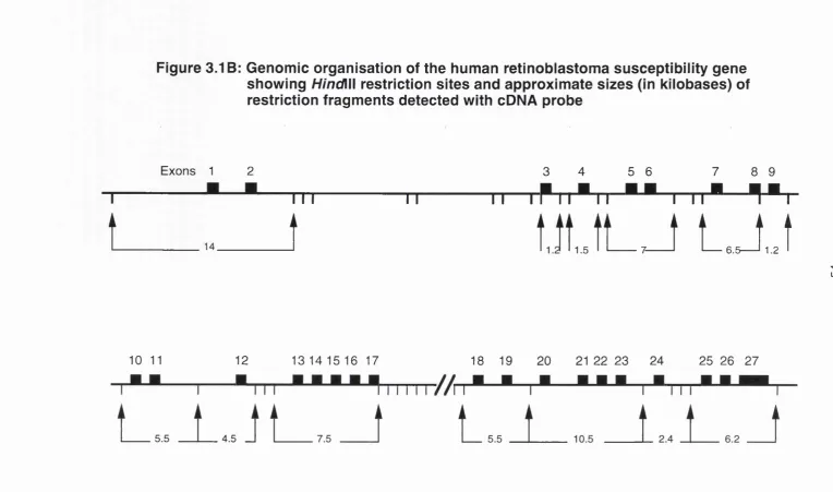
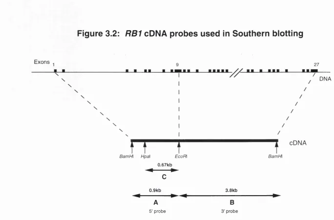
3.1 A&B Genomic map of RB1 and restriction sites for EcoRl and
HindWl...53&S4 3.2 cDNA probes used for RB1 Southern blotting... 61 3.3A&B Germline bands of RB1 in normal controls detected by
Southern blotting analysis (S' probe and 3' probe)... 6S&66 3.4 BamH\ polymorphism in the RB1 gene... 68 3.5 Polymorphisms in the RB1 gene... 70 3.6 Sensitivity of Southern blotting using BamH\ polymorphic
site as marker... 71 3.7 Representative Southern blots showing rearranged bands
consistently detected in 5 AML patients using digestion
with both EcoRl and HindWl... 72 3.8 Novel bands detected in single enzyme digest... 74
3.9 PGR probes used in Southern blotting analysis of AML
patients with the 'common' rearrangements...77 3.10 Southern blotting using the RB1 promoter as a probe 78 3.11 A&B Southern blotting analysis using 4 different probes of
2 AML patients with common rearrangements... 79&80
4.1 RB protein structure showing phosphorylation sites and
4.4 Spectrum of pRB expression in AML patients...98 4.5 Western blot of 8 patients using Pab-2 anti-RB antibody 99 4.6 Kaplan-Meier survival curves of patients with normal
and abnormal pRB... 101 4.7 Possible role of RB1 mutation in leukaemia progression 108
5.1 Genomic organisation of p 76 gene... 113 5.2A&B PCR of exon 1 or 2 of the p 76 gene... 119 5.3A&B Multiplex PCR of exon 1 or exon 2 using globin as
internal control... 121 5.4 Multiplex PCR of exons 1 and 2 using globin as
an internal control... 123 5.5 Amplification of non-leukaemic cells in leukaemic samples.. 125 5.6 Germline p i 6 bands in 4 different enzymes using p76
and c-fms probes... 128 5.7 Quantitative Southern blotting: Optimisation of p76
and c-/ms probes... 129 5.8 Southern blot and graph showing the sensitivity of p76
detection and linearity of signal intensity... 132 5.9 Representative Southern blot of 3 AML patients using
EcoRl digests and probed with p 76 and c-fms probes 134
6.1 Basis of SSCP analysis technique... 141 6.2 RT-PCR-SSCP analysis of exon 2 p76 gene using
3 different gel running conditions... 144 6.3A&B SSCP analysis of 16 AML patients showing variability
in the mobility shifts using 6 different RT-PCR reactions 147
7.1 Specificity of p i 6 western blotting... 155 7.2 Variable p i 6 expression in AML patients... 158 7.3 p i 6 expression in cell lines with and without
differentiating agents... 160 7.4 Absence of p i 6 expression in J6 and HL60 cell lines... 161 7.5 CDK expression in 9 samples with abnormal
p i 6 expression... 162 7.6 Southern blot showing méthylation status of CpG island
in 6 cases with reduced p i 6 expression... 164 7.7 Feedback loop between pRB and pi 6... 166
LIST OF TABLES
Table no. Title Page
1.1 The different cyclins, their preferential CDK partners
and site of action in the cell cycle... 28
3.1 Frequency of RB1 gene rearrangements in various
primary solid tumours... 55 3.2 Frequency of RB1 gene rearrangements in AML... 56 3.3 Breakdown of AML patients according their FAB types 57 3.4 RB1 Germline bands detected by Southern blotting 67 3.5 AML patients with their respective additional bands
seen on different enzyme digests... 73
4.1 Frequency of pRB inactivation in solid tumours... 92 4.2 Studies of pRB expression in AML... 92 4.3 Spectrum of abnormal protein expression in
AML patients... 97 4.4 Comparison of complete remission rate and median
survival in patients with normal and abnormal pRB... 100 4.5 Frequency of pRB abnormalities in AML cases
according to their FAB types... 105
5.1 Frequency of p16 gene deletions in primary
solid tumours... 112 5.2 p16 gene deletions in haemopoietic malignancies... 114 5.3 Predicted range of intensity of p16 band in
homozygous and heterozygous deletions... 137
6.1 Results of SSCP analysis of exon 2-3 of the p16
gene using 7 different RT-PCR reactions... 145
7.1 Correlation between p i 6 protein overexpression and
pRB expression in 60 AML cases... 159
CHAPTER ONE
INTRODUCTION
1.1 Carcinogenesis
Genetic alterations play a central role in the development of cancer (Klein,
1987; Bishop, 1987) and tumour formation is believed to be a multi-step process (Knudson, 1971; Pitot et al., 1991) involving the accumulation of many genetic mutations, whereby critical molecular targets are deregulated culminating in the malignant transformation and the clonal expansion of a cell. Central to this theory are the roles of proto-oncogenes and tumour suppressor genes whose activation and inactivation respectively cause disruption of critical events in cell division, cell differentiation and cell death or apoptosis. The oncogenes and the tumour suppressor genes may well be likened to the accelerator and the brake pedal of a fast car cruising on a busy motorway. A stuck accelerator or failed brakes could lead to loss of control of the vehicle, a potential disaster for the occupants. Within the cellular context, this would mean continued and dysregulated cell proliferation leading to malignancy.
Any attempt to understand the process of carcinogenesis requires the identification of these genes and knowledge of their fundamental roles and behaviour both in health and disease. The advances in cytogenetics and molecular biology have allowed identification, detailed characterisation and analysis of many of these genes. Developments in protein biochemistry have also enabled the nature and cellular functions of the products of these genes to be elucidated. Within this framework, it is equally crucial to analyse the interaction between genes as well as protein-protein interaction with regard to their specific and accumulative contribution to tumour initiation and/or progression so as to enable us to understand further the biology of disease and to design novel approaches to cancer therapy in the future.
The multi-step model of carcinogenesis was put forward based on both cancer epidemiology data and experimental observations. There is an increase in cancer incidence with age, suggesting that multiple genetic alterations accumulate with time (Cook at a!., 1969). Studies in transgenic mice have shown that overexpression of the oncogene c-myc alone does not cause the development of breast tumours until late in life. However when H-ras, another proto-oncogene, is co-expressed the mice develop tumours at a much earlier
age (Stewart et al., 1984), confirming the prediction that supplying multiple activated or unregulated oncogenes should shorten the lag period to initiate tumours.
The overall process of carcinogenesis can be divided into at least 3 phases: initiation, promotion and progression. The initiation phase is the result of an irreversible genetic alteration(s) in the DNA. It is believed that the reversible stage of tumour promotion does not involve structural changes in DNA, but rather the inappropriate expression of downstream genes mediated through
promoter-receptor interactions. Finally, the irreversible phase of tumour progression is characterised by karyotypic instability and malignant growth of cells (Pitot, 1993).
1.2 Haemopoiesis and the molecular basis of leukaemia
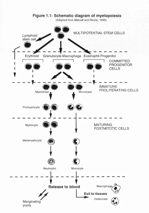
Haemopoiesis is a process of self renewal and differentiation of pluripotential stem cells. It is believed that a common pluripotential stem cell gives rise to a series of progenitor cells for each of the main lineages. A representative schematic diagram of myelopoiesis is depicted in Figure 1.1 (Metcalf and Nicola, 1995). The clonal selection and malignant proliferation of a haemopoietic progenitor cell is the basic process which gives rise to leukaemia. For example, acute myeloid leukaemia (AML) probably arises from a single abnormal myeloid progenitor cell. This disease is characterised by (1) the accumulation of abnormal (leukaemic) blast cells, principally in the bone marrow, and (2) impaired production of normal blood cells. In line with the multi-step model proposed by Knudson, leukaemogenesis, which is considered to be a special type of carcinogenesis, is thought to be at least a two-step phenomenon (Knudson, 1973; Farber and Cameron, 1980). First, a critical mutation of a proto-oncogene in a single cell leads to clonal haemopoiesis, and progression to AML is accompanied by further genetic changes, perhaps involving one or more oncogenes or tumour suppressor genes, often reflected by chromosomal abnormalities or DNA rearrangements. The activation of oncogenes and/or inactivation of tumour suppressor genes is believed to endow the leukaemic cell clone with a proliferative advantage or inhibit its normal differentiation and subsequent death (Williams eta!., 1995).
Structural aberrations in specific gene families have been shown to be consistently associated with specific phenotypes of leukaemia and a selection of these is discussed in the following paragraphs. Most of these
Figure 1.1: Schematic diagram of myelopoiesis
(Adapted from Metcalf and Nicola, 1995)
f
MULTIPOTENTIAL STEM CELLS Lymphoid
stem celL
T
Erythroid Granulocyte-Macrophage Eosinophil Progenitor
I I I I I I COMMITTED
PROGENITOR
I - - 1
_________________
^ ^ ^ ^ IMMATURE
PROLIFERATING CELLS
DXCmCXG
Myeloblast
Promyelocyte
t
t
D X G H X 0
Monoblast
i
MyelocyteMetamyelocyte
I
i
I
Neutrophil
t
€)
I
Monocyte
MATURING
POSTMITOTIC CELLS
Macrophage
Release to blood
Exit to tissues Osteoclast Marginating
pools
leukaemogenic genes are normal genes which have become oncogenic as a result of a genetic change which can either be a point mutation, gene rearrangement, deletion, amplification or fusion to other genes giving rise to deregulation or abnormal expression. Some of these genetic changes are gross, hence they can be seen at the chromosomal level by cytogenetical
analysis while others require detection by molecular methods such as Southern blotting. Yet a further subset of microscopic mutations will only be detected by sequencing the involved gene.
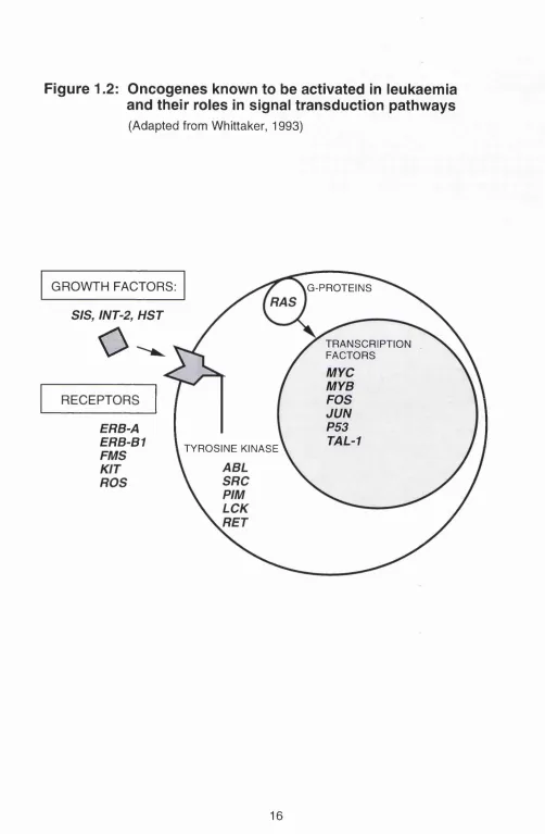
Many of the proto-oncogene products which are potentially leukaemogenic provide the vital elements in the signal transduction pathway (reviewed by Butturini and Gale, 1990; Cantley et al., 1991; Sawyers et al., 1991 ; Cline, 1994). A diagrammatic representation of some of the proto-oncogenes involved in signal transduction is shown in Figure 1.2. Some encode growth factors, like sis and hst which are genes for the platelet derived growth factor beta (PDGF-p) and the fibroblast growth factor respectively. Others like fms
and erb-b1 encode for receptors. The binding of growth factors to their receptors triggers a cascade of events involving protein kinases some of which are encoded by oncogenes such as abl, pirn, src, ras and ret. The ultimate event is the transduction of the signals to the nucleus where the genes controlling transcription, cell proliferation and cell differentiation reside, such as myc, myb, fos, and lun. Hence, within the signal transduction pathway itself each level of control may be disrupted which can be potentially disastrous for the cell.
The t(8;14)(q24;q32) translocation provides the paradigm for oncogene activation arising from a genetic event resulting in a specific leukaemia phenotype. This translocation which is characteristic of B urkitt's leukaemia/lymphoma, brings into juxtaposition the myc proto-oncogene at 8q24 with the immunoglobulin heavy chain (IgH) locus at chromosome 14 (Dalla-Favera et a!., 1982). The myc gene which normally functions as a transcription factor, is overexpressed as a result of this translocation event resulting in the excessive transcription of downstream target genes, leading to oncogenesis. Another well known example of translocation resulting in a phenotype-specific leukaemia is the t(9;22)(q34;q11) translocation present in about 95% of chronic myeloid leukaemia (CML) patients. This event translocates the abl proto-oncogene to a specific site at 22q11 known as the major breakpoint cluster region (M-BCR) (Heisterkampe et a!., 1983, Shtivelman e ta !., 1985). The resultant chimeric gene, named bcr-abi, is
Figure 1.2: Oncogenes known to be activated In leukaemia
and their roles In signal transduction pathways
(Adapted from Whittaker, 1993)
GROWTH FACTORS: G-PROTEINS
StS, INT-2, HST
o
TRANSCRIPTIONFACTORS
MYC MYB FOS JUN P53
TAL-1 ERB-A
ERB-B1 TYROSINE KINASE
transcribed as a chimeric mRNA which in turn translates into a fusion protein p210 which has an inappropriately high level of tyrosine kinase activity as compared with the product of the normal abl gene (Lugo eta!., 1990). Hence, the change in the protein expression of the leukaemogenic genes can provide a specific marker for analysing primary leukaemias.
Another unique leukaemogenic event is the t(15;17)(q22;q21) translocation
which is found in almost all cases of acute promyelocytic leukaemia (APML) (de The eta!., 1990). The gene at the breakpoint on chromosome 17 encodes the retinoic acid receptor a chain while the gene on chromosome 15 has been
called PML, for promyelocytic leukaemia. One of the postulations for the resultant effect of the translocation is that dysfunctional PML proteins are produced leading to an arrest of differentiation of promyelocytes (Mu et a!.,
1994). Interestingly, in APML, treatment with all-trans retinoic acid results in remission in the majority of patients not only at the phenotypic level but also in the disappearance of the rearrangement at the gene level (Chomienne et a!.,
1989). The t(15;17) translocation provides further evidence for the specific causal relationship between genetic events and certain leukaemias and highlights the potential of harnessing this knowledge in the management of the patient.
Another proto-oncogene which has been reported to be mutated in certain leukaemias is the fms gene. This gene encodes the receptor for the macrophage and monocyte specific growth factor CSF1 (Sherr et a!., 1985). The binding of CSF1 to the receptor activates its tyrosine kinase resulting in autophosphorylation of the receptor as well as phosphorylation of other proteins. This cascade of events ultimately triggers events which leads to mitogenesis. Point mutations and deletions of fms have been described in myelodysplasia and AML and in some cases overexpression of fms has also been reported (Ridge eta!., 1990; Tobal eta!., 1990).
The involvement of ras oncogenes in haemopoietic malignancies has also been extensively studied. These genes encode the G-proteins, a class of transcription factors involved in signal transduction. Mutations of the ras gene family (H, K and N-ras) result in abnormal protein products which have transforming activities in certain cells. In leukaemias, ras mutations have been found in up to 30% of AML cases, 40% of patients with myelodysplastic syndromes (MDS), 30% of multiple myeloma patients, 20% of ALL and has
also been reported in the acute phase of CML (Bos et al., 1987; Lyons at a!.,
1988; Neri eta!., 1989; Janssen eta!., 1987).
Genes which control for programmed cell death (apoptosis) also play a major part in haemopoietic malignancies. The bcl-2 gene is the prototype repressor of cell death (Hockenberry eta!., 1990). The gene is characteristically involved in human follicular lymphoma through the translocation t(14:18), which juxtaposes the bcl-2 gene and the immunoglobulin heavy chain gene locus on
chromosome 14 (Tsujimoto at al., 1985). This results in deregulated overexpression of bcl-2 mRNA and its protein. Studies in transgenic mice
models have shown that the presence of this chimeric bcl-2-lgH resulted in follicular hyperplasia in the lymphoid system (McDonnell at al., 1989; Korsmeyer, 1992). bcl-2 has the unique role of promoting cell survival by repressing apoptosis, rather than promoting cell proliferation.
Loss of tumour suppressor genes in leukaemia has also been widely reported. For example, a diverse variety of mutations of the p53 gene have been reported in many different leukaemias although none of the studies has been conclusive. The main evidence for p53 involvement in leukaemia is provided by CML where 30% of cases in blast crisis has been reported to have p53
mutations (Ahuja at al., 1989), suggesting a role during progression of the disease. For AML, some groups have failed to detect p53 protein expression whilst others have found overexpression up to 10-100 fold greater than normal marrow cells (Prokocimer at al., 1986; Smith at al., 1986; Fenaux at al., 1992; Koeffler at al., 1986). Point mutations and deletions have also been described in many leukaemic cell lines. The role of another tumour suppressor gene, the retinoblastoma gene, in leukaemias will be discussed later.
There are also several constitutional abnormalities that predispose to leukaemia. These include Down's syndrome (Trisomy 21) and Klinefelter's syndrome, both of which are associated with an increased risk of AML. Patients who have DNA repair disorders or fragile chromosomes also have an increased susceptibility to acute leukaemia, such as Fanconi's anaemia. Bloom's syndrome and ataxia telangiectasia. The putative gene for ataxia telangiectasia has recently been cloned and sequenced (Savitsky at al.,
1995).
Accumulative genetic events do not only contribute to the initiation of leukaemia. It is believed that further molecular lesions occur even after a cell
clone has been transformed or specifically during progression. Hence, a new clone of even more aggressive leukaemic cells may arise as a consequence of this sub-clonal selection. This could probably explain the progression of many leukaemias and lymphomas from a relatively indolent phase to a more aggressive phase. Examples include the acute blastic transformation in CML, for which the role of p53 was described earlier, and the evolution of chronic lymphocytic leukaemia into an aggressive lymphoma. It is crucial that the genetic events responsible for this change in tumour behaviour are defined so that novel leukaemia therapies may be designed to combat leukaemia progression.
The preceding simplistic review of the involvement of oncogenes and tumour
suppressor genes in haemopoietic malignancies highlights the critical role of genetic events in leukaemogenesis. Some of these events are phenotype specific whilst others appear to occur in a diverse group of leukaemias. Analysis of these events can be carried out at the DNA, RNA or the protein level of the genes concerned. An important inference is the fact that these various oncogenes and tumour suppressor genes encode for products which are the major players in signal transduction, cell proliferation and cell death.
It must be borne in mind that the concept of accumulative genetic alterations in carcinogenesis as a whole, or leukaemogenesis specifically, makes it difficult to assign the exact role for each oncogene and tumour suppressor gene in leukaemia initiation or progression. For example, although mutation of the ras gene is the most commonly detected abnormality in AML (Bos et a/., 1987; Farr et al., 1988), it may not be the initial fundamental abnormality in those 30% of cases since it may be present only in a subset of the leukaemic cells (Bashey et a!., 1992). Furthermore, AML patients with N-ras mutations have no characteristic phenotypic features, clinical patterns, or response to therapy (Radich et a/., 1990). Hence, unless it is possible to show a direct causal relationship between the alteration of an oncogene or a tumour suppressor gene with a particular leukaemia phenotype, assigning specific roles for these genes in leukaemia initiation or progression must be exercised with caution. It is possible that all of them are capable of playing both roles in a pleiotropic manner although some may show specificity for the development of specific leukaemia phenotypes.
1.3 T umour suppressor genes
In the mid 1980s, several lines of evidence were put forward to support the idea that loss of genetic information is a critical step in tumorigenesis. These genes were postulated to function as negative regulators of cell proliferation and the loss of these genes resulted in removal of the existing brakes to cell growth, thereby endowing cells with a growth advantage.
Somatic cell genetics provided the first firm evidence indicating the importance of loss of genetic information from tumour cell genomes. It was shown that fusion of tumour cells with normal non-tumorigenic cells resulted in hybrids which were generally non-tumorigenic (Harris et a/., 1969). This elegant experiment suggested that the normal partners in the fusion were supplying genes that reimposed normal growth control on the malignant cell. A more conclusive experiment was conducted by Weissman et al (1987), where it was shown that the introduction of a normal chromosome 11 into a Wilm's tumor cell line resulted in the loss of tumorigenicity.
The second line of evidence pointing towards genetic loss during tumorigenesis derived from karyotypic analysis of specific human tumours. It was nearly 17 years ago that cytogeneticists observed that retinoblastoma tumour cells had interstitial deletions affecting the q14 band of chromosome 13 (Yunis and Ramsay, 1978). Similar analysis on Wilm's tumour cells revealed a specific loss of genetic material associated with the p i 3 band of chromosome 11 (Riccardi eta !., 1980). These hereditary cancers have revealed a new class of gene that is important in the pathogenesis of cancer. These genes, which are clearly different from oncogenes, have been called tumour suppressor genes (TSG) or anti-oncogenes, because of their potential to induce cancer in a recessive mode i.e. one normal allele being sufficient to protect against a particular cancer. TSGs have been shown to be highly conserved genes that are thought to be involved in the regulation of normal cell growth. The genes are considered to function as housekeeping genes which are ubiquitously expressed. As opposed to oncogenes, loss of TSGs through deletions or mutations will result in loss of the gene function resulting in an increased risk of developing cancer (Marshall, 1991).
p53 and RB1 are the two best characterised tumour suppressor genes, their inactivation lead to deregulated cell proliferation and is a key factor in human tumorigenesis. Both are frequently inactivated in many naturally occurring
human malignancies including sarcomas, small cell lung carcinomas, oesophageal carcinomas and cervical carcinomas amongst others. The inactivation of both of these TSGs can be the result of mutations at the gene level and what is most fascinating is that both are also inactivated through
being targeted by viral oncoproteins (Lane, 1992 and 1994; Bargonetti et al.,
1991). Studies in transgenic mice using single or double knockout systems have also shown that these two tumour suppressors may be involved in the same cell cycle regulatory mechanism and that each of them may compensate for the loss of the other's tumour suppressor function, at least in vitro (Picksley and Lane, 1994; Williams et ai., 1994, Harvey et ai., 1995).
p53 is a sequence specific DNA-binding protein which functions as a transcription factor. It was first discovered by virtue of its association with SV40 T-antigen (Lane and Crawford, 1979), a viral oncoprotein which binds to pRB as well. p53 is also the commonest target for genetic alterations across all types of human cancer (Hollstein et ai., 1991). Allelic losses or mutations of
p53 gene occur either early, as in the Li-Fraumeni syndrome (Malkin et ai.,
1990), or late, as in colorectal cancer, during the process of carcinogenesis. It has been nicknamed the guardian of the genome by virtue of its property of blocking the division of cells that have sustained DNA damage and in some cases triggering cell death by apoptosis (Lane, 1992). The potent tumour suppressor properties of p53 are shown by the fact that introduction and expression of wild-type p53 protein inhibits the growth of tumour cells whatever their other molecular genetic changes may be (Baker et ai., 1990).
A number of genes involved in the control of cell growth are now known to be transcribed in a p53-dependent manner. Following DNA damage, p53 protein expression is induced, it binds to specific DNA sequences and activates the expression of these target genes (Vogelstein and Kinzler et ai., 1992). Two of the well characterised target genes include MDM2, an oncogene from murine double minute chromosomes (Fakharzadeh et ai., 1991), and p21, which encodes for a recently discovered cyclin dependent kinase inhibitor protein. In fact, p21 has been suggested to mediate p53-induced growth arrest and apoptosis following DNA damage (El-Deiry et ai., 1994; Dulic et ai., 1994).
The characterisation of p 5 3 and RB1, both in terms of their genetic configuration and biological activities, led to the inevitable convergence of cell cycle and cancer research. It is easy to understand that the disruption of cell cycle control is the result of the breakdown in the function of the molecules
which control the regulatory pathways of cell growth. It is thus crucial to define the events in the cell cycle and elucidate the function of the genes which
control cell proliferation to understand fully the impact of deregulation in disease states.
1.4 The human retinoblastoma susceptibility gene {RB1)
The human retinoblastoma susceptibility gene has become the paradigm for the class of recessively acting tumour suppressor genes, whereby the functional loss of both alleles is critical for tumour formation. It was identified
as the gene responsible for the childhood cancer retinoblastoma (Cavanee et al., 1983; Godbout et a!., 1983) as well as giving a predisposition to osteosarcoma (Abramson et a!., 1984; Draper e ta !., 1986; Hansen et al.,
1985). Retinoblastoma is a malignant tumour affecting the developing retina usually recognised before the age of 5 years and has had a reasonably high cure rate for much of this century. It affects between 1 in 15,000 and 1 in 34,000 live births. 30-40% of patients with retinoblastoma have a heritable predisposition to the tumour as well as to a number of other cancers, especially osteosarcoma.
The location of the retinoblastoma gene has been defined in three ways: (a) the existence, in about 5% of patients, of cytologically visible deletions of chromosome 13 with a minimum region of overlap in the q14 region (Yunis and Ramsay, 1978), (b) genetic linkage analysis in affected families, between the RB1 phenotype and a protein polymorphism of the enzyme esterase D (ESD), the gene for which also lies in chromosome 13q14 (Sparkes et al.,
1980), (c) the development of loss of heterozygosity for loci on chromosome 13, again with a minimum region of overlap in the q14 region, in sporadic tumours (Dryja et al., 1986). Based on these data, a search was made for a gene expressed in fetal retinal cells but inactivated in retinoblastoma tumours, culminating in the cloning of a candidate gene called 4.7R (Friend et al., 1986; Fung et al., 1987; Lee et al., 1987a). Subsequent work by many groups has authenticated this candidate gene, now called RB1. The genomic structure
and organisation of the RB1 gene will be discussed in chapter 3.
Two forms of retinoblastoma are distinguished on a genetic basis, the hereditary form (comprising 30-40% of all cases) and the non-hereditary form. The hereditary form is an autosomal dominant trait and 90% of carriers eventually develop retinoblastoma. These individuals also have a high risk of
developing additional primary neoplasms later in life. In contrast, patients with non-hereditary retinoblastoma have no increased risk of second cancers. Because of this clear-cut heritability, retinoblastoma has been a prototype for the study of genetic determination in cancer. It also provided the best model for a tissue specific neoplasm arising as a consequence of deletion or mutation of both copies of the gene.
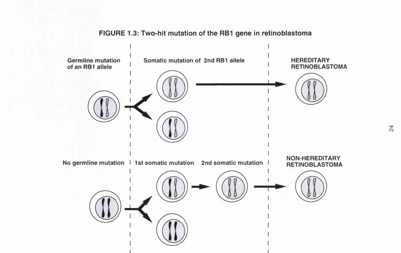
It is likely that all retinoblastoma tumours have both RB1 alleles mutated, as originally postulated by Knudson (1971) and proven experimentally later by
Cavanee ef a /(1983). In fact, no retinoblastoma tumours or cell lines analysed to date have been shown to have an intact RB protein. The classic 'two-hit hypothesis' proposed by Knudson states that two mutational events are necessary for tumorigenesis not only for the hereditary cases but also for the non-hereditary cases (Figure 1.3). This hypothesis was based primarily upon statistical data indicating that individuals with the hereditary form of retinoblastoma developed tumours earlier than those with non-hereditary disease (Knudson, 1971). In the inherited form of the disease, one mutation is acquired from the parent and the other occurs during somatic development. In the sporadic form, both mutations occur in the same cell clone during somatic development. The two events were regarded as involving the same gene for both the hereditary and non-hereditary forms. Structural analysis of the RB1
locus in retinoblastoma and many types of adult tumours has shown that both copies of the wild type RB1 alleles are deleted in these tumours.
Once the retinoblastoma gene was defined, intense research was dedicated to characterise the function(s) of the RB protein.
1.4.1 The RB protein (pRB) and cell cycle control
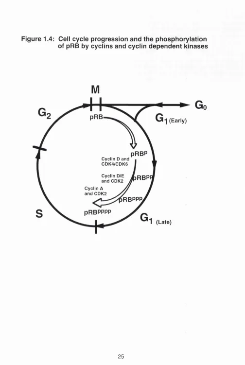
In order to proliferate, a cell must traverse through the cell cycle (Figure 1.4). The cell cycle is made up of a series of processes controlled by an intricate network of extracellular factors and intracellular signalling pathways. These
pathways in turn are regulated by both positive and negative growth signals from the environment. From the resting state (Go) a cell goes through four phases to complete the cycle: G i, a phase during which the cell prepares to synthesise DNA; S, where DNA synthesis takes place; G2, a period in which preparations are made for cell division; and eventually M phase where mitosis and subsequent cell division occurs. After completion of mitosis, cells can either enter a new cycle or return to a quiescent state. Cells that are not
FIGURE 1.3: Two-hit mutation of the RBI gene in retinoblastoma
Germline mutation of an RBI allele
Somatic mutation of 2nd RBI allele
k
I
No germline mutation I 1st somatic mutation 2nd somatic mutation
I
K
HEREDITARY
RETINOBLASTOMA
Figure 1.4: Cell cycle progression and the phosphorylation
of pRB by cyclins and cyclln dependent kinases
M
pRB
1
(Early)pRBP
Cyclin D and CDK4/CDK6
Cyclin D/E
and CDK2
RBPI
Cyclin A and CDK2
RBPPP.
1
(Late)Go
proliferating may be either terminally differentiated, and thus unable to re enter the cell cycle, or remain in Go- To maintain a controlled proliferation rate and to coordinate the timing and order of cell cycle events, there are in-built mechanisms which function as checkpoints. These checkpoints are also critical for the fidelity in the transmission of genetic information. The restriction point, R , is a crucial control checkpoint in late G i. Cells which pass through this point are committed to complete the cycle, even if growth factors are subsequently withdrawn (Pardee et al., 1974). One of the characteristics of a transformed cell is the partial or complete loss of this control checkpoint (Pardee at al., 1978). A key cellular protein which regulates progression through the Gi phase of the cell cycle is the retinoblastoma protein (pRB). By
sitting and functioning at a most crucial time window during which the cell makes most of its decisions about growth versus quiescence, pRB is likely to function as a regulator in cell proliferation (Weinberg, 1989).
The RB protein (pRB) was identified by antibodies which had been raised against three peptides based on the predicted amino acid sequence from the
RB1 cDNA. These polyclonal antibodies immunoprecipitated a protein of about 110 kDa from normal cells. The pRB was localised primarily to the nucleus by cell fractionation and immunostaining analysis and later shown to be constitutively expressed in most cultured cells and normal tissues (Lee at al., 1987b; Bernards at al., 1989; Mancini at al., 1994). In populations of asynchronously proliferating cells, SDS-containing polyacrylamide gel electrophoresis and western blotting analysis detect pRB bands ranging in size from approximately 110 to 116 kilodaltons. This heterogeneity in the molecular sizes detected reflects the multiple phosphorylation states of the protein (Burke at al., 1992). Specifically, in Go or in early G i, the hypophosphorylated form of pRB is found exclusively. In late G i, pRB becomes highly phosphorylated and maintains this configuration throughout the remainder of the cell cycle, losing its multiple phosphates only upon emergence from mitosis via the actions of phosphoprotein phosphatases.
Several lines of evidence indicate that pRB plays a pivotal role as a growth suppressor. It has been shown through gene transfer experiments using malignant cell lines lacking in RB1 that some of the neoplastic properties are reverted by introduction of the RB1 gene either by retroviral infection (Huang
at al., 1988), microinjection (Goodrich at al., 1991) or transfection (Qin at al.,
1992). The common observation for all these experiments is that the tumour cells demonstrate a reduced growth rate which is attributable to Gi arrest.
Clues as to how pRB carries out this inhibitory function stem from observations that pRB is phosphorylated in a cell cycle dependent manner (Chen etal., 1989; Mihara etal., 1989; De Caprio et al., 1989). This reversible phosphorylation event is believed to be the mechanism for affecting the inactivation of the growth inhibitory functions of pRB as suggested by three
further experimental findings. Firstly, transforming viral oncoproteins such as the E l A proteins of adenovirus, the SV40 T antigen and the E7 protein of papillomavirus, bind only to the hypophosphorylated pRB to eliminate its function (Ludlow et a!., 1989). The binding of pRB to this set of oncoproteins occurs at a specific domain called the 'pocket' region of the pRB. Two other members of the pRB family, pi 07 and p i 30, have similar pocket regions, form complexes with E1A (Ewen et a!., 1991; Hannon etal., 1993; Li et al., 1993) and have been shown to have growth suppressive properties similar to pRB (Claudio et al., 1994). Secondly, hypophosphorylated pRB binds and seemingly controls several other cellular proteins (see below). In fact, no cellular protein is known to bind to the hyperphosphorylated form of pRB which is considered to be inactive. Thirdly, in vitro studies have shown that conditions that cause phosphorylation of pRB favour cell proliferation (Cobrinik etal., 1992).
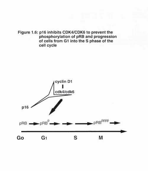
More than a dozen potential phosphorylation sites are present within pRB either on serine or threonine residues, suggesting that either multiple kinases are involved in the phosphorylating event or a kinase working sequentially. It is now known that a kinase complex consists of a regulatory subunit called cyclin and a catalytic subunit called cyclin dependent kinase (CDK). To date at
least 8 different cyclins and 7 different CDKs are known to exist (reviewed by Pines, 1995). Table 1 provides a list of the cyclins and CDKs as well as the preferential pairing and the observed site of action for each heterodimer in the cell cycle.
The first kinase demonstrated to phosphorylate pRB in vitro is cdc2, also called CDK1 (Lin et al., 1991). Many different CDKs have since been shown to phosphorylate pRB in vitro at the same sites as those phosphorylated in
vivo. It is not clear though as to which kinase(s) phosphorylate pRB in vivo.
One possible scenario is that there is a sequential wave of phosphorylation affected by different cyclin-CDK complexes as the cell trasverses through the Gi phase of the cell cycle (Peters, 1994). The CDK4 and CDK6 however have been implicated as the major kinases in the phosphorylation of pRB, working in concert with the Gi cyclins, namely cyclins D1, D2 and D3_ (Kato et ai..
1993; Ewen et al., 1993). Cyclin E-CDK2 also contributes to pRB phosphorylation possibly in late Gi as evidenced by observations that the levels of cyclin E mRNA and protein increase in mid-to-late Gi (Lew et a!.,
1991; Koff et a!., 1991). Further evidence came from experiments using cell lines transfected with either cyclin D1, cyclin E or both. The ectopic expression of each cyclin independently causes pRB phosphorylation. Furthermore, expression of both cyclins shortens the Gi phase significantly greater than when only cyclin D1 is expressed (Resnitzky and Reed, 1995).
Table 1.1: The different cyclins, their preferential CDK partners and site of action in the cell cycle (Pines, 1995)
Cyclin CDK Site of action
A CDK1,CDK2 S/G2
B CDK1 Mitosis
0 ? ?
D CDK2, 4, 5, 6 GI
E CDK2 G1/S
F ? G2?
G ? ?
H CDK7 All
The classical cyclins such as cyclins A and B undergo periodic accumulation and destruction in phase with the cell cycle, suggesting this as the mechanism for regulating the activities of various cyclin-CDK complexes. In contrast, in continuously proliferating cells the level of D-type cyclins remains less variable during the cell cycle. The presence of three types of cyclin D suggests that there is redundancy in their individual actions but studies have shown that the expression of each particular member is tissue specific (Inaba et a!., 1992). For example, T-lymphocytes express predominantly cyclin D3 and to a lesser degree cyclin D2 but lack cyclin D1 (Sutherland etal., 1993; Tam etal., 1994).
Evidence pointing to cyclin D1 as the cyclin responsible for inducing the phosphorylation of pRB came from observation that the levels of cyclin D1 peak in m id-to-late G i, which approxim ates the tim e phase for phosphorylation of pRB (Motokura ef a/.,1991). Direct interaction of pRB with cyclin D1 has been shown in vitro (Dowdy eta l., 1993) and experiments
using the insect cell system have demonstrated that cyclin D1-CDK4 complexes phosphorylate pRB (Kato et al., 1993). Cyclin D1 has a rapid turnover time with a half life of about 15-30 minutes and its steady state levels decline rapidly upon removal of extracellular mitogen (Matsushime at a!.,
1991). This suggests that there is a narrow window within the Gi phase at the time of anticipation of pRB phosphorylation, whereby the cell increases the level of cyclin D1 until it reaches a certain threshold level. The D-type cyclins have also been shown to bind directly to pRB in vitro via a conserved LXGXE motif, where X is any amino acid, and this is thought to account for the ability of D-cyclins to reverse the pRB-mediated growth arrest of SAOS-2 cells
(Hinds at ai., 1992; Ewen at ai., 1993). The interaction between cyclin D1 and pRB has been further explored by analysing tumour cell lines lacking pRB. The level of cyclin D1 is dramatically reduced in these cell lines but upon transfection of a functional RB1 gene expression of cyclin D1 is induced (Muller at ai., 1994). This observation suggests that pRB stimulates the expression of cyclin D1, a key component in the cell cycle itself, and that a regulatory loop exists between pRB and cyclin D1 (Bates at ai., 1994).
Another mechanism for regulating the effector functions of pRB is through its modulation of gene transcription. Many cellular genes have been identified as targets of transcriptional regulation by pRB. These include c-fos, c-myc,
transforming growth factor beta-1 {TGF-p1), and the nau genes (Chen at ai.,
1994). What is most fascinating is that pRB can both positively and negatively regulate the promoters of TG F-pi, c-fos, c-myc through a common motif
called the retinoblastoma control element (RCE) (Kim at ai., 1991).
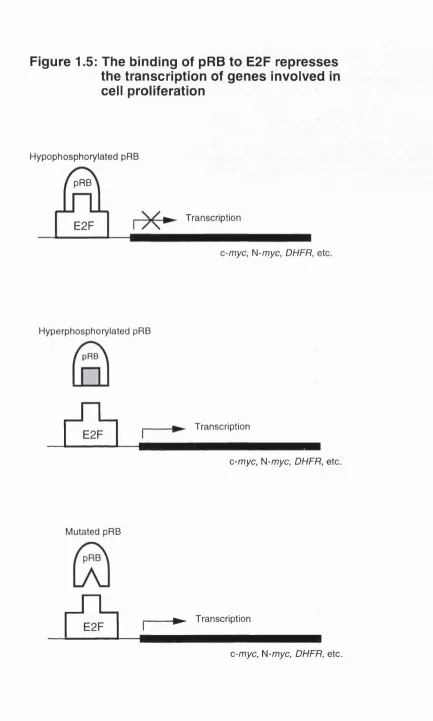
Recent studies have also shown that pRB directly interacts with many transcription factors. The best characterised of these is E2F, which actually denotes a group of 5 distinct transcription factors, namely E2F1-5, which are all targeted to variants of the consensus nucleotide sequence TTTCGCGC (Nevins, 1992; La Thangue, 1994). The site is found in the promoter region of many genes important for cell proliferation such as c-myc, N-myc, B-myb,
DMA polymerase-a, dihydrofolate reductase {DHFR), thymidine kinase, RB1
and the promoter of the E2F-1 gene itself. Complexes of pRB-E2F were initially detected in various cell extracts by several independent approaches (Chellapan at ai., 1991; Chittenden at ai., 1991; Bandara and La Thangue, 1991). The binding of hypophosphorylated pRB to E2F is believed to cause a repression in the transcription of these genes. Phosphorylation of pRB causes it to lose its grip on E2F hence allowing transcription to occur (Figure 1.5). Of
Figure 1.5: The binding of pRB to E2F represses
the transcription of genes involved in
cell proliferation
Hypophosphorylated pRB pRB
► Transcription E2F
c-myc, N-myc, DHFR, etc.
Hyperphosphorylated pRB
e
£
i
Transcriptionc-myc, N-myc, DHFR, etc.
Mutated pRB
r£i
Transcriptionc-myc, N-myc, DHFR, etc.
the five known E2Fs, only three, E2F-1, E2F-2, and E2F-3, are believed to be controlled directly by pRB whilst E2F-4 and E2F-5 are believed to be under the control of the pRB related proteins p107 and p130 (Ginsburg etal., 1994, Cobrinik etal., 1993; Dyson etal., 1993; Shirodkar etal., 1992).
So, if pRB is inactivated in a cell by its phosphorylation or rendered defective through deletion/mutation of the gene, the E2F transcription factors could be
constantly turned on, causing derepression of the genes required for S-phase entry and eventually leading to deregulated cell growth. This could endow the cell with a proliferative advantage, contributing to a neoplastic state.
Apart from E2F, pRB also binds to other transcription factors. These include Elf-1, Myo-D, PU-1, ATF2 and c-abl. Some of these have roles in differentiation. Elf-1 and PU-1 belong to the ets family of transcription factors and they are specific for T- and B-lymphocytes respectively (Wang et al.,
1993; Hagemeier et al., 1993). The transactivating potential of both of these factors is suppressed by pRB. By contrast, the transcriptional activation of ATF2 is enhanced by pRB (Kim et al., 1992). The most intriguing of these
interactions is shown by the modulation of c-abl by pRB. c-abl is a tyrosine kinase with oncogenic potential and pRB is reported to bind directly to the active catalytic domain of c-abl, thus blocking its kinase activity which is activated upon phosphorylation of pRB (Welch and Wang, 1993).
The preceding discussion underscores the diversity of pRB functions as a positive and negative regulator of cellular effectors which regulate cell proliferation.
1.4.2 The role of pRB in differentiation
There is accumulating evidence that pRB also has an important role in cell
differentiation. The most compelling evidence comes from RB1 gene knockout studies (Lee et al., 1992; Jacks et al., 1992; Clarke et al., 1992). These studies also suggested that pRB is not required for the function of many tissues. Homozygous RB1 deficient mice develop normally until day 12 of gestation. The apparently normal development of all tissue types up to this time clearly implies that much of normal differentiation does not require pRB. On day 13, when the homozygous RB1 negative embryos begin to show abnormalities, only two tissues, part of the central nervous system and the haemopoietic system, show macroscopic defects. The RB1 -/- mice dies with
a failure of erythropoietic and neuronal differentiation, manifesting increased mitotic index and cell death in these tissues. It is possible therefore that the target for pRB might be terminal differentiation of certain tissues as further suggested by observations that RB1 mRNA levels increases when
erythroleukaem ia, myoblast or embryonic carcinom a cells undergo differentiation (Coppola etal., 1990; Richon etal., 1992; Slack etal., 1993).
Other indirect evidence for the role of pRB in differentiation is provided by studies of the interaction of pRB with cellular proteins involved in differentiation. For example, muscle development involves the direct interaction of pRB with muscle-specific basic helix-loop-helix (bHLH) factors of the Myo-D family (Gu et al., 1993). It seems that the interaction of pRB and bHLH factors promotes the permanent withdrawal of muscle cells from the cell cycle and the activation of the myogenic differentiation pathway. A recent study by Kreider et al (1995) showed that another helix-loop-helix protein known as Id was able to inhibit the differentiation of a murine myeloid precursor cell line, 32DC13(G). It will be interesting to see whether bHLH, which interacts with pRB, can inhibit myeloid differentiation in human cells.
Hence, the role of pRB may not be solely related to the control of cell cycle progression but also the control of differentiation of specific tissues. In terms of tumour biology, the inactivation of pRB could potentially block one of the differentiation mechanisms in the malignant cell clone. For certain malignancies such as acute leukaemia where there is a maturational arrest, this blockade may be a key event in leukaemia progression.
1.4.3 The role of pRB in apoptosis
The preservation of tissue homeostasis and the prevention of carcinogenesis require the maintainence of an equilibrium between cellular proliferation and cell death or apoptosis. Apoptosis is a genetically controlled process, and amongst the genes important for the regulation of apoptosis are p53 (Lowe et al., 1993) and c-myc (Evan et al., 1992). It has also been shown in vitro that cellular proteins such as c-myc (Filers et al., 1991) and E2F have dual functions as positive regulators of apoptosis and cellular proliferation. Interestingly, pRB binds to both these proteins and the resulting effect of this interaction is growth suppression. It is possible that pRB can also act as a negative regulator of apoptosis as well. Evidence for this was recently put forward by Hass-Kogan et al {^ 995) in a study analysing ionising
induced apoptosis occurring in SAOS-2 cells, which have defective pRB. It was shown that transfection of wild-type RB1 into these cells conferred a significant degree of protection against apoptosis when compared to untransfected cells. Since SAOS-2 cells lack p53 as well, this result also suggests that the ability of pRB to protect cells from apoptosis does not require wild-type p53 expression. Further evidence which suggests that pRB may inhibit apoptosis comes from studies of RB 7-deficient mice. These mice exhibit widespread apoptosis not seen in normal mice (Clarke etal., 1992).
1.4.4 The role of RB1 in tumour formation and progression
It can be predicted from its biochemical and biological properties that inactivation or absence of pRB, either through deletions and mutations or via excessive phosphorylation, can predispose a cell to malignant transformation. A cell which has a defective pRB will not encounter the control normally imposed by pRB and hence will have a growth advantage, may fail to differentiate or undergo apoptosis and may continue to divide inappropriately which may eventually lead to malignant transformation. Likewise, a malignant
cell which acquires RB1 mutation will have a further growth advantage, leading to a more progressive disease.
The first indication that the RB1 gene may contribute to cancers other than retinoblastoma was the development of secondary non-ocular malignancies, which include both sarcomas and gliomas, in individuals who have inherited a germline RB1 mutation (Abramson et a/., 1984; Meadows et al., 1985). Osteosarcoma, the most common second malignancy, occurs at a several hundred-fold increased incidence in retinoblastoma survivors (Draper et al.,
1986).
The first study of RB1 changes in tumours not usually associated with the development of retinoblastoma was that of small cell lung cancer (SOLO). Structural changes in the RB1 gene were seen in a primary SOLO and four SOLO cell lines as well as a pulmonary carcinoid cell line. Moreover, the absence of RB1 transcripts was noted in 60% of the SCLC lines as well as a pulmonary carcinoid cell lines (Harbour etal., 1988).
Further direct evidence for the oncogenic properties of pRB is provided by gene knockout studies. Homozygous RB1 -/- mice die at 12-16 days. However, heterozygous RB1 +/- mice have been successfully bred (Clarke et
al., 1992; Jacks et al., 1992; Lee et al., 1992) and they develop pituitary tumours with almost 100% penetrance. Although it is not retinoblastoma which develops in these mice, the resultant evolution of a tumour is consistent with the proposed role of RB1 as a tumour suppressor gene.
Inactivation of pRB has been shown to be a key factor in the initiation and/or progression of several other common human malignancies including small cell lung carcinoma (Harbour et al., 1988; Horowitz etal., 1990), non-small cell lung carcinoma (Reissmann et al., 1990; Xu et al., 1991a), breast carcinoma (Lee et al., 1988; T'Ang et al., 1988), bladder carcinoma (Horowitz et al.,
1989), osteogenic sarcomas (Toguchida etal., 1988), sarcomas (Canoe etal.,
1990), prostate carcinoma (Bookstein e ta l., 1990), renal cell carcinoma (Ishikawa eta l., 1991), oesophageal carcinoma (Boynton etal., 1991) and parathyroid carcinoma (Cryns etal., 1994).
The broad range of malignancies in which pRB inactivation occurs suggests a pleiotropic role rather than a specific one. It is important to remember that most of these malignancies develop in individuals without a history of retinoblastoma. Why hereditary mutations in RB1 predispose so strongly to retinoblastoma and not to other tumour types is unclear. The key issue is whether loss of RB1 gene function is related to initiation of the tumour or with progression of disease. Assigning the exact role of RB1 in the genesis of these tumours is not easy. It is probably adequate to say that RB1 mutations contributes to the accumulative genetic events leading to cancer. The fact that these mutations are only seen in a subset of these malignancies suggests that pRB inactivation is not the rate limiting step in the development of these
cancers nor is it the initiating event (Benedict etal., 1990).
The availability of purified high-affinity polyclonal antibodies has provided a more sensitive method for analysing inactivation of pRB. It has also enabled
researchers to examine the status of pRB at the single cell level in each type of malignancy using techniques such as immunostaining and flowcytometry. This has allowed the screening of subpopulations of tumour cells with different pRB expression. For example, histochemical staining of soft tissue sarcomas showed that there was a sub-population of tumour cells with normal pRB and another with non-detectable pRB (Cance et al., 1990). This heterogeneity in pRB expression could be explained by a further genetic alteration occurring in a sub-clone of tumour cells, allowing this particular sub-clone a further growth advantage.
It has been shown in high grade human sarcomas that the absence of pRB predicts for a poorer prognosis both in therapeutic response and survival rates (Cance et al., 1990). In other studies, altered pRB expression is more commonly associated with the invasive phenotype of bladder cancer (Presti et a!., 1991; Cairns e t a l1991) and non-small cell lung cancer (Xu etal., 1991).
The above observations indicate that in tumours other than retinoblastoma,
RB1 deletions are likely to be involved in tumour progression.
1.5 The role of cyclin dependent kinase inhibitors (CKIs) in cell cycle regulation
The activity of CDKs is controlled both by their association with cyclins and the complex phosphorylation and dephosphorylation of the CDK polypeptide itself, hence providing another tier of control (Norbury and Nurse, 1992).
The fact that the cyclin D-CDK complex mediates the phosphorylation of pRB predicts that the prevention of this phosphorylation event can be executed by mechanisms which either impinge on the pRB itself or through inhibition of CDK activity. Studies to date suggest that the latter mechanism seems to be more likely. The identification of CDK inhibitors (CKIs) over the last two years has introduced another level of regulation within an already complex network. These inhibitory proteins either associate with the cyclin-CDK complex or bind
to the CDK subunit alone resulting in the impediment of its kinase activity, hence providing the cell with another set of brakes in the cell cycle. Some CKIs are modulated in response to extracellular signals such as TGF-p, cyclic
AMP (cAMP) and contact inhibition, others appear to function in intrinsic steps of the cell cycle such as mediating radiation induced growth arrest and
apoptosis, while a few others function as tumour suppressors.
p21 and p i 6 were the first CKIs found in the mammalian system. The discovery of these proteins made an outstanding impact on cell cycle research due to their links with the tumour suppressor genes p53 and RB1. The emergence of p21 and p i 6 was followed by the discovery of other cell cycle inhibitory proteins. They can be loosely grouped into: (a) the p15/p16/p18/p19 group which specifically inhibit cyclin D-CDK4 and cyclin D-CDK6 complexes, and (b) the p21/p27/p57 group which interact with all the known cyclin-CDK complexes.