UNIVERSITY O F LONDON
An Investigation into the Clinical Potential and Applications of Ophthalmic Diode Lasers
A thesis submitted for the degree of Doctor of Medicine
John Dominic Anthony McHugh MBBS (Lond) FRCSEd FCOphth DO RCS
Allerton Vision Research Fellow The Institute of Ophthalmology
ProQuest Number: U049409
All rights reserved
INFORMATION TO ALL USERS
The qu ality of this repro d u ctio n is d e p e n d e n t upon the q u ality of the copy subm itted.
In the unlikely e v e n t that the a u th o r did not send a c o m p le te m anuscript and there are missing pages, these will be note d . Also, if m aterial had to be rem oved,
a n o te will in d ica te the deletion.
uest
ProQuest U049409
Published by ProQuest LLC(2017). C op yrig ht of the Dissertation is held by the Author.
All rights reserved.
This work is protected against unauthorized copying under Title 17, United States C o d e M icroform Edition © ProQuest LLC.
ProQuest LLC.
789 East Eisenhower Parkway P.O. Box 1346
TABLE OF CONTENTS
Title p a g e ... 1
Table of contents ... 2
l i s t of fig u re s... 8
List of tables ... 13
Abstract of thesis ... 15
A cknow ledgem ents... 17
SECTION I IN T R O D U C T IO N ... 18
CHAPTER 1: HISTORICAL ASPECTS OF OPHTHALMIC PHOTOCOAGULATION . . 19
1.1 Solar re tin o p a th y ... 20
1.2 Therapeutic photocoagulation... 20
13 Ruby l a s e r ... 21
1.4 Argon l a s e r ... 21
1.5 Krypton l a s e r ... 22
1.6 Dye l a s e r ... 22
1.7 Nd:YAG l a s e r ... 23
1.8 Carbon dioxide (C 02) laser ... 24
1.9 Excimer l a s e r ... 24
1.10 Diode la s e r s ... 24
1.11 Conclusions ... 26
2 3 Stimulated e m is s io n ... 30
2.4 L a s e r s ... 30
2.5 General characteristics of lasers ... 32
2.5.1 Monochromatidty ... 32
2.53 D irectionality... 32
2.5.3 C o h eren ce... 32
2.6 Mode of operation of lasers ... 33
2.6.1 Continuous wave lasers ... 33
2.63 Pulsed l a s e r s ... 33
2.7 Laser delivery systems ... 34
2.8 Mode of action of diode lasers ... 34
2.8.1 Sem iconductors... 34
2.8.2 Semiconductor ju n ctio n s... 37
2.83 Laser diodes ... 37
2.8.4 Heterojunction diodes ... 37
2.9 Characteristics of laser d io d e s ... 38
2.9.1 Temporal behaviour... 38
2.9.2 Current-output characteristics... 38
2.93 Output beam p ro p e rtie s ... 42
2.10 Efficiency and reliability of laser d io d e s ... 42
3.2 Study design ... 48
3.2.1 Technological co n sid eratio n s... 48
3.2.2 Biophysical considerations ... 48
3.23 Clinical considerations... 49
SECTION H: DESIGN O F INSTRUM ENTATION... 50
CHAPTER 4: INSTRU M EN TATIO N... 51
4.1 Retinal studies ... 52
4.1.1 Diode lasers used in retinal s tu d ie s... 52
4.1.2 Argon and krypton l a s e r s ... 52
4.2 Diode lasers used in macular and trabecular histological studies and in the clinical t r i a l s ... 53
SECTION III: HISTOLOGICAL S T U D IE S ... 64
CHAPTER 5" MATERIALS AND METHODS OF HISTOPATHOLOGICAL STUDIES . 65 5.1 Retinal exposures... 66
5.1.1 Animal studies ... 66
5.1.2 Human stu d ies... 66
5.2 Human macular exposures ... 67
5 3 Microscopic studies on retinal tissue ... 68
5.4 Human trabecular meshwork e x p o su res... 69
5.5 Microscopic studies on trabecular tis su e ... 69
5.6.4 Histopathological appearances of retinal laser
b u m s ... 76
5.6.5 R a b b it... 76
5.6.6 H u m a n ... 84
5.7 Results of human macular ex p o su res... 95
5.7.1 Ophthalmoscopic appearances ... 95
5.7.2 Light m icroscopy... 95
5.73 Electron microscopy... 109
5.8 Results of human trabecular exposures ... 115
5.8.1 Clinical d a t a ... 115
5.8.2 lig h t microsajpy ... 115
5.83 Electron microscopy ... 115
CHAPTER 6: DISCUSSION OF HISTOPATHOLOGICAL S T U D IE S ... 126
6.1 Retinal photocoagulation ... 127
6.2 Macular photocoagulation... 135
6 3 Human trabecular photocoagulation ... 144
SECTION IV: CLINICAL S T U D IE S ... 149
CHAPTER 7: METHODS AND RESULTS OF DIODE LASER CLINICAL TRIALS . . . 150
7.1 Study d esign... 151
7.2.5 Post-treatment review ... 153
7 3 Results of treatment of retinal vasculopathies... 154
73.1 Use of instrum entation... 154
1 3 2 The laser b u r n s ... 155
7 3 3 Clinical re s u lts... 159
7.4 Diode laser therapy for chronic open angle glaucom a... 186
7.4.1 Aims of study ... 186
7.4.2 Inclusion c r i t e r i a ... 186
7.4.3 Patient evaluation ... 186
7.4.4 Treatment protocol ... 187
7.43 Post-treatment review ... 187
1 5 Results of diode laser trabeculoplasty ... 187
7.5.1 Use of instrum entation... 187
7.5.2 The laser b u r n s ... 188
7.53 Clinical re s u lts... 188
CHAPTER 8: DISCUSSION OF CLINICAL S T U D IE S ... 196
8.1 Retinal vascular diseases... 197
8.1.1 Proliferative diabetic retinopathy ... 197
8.1.2 Exudative diabetic retinopathy ... 202
8.1.3 Retinal vein th ro m b o sis... 204
8.2 Glaucoma ... 205
SECTION V:
CONCLUSIONS ARISING FROM THE FINDINGS OF TH E PROJECT . 214 CHAPTER 9:
OPHTHALMIC DIODE LASERS- CURRENT WORK
AND FUTURE P L A N S ... 215
9.1 Discussion of the study findings... 216
9.2 Current stu d ies... 217
9.3 Future p ro jects... 218
9.4 C onclusions... 218
SECTION VI: REFERENCES ... 219
APPENDIX I: Consent form and pro formas for pilot clinical studies (retinal vascular d is e a s e s )... 243
APPENDIX II: The Hammersmith system for grading diabetic retinopathy . 248 APPENDIX III: Protocol for a clinical trial to compare diode and argon laser treatment of proliferative diabetic retinopathy ... 250
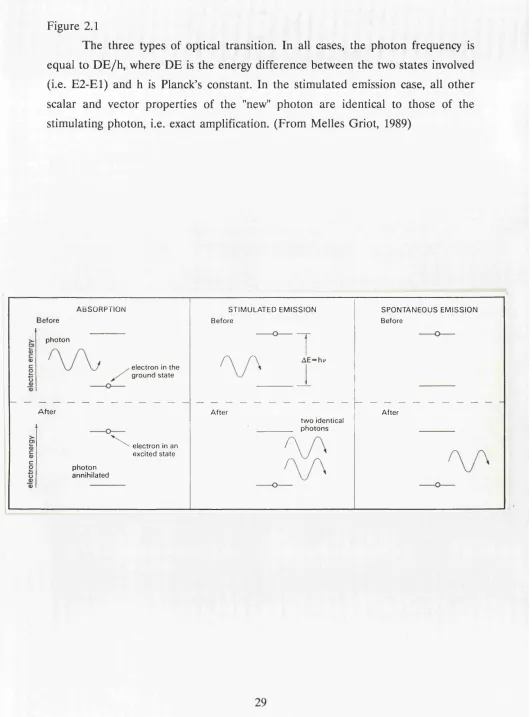
LIST OF FIGURES 2.1
The three types of optical transition
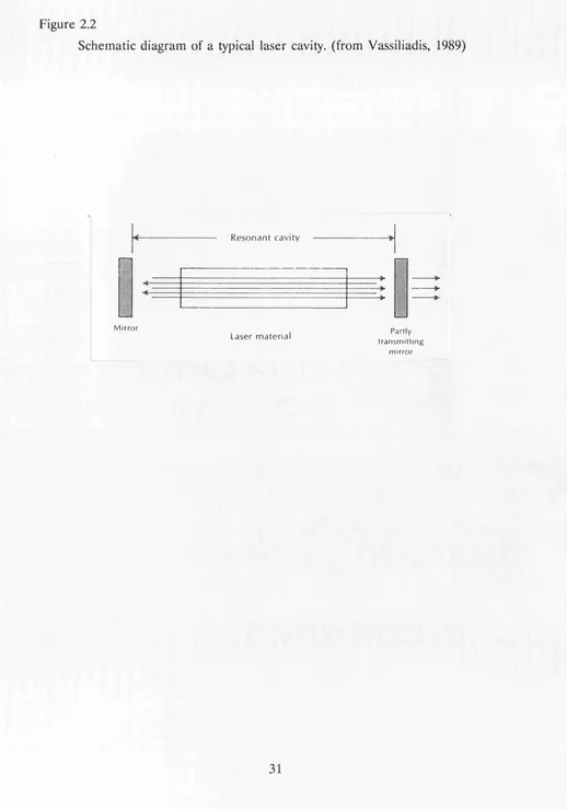
2.2
Schematic diagram of a typical laser cavity 2.3
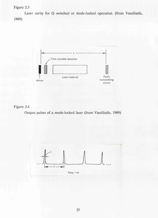
Laser cavity for Q switched or mode-locked operation 2.4
Output pulses of a mode-locked laser 2.5
Energy band diagrams for an insulator, a metallic conductor and a semiconductor
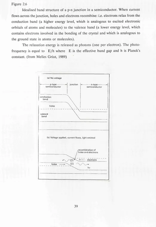
2.6
Idealised band structure of a p-n junction in a semiconductor 2.7
Structure of a simple gallium arsenide laser diode
2.8
A double-heterojunction laser diode 2.9
Beam output power versus input current for a typical gallium aluminium arsenide laser diode
2.10
Section of a typical laser diode and its integral monitor photodiode
2.11
Irradiance profile of a Gaussian T EM 00 mode in a laser diode 4.1
Hand-held diode laser 4.2
5.1
Macrophotographs of diode laser lesions in the eyes of rabbit (a, b) and human (c) 5.2 (a)
Radiant power and lesion size in rabbit retina 5.2 (b)
Radiant power and lesion size in human retina 5.3
Light micrograph of diode laser lesions produced by varying levels of radiant exposure in rabbit retina 5.4
Transmission electron micrographs of the basal region of the pigment epithelium, Bruch’s membrane and choriocapillaris in rabbit retina at the centre of an area irradiated by a diode laser
5.5
Light micrographs of diode laser lesions produced in human retina
5.6
Light micrographs of areas of laser exposure located over major retinal vessels, (a) diode, (b) krypton and (c) argon
5.7
Light micrographs of pigment epithelium and underlying choroid in areas of (a) diode, (b) krypton and
(c) argon laser exposures in human 5.8
Transmission electron micrographs of outer retina
5.10 5.11 5.12 5.13 5.14 5.15 5.16 5.17 5.18
Light micrographs of the macular region of a 65 year old human retina 18 hours after exposure to
diode laser radiation 97
High power light micrographs of the pigment epithelium
and choroid of lesion shown in figures 5.10 (a) and (c) 99
Light micrograph of the macular region of a human retina
following argon and krypton irradiation 103
Light micrographs of the macular region of a 68 year old human retina 5 weeks following exposure
to diode laser radiation 105
High power light micrographs of the outer retina,
5 weeks after diode laser irradiation 107
Electron micrographs of the macular region of a 65 year old human retina 18 hours after diode
laser irradiation 111
Electron micrographs of retinal pigment epithelium and choroid of the macular region of a 68 year human
retina, 5 weeks after exposure to a diode laser 113
Light micrographs of the trabecular meshwork 18 hours after
irradiation with (a) argon and (b) diode lasers 117
Scanning electron micrographs of sites of argon laser
5.19
5.20
Scanning electron micrographs of areas of trabecular
meshwork 18 hours after irradiation with the diode laser 121
Transmission electron micrographs of trabecular
meshwork 18 hours after irradiation by (a) and (b) argon and
(c) and (d) diode lasers 123
6.1
Transmission of radiation through the ocular media 128 7.1 (a)
Appearance of diode laser-induced burns in a heavily
pigmented fundus 156
7.1 (b)
Appearance of acute lesions produced in a
lightly pigmented fundus 156
7.2
Diode laser burns 6 weeks following treatment 158 7.3 (a)
Effect of diode laser treatment on diabetic disc new vessels 160 7.3 (b)
Effect of diode laser treatment on diabetic peripheral new vessels 160 7.4
7.5:
7.6
Visual acuity and diode laser treatment in
proliferative diabetic retinopathy 163
Case report 1: Proliferative diabetic retinopathy 168
7.9:
Case report 4: Central retinal vein occlusion 183
7.10
Intraocular pressures before and at 2 weeks
following diode laser trabeculoplasty (DLT) 191
7.11
Intraocular pressures before and at 6 months
LIST OF TABLES
Relationship between gross ophthalmoscopic appearances and radiant power of diode laser
Approximate percentages of transmission and absorption of ocular components for various wavelengths
used for retinal photocoagulation
Diode laser panretinal photocoagulation for proliferative diabetic retinopathy: mean number of burns applied
Diode laser panretinal photocoagulation: Success and distribution of new vessels
Changes in intraocular pressure induced by diode laser trabeculoplasty (DLT)
VIII
Mean reduction in IOP following diode laser trabeculoplasty
ABSTRACT OF THESIS
A study has been carried out of transpupillary retinal and of trabecular photocoagulation with infrared diode lasers. There were three phases to the study:
A. Design and construction of instrumentation.
B. Histopathological studies of lesions produced by diode lasers in rabbit retina, human peripheral retina and macula and on human trabecular meshwork. The appearances of the lesions were compared with those produced by lasers which emitted at other wavelengths.
C. Pilot clinical trials in which diode lasers were used in the treatment of the following conditions:
1. Proliferative diabetic retinopathy 2. Exudative diabetic retinopathy
3. Branch retinal vein thrombosis complicated by neo vascularisation of the optic disc or of the retina
4. Central retinal vein thrombosis complicated by established or threatened rubeosis iridis, or by optic disc neovascularisation
5. Chronic open angle glaucoma RESULTS
A. INSTRUMENTATION
For initial histopathological and clinical studies, a diode laser emitting at 810 nm and with a power output of 800 mW was constructed, which was incorporated in a modified hand-held direct ophthalmoscope.
In a subsequent phase of development, a diode laser was assembled that could be attached to a standard slit lamp microscope and which had an eventual power output of 1.4 W. The project was completed using this modality, as it was felt that there was greater flexibility and ease of use in the treatment of patients.
B. HISTOPATHOLOGICAL STUDIES
of the macula, in which inner retinal damage was observed, which was associated with absorption by macular pigments.
Trabecular photocoagulation with a diode laser produced a pattern of damage to trabecular beams. The histological appearances were similar to those seen in relation to argon blue-green exposures, although the diode laser lesions extended more deeply into the trabeculum.
C. PILOT CLINICAL STUDIES WITH DIODE LASERS
106 eyes in 85 patients were given diode laser therapy for proliferative diabetic retinopathy, exudative diabetic retinopathy, branch and central retinal vein thrombosis and chronic simple glaucoma.
Regression of neovascularisation was observed in 33 of 47 eyes (70%) with proliferative diabetic retinopathy, and in all 11 eyes treated for branch vein thrombosis. Six eyes were successfully treated for established or incipient rubeosis iridis, following central vein thrombosis.
Focal photocoagulation applied to 22 eyes for exudative diabetic maculopathy resulted in a reduction in the number of microvascular abnormalities and partial resorption of exudates.
Laser trabeculoplasty carried out on 20 eyes for glaucoma resulted in a mean ocular hypotensive effect of 10.2 mm Hg, at 2 weeks following treatment and of 9.55 mm Hg, at 6 months.
ACKNOWLEDGEMENTS
My financial support for this project was provided entirely by the Lady Allerton Vision Research Trust, and I would like to express my appreciation of its generosity.
I am indebted to my supervisor, Professor John Marshall PhD who was an invaluable source of constructive suggestions and critical advice in relation to every aspect of this study.
Mr Timothy ffytche FRCS and Mr Peter Hamilton FRCS, consultant ophthalmologists, allowed me to work in their clinics, from which most of the patients for the clinical trials were recruited. I greatly benefited from their clinical expertise when implementing the trials and their enthusiasm for the project was a great source of encouragement.
I was privileged to collaborate in the technical aspects of the project with Dr Tony Raven and Dr Robin Lee of Scientific Generics, Cambridge and with the team led by Mr CR Keeler of Keeler Holdings Ltd, Windsor.
The histological phase of the study required education in a wide range of techniques to which I was hitherto unfamiliar. I would like to express my appreciation to Professor Marshall’s technicians, Mr Steven Rothery and Mrs Ann Patmore for their invaluable help in this regard.
CHAPTER 1
The application of semiconductor lasers to ophthalmic photocoagulation is a relatively novel concept. Before detailing the aims of this project, it may therefore be helpful to outline the historical background and some technical aspects relating to ophthalmic laser therapy.
1.1 SOLAR RETINOPATHY
The damaging consequences of ocular exposure to high energy sources of optical radiation have been recognised for centuries. In Plato’s "Phaedo", he records Socrates’ advice regarding the potentially harmful effects of sunlight:
"I decided that I must be careful not to suffer the misfortune which happens to
people who look at the sun and watch it during an eclipse. For some o f them ruin their
eyes unless they look at its image in water or something o f the sort." (Plato, c. 380 B.C.)
The first description of a central scotoma due to a solar retinopathy is attributed to Theophilus Bonetus (1620-1689) (cited by Hamm, 1947). The development of the ophthalmoscope was followed by several descriptions of the appearance of macular burns which occurred as a result of viewing solar eclipses (Birch-Hirschfeld, 1912; Blessig, 1912; Cords, 1912). Retinal lesions were produced by sunlight, or by a carbon arc by a number of investigators (Czerny, 1867; Deutschmann, 1882; Widmark, 1893).
Maggiore examined the histopathological appearance of radiation burns produced in human eyes due for enucleation and described retinal oedema in the sites of irradiation (Maggiore, 1927).
1.2 THERAPEUTIC PHOTOCOAGULATION
Moran-Salas and Meyer-Schwickerath performed experiments concurrently in the 1940’s to examine the therapeutic potential of retinal photocoagulation (Meyer-Schwickerath, 1949; Moran-Salas, 1950).
The development of the xenon arc photocoagulator provided a source of broad band optical radiation which was effective in producing full-thickness chorioretinal lesions (Meyer-Schwickerath, 1960). Xenon arc devices are of proven effectiveness in the treatment of proliferative diabetic retinopathy (British Multicentre study group, 1984; Stenkula, 1984) and are still commonly used for retinal therapeutic procedures.
13 RUBY LASER
The advent of the ruby laser in 1960 (Maiman, 1960) aroused interest amongst ophthalmologists and this rapidly resulted in the investigation of its potential in the treatment of ocular conditions. Early work on animals by Zaret in 1961 (Zaret, 1961) was followed within two years by therapeutic regimens in humans (Campbell et al, 1963; Zweng et al, 1964). Although the ruby laser (emitting at 694.3 nm) was relatively effective in producing chorioretinal adhesions in detachment surgery, results were disappointing in treating retinal vascular conditions (Campbell et al, 1963; Aiello et al, 1968; Taylor, 1970).
In the mid-sixties the failure of the ruby laser was attributed to an inappropriate wavelength and the resultant lack of absorption of laser energy in retinal vessels. Recent work has emphasised the role of the retinal pigment epithelium in the treatment of retinal vascular diseases (Glaser 1980; Wong, et al, 1987; Miller et al, 1986). The pigment epithelium is a tissue with broad band absorption characteristics and the major site of energy degradation in retinal photocoagulation. The inadequacies of the ruby laser are now thought to be due to the pulse duration rather than wavelength, as the short pulse duration of early instruments produced a high risk of choroidal haemorrhage.
1.4 ARGON LASER
recurrence of membranes and visual deterioration (Macular Photocoagulation Study Group, 1982; The Moorfields Macular Study Group, 1982).
Argon lasers were also found to be useful in the treatment of several forms of glaucoma. The technique of laser trabeculoplasty was suggested by Wise and Witter in 1979 (Wise and Witter, 1979). Several studies have subsequently proved the benefit of this form of technique for the treatment of chronic open angle glaucoma (Wise, 1981; Schwartz, 1981). Laser iridotomies for closed angle glaucoma were performed initially by the ruby laser (Beckman and Sugar, 1973), and then by the argon laser (Abraham and Miller, 1975). In these situations the laser provided a viable alternative to operative intervention.
In recent years concern about the adverse photochemical effects of blue light on retinal photoreceptors of both the patient and the ophthalmologist has led to the development of argon systems in which the blue output can be virtually eliminated. For the patient, this can be achieved either through the use of prisms in the beam path which deflect the argon blue line; for the surgeon it can be minimised by the incorporation of a filter within each eye piece, which attenuates the transmission of shorter wavelengths.
1.5 KRYPTON LASER
In the mid-seventies a new red light emitting laser, the krypton laser (647nm) was introduced and it has been found to be as effective as argon in the treatment of retinal conditions (Blankenship, 1986). It also had advantages when compared with argon blue in that its longer wavelength resulted in virtually no absorption by macular xanthophyll pigment (Marshall and Bird, 1979; Yannuzzi and Shakin, 1982). Further, the lower photon energy at longer wavelengths in the visible spectrum reduced the potential for inducing photochemical damage (Ham et al, 1976).
1.6 DYE LASER
However, because melanin is the prime absorber at every wavelength emitted by the dye laser, thermal damage resulting from the source must be similar at each wavelength. Temperature profiles will only vary with wavelength in the choroid and will only be significant at pulse durations of 1 ms or less. All retinal profiles will be virtually identical at all wavelengths and hence no selective effect can be achieved. Support for this argument has been provided by studies which have demonstrated that the thermal damage resulting from dye laser exposure is widespread throughout the outer retina and have failed to support the concept of selectivity of targets (Smiddy et al, 1988; Brooks et al, 1989). Recent comparisons of the treatment of neovascular membranes with several dye laser wavelengths have not demonstrated any difference in clinical efficacy (Haut et al, 1987; Brancato et al, 1988).
It is probable that the real future of the dye laser lies in the development of much more target-specific chromophores or fluorophores and the use of irradiances and pulse durations that will cause photochemical rather than thermal damage. The possible development of fluorescent, or chromatically labelled monoclonal antibodies offers promise for selective use of dye lasers. The use of the dye laser has already shown promising results in relation to photodynamic therapy. This method involves sensitisation of an intraocular tumour with haematoporphyrin derivative (HPD). Subsequent irradiation of the tumour with light of a wavelength of approximately 630 nm results in activation of the HPD and release of cytotoxic singlet oxygen radicals. L’Esperance has reported successful tumour destruction utilising the wavelength selectivity of a dye laser in conjunction with HPD (L’Esperance, 1985).
1.7 Nd:YAG LASER
1.8 CARBON DIOXIDE (C 02) LASER
The C 02 laser (which emits at 10,600 nm) has been used with limited success in the treatment of tumours of the ocular adnexa, and in carrying out glaucoma trephination procedures (Beckman 1971; Beckman, 1979). The use of this laser in ophthalmology still remains restricted to a relatively few centres, because of its lack of precision as a cutting tool.
1.9 EXCIMER LASER
The recently introduced excimer lasers interact with tissues by a process that has been termed "photoablation". They are demonstrating exciting possibilities as a mode of therapy for several anterior segment conditions, for example glaucoma and corneal opacification. The correction of refractive errors has also been carried out with the excimer laser by altering the corneal profile, "photorefractive keratectomy" (Marshall, 1986).
1.10 DIODE LASERS
Continuous wave argon, krypton and dye lasers, while broadly similar in terms of both the damage they induce to the retina, and in their therapeutic efficacy also have several inherent disadvantages.
Laser energy is generated within a relatively bulky gas-filled tube; electrical energy consumption is high, and the efficiency of electrical-optical conversion is low. Many systems require a three-phase power supply and forced cooling facilities dependent on circulating air or water. Heat dissipating factors tend to increase the size of the equipment and these together with their special electrical requirements mean that in most situations rooms need to be adapted for the permanent installation of laser apparatus. Additionally, maintenance costs are high with the average tube life being 2-3 years, and the cost of replacement being about 20% of the total cost of the laser. Advances in semi-conductor technology have allowed the development of infrared diode lasers (750-950 nm) measuring a few millimetres in size. These are used in compact disc players and have important applications in the fields of optical printing and communications.
powered by either a standard 13 amp power supply or a 6 volt battery and no ancillary cooling facilities are needed.
Currently, most high powered diodes emit in the infrared region of the spectrum (810 nm). This wavelength is just outside the visible spectrum and therefore a permanent protective filter, a dielectric mirror, could be incorporated in a clinical system to protect the operator. This would also remove the requirement for a mechanical shutter and as it would only attenuate invisible wavelengths it would allow the ophthalmologist an uninterrupted, unimpeded view of the retina throughout therapy.
Brancato and Pratesi (Brancato and Pratesi, 1987) and Puliafito (Puliafito, 1987) working independently, successfully produced chorioretinal lesions in rabbit retinas with diode lasers. The former used a transpupillary route while the latter used an intraocular fibre optic. The lesions produced by Brancato were described as being similar to those produced by current clinical lasers. In this early report, the emission of the laser diode was of relatively low power and as a result, an exposure time of six to ten seconds was required even to produce low intensity ophthalmoscopically visible burns. However in a later report intense white lesions were produced in rabbit with an output of 120 mW and exposure durations of between 0.3 and 1 second (Brancato and Pratesi, 1988).
The project described in this thesis resulted in the first histopathological report of retinal photocoagulation by a diode laser in human eyes (McHugh et al, 1988). Microscopic analysis of the lesions demonstrated them to be similar to those produced by conventional clinical photocoagulators and in particular to those induced by krypton lasers. The physical parameters of the exposures such as power levels, exposure durations and spot sizes were also similar.
in the treatment of glaucoma (Mchugh et al, 1990 (II)).
Developments of diode lasers include the possibility of diode-pumped Nd:YAG lasers, and of mode-locked diode laser which would themselves have a photodisruptive mechanism of action.
1.11 CONCLUSIONS
CHAPTER 2
BASIC PRINCIPLES OF LASERS
2.1 GENERAL CONSIDERATIONS
The term "LASER" is an acronym for "Light Amplification by the Stimulated Emission of Radiation". The physical principles underlying the generation of laser energy are related to the quantum theory of radiation, which was originally proposed by Albert Einstein in 1905. According to quantum theory, optical radiation consists of discrete "quanta" of energy called photons, whose energy is given by the equation,
E = hS)
where E is the energy of the quantum, h is a proportionality constant called Planck’s constant and *9 is the frequency of the light. According to Einstein, the minimum amount of radiant energy that can interact with matter is equal to the energy of a single photon.
An elaboration of the theory was postulated by Neils Bohr in 1913. He stated that each electron in an atom or molecule occupies a specific energy level. The majority of electrons occupy the lowest energy level available to them and this is called the ground state. This represents the most stable level for a collection of atoms or molecules.
Electrons may move between energy levels by several processes. One way in which this can occur is by optical transition. In this case, a photon of frequency ^ interacts with an electron. Provided that the energy of the photon is equal to the difference in two energy levels, it can stimulate an electron to undergo transition from one energy level to another via so-called stimulated transition. An electron having undergone such a transition is described as an excited electron. There are several possible consequences to stimulated transition (figure 2.1).
2.2 SPONTANEOUS EMISSION
An electron may be elevated from a stable lower energy level E l to a higher energy level E2, by for example absorption of a photon. After a period of time the electron will fall back to its lower energy level, with emission of a photon of light of energy
[E2-E1]
Figure 2.1
The three types of optical transition. In all cases, the photon frequency is equal to D E /h , where DE is the energy difference between the two states involved (i.e. E2-E1) and h is Planck’s constant. In the stimulated emission case, all other scalar and vector properties of the "new" photon are identical to those of the stimulating photon, i.e. exact amplification. (From Melles Griot, 1989)
A B S O R P T I O N ST IM U L A TE D E M I S S I O N S P O N T A N E O U S E M I S S I O N B ef o re
B e f o r e B e f o r e
p h o t o n
e l e c t r o n in t h e g r o u n d s t a t e
A fter A f t e r A fte r
t w o id en tic al p h o t o n s e l e c t r o n in an
e x c i t e d s t a t e p h o t o n
2 3 STIMULATED EMISSION
An electron which is already in an upper level can be stimulated to undergo transition to the ground state by absorption of a photon. An electron falling to a lower energy level is accompanied by emission of the incident photon and a photon emitted which is of energy
[E2-E1]
The "new" photon emitted has identical properties to the incident photon in terms of frequency, phase, polarisation and direction of propagation. Such light emitted due to stimulated emission is referred to as coherent light.
In order that stimulated emission occurs within a medium, it is necessary that the majority of electrons are excited or in a higher energy level before light enters the medium. This is a condition referred to as "population inversion". In this situation, the introduction of a beam of light of the appropriate wavelength would result in amplification of the beam following stimulation of the excited electrons into emitting photons which are in phase with the incident photons.
2.4 LASERS
There are three basic requirements for the production of laser radiation: 1. An excitable medium which has transition characteristics which allow population inversion.
2. An energy source to excite the lasing medium.
3. A system for reflecting spontaneously emitted photons repeatedly through the lasing medium (a resonator).
To create population inversion, energy must be supplied to the medium. In the case of gas lasers, such as argon or krypton lasers the external energy is supplied in the form of an electric current passing through the gas. An electrical supply also provides the energy source for diode lasers (vide infra). In the case of the ruby, or neodymium-YAG laser, the energy is supplied in the form of optical radiation by a flash lamp.
Figure 2.2
Schematic diagram of a typical laser cavity, (from Vassiliadis, 1989)
R e s o n a n t c a v i t y
M i r r o r
L a s e r m a t e r i a l Pa rtly
threshold energy has been reached. After population inversion has been achieved, spontaneous emission will occur from excited electrons. Some of these will be reflected between the mirrors bounding the cavity and these will be of the correct wavelength to effect stimulated emission. Multiple reflections of the emitted photons increase the probability of light amplification. Thus stimulated emission becomes the predominant mechanism of photon propagation and a coherent beam is quickly generated. This represents the basis of laser action.
2 5 GENERAL CHARACTERISTICS O F LASERS 2.5.1 MONOCHROMAT1CITY
The light that is emitted from a laser is of one or a few individual wavelengths (colours), since there are only a limited number of efficient electronic transitions from excited energy levels in an atom or molecule to lower energy levels. Thus the argon laser emits over several wavelengths (lines) in the blue-green part of the spectrum, with the dominant lines being 488 nm and 514.5 nm.
In the case of clinical dye laser systems, use of a birefringent filter allows tuning of their potentially broad spectral output to an effectively monochromatic range.
2.5.2 DIRECTIONALITY
Since a laser’s resonant cavity amplifies only electrons travelling along its long axis, a laser beam is highly directional and it has little divergence compared with conventional light sources. Low divergence permits effective focusing of the beam into a small spot whose irradiance (i.e. power per unit area) is high. This is of clinical relevance, since most ophthalmic laser applications require high focal irradiances.
2.5.3 COHERENCE
2.6 MODE OF OPERATION OF LASERS
Depending on the lasing medium and the mode of excitation, a laser may be operated in one of several ways.
2.6.1 CONTINUOUS WAVE LASERS
In continuous wave (CW) mode, there is constant stimulation of the atoms or molecules in the medium and laser radiation is therefore emitted continuously. Examples of lasers that are typically operated in the CW mode are argon, krypton, dye and carbon dioxide lasers. The diode laser is also classed as a CW laser.
2.6.2 PULSED LASERS
Pulsed operation may be obtained when laser materials such as ruby, or neodymium YAG are pumped with flash lamps. The output of these lasers consists of a train of pulses of durations of the order of milliseconds. The duration of the emitted pulses can be made shorter still, with consequently higher energy by either of two processes.
a) Q Switching (figure 2.3)
The "Q", or quality factor of a resonant cavity is related to the ratio of energy storage to energy dissipation in a medium. If a time variable absorbing cell (a "Q-switch") is added to the laser cavity, thereby obscuring one of the mirrors, laser generation is inhibited and a high level of population inversion occurs. If the cell is suddenly made transparent, laser action occurs, resulting in the generation of a high power pulse of light of between 2-14 nanosecond (1 nanosecond = 10~9 s) duration. The most commonly used Q-switch in photodisruptors is a "Pockels cell", an electro- optical device which is opaque until an electrical signal switches it to a transparent condition shortly after flash lamp action begins.
b) Mode Locking (figure 2.4)
This results in the emission of a train of laser pulses in which the longitudinal modes of the laser are coupled together. The duration of these pulses is typically of the order of 30 picoseconds ( 1 picosecond = W 12 s) and the peak power of individual mode-locked laser pulses is approximately one thousand times greater than that of a Q-switched laser pulse. Certain practical differences exist between Q-switched and mode locked systems. Mode-locked lasers require a longer laser cavity to provide enough longitudinal modes and a dye cell that needs regular maintenance and replacement of dye. The dye tends to bleach unevenly, producing an irregular distribution of energy in the focal spot. The range of energies available in current mode-locked systems is limited to 5 mJ or less and can be varied only by the use of filters. In contrast, Q-switched systems have a greater range of pulse energy, use a solid state Pockels cell that requires little maintenance and are small enough to be incorporated in a slit-lamp microscope. Q-switched lasers therefore tend to be favoured in clinical practice.
2.7 LASER DELIVERY SYSTEMS
The optical delivery systems employed in ophthalmic lasers are typically either a fibre optic cable, or an articulated mirror-arm. These are most commonly attached to a slit-lamp microscope’s objective lens. Lasers adapted for operative endophotocoagulation will have a specialised tip attached to the end of a fibre optic.
2.8 MODE OF ACTION OF DIODE LASERS
The relative novelty of diode lasers in ophthalmology and their many unique aspects indicate a separate consideration of these devices.
2.8.1 SEMICONDUCTORS (figure 2.5)
Figure 2.3
Laser cavity for Q switched or mode-locked operation, (from Vassiliadis, 1989)
I
M ir r o r
■ T i m e v a r i a b l e a b s o r b e r
L a s e r m a t e r i a l Pa r tl y t r a n s m i t t i n g
m i r r o r
Figure 2.4
Output pulses of a mode-locked laser (from Vassiliadis, 1989)
Figure 2.5
Energy band diagrams for an insulator, a metallic conductor and a semiconductor. Eg is the band gap, kT is the thermal excitation energy and Ef is the Fermi energy (a measure o f the electron population distribution in the crystal). Thermal excitation of electrons into the conduction band in the semiconductor permits limited conduction, (from Melles Griot, 1989)
c o n d u c t i o n b a n d
E r
---E0, b a n d g a p
c o n d u c t i o n \ b a n d \ v a l e n c e b a n d
^ valence band
LLl
---oc
1 & filled band
INSULATOR
filled band
C O N D U C T O R (m e ta l)
*
1
c o n d u c t i o n b a n d jkT=E„ oc e ---
E,---I E,---I E,---I 0
j valence band j
filled band . y
small input of energy to enable electrons to move to the higher level and for conduction to occur.
The electronic properties of semiconductors can be modified by "doping". Doping refers to the introduction of "foreign" atoms into the crystal lattice during its synthesis. This may have the effect of producing either an excess of electrons, or a relative deficit of electrons in the lattice. In technical jargon, this latter phenomenon is referred to as an excess of "holes". Semiconductors which are doped to produce excess electrons are termed n-type semiconductors and those that are doped to produce an excess of holes are termed p-type semiconductors.
2.8.2 SEMICONDUCTOR JUNCTIONS (figure 2.6)
If a p-type and an n-type semiconductor are combined, the interface between the semiconductors is called a p-n junction. If a voltage is applied to the junction, current flows through the junction. Electrons flow into the p-type region and holes move into the n-type region.
This flow can be considered as the recombination of electrons and holes in the junction region. Each electron-hole recombination is accompanied by spontaneous emission of a photon of energy h\x This is the mechanism by which a light-emitting diode (LED) operates.
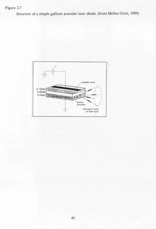
2.83 LASER DIODES (figure 2.7)
If the n-type and p-type semiconductors at the p-n junction are sufficiently well doped and a high enough current applied, then population inversion of electrons can be induced in the junction region.
For laser production, the junction is enclosed within an optical cavity. The opposite facets of the crystal are optically polished and coated with suitably reflecting dielectric layers. This represents the basic structure of diode lasers.
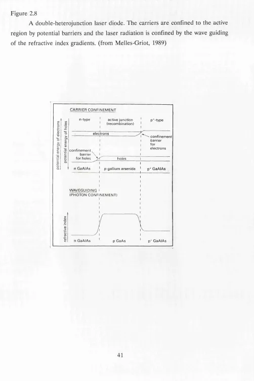
2.8.4 HETEROJUNCTION DIODES
although the emission of high power diodes (1 watt) is at present restricted to the near infrared region (780-840 nm).
The active layer of a gallium aluminium arsenide laser diode, is 0.2 microns thick and composed of GaAs and it is sandwiched between two layers of GaAlAs 1 micron thick. The dimensions of a laser diode are typically 0.5 x 0.2 x 0.1 mm- i.e. the size of a grain of salt.
2.9 CHARACTERISTICS OF LASER DIODES 2.9.1 TEMPORAL BEHAVIOUR
Laser diodes can be driven to run in a pulsed or continuous mode. High power laser diodes used in ophthalmology are CW lasers. Ion lasers will be generating laser light continuously throughout any treatment regime. The individual doses applied to the patient’s eye are achieved by a mechanical shutter which opens and shuts in relation to control signals from the foot switch. In the diode laser, the foot switch activates the laser and laser light is generated in CW mode for the selected duration of exposure. The diode laser is not active between exposures and this clearly has implications regarding the working life of such devices in comparison to ion lasers.
2.9.2 CURRENT-OUTPUT CHARACTERISTICS
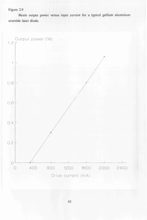
The output power of a laser diode is a function of the current flowing across the active junction. Figure 2.9 shows a typical plot of beam output power versus input current. Above the lasing threshold, there is a linear region of operation, allowing a high degree of confidence in the ability to modulate the laser output in a controlled fashion.
Figure 2.6
Idealised band structure of a p-n junction in a semiconductor. When current flows across the junction, holes and electrons recombine: i.e. electrons relax from the conduction band (a higher energy level, which is analogous to excited electronic orbitals of atoms and m olecules) to the valence band (a lower energy level, which contains electrons involved in the bonding of the crystal and which is analogous to the ground state in atoms or molecules).
The relaxation energy is released as photons (one per electron). The photo frequency is equal to E /h where E is the effective band gap and h is Planck’s constant, (from Melles Griot, 1989)
(a) N o v o l t a g e
j u n c t i o n p - t y p e
---s e m i c o n d u c t o r
n - t y p e ---s e m i c o n d u c t o r c o n d u c t i o n
b a n d h o l e s v a l e n c e
b a n d
(b) V o lt a g e a p p l i e d , c u r r e n t fl o w s , light e m i t t e d
r e c o m b i n a t i o n o f h o l e s a n d e l e c t r o n s
Figure 2.7
Structure of a simple gallium arsenide laser diode, (from Melles Griot, 1989)
m e ta ll ic strip
p* GaAs p GaAs
n GaAs b>—
a c t iv e ju n c t i o n
Figure 2.8
A double-heterojunction laser diode. The carriers are confined to the active region by potential barriers and the laser radiation is confined by the wave guiding of the refractive index gradients, (from Melles-Griot, 1989)
C A R R IE R C O N F I N E M E N T
a c t i v e j u n c t i o n ( r e c o m b i n a t i o n )
n - t y p e p * - t y p e e l e c t r o n s
c o n f i n e m e n t ba rr ie r fo r e l e c t r o n s c o n f i n e m e n t
barrie r
fo r h o le s h o l e s
n G a A I A s p g a ll iu m a r s e n i d e p* G a A I A s
W A VE G U ID IN G I ( P H O T O N C O N F IN E M E N T )
2.93 OUTPUT BEAM PROPERTIES a) Transverse mode structure
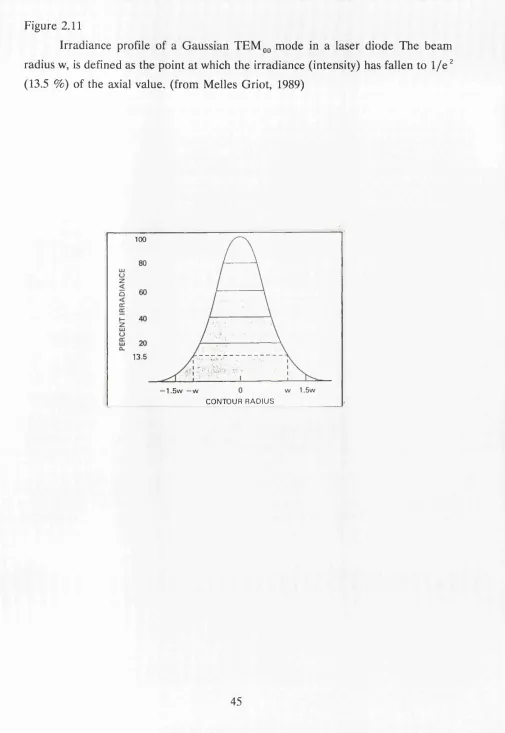
Because the junctions in diode lasers have a strange aspect position in that they are extremely narrow in the p-n axis, but relatively extensive in the long axis, the beams of light that they emit tend to be elliptical in cross section. This means that the energy distribution across the beam may be Gaussian on each meridian, but will vary in isoenergy points. The output of laser diodes consists of the lowest order transverse mode. This mode has a transverse intensity profile which is pseudo-Gaussian in shape (figure 2.11), which is analogous to that of many gas lasers.
b) Beam divergence and asymmetry
The divergence of a coherent Gaussian beam is inversely proportional to the radius of the beam waist from which it is diverging. The active region of a laser diode junction is small and asymmetrical. Consequently, the divergence of a laser diode beam is quite high. Beam divergence is typically between 12 and 30 degrees. When coliimation of the beam is required, it is necessary to expand the minor axis of the output, in order to eliminate asymmetrical divergence.
2.10 EFFICIENCY AND RELIABILITY O F LASER DIODES The electrical-optical conversion efficiency of laser diodes is high. Typically, about 20% of input power is converted to laser radiation. By comparison, the efficiency of a Nd-YAG laser is less than 4% and that of an argon or krypton laser is 0.05%. Two implications of the efficiency of diode lasers are that a one watt laser diode can be run by a 6 volt battery and that no external cooling system is necessary, since excess heat production is low.
Figure 2.9
Beam output power versus input current for a typical gallium aluminium arsenide laser diode.
O u t p u t p o w e r (W)
1.2
0,8
0,6
0,4
0,2
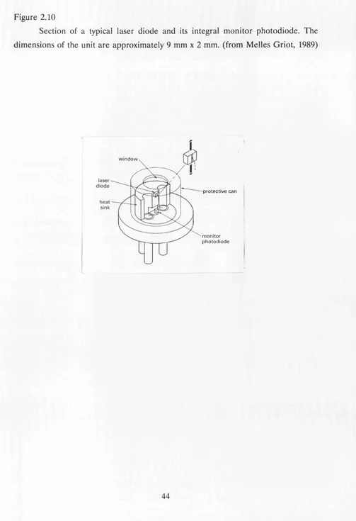
Figure 2.10
Section of a typical laser diode and its integral monitor photodiode. The dimensions of the unit are approximately 9 mm x 2 mm. (from M elles Griot, 1989)
w i n d o w
p r o t e c t i v e c a n
Figure 2.11
Irradiance profile of a Gaussian T E M 00 mode in a laser diode The beam radius w, is defined as the point at which the irradiance (intensity) has fallen to 1 / e 2 (13.5 %) of the axial value, (from Melles Griot, 1989)
100
13.5
— 1.5w —w
2.11 DEVELOPMENTS AND APPLICATIONS OF DIODE LASERS The development of mode-locked diode lasers with very high peak powers in the nanosecond or picosecond pulse range may result in exploration of their potential for photodisruptive treatment of ocular structures. Investigations are already being performed into the advantages of diode-pumped YAG lasers over the currently used flashtube as an energising source.
The wavelength of emission of a diode laser is influenced by the band gap within the semiconductor and this in turn depends upon its constituent materials. Compounds are being studied which can produce coherent radiation in the visible spectrum. A G a/A l/Ind/G aA s system can lase in the 580-680 nm range. A system comprising ZnSSe/GaAs could be developed to emit blue laser radiation. Other compounds are being developed which emit in the near and mid-infrared regions. If high powered lasers were available composed of these alternative compounds, then investigations could be performed into the ophthalmic applications of diode lasers which emit at a range of wavelengths.
2.12 CONCLUSIONS
CHAPTER 3
3.1 STUDY AIM
The essential aim of this project has been to develop a clinical diode laser and to examine its effectiveness as a therapeutic device in ophthalmology.
3 2 STUDY DESIGN
The design of this study required consideration of several problems. These problems may be categorised into technological, biophysical and clinical aspects.
3.2.1 TECHNOLOGICAL CONSIDERATIONS
The relative novelty of laser diodes implied the necessity for the design of an optical system that would allow effective transpupillary delivery of laser radiation to particular target structures within the eye. This system had to incorporate a number of features:
a) A visible aiming beam, that was coaxial with the infrared treatment beam.
b) A focusing and illumination system which was either an integral part of the laser device, or which was independently accessible, for example from a slit lamp microscope.
c) A means of preventing exposure of the operator to the treatment beam.
d) The facility to adjust the output characteristics of the laser, for example, laser power, spot size and pulse duration in a controlled fashion.
3.2.2 BIOPHYSICAL CONSIDERATIONS
Data on the histopathology of human ocular laser photocoagulation had been restricted to lasers which emit in the visible wavelengths (488-694 nm) (Marshall, 1967 and 1979) and lasers emitting at 1064nm (Van der Zypen, 1985).
a) The retinal mid-periphery b) The macula
c) The trabecular meshwork
3.23 CLINICAL CONSIDERATIONS
Prior to the present study, no clinical assessment had been performed of the ability of diode lasers to treat ocular conditions. Trials therefore had to be designed which would address the following elements of laser therapy:
a) The suitability and comparability of treatment regimens commonly used with conventional gas lasers, when applied to treatment with diode lasers. These aspects would be assessed by the rate and extent of regression of treated lesions.
b) The visual outcome of therapy. c) Laser-related side effects.
d) The ergonomics and the general reliability of diode lasers.
This aspect of the investigation was conducted in the form of pilot studies which examined the effects of diode laser photocoagulation in the treatment of the following conditions:
• Proliferative diabetic retinopathy • Exudative diabetic retinopathy
• Branch retinal vein throm bosis com plicated by neovascularisation of the optic disc, or of the retina
• Central retinal vein thrombosis complicated by established or threatened rubeosis iridis, or by optic disc neovascularisation.
• Chronic open angle glaucoma.
SECTION II
4.1 RETINAL STUDIES
4.1.1 DIODE LASERS USED IN RETINAL STUDIES Two lasers were used in the retinal histopathological studies.
a) For the initial exposures in rabbits a model SDL2420-H1 laser diode with 250 mW rated output and wavelength of 810 nm was used.
b) For subsequent rabbit and human exposures the laser was uprated to a SDL2430 laser with double the output power (500 mW). In some cases, to achieve higher power levels two laser outputs were combined to provide 800 mw output.
The diverging output beam from the diode was first collimated and then combined with a helium-neon beam for visual alignment and focusing. A simple lens arrangement allowed adjustment of the vergence of both laser diode and helium-neon beams for retinal focusing. Viewing of the retina was provided by a modified direct ophthalmoscope (figure 4.1 (a) and (b)). The laser diode was driven by a Spectra Diode Labs SDL800M laser diode driver with pulse duration and amplitude controlled by an external pulse generator. Power and energy levels at the eye were calibrated with a UDTS390 photometer and referenced to an EG and G radiometer 581. Both lasers operated in a continuous wave mode and exposure times of between 0.2 and 1.2 seconds were selected. The spot size of the treatment beam at the retina was 200 microns.
4.1.2 ARGON AND KRYPTON LASERS
4.2 DIODE LASERS USED IN MACULAR AND TRABECULAR HISTOLOGICAL STUDIES AND IN TH E CLINICAL TRIALS
Spectra Physics SDL2430 laser diodes were used with a spectral emission at 810 nm (figure 4.2). and with a maximum output power of up to 1.4 W. The laser diodes were driven by a eye were calibrated with a UDTS390 photometer and again referenced to an EG and G radiometer 581. Measurements demonstrated that transmission losses within the system were less than 5%. The laser operated in a continuous mode, but was a power on demand system.
Two forms of delivery system were employed in these phases of the study: a) A development of the original hand held version, which utilised a direct ophthalmoscope.
b) A model which could be attached to the tonometer stand of a standard Haag-Streit 900 slit lamp microscope (figure 4.3 (a)-(d)).
In the slit lamp version an aiming beam was provided by a red-emitting (680 nm) low power (300 microwatts) laser diode. Viewing of the retina was accomplished with the optics and illumination source of the slit lamp microscope in conjunction with a fundus contact lens.
In order to establish compatibility with the diode delivery system, three lenses were used in the clinical studies. These were the Goldmann three mirror; the Mainster lens; or the Rodenstock panfundoscope lens. Laser spot size was variable in 50 micron steps between 100 and 500 microns, but as with all lasers the absolute spot size at the retina varied slightly with choice of contact lens. The maximum output power of the laser was 1.4 watts. The exposure duration could be varied between 10 and 990 ms.
Figure 4.1 HAND-HELD DIODE LASER
(a) Hand-held version of diode laser, utilising a direct ophthalmoscope. (b) Section through hand-held version of the diode laser showing the major optical elements.
Key to figure 4.1 (b)
IR : Infrared treatment laser diode Aim : Aiming laser
Figure 4.3 SLIT-LAMP MOUNTED DIODE LASER
(a) Version of diode laser which is attachable to a standard slit lamp microscope, shown with instrument console.
(b) Diode laser shown mounted on Haag-Streit slit lamp microscope.
(c) Section through slit lamp-mounted diode laser which shows the principal optical components in the assembly (see key).
(d) Top: Optical arrangement of treatment laser diodes. A diverging beam from IR1 passes through a collimating lens (producing a parallel beam), two anamorphic prisms (expanding the beam) and a total internal reflection block. It then passes into a polarising beam combiner, where it joins the beam from IR2.
A diverging beam passes from IR2 and passes through a collimating lens, two anamorphic prisms and a half wave plate (rotating the beam through 90 degrees) and then into a polarising beam combiner. The combined beams are reflected by the dielectric mirror into the vertical telescope.
Bottom: Optical arrangement of treatment and aiming laser diodes. A diverging beam passes from the red laser diode and passes through a collimating lens. It is then reflected by a total internal reflection prism and passes through the dielectric mirror, after which it combines coaxially and collinearly with the infrared treatment laser path and is directed into the vertical telescope.
Key to figure 4.3 (c) and (d)
A l, A2 and A3 : Collimating lenses IR1 and IR2 : Infrared treatment lasers Aim : Aiming laser diode
X : Combined aiming and treatment laser beams
1 and 2 : Anamorphic prisms 3 : Half wave plate
Figure 4.3 (c)
P r o t e c t i ve f i I te r s
O p e r a to r
-*>—
Work i ng 0 i s t a n c e
s e l e c t o r lens
v i ew A
v i ew
Laser
di oaes
Laser head
a I i
Figure 4.3 (d)
v i e w A
A2
I R2
A3
AIM
In the clinical diode laser, filters with dielectric coatings were introduced into the laser head to prevent laser irradiation at 810 nm from entering the operator’s eye. These filters consisted of layers of high and low refractive index dielectric materials which have the property of allowing transmission of all wavelengths shorter than the emission wavelength of the treatment laser and the reflection of near- infrared wave fronts. These wave fronts are rendered in phase with each other by the coatings, which results in constructive interference. Dielectric filters therefore exhibit a high level of reflectance for the incident infrared beam from the treatment laser. A filter designed to produce this effect is known as a "hot mirror". The incorporation of a hot mirror into the laser head obviates the need for a mechanical safety shutter during the treatment exposures and it allows an unimpeded retinal view throughout all treatment sessions.
A photometer was used to monitor the level of infrared radiation passing back through the surgeon’s eye pieces during exposure at maximum power. Less than 20 microwatts was measured at the eyepieces, which is well within the safety limits for laser exposure.
SECTION HI
CHAPTER 5
5.1 RETINAL EXPOSURES 5.1.1 ANIMAL STUDIES
Three six month old Dutch rabbits were used in this study. Animals were anaesthetised with intramuscular fentanyl and intravenous valium. Cyclopentolate 1% was instilled in both eyes for mydriasis. The animals were placed on a positioning board, with the target eye held open with a lid speculum.
Transpupillary photocoagulation was performed to both eyes using a diode laser at power levels which varied between 100 mW and 400 mW. The laser spot size was 200 microns and the exposure times were varied between 0.20 s and 1.2 s. The position and ophthalmoscopic visibility of all the lesions produced were recorded.
The rabbits were sacrificed immediately following the last exposure with an overdose of intravenous sodium pentobarbitone and after death, the eyes were removed for histological examination.
5.1.2 HUMAN STUDIES
Subsequent to the granting of permission by the relevant local ethical committee, three patients agreed to take part in the diode laser study. All three patients were going to undergo enucleation because of malignant melanoma. Two had ciliary body malignant melanomas and one a juxtapapillary melanoma. All had partial retinal detachments which involved the macula and in two cases the tumour partially obscured the pupil. In these cases the loss of pupillary area would result in a proportional attenuation of the radiation incident upon the retina (because of the large cone angle of the incident beam) and therefore the figures of radiant power in table I may be erroneously high for a given retinal reaction.
patient during photocoagulation. Immediately following treatment, colour fundal photography was performed. Eighteen hours following laser exposure, enucleation was performed under general anaesthetic and the eye prepared for histopathological study.
For comparison, fresh sections were prepared from tissue derived from an earlier study in which sample argon (200 microns, 300 mW), and krypton lesions, (200 microns, 600 mW) had been produced with exposure durations of 0.2 to 0.5 seconds in a melanoma eye (Marshall and Bird, 1979).
5.2 HUMAN MACULAR EXPOSURES
A submission was made to the local ethical committee, which gave approval for the study to proceed. For the diode exposures two patients, each with a choroidal melanoma which required enucleation were given a full explanation of the nature of the trial prior to giving consent to the procedure being carried out. The first subject was a 68 year old male, with a tumour of the left eye which was situated nasal to the optic disc. Although there was subretinal fluid causing a serous detachment of the nasal retina, at the time of examination and treatm ent the macula had a normal ophthalmoscopic appearance (but see histology section).
Mydriasis was accomplished with cyclopentolate 1% and following topical anaesthesia, a Goldmann contact lens was applied. A total of 63 exposures were applied to the macula in a "grid" pattern that was centred on the fovea, although no burns were applied to the foveola. The spot size was 300 microns and the exposure duration 0.25 seconds. The output was varied between 400 and 800 mW. Enucleation was performed five weeks subsequent to the exposures. No discomfort was reported by the patient during photocoagulation.
In an eye from an earlier study (Marshall and Bird, 1979), sample argon (50 microns, 87 mW), and krypton lesions, (50 microns, 200 mW) had been produced with exposure durations of 0.2 to 0.5 seconds. The argon exposures were made in a line superonasal to the inferotemporal axis and which traversed the fovea. A similar number of krypton exposures were made at similar distances from the fovea at right angles to the line of argon burns. This eye had been enucleated 20 hours after photocoagulation, again due to the presence of a malignant melanoma of the anterior choroid. Microscopic analysis was carried out on ocular tissue from this experiment using an identical preparative procedure to that used for diode-irradiated specimens.
5 3 MICROSCOPIC STUDIES ON RETINAL TISSUE
Immediately following enucleation, a 5 mm penetrating incision was made at the ora serrata and the eyes immersed in 100 mis of fixative. This initial solution contained 2.5% glutaraldehyde buffered in 0.1 M sodium cacodylate containing 10 mg/ml calcium chloride and with a final pH of 7.4. In rabbits the anterior half of the globe was removed, together with the lens, iris and vitreous and discarded. In humans the globe was hemisected, usually obliquely and in such a fashion that the irradiated area was totally isolated from that portion containing the tumour so that routine diagnostic procedures could be carried out on the latter. The retinal lesions of both species were photographed under glutaraldehyde onto Kodak Ektachrome EPY 135 film using a macrophotographic system (Olympus).
Photography was performed using Kodak Technical PAN 2415 film.
Sections for transmission electron microscopy (TEM) were cut using diamond knives in a Reichert OMU4 ultramicrotome. They were mounted on 200 mesh copper grids and stained with uranyl acetate and lead citrate before being examined in an AE1 801 transmission electron microscope. Sections were photographed onto Ilford Technical Film.
5.4 HUMAN TRABECULAR MESHWORK EXPOSURES
A submission was made to the local ethical committee, which gave approval for the study to proceed. A full explanation of the nature of the trial was given to the patient, who then gave consent for the procedure. The subject was a 65 year old female with a right ciliary body malignant melanoma. Gonioscopy prior to treatment and subsequent microscopic examination confirmed that the tumour did not involve the trabeculum.
Topical anaesthesia was instilled and a Goldmann contact lens was applied to the eye. Three quadrants of the trabecular meshwork were irradiated with the diode laser. A total of 70 burns were applied. The target area was the central, pigmented portion of the trabeculum and the aim was to produce a blanching reaction. The spot size was 100 microns, the exposure duration was 200 ms and the required output power varied between 750 and 1200 mW.
For comparison, an argon blue-green laser was used to apply a further 25 bums to the remaining quadrant of the same eye. In common with the diode exposures, the end point was a visible blanching of the pigmented region of the trabeculum. The spot size was 100 microns, the exposure duration was 200 milliseconds and the power needed to produce a visible reaction varied between 500 and 1000 milliwatts. Gas bubble formation was not observed following either diode or argon irradiation. Enucleation was performed 18 hours following treatment.
5.5 MICROSCOPIC STUDIES ON TRABECULAR TISSUE
out on the latter.
Meridional sections of the iridocorneal angle were isolated under a dissecting microscope. The samples were trimmed in such a way that the laser employed to irradiate a particular portion of trabeculum and the power that was used could be identified.
Tissue for light and transmission electron microscopy was washed briefly in 0.1 M sodium cacodylate buffer containing 7.5% sucrose and post-fixed for one hour in 2% osmium tetroxide buffered in 0.1 M sodium cacodylate. Samples were dehydrated through a graded series of concentrations of ethanol in water and embedded in araldite via epoxypropane.
Sections for light microscopy (LM) were cut at 1 micron on glass knives mounted in a Huxley Mark 1 ultramicrotome and stained with toluidine blue.
Silver sections for transmission electron microscopy (TEM) were cut using diamond knives in a Reichert OMU4 ultramicrotome. They were mounted on 200 mesh copper grids and stained with uranyl acetate and lead citrate before being examined in an AE1 801 transmission electron microscope.
Specimens for scanning electron microscopy were post-fixed overnight in 2% osmium tetroxide buffered in 0.1 M sodium cacodylate. They were dehydrated through a series of ascending concentrations of acetone before being critical-point dried (Samdri 780). Dried samples were coated with a 30 nm layer of gold in a sputter coater (Emscope) prior to being examined in a Hitachi 520 scanning electron microscope and photographed onto Kodak Tri-X PAN 5-TX 120 film.
5.6 RESULTS OF RETINAL STUDIES
5.6.1 MACROSCOPIC FEATURES OF RETINAL BURNS