R E V I E W
Open Access
X-linked disorders with cerebellar dysgenesis
Ginevra Zanni
*and Enrico S Bertini
Abstract
X-linked disorders with cerebellar dysgenesis (XLCD) are a genetically heterogeneous and clinically variable group of disorders in which the hallmark is a cerebellar defect (hypoplasia, atrophy or dysplasia) visible on brain imaging, caused by gene mutations or genomic imbalances on the X-chromosome. The neurological features of XLCD include hypotonia, developmental delay, intellectual disability, ataxia and/or other cerebellar signs. Normal cognitive development has also been reported. Cerebellar dysgenesis may be isolated or associated with other brain malformations or multiorgan involvement. There are at least 15 genes on the X-chromosome that have been constantly or occasionally associated with a pathological cerebellar phenotype. 8 XLCD loci have been mapped and several families with X-linked inheritance have been reported. Recently, two recurrent duplication syndromes in Xq28 have been associated with cerebellar hypoplasia. Given the report of several forms of XLCD and the excess of males with ataxia, this group of conditions is probably underestimated and families of patients with
neuroradiological and clinical evidence of a cerebellar disorder should be counseled for high risk of X-linked inheritance.
Disease names and synonyms
X-linked Congenital Ataxias
X-Linked Disorders/Syndromes with Cerebellar Dysgenesis
Definition and classification
The term X-linked disorders with cerebellar dysgenesis (XLCD) is used here to describe an emerging group of rare conditions in which the hallmark is a cerebellar defect, visible on neuroimaging, caused by gene muta-tions or genomic imbalances on the X-chromosome. A classification of this group of rare disorders is still diffi-cult because in most cases the pathogenesis is unknown. The best characterized forms are X-linked syndromes with associated cerebellar hypoplasia due to OPHN1 or CASKgene mutations. In other X-linked disorders like Fragile X, Oral-facial-digital type I or Opitz GBBB syn-dromes, cerebellar defects are increasingly being recognized.
Epidemiology
The overall incidence of congenital cerebellar malforma-tions is high, 1/4,000-5,000 live births, the contribution of X-linked forms is probably underestimated.
Clinical and molecular description
a. X-linked non-progressive congenital ataxias
Congenital ataxias (CA) were first defined by Batten in 1905 [1] as “cases in which ataxia has been noted early in life and in which there is a tendency to gradual improvement”. This heterogeneous group of disorders accounts for 10% of non-progressive infantile encepha-lopathies [2]. The first X-linked form was described in a large family of Eastern Russian descent with seven affected males over three generations. All patients had markedly delayed early developmental milestones, cere-bellar ataxia, external ophthalmoplegia, pyramidal signs, normal cognitive development and a non-pro-gressive course. Neuroimaging studies revealed marked hypoplasia/atrophy of the cerebellar vermis and hemi-spheres. Linkage studies mapped the gene to a large genetic interval on Xp11.21-Xq24 with a maximum lod score of 4.66 at DXS1059 [3]. A second family was reported with a similar phenotype, the propositus and his maternal uncle manifesting severe hypotonia at birth, psychomotor delay, slow eye movements, non progressive ataxia and normal intelligence [4]. Neuroi-maging studies revealed global cerebellar hypoplasia/ atrophy not evident in the first years of life. Despite the cerebellar changes, the clinical course was non-progressive, as it is expected in this group of ataxias. A genetic interval, partially overlapping with the first * Correspondence: [email protected]
Unit of Molecular Medicine, Departement of Neurosciences, Bambino Gesù ediatric Research Hospital, 4 Piazza S. Onofrio, 00165 Rome, Italy
reported family, was identified by exclusion mapping. A large family of Norwegian descent with six affected males over three generations with non-progressive X-linked congenital ataxia and normal cognitive develop-ment was reported [5]. Hypotonia and overall delay of motor development were noted since birth. Nystagmus, dysarthria and trunkal ataxia were present. Neuroima-ging studies showed global hypoplasia/atrophy of the cerebellum, predominant in the vermis (Figure 1a-c) The condition was mapped to a genetic interval of 12 Mb at Xq25-q27.1 with a maximum lod score of 3.44 at DXS1192 not overlapping with the previously identi-fied locus, indicating that there are at least two genes responsible for this rare forms of X-linked congenital ataxia.
b. X-linked genes/syndromes with cerebellar dysgenesis Oligophrenin-1 syndrome
TheOPHN1syndrome (MIM 300486) was first described in a 12 year-old girl, carrier of ade novotranslocation t (X;12)(q11;q15) which was found to disrupt the Oligo-phrenin-1(OPHN1)gene at Xq12 and encodes a RhoGT-Pase activating protein (RhoGAP) involved in synaptic morphogenesis and function [6-8]. The girl had congeni-tal hypotonia, severe developmencongeni-tal delay, moderate cog-nitive impairment, early-onset complex partial seizures and bilateral divergent strabismus and dysmetria. At a later reevaluation, Brain MRI showed a posterior vermis dysgenesis including partial agenesis of lobules VI and VII associated with right vermian parasagittal cleft and mild cerebellar hemisphere hypoplasia. A consistent
dilatation of the lateral cerebral ventricles and mild corti-cal atrophy was also present. A similar clinico-radiologi-cal phenotype (Figure 1a-c) was found in the affected males of a family carrying a single nucleotide deletion in exon 19 of theOPHN1, initially diagnosed with non-syn-dromic X-linked mental retardation [9]. Another deletion comprising exon 19 of theOPHN1gene, was identified in a family with five affected males showing moderate to severe mental retardation, infantile-onset epilepsy, hypo-tonia, strabismus and ataxia. Cryptorchidism and genital hypoplasia were present in all affected males. Neurora-diological findings included cortical atrophy, enlargement of the cerebral ventricles, bilateral hypoplasia of the head of the caudate nucleus, lower vermis and cerebellar hemi-sphere hypoplasia. One carrier sister had mild learning disabilities and strabismus but her brain MRI was normal [10]. A stop mutation in exon 3 and an 8 base-pair inser-tion in exon 9 of theOPHN1gene were identified in two other families with a similar phenoype [11]. In a family with a 2 base-pair deletion in exon 8 ofOPHN1leading to a premature stop codon, the affected males showed developmental and cognitive delay with IQ ranging from 46 to 54, strabismus, early-onset generalized tonic-clonic seizures, abnormal behaviour and a characteristic facial phenotype with long face, prominent forehead, infraorbi-tal creases, deep set eyes, upturned philtrum and large ears. The obligate carrier females showed only mild cog-nitive impairment and subtle facial changes. Brain MRI studies performed in the propositus at age 3 months showed underdeveloped frontal lobes, loss of brain volume with enlarged lateral ventricles, prominent subar-achnoid spaces and a particular square shape of the fron-tal horns. Cerebellar hypoplasia and vermis dysgenesis with a large cisterna magna and a retrocerebellar cyst were noted [12].
Ade novotranslocation disrupting theOPHN1gene in a female patient and intragenic deletions ofOPHN1detected by arrayCGH, in a male patient and in two families in which affected individuals all showed the characteristic clinico-radiological phenotype associated withOPHN1 syndrome were also reported [13-16]. In one family with six affected males with intellectual deficit, minor facial anomalies, cubitus valgus, with or without sensorineural deafness, a deletion of exons 16-18 in theOPHN1gene was identified [17]. ScreeningOPHN1in a large cohort of patients led to the identification of mutations in 12% of individuals with intellectual deficit and cerebellar hypopla-sia, suggesting that the screening of this gene should be implemented in boys with vermis hypoplasia, enlarged cer-ebral ventricles and developmental delay [18].
CASK syndrome
CASKsyndrome (MIM 300749) was first described in a 4 year-old girl, carrier of an apparentlyde novoparacentric inversion 46X, inv(X)(p11.4p22.3) disrupting the
acuity. Unsteady gait and seizures were present in some but not all affected individuals. In 2 additional families with apparently non-syndromic XLMR, novel missense mutations in exon 8 and 27 of theCASKgene were iden-tified [23]. Thus the phenotypes associated withCASK mutations range from mild MR with or without congeni-tal nystagmus, to severe cognitive impairment associated with cerebellar and pontine hypoplasia and abormalities of cortical development.
Christianson syndrome
Christianson syndrome (MIM 300243) was first described in a South African family of Norwegian descent in which 16 affected males manifested profound MR, absence speech, early-onset generalized tonic-clonic epilepsy, bilat-eral ophthalmoplegia and truncal/gait ataxia. Three of the 10 obligate carrier females had mental retardation. In the patients, developmental milestones were severely retarded and hypotonia was present. The chest was narrow, limb muscles were poorly developed, and joints were stiff and contractured. MRI scan showed a prominent cisterna magna, enlarged 4th ventricle, cerebellopontine and supra-cerebellar cisterna, hypoplasia/atrophy of the cerebellum predominantly of the vermis and pons. Most of the affected males in this family died prematurely, around age 25-30 years. Neuropathological examination showed an atrophic cerebellum, especially of the vermis. The molecu-lar layer displayed microcystic changes and there was widespread neuronal loss in the granular and PC layers, in the pons and in the hippocampi. The condition was mapped by linkage analysis studies to Xq24-q27 [24]. In this family, a 2 base-pair deletion within the coding sequence of the Solute Carrier family 9 member 6 (SLC9A6)was identified [25].SLC9A6is located at Xq26.3 and encodes a sodium/hydrogen exchanger protein 6 (NHE6) which regulates the endoluminal pH of early endosomes involved in the trafficking of proteins essential for growth and maintainance of dendritic spines [26]. Additional mutations ofSLC9A6: a 6 base-pair deletion, a nonsense mutation R468X, a splice site mutation causing skipping of exon 3, were identified in three other families with an Angelman-like phenotype characterized by devel-opmental delay, postnatal microcephaly and epilepsy occurring between 9 and 26 months of age. Patients exhib-ited ataxia, hyperkinetic movements and frequent smiling with episodes of unprovoked laughter, open mouth, pro-fuse drooling and swallowing difficulties, gastroesophageal reflux and thin body habitus. MRI studies performed 3 years later in one affected child showed a mild progression of the cerebellar atrophy. Proton Magnetic Resonance spectroscopy (MRS) showed elevated glutamate/glutamine in the basal ganglia of affected individuals. An additional family of 6 affected males with severe mental retardation, absent speech, autism spectrum disorder, epilepsy, late-onset ataxia, weakness and dystonia with stereotyped hand
movements was reported and an in-frame 9 base-pair deletion inSLC9A6was identified. Brain MRI showed moderate ventricular enlargement and sulcal widening due to atrophy of the brain, thin corpus callosum but normal cerebellum and brainstem. Post-mortem examination of two affected males showed widespread neuronal loss and deposits of the microtubule-binding protein tau in cortical and sub-cortical regions which are generally the defining neuropathological characteristics of a number of adult-onset neurodegenerative disorders but had never been seen before in early- onset cognitive delay or autism [27]. Fragile × syndrome
The Fragile × (FRAXA) syndrome (MIM 300624), is the most frequent cause of monogenic mental retardation with an estimated prevalence of 1/4000 males and 1/ 8000 females. In addition to cognitive deficits, the phe-notype of the syndrome include mild facial anomalies (prominent jaw, high forehead, large ears), and macro-orchidism. Many patients also manifest attention deficit hyperactivity disorder and autistic-like behavior. The syndrome is caused by a massive (more than 200) CGG triplet expansion in the 5’ untranslated region of the FMR1gene which results in transcriptional silencing of the gene [28]. Loss of function of the FMRP protein leads to abnormalities of dendritic spine maturation associated with altered synaptic plasticity and cerebellar motor learning (eyeblink conditioning) also seen in FRAXA patients [29]. A comparative study of the pos-terior fossa of a group of mutated patients and a group of age-matched controls revealed a significant reduction of the posterior vermis (lobules VI-VII) in the Fragile × population [30,31]. These data were recently confirmed by MRI morphometric studies in 3D [32].
Fragile X-associated Tremor/Ataxia syndrome
begin at or before birth [34]. Neuroimaging studies of affected individuals show global loss of brain volume, in particular cerebellar and cortical atrophy and hyperin-tensity white matter lesions around the periventricular area and in the middle cerebellar peduncles [35]. Hoyeraal-Hreidarsson syndrome
Hoyeraal-Hreidarsson syndrome (HHS) (MIM 300240) is a rare X-linked recessive disorder, characterized by intrauterine growth retardation, microcephaly, intellec-tual deficit, bone marrow failure, cancer predisposition and cerebellar hypoplasia [36]. It is caused by mutations ofDCK1gene located on Xq28, encoding the nucleolar protein dyskerin which interacts with the human telo-merase RNA complex [37]. Detailed neuroimaging stu-dies of an HHS patient showed global cerebellar hypoplasia, a small brainstem, thin corpus callosum and cerebral calcifications [38].
X-linked sideroblastic anemia with ataxia
X-linked sideroblastic anemia with ataxia (XLSA/A) (MIM 301310) is a rare X-linked disorder characterised by early onset slowly progressive ataxia associated with a sideroblastic anemia due to mitochondrial iron accu-mulation. It is caused by mutation of the ABC7 gene which maps to Xq13 and encodes an ATP-binding mitochondrial iron transporter located in the inner mitochondrial membrane, involved in mitochondrial iron homeostasis as well as in the maturation of cytoso-lic iron-sulfur proteins [39]. Neuroimaging studies showed global cerebellar hypoplasia with signs of atro-phy in affected individuals [40].
Oral-Facial-Digital type I/X-linked Joubert syndrome Oral-Facial-Digital type I (OFDI) (MIM 311200) is a rare male lethal X-linked dominant syndrome charac-terised by malformations of the mouth, face and digits in affected females. CNS anomalies have been found in 40% of ODFI patients, in particular vermis hypoplasia, corpus callosum agenesis, hydrocephalus and periventri-cular heterotopia [41].OFD1maps to Xp22 and encodes a ciliary protein.OFD1 mutations have been identified in two families with retinitis pigmentosa, postaxial poly-dactyly and the molar tooth sign characteristic of Jou-bert and JouJou-bert-related syndromes [42,43].
X-linked Opitz/GBBB syndrome
Opitz GBBB syndromes (MIM 300000) are X-linked reces-sive or autosomal dominant conditions characterised by developmental delay and midline malformations: hyperte-lorism, cleft lip/palate oesophagolaryngotracheal defects, imperforate anus and hypospadias [44,45]. The X-linked form is caused by mutations in theMID1gene which maps to Xp22.22 and encodes a microtubule-associated protein involved in ribonucleoprotein complex stabiliza-tion and ubiquitinastabiliza-tion [46,47]. Anterior vermis hypoplasia and corpus callosum abnormalities were found in more than one third of patients withMID1mutations [48,49]
and lack ofmid1in the mouse causes abnormal develop-ment of the anterior cerebellar vermis [50].
X-linked heterotaxy
X-linked heterotaxy (MIM 306955) is caused by muta-tions of the Zinc finger transcription factorZIC3located in Xq26 and encodes a transcription factor involved in the establishment of the left/right axis in early embryo-nic development [51]. Vermis hypoplasia is found in X-linked heterotaxy and has been reported in males hemi-zygous forZIC3mutations [52,53]. Cerebellar dysgenesis predominant in the flocculo-nodular lobe was described inzic3deficient mice [54].
X-linked cortical dysgenesis
The gene DCX located in Xq22 and encodes the Dou-blecortin protein, mutated in 40% of type I lissence-phaly. This cortical dysgenesis is associated with severe mental retardation and seizures in affected males and subcortical laminal heterotopia in heterozygous females [55,56]. Doublecortin is a microtubule associated protein which participates in the assembly and stabilization of microtubules in migrating neuronal cells. 25% of male patients withDCXmutations have mild cerebellar hypo-plasia, predominant in the vermis [57,58].
TheARXgene located in Xp21, encodes an Aristaless-related homeobox transcription factor involved in multi-ple aspects of cortical development [59,60]. In a severe case of X-linked lissencephaly, absent corpus callosum and ambiguous genitalia (XLAG) due to a mutation in ARX, neuroimaging studies showed a hypoplastic vermis and enlarged 4th ventricle [61].
The gene FLNAmaps to Xq28 and encodes Filamin 1, a protein interacting with F-actin, involved in brain neu-rogenesis and neuronal migration [62].FLNA mutations cause several different syndromes with a predominant skeletal phenotype: Oto-palato digital type I and II, Mel-nick Needles dysplasia, frontometaphyseal dysplasia, FG-like syndrome [63]. Mutations of FLNAhave also been found in a high proportion of individuals with bilateral periventricular nodular heterotopia, megacisterna magna, cardiovascular malformations and epilepsy. Almost all patients with FLNA mutations had mild to moderate cerebellar hypoplasia [64].
Rett syndrome
stereotyped hand movements, breathing irregularities, seizures, epilepsy, loss of speech, hypotonia, ataxia and hyperreflexia. MRI studies of young Rett patients have demonstrated the presence of a mild cerebellar hypopla-sia especially of lobules I-IV, whereas in adult patients a progressive cerebellar atrophy was detected and also confirmed by post-mortem neuropathologic studies of the cerebellum [66,67].Mecp2mouse mutants show cer-ebellar and hyppocampal-based learning deficits and develop gait incoordination [68].
Xq28 microduplication syndromes
Duplications of theMECP2 region (MIM300260), cause a syndrome of infantile hypotonia, profound intellectual deficit, autistic features and recurrent infections [69]. Neuroradiological evidence of progressive cerebellar atrophy, emerging in the second decade of life was recently reported in males of three families carrying duplications of theMECP2region [70].
In 4 families with X-linked intellectual disabilities and a copy number gain of an identical 0.3 Mb region in chro-mosome band Xq28 including 19 annotated genes includ-ing FLNAandGDI1 (an X-linked mental retardation gene) but notMECP2, affected patients presented with hypotonia, severely delayed psychomotor development, mild ataxic gait, strabismus or nystagmus but no other cerebellar signs. MRI of the brain showed mildly enlarged fourth ventricle and ventricular dilatation in two unrelated patients and a classical Dandy Walker malformation with cerebellar hypoplasia and agenesis of the corpus callosum
in two other affected males. Minor facial anomalies were present in all patients [71]. A fifth family with 3 affected males carrying an identical Xq28 duplication and showing mild to moderate intellectual disability associated with ver-mis hypoplasia was reported [72].
X-linked hydrocephalus
X-linked hydrocephalus with aqueductal stenosis (MIM 30700) is the most common form of hereditary hydroce-phalus with an incidence of 1/30,000 in male neonates. Most cases of X-linked hydrocephalus, are caused by mutations in theL1CAM gene, which encodes a cellular adhesion protein implicated in the regulation of vesicle cycling through its interaction with proteins of clathrin-mediated endocytosis [73]. In patients with loss of func-tion mutafunc-tions ofL1CAM, global vermis hypoplasia or anterior vermis hypoplasia has been observed [74].
In a few families with X-linked mental retardation and hydrocephalus mutations of AP1S2(sigma subunit of the clathrin adaptor protein complex 1) were identified. Brain MRI imaging of affected males in one family showed ventricular dilatation without aqueductal steno-sis, calcifications of the basal ganglia, and mild vermis hypoplasia [75,76]. Loss of function of the clathrin adap-tor AP1 determines defects in synaptic vesicle cycling with accumulation of large endosome-like vacuoles, defects which are paired with reduced motor coordina-tion and long term spatial memory in mutant mice [77].
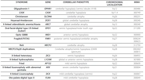
A list of genes implicated in X-linked cerebellar dys-genesis is provided in Table 1.
Table 1 List of genes implicated in X-linked cerebellar dysgenesis.
SYNDROME GENE CEREBELLAR PHENOTYPE CHROMOSOMAL
LOCALIZATION
MIM#
Oligophrenin-1 OPHN1 cerebellar hypoplasia (vermis lobules VI-VII) Xq12 300486
CASK CASK cerebellar hypoplasia Xp11.4 300749
Christianson SLC9A6 cerebellar atrophy Xq26 300231
Hoyeraal-Hreidarsson DCK1 global cerebellar hypoplasia Xq28 300240 X-linked sideroblastic anemia/Ataxia ABC7 global cerebellar atrophy Xq13 300240
Oral-facial-digital type I/X-linked Joubert
OFD1 vermis hypoplasia/molar tooth sign Xp22 311200
Opitz GBBB MID1 anterior vermis hypoplasia Xp22 300000
FragileX/FXTAS FMR1 posterior vermis hypoplasia/cerebellar atrophy Xq27 300624/ 300623
Rett MECP2 cerebellar atrophy Xq28 312750 MECP2/Xq28 duplications - Cerebellar atrophy/vermis hypoplasia (DWM
spectrum)
Xq28 300260/
300815
X-linked heterotaxy ZIC3 vermis hypoplasia Xq26 306955
X-linked hydrocephalus L1CAM global or anterior vermis hypoplasia Xq28 307000
Fried AP1S2 mild vermis hypoplasia Xp22 300630
X-linked lissencephaly with abnormal genitalia
ARX severe cerebellar hypoplasia Xp21 300004
c. X-Linked loci/syndromes with cerebellar dysgenesis - A four generation family of Dutch descent with nine affected males presenting with severe cognitive delay, early hypotonia with progression to spasticity and con-tractures, choreoatetosis, seizures and a long narrow face with coarse features was reported [78]. Brain ima-ging studies revealed a cystic enlargement of the 4th ventricle with cerebellar hypoplasia and iron accumula-tion in the basal ganglia with neuroaxonal dystrophy. The disease was mapped to the long arm of the X-chro-mosome with a maximum lod score of 2.19 at DXS425 in Xq25-q27 [79].
- A family of Italian descent has been reported with 7 affected males, two of them showing severe congenital ataxia, generalized hypotonia, psychomotor delay, recur-rent bronchopulmonary infections later developing myo-clonic encephalopathy and macular degeneration [80]. Serial neuroimaging studies showed cerebellar vermis and corpus callosum hypoplasia, a cyst of the septum pellucidum and persistence of the cavum vergae. The other five affected males in the family had recurrent bronchopneumonia with severe congenital hypotonia and died within the first years of life. Immunologic investigations only showed a reduced level of IgG2 sub-class in one sib. The condition was mapped to the short arm of the X-chromosome with a maximum lod score of 2.48 at DXS7099 in Xp22.33-pter [81].
- A family with a syndromic form of X-linked mental retardation characterized by absent speech, truncal ataxia, contractures and hypoplasia of the cerebellum and the brainstem revealed at neuroimaging studies was reported. Only one of the affected males developed sei-zures and showed changes in the basal ganglia suggest-ing iron deposits. The condition was mapped to Xq23-q24 [82].
- A family in which affected males showed congenital cerebellar hypoplasia, microcephaly, short stature, pro-found developmental delay, blindness, deafness and sei-zures was described. Death occurred in infancy or early childhood. This syndrome was mapped to Xq24-Xq27 [83]. - A family with 7 affected males presenting moderate to severe intellectual impairment and infantile spams was reported. Neuroimaging studies showed cerebellar atrophy or corpus callosum hypoplasia or both, in half of the affected individuals. The condition was mapped to Xp11.4-p22.11 [84].
- A family with 3 males in two generations affected with moderate intellectual deficit and prominent gla-bella, synophris, prognathism, generalized hirsutism and bilateral single palmar creases was described. All patients developed seizures in childhood and two of them showed a progressive gait disturbance and pro-longed nerve conduction velocity and hypogammaglobu-linemia. Neuroimaging studies in one patient revealed
cerebellar atrophy. The disorder was mapped to Xq21.33-Xq23 [85].
Note: The clinical features of other XLCD for which no mapping data are available [86-93] have been reviewed by [5].
Diagnosis
Family history is important to exclude autosomal or other forms of inheritance. In sporadic males or docu-mented X-linked familiar cases, genetic testing, with the possible exception ofFMR1, should be performed only in well selected patients with a specific clinical and neu-roradiological phenotype as described in each paragraph of the present review. A precise nosological classification and a correlation between the neuroradiological pheno-types (cerebellar hypoplasia, atrophy or dysplasia, brain-stem and cerebellar hypoplasia, molar tooth sign) and the genotype is still lacking but will certainly improve as the genetic causes of these conditions are being identi-fied and also thanks to the advancement of neuroima-ging techniques (eg. tractography, voxel-based morphometry). On the X-chromosome, a high incidence of causal deletions and microduplications (pathogenic copy number variations) was found, demonstrating that dysregulated expression of tightly regulated genes can disturb normal brain and cognitive development [94]. At least 1/3 of cases withOPHN1or CASKsyndrome were found to be carriers of a chromosomal or subchromoso-mal abnorsubchromoso-mality. It is therefore recommended, in patient with a neuroradiologically documented cerebellar disorder, especially when two or more dysmorphic fea-tures are present, to perform standard karyotype fol-lowed by array-CGH studies.
Genetic counseling and prenatal diagnosis
Given the report of several forms of X-linked cerebellar disorders and the excess of males with ataxia [95,96], families of children with congenital cerebellar hypopla-sia, atrophy or Dandy-Walker malformation with or without intellectual disability, should be counseled for high risk of X-linked inheritance. The identification of the molecular defect in couples at risk, allows early pre-natal testing whereas foetal brain neuroimaging may prove uninformative.
learning problems is important because most of these conditions are syndromic. A cognitive and behavioural assessment (Wechsler scales, Vineland) is essential because X-linked cerebellar disorders are frequently associated with mild to severe developmental/cognitive delay. A Metabolic work-up (lactate/pyruvate ratio in blood and spinal fluid, lysosomal enzymes, isoelectrofo-cusing of transferrin, creatin kinase, very long chain fatty acids, plasma and urinary aminoacids and urinary organic acids chromatography) together with nerve con-duction studies should be performed to exclude meta-bolic or neuromuscular causes (pyruvate dehydrogenase deficiency, X-linked adrenoleukodystrophy, X-linked dystrophinopathies). Neuroimaging studies of the cere-bellum with particular attention to the brainstem and cerebral cortex, can be fundamental for the differential diagnosis between different X-linked cerebellar syn-dromes and other non cerebellar X-linked disorders that can present with ataxic symptoms (e.g Arts syndrome, characterized by ataxia, deafness and neuropathy). A clinical and neuroradiological follow-up is also necessary for the assessment of the natural history of the condi-tion. Rehabilitation strategies should be planned for motor, cognitive and behavioral difficulties and specific manifestations such as visual impairment and seizures.
Acknowledgements
The author is grateful to John M Opitz (University of Utah), Eugen Boltshauser (University of Zuerich) and Jamel Chelly and his research group at the Institut Cochin (UMR8104, Université Paris Descartes) for help-ful discussions and advices. This work was supported by a grant from Telethon Italy (projects GGP08145 and 492B) and the Italian Ministry of Health.
Abbreviations
CA: Congenital ataxia; DWM: Dandy Walker malformation; XLCD: X-linked Cerebellar Dysgenesis.
Authors’contributions
The present review was conceived by both authors, GZ drafted the manuscript. All authors read and approved the final manuscript.
Competing interests
The authors declare that they have no competing interests.
Received: 2 November 2010 Accepted: 15 May 2011 Published: 15 May 2011
References
1. Batten FE:Ataxia in childhood.Brain1905,28:484.
2. Steinlin M:Nonprogressive congenital ataxias.Brain Dev1998,20:199-208. 3. Illarioshkin SN, Tanaka H, Markova ED, Nikolskaya NN,
Ivanova-SmolenskayaI A, Tsuji S:X-linked nonprogressive congenital cerebellar hypoplasia: clinical description and mapping to chromosome Xq.
Ann Neurol1996,40:75-83.
4. Bertini E, des Portes V, Zanni G, Santorelli F, Dionisi-Vici C, Vicari S, Fariello G, Chelly J:X-linked congenital ataxia: a clinical and genetic study.Am J Med Genet2000,92:53-56.
5. Zanni G, Bertini E, Bellcross C, Nedelec B, Froyen G, Neuhäuser G, Opitz JM, Chelly J:X-linked congenital ataxia: a new locus maps to Xq25-q27.1.
Am J Med Genet2008,146:593-600.
6. Bienvenu T, Der-Sarkissian H, Billuart P, Tissot M, DesPortes V, Brulst T, Chabrolle JP, Chauveau P, Cherry M, Kahn A, Cohen D, Beldjord C, Chelly J, Cherif D:Mapping of the X-breakpoint involved in a balanced X;12 translocation in a female with mild mental retardation.Eur J Hum Genet 1997,5:105-109.
7. Billuart P, Bienvenu T, Ronce N, des Portes V, Vinet MC, Zemni R, Carrié A, Beldjord C, Kahn A, Moraine C, Chelly J:Oligophrenin-1encodes a rhoGAP protein involved in X-linked mental retardation.Nature1998,392:923-926. 8. Khelfaoui M, Pavlowsky A, Powell AD, Valnegri P, Cheong KW, Blandin Y,
Passafaro M, Jefferys JG, Chelly J, Billuart P:Inhibition of RhoA pathway rescues the endocytosis defects in Oligophrenin1 mouse model of mental retardation.Hum Mol Genet2009,18:2575-83.
9. des Portes V, Boddaert N, Sacco S, Briault S, Maincent K, Bahi N, Gomot M, Ronce N, Bursztyn J, Adamsbaum C, Zilbovicius M, Chelly J, Moraine C:
Specific clinical and brain MRI features in mentally retarded patients with mutations in theOligophrenin-1gene.Am J Med Genet2004,
124:364-371.
10. Bergmann C, Zerres K, Senderek J, Rudnik-Schoeneborn S, Eggermann T, Häusler M, Mull M, Ramaekers VT:Oligophrenin 1(OPHN1) gene mutation causes syndromic X-linked mental retardation with epilepsy, rostral ventricular enlargement and cerebellar hypoplasia.Brain2003,
126:1537-1544.
11. Philip N, Chabrol B, Lossi AM, Cardoso C, Guerrini R, Dobyns WB, Raybaud C, Villard L:Mutations in theoligophrenin-1gene (OPHN1) cause × linked congenital cerebellar hypoplasia.J Med Genet2003,40:441-446. 12. Chabrol B, Girard N, N’Guyen K, Gérard A, Carlier M, Villard L, Philip N:
Delineation of the clinical phenotype associated withOPHN1mutations based on the clinical and neuropsychological evaluation of three families.Am J Med Genet2005,138:314-317.
13. Menten B, Buysse K, Vermeulen S, Meersschaut V, Vandesompele J, Ng BL, Carter NP, Mortier GR, Speleman F:Report of a female patient with mental retardation and tall stature due to a chromosomal rearrangement disrupting theOPHN1gene on Xq12.Eur J Med Genet 2007,50:446-454.
14. Froyen G, Van Esch H, Bauters M, Hollanders K, Frints SG, Vermeesch JR, Devriendt K, Fryns JP, Marynen P:Detection of genomic copy number changes in patients with idiopathic mental retardation by high-resolution X-array-CGH: important role for increased gene dosage of XLMR genes.Hum Mutat2007,28:1034-42.
15. Madrigal I, Rodríguez-Revenga L, Badenas C, Sánchez A, Milà M:Deletion of theOPHN1gene detected by aCGH.J Intellect Disabil Res2008,
52:190-194.
16. Owain M, Kaya N, Zaidan H, Hashmi N, Bakheet A, Al-Muhaizea M, Chedrawi A, Basran R, Milunsky A:Novel intragenic deletion inOPHN1in a family causing XLMR with cerebellar hypoplasia and distinctive facial appearance.Clin Genet2010,4:1-8.
17. Hall BD, Bartel F, Friez MJ, Skinner C, Schwartz CE:Oligophrenin-1mutation (exons 16-18) etiology of XLMR/cubitus valgus syndrome: report of six males in one family.30th Annual David W Smith Workshop on Malformations and Morphogenesis.
18. Zanni G, Saillour Y, Nagara M, Billuart P, Castelnau L, Moraine C, Faivre L, Bertini E, Durr A, Guichet A, Rodriguez D, des Portes V, Beldjord C, Chelly J:
Mutations ofOligophrenin 1frequently cause X-linked mental retardation with cerebellar hypoplasia.Neurology2005,65:1364-1369. 19. Stevenson D, Laverty HG, Wenwieser S, Douglas M, Wilson JB:Mapping
and expression analysis of the humanCASKgene.Mamm Genome2000,
11:934-7.
20. Najm J, Horn D, Wimplinger I, Golden JA, Chizhikov VV, Sudi J, Christian SL, Ullmann R, Kuechler A, Haas CA, Flubacher A, Charnas LR, Uyanik G, Frank U, Klopocki E, Dobyns WB, Kutsche K:Mutations ofCASKcause an X-linked brain malformation phenotype with microcephaly and hypoplasia of the brainstem and cerebellum.Nat Genet2008,40:1065-7. 21. Hsueh YP:Calcium/calmodulin-dependent serine protein kinase and
mental retardation.Ann Neurol2009,66:438-43.
22. Piluso G, D’Amico F, Saccone V, Bismuto E, Rotundo IL, Di Domenico M, Aurino S, Schwartz CE, Neri G, Nigro V:A missense mutation inCASK
causes FG syndrome in an Italian family.Am J Hum Genet2009,
23. Hackett A, Tarpey PS, Licata A, Cox J, Whibley A, Boyle J, Rogers C, Grigg J, Partington M, Stevenson RE, Tolmie J, Yates JR, Turner G, Wilson M, Futreal AP, Corbett M, Shaw M, Gecz J, Raymond FL, Stratton MR, Schwartz CE, Abidi FE:CASKmutations are frequent in males and cause X-linked nystagmus and variable XLMR phenotypes.Eur J Hum Genet 2010,18:544-52.
24. Christianson AL, Stevenson RE, van der Meyden CH, Pelser J, Theron FW, van Rensburg PL, Chandler M Schwartz CE:X linked severe mental retardation, craniofacial dysmorphology, epilepsy, ophthalmoplegia, and cerebellar atrophy in a large South African kindred is localised to Xq24-q27.J Med Genet1999,36:759-766.
25. Gilfillan GD, Selmer KK, Roxrud I, Smith R, Kyllerman M, Eiklid K, Kroken M, Mattingsdal M, Egeland T, Stenmark H, Sjøholm H, Server A, Samuelsson L, Christianson A, Tarpey P, Whibley A, Stratton MR, Futreal PA, Teague J, Edkins S, Gecz J, Turner G, Raymond FL, Schwartz C, Stevenson RE, Undlien DE, Strømme P:SLC9A6mutations cause X-linked mental retardation, microcephaly, epilepsy, and ataxia, a phenotype mimicking Angelman syndrome.Am J Hum Genet2008,82:1003-10.
26. Roxrud I, Raiborg C, Gilfillan GD, Strømme P, Stenmark H:Dual degradation mechanisms ensure disposal of NHE6 mutant protein associated with neurological disease.Exp Cell Res2009,315:3014-27.
27. Garbern JY, Neumann M, Trojanowski JQ, Lee VM, Feldman G, Norris JW, Friez MJ, Schwartz CE, Stevenson R, Sima AA:A mutation affecting the sodium/proton exchanger,SLC9A6, causes mental retardation with tau deposition.Brain2010,133:1391-402.
28. Verkerk AJ, Pieretti M, Sutcliffe JS, Fu YH, Kuhl DP, Pizzuti A, Reiner O, Richards S, Victoria MF, Zhang FP:Identification of a gene (FMR-1) containing a CGG repeat coincident with a breakpoint cluster region exhibiting length variation in fragile × syndrome.Cell1991,65:905-14. 29. Koekkoek SK, Yamaguchi K, Milojkovic BA, Dortland BR, Ruigrok TJ, Maex R,
De Graaf W, Smit AE, VanderWerf F, Bakker CE, Willemsen R, Ikeda T, Kakizawa S, Onodera K, Nelson DL, Mientjes E, Joosten M, De Schutter E, Oostra BA, Ito M, De Zeeuw CI:Deletion ofFMR1in Purkinje cells enhances parallel fiber LTD, enlarges spines, and attenuates cerebellar eyelid conditioning in Fragile × syndrome.Neuron2005,47:339-52. 30. Reiss AL, Freund L, Tseng JE, Joshi PK:Neuroanatomy in fragile × females:
the posterior fossa.Am J Hum Genet1991,49:279-88. 31. Lee AD, Leow AD, Lu A, Reiss AL, Hall S, Chiang MC, Toga AW,
Thompson PM:3D pattern of brain abnormalities in Fragile × syndrome visualized using tensor-based morphometry.Neuroimage2007,34:924-38. 32. Gothelf D, Furfaro JA, Hoeft F, Eckert MA, Hall SS, O’Hara R, Erba HW,
Ringel J, Hayashi KM, Patnaik S, Golianu B, Kraemer HC, Thompson PM, Piven J, Reiss AL:Neuroanatomy of fragile × syndrome is associated with aberrant behavior and the fragile × mental retardation protein (FMRP).
Ann Neurol2008,63:40-51.
33. Loesch DZ, Litewka L, Churchyard A, Gould E, Tassone F, Cook M:Tremor/ ataxia syndrome and fragile × premutation: diagnostic caveats.J Clin Neurosci2007,14:245-8.
34. Garcia-Arocena D, Hagerman PJ:Advances in understanding the molecular basis of FXTAS.Hum Mol Genet2010,19:R83-R89. 35. Ishii K, Hosaka A, Adachi K, Nanba E, Tamaoka A:A Japanese case of
Fragile X-associated Tremor/Ataxia syndrome (FXTAS).Inter Med2010,
49:1205-1208.
36. Hoyeraal HM, Lamvik J, Moe PJ:Congenital hypoplastic thrombocytopenia and cerebral malformations in two brothers.Acta Paediatr Scand1970,
59:185-91.
37. Yaghmai R, Kimyai-Asadi A, Rostamiani K, Heiss NS, Poustka A, Eyaid W, Bodurtha J, Nousari HC, Hamosh A, Metzenberg A:Overlap of dyskeratosis congenita with the Hoyeraal-Hreidarsson syndrome.J Pediatr2000,
136:390-393.
38. Kuwashima S:Hoyeraal-Hreidarsson syndrome: magnetic resonance imaging findings.Jpn J Radiol2009,27:324-7.
39. Allikmets R, Raskind WH, Hutchinson A, Schueck ND, Dean M, Koeller DM:
Mutation of a putative mitochondrial iron transporter gene (ABC7) in X-linked sideroblastic anemia and ataxia (XLSA/A).Hum Mol Genet1999,
8:743-749.
40. Bekri S, Kispal G, Lange H, Fitzsimons E, Tolmie J, Lill R, Bishop DF:Human
ABC7transporter: gene structure and mutation causing X-linked sideroblastic anemia with ataxia with disruption of cytosolic iron-sulfur protein maturation.Blood2000,96:3256-64.
41. Thauvin-Robinet C, Cossee M, Cormier-Daire V, Van Maldergem L, Toutain A, Alembik Y, Bieth E, Layet V, Parent P, David A, Goldenberg A, Mortier G, Heron D, Sagot P, Bouvier AM, Huet F, Cusin V, Donzel A, Devys D, Teyssier JR, Faivre L:Clinical, molecular, and genotype-phenotype correlation studies from 25 cases of oral-facial-digital syndrome type 1: a French and Belgian collaborative study.J Med Genet2006,43:54-61. 42. Coene KL, Roepman R, Doherty D, Afroze B, Kroes HY, Letteboer SJ,
Ngu LH, Budny B, van Wijk E, Gorden NT, Azhimi M, Thauvin-Robinet C, Veltman JA, Boink M, Kleefstra T, Cremers FP, van Bokhoven H, de Brouwer AP:OFD1is mutated in X-linked Joubert syndrome and interacts with LCA5-encoded lebercilin.Am J Hum Genet2009,
85:465-81.
43. Brancati F, Dallapiccola B, Valente EM:Joubert syndrome and related disorders.Orphanet Journal of Rare Diseases2010,5:20.
44. Opitz J, Frias JL, Gutenberger JE, Pellett JR:The G syndromeof multiple congenital anomalies.Birth Defects Orig Art Ser1969,2:95-101.
45. Opitz JM, Summitt RL, Smith DW:The BBB syndrome familial telecanthus with associated congenital anomalies.Birth Defects: Orig Art Ser1969,
2:86-94.
46. Quaderi NA, Schweiger S, Gaudenz K, Franco B, Rugarli EI, Berger W, Feldman GJ, Volta M, Andolfi G, Gilgenkrantz S, Marion RW, Hennekam RC, Opitz JM, Muenke M, Ropers HH, Ballabio A:Opitz GBBB syndrome, a defect of midline development, is due to mutations in a new RING finger gene on Xp22.Nat Genet1997,17:285-291.
47. Aranda-Orgillés B, Trockenbacher A, Winter J, Aigner J, Köhler A, Jastrzebska E, Stahl J, Müller EC, Otto A, Wanker EE, Schneider R, Schweiger S:The Opitz syndrome gene productMID1assembles a microtubule-associated ribonucleoprotein complex.Hum Genet2008,
123:163-76.
48. Pinson L, Auge J, Audollent S, Mattei G, Etchevers H, Gigarel N, Razavi F, Lacombe D, Odent S, Le Merrer M, Amiel J, Munnich A, Meroni G, Lyonnet S, Vekemans M, Attie-Bitach T:Embryonic expression of the humanMID1gene and its mutations in Opitz syndrome.J Med Genet 2004,41:381-6.
49. Fontanella B, Russolillo G, Meroni G:MID1mutations in patients with X-linked Opitz GBBB syndrome.Hum Mutat2008,29:584-94. 50. Lancioni A, Pizzo M, Fontanella B, Ferrentino R, Napolitano LM, De
Leonibus E, Meroni G:Lack ofMid1, the mouse ortholog of the Opitz syndrome gene, causes abnormal development of the anterior cerebellar vermis.J Neurosci2010,30:2880-7.
51. Gebbia M, Ferrero GB, Pilia G, Bassi MT, Aylsworth A, Penman-Splitt M, Bird LM, Bamforth JS, Burn J, Schlessinger D, Nelson DL, Casey B:X-linked situs abnormalities result from mutations inZIC3.Nat Genet1997,
17:305-8.
52. Debrus S, Sauer U, Gilgenkrantz S, Jost W, Jesberger H-J, Bouvagnet P:
Autosomal recessive lateralization and midline defects: Blastogenesis recessive 1.Am J Med Genet1997,68:401-404.
53. Grinberg I, Millen KJ:TheZICgene family in development and disease.
Clin Genet2005,67:290-296.
54. Aruga J, Ogura H, Shutoh F, Ogawa M, Franke B, Nagao S, Mikoshiba K:
Locomotor and oculomotor impairment associated with cerebellar dysgenesis inZic3-deficient (Bent tail) mutant mice.Eur J Neurosci2004,
20:2159-2167.
55. Des Portes V, Pinard JM, Billuart P, Vinet MC, Koulakoff A, Carrie A, Gelot A, Dupuis E, Motte J, Berwald-Netter Y, Catala M, Kahn A, Beldjord C, Chelly J:
A novel CNS gene required for neuronal migration and involved in X-linked subcortical laminar heterotopia and lissencephaly syndrome.Cell 1998,92:51-61.
56. Gleeson JG, Allen KM, Fox JW, Lamperti ED, Berkovic S, Scheffer I, Cooper EC, Dobyns WB, Minnerath SR, Ross ME, Walsh CA:Doublecortin, a brainspecific gene mutated in human X-linked lissencephaly and double cortex syndrome, encodes a putative signaling protein.Cell1998,
92:63-72.
57. Ross ME, Swanson K, Dobyns WB:Lissencephaly with cerebellar hypoplasia (LCH): a heterogeneous group of cortical malformations.
Neuropediatrics2001,32:256-263.
59. Kitamura K, Yanazawa M, Sugiyama N, Miura H, Iizuka-Kogo A, Kusaka M, Omichi K, Suzuki R, Kato-Fukui Y, Kamiirisa K, Matsuo M, Kamijo S, Kasahara M, Yoshioka H, Ogata T, Fukuda T, Kondo I, Kato M, Dobyns WB, Yokoyama M, Morohashi K:Mutation ofARXcauses abnormal
development of forebrain and testes in mice and X-linked lissencephaly with abnormal genitalia in humans.Nat Genet2002,32:359-369. 60. Bienvenu T, Poirier K, Friocourt G, Bahi N, Beaumont D, Fauchereau F,
Ben Jeema L, Zemni R, Vinet MC, Francis F, Couvert P, Gomot M, Moraine C, van Bokhoven H, Kalscheuer V, Frints S, Gecz J, Ohzaki K, Chaabouni H, Fryns JP, Desportes V, Beldjord C, Chelly J:ARX, a novel Prd-class-homeobox gene highly expressed in the telencephalon, is mutated in X-linked mental retardation.Hum Mol Genet2002,
11:981-991.
61. Kato M, Das S, Petras K, Kitamura K, Morohashi K, Abuelo DN, Barr M, Bonneau D, Brady AF, Carpenter NJ, Cipero KL, Frisone F, Fukuda T, Guerrini R, Iida E, Itoh M, Lewanda AF, Nanba Y, Oka A, Proud VK, Saugier-Veber P, Schelley SL, Selicorni A, Shaner R, Silengo M, Stewart F, Sugiyama N, Toyama J, Toutain A, Vargas AL, Yanazawa M, Zackai EH, Dobyns WB:Mutations ofARXare associated with striking pleiotropy and consistent genotype-phenotype correlation.Hum Mutat2004,23:147-159. 62. Fox JW, Lamperti ED, Eksioglu YZ, Hong SE, Feng Y, Graham DA, Scheffer IE, Dobyns WB, Hirsch BA, Radtke RA, Berkovic SF, Huttenlocher PR, Walsh CA:
Mutations infilamin 1prevent migration of cerebral cortical neurons in human periventricular heterotopia.Neuron1998,21:1315-1325. 63. Robertson SP:Filamin A: Phenotypic diversity.Curr Opin Genet Dev2005,
15:301-307.
64. Parrini E, Ramazzotti A, Dobyns WB, Mei D, Moro F, Veggiotti P, Marini C, Brilstra EH, Dalla Bernardina B, Goodwin L, Bodell A, Jones MC, Nangeroni M, Palmeri S, Said E, Sander JW, Striano P, Takahashi Y, Van Maldergem L, Leonardi G, Wright M, Walsh CA, Guerrini R:Periventricular heterotopia: phenotypic heterogeneity and correlation withFilamin A
mutations.Brain2006,129:1892-1906.
65. Amir RE, Van den Veyver IB, Wan M, Tran CQ, Francke U, Zoghbi HY:Rett syndrome is caused by mutations in X-linkedMECP2, encoding methyl-CpG-binding protein 2.Nat Genet1999,23:185-188.
66. Murakami JW, Courchesne E, Haas RH, Press GA, Yeung-Courchesne R:
Cerebellar and cerebral abnormalities in Rett syndrome: a quantitative MR analysis.Am J Roentgenol1992,159:177-183.
67. Bauman ML, Kemper TL, Arin DM:Microscopic observations of the brain in Rett syndrome.Neuropediatrics1995,26:105-108.
68. Pelka GJ, Watson CM, Radziewic T, Hayward M, Lahooti H, Christodoulou J, Tam PP:Mecp2deficiency is associated with learning and cognitive deficits and altered gene activity in the hippocampal region of mice.
Brain2006,129:887-898.
69. Van Esch H, Bauters M, Ignatius J, Jansen M, Raynaud M, Hollanders K, Lugtenberg D, Bienvenu T, Jensen LR, Gecz J, Moraine C, Marynen P Fryns JP, Froyen G:Duplication of theMECP2region is a frequent cause of severe mental retardation and progressive neurological symptoms in males.Am J Hum Genet2005,77:442-453.
70. Reardon W, Donoghue V, Murphy AM, King MD, Mayne PD, Horn N, Birk Moller L:Progressive cerebellar degenerative changes in the severe mental retardation syndrome causedby duplication of MECP2 and adjacent loci on Xq28.Eur J Pediatr2010,169:941-949.
71. Vanderwalle J, van Esch H, Govaerts K, Verbeeck J, Zweier C, Madrigal I, Mila M, Pijkels E, Fernandez I, Kohlhase J, Spaich C, Rauch A, Fryns JP, Marynen P, Froyen G:Dosage-dependent severity of the phenotype in patients with mental retardation due to a recurrent copy-number gain at Xq28 mediated by an unusual recombination.Am J Hum Genet2009,
85:809-822.
72. Martinez F, Monfort S, Rosello MP, Oltra S, Quiroga R, Orellana C:
Association of Xq28 recurrent duplication with Dandy Walker malformation: a putative role for filamin A as a predisposing factor.
Eur J Hum Genet2010,18(S1):274.
73. Thelen K, Kedar V, Panicker AK, Schmid RS, Midkiff BR, Maness PF:The neural cell adhesion molecule L1 potentiates integrin-dependent cell migration to extracellular matrix proteins.J Neurosci2002,22:4918-31. 74. Kanemura Y, Okamoto N, Sakamoto H, Shofuda T, Kamiguchi H,
Yamasaki M:Molecular mechanisms and neuroimaging criteria for severe L1 syndrome with X-linked hydrocephalus.J Neurosurg2006,105:403-412. 75. Tarpey PS, Stevens C, Teague J, Edkins S, O’Meara S, Avis T, Barthorpe S,
Buck G, Butler A, Cole J, Dicks E, Gray K, Halliday K, Harrison R, Hills K,
Hinton J, Jones D, Menzies A, Mironenko T, Perry J, Raine K, Richardson D, Shepherd R, Small A, Tofts C, Varian J, West S, Widaa S, Yates A, Catford R, Butler J, Mallya U, Moon J, Luo Y, Dorkins H, Thompson D, Easton DF, Wooster R, Bobrow M, Carpenter N, Simensen RJ, Schwartz CE, Stevenson RE, Turner G, Partington M, Gecz J, Stratton MR, Futreal PA, Raymond FL:Mutations in the gene encoding the Sigma 2 subunit of the adaptor protein 1 complex,AP1S2, cause X-linked mental retardation.Am J Hum Genet2006,79:1119-1124.
76. Saillour Y, Zanni G, Des Portes V, Heron D, Guibaud L, Iba-Zizen MT, Pedespan JL, Poirier K, Castelneau L, Julien C, Franconnet C, Bonthron DT, Porteous MM, Chelly J, Bienvenu T:Mutations in theAP1S2gene encoding the sigma 2 subunit of the adaptor protein 1 complex AP1S2 are associated with syndromic X-linked mental retardation with hydrocephalus and calcifications in basal ganglia.J Med Genet2007,
44:739-744.
77. Glyvuk N, Tsytsyura Y, Geumann C, D’Hooge R, Hüve J, Kratzke M, Baltes J, Boening D, Klingauf J, Schu P:AP-1/sigma1B-adaptin mediates endosomal synaptic vesicle recycling, learning and memory.EMBO J2010,
29:1318-1330.
78. Pettigrew AL, Jackson LG, Ledbetter DH:New X-linked mental retardation disorder with Dandy-Walker malformation, basal ganglia disease, and seizures.Am J Med Genet1991,38:200-207.
79. Huang TH, Hejtmancik JF, Edwards A, Pettigrew AL, Herrera CA, Hammond HA, Caskey CT, Zoghbi HY, Ledbetter DH:Linkage of the gene for an X-linked mental retardation disorder to a hypervariable (AGAT)n repeat motif within the human hypoxanthine phosphoribosyltransferase (HPRT) locus (Xq26).Am J Hum Genet1991,49:1312-1319.
80. Bertini E, Cusmai R, de Saint Basile G, Le Deist F, Di Capua M, Gaggero DR, Dionisi-Vici C, Santillo C, Caniglia M:Congenital X-linked ataxia, progressive myoclonic encephalopathy, macular degeneration and recurrent infections.Am J Med Genet1991,43:443-51.
81. Des Portes V, Bachner L, Bruls T, Beldjord C, Billuart P, Soufir N, Bienvenu T, Vinet MC, Malaspina E, Marchiani V, Bertini E, Kahn A, Franzoni E, Chelly J:
X-linked neurodegenerative syndrome with congenital ataxia, late-onset progressive myoclonic encephalopathy and selective macular degeneration, linked to Xp22.33-pter.Am J Med Genet1996,64:69-72. 82. Arena JF, Schwartz C, Stevenson R, Lawrence L, Carpenter A, Duara R,
Ledbetter D, Huang T, Lehner T, Ott J:Spastic paraplegia with iron deposits in the basal ganglia: a new X-linked mental retardation syndrome.Am J Med Genet1992,43:479-90.
83. Gustavson KH, Annerén G, Malmgren H, Dahl N, Ljunggren CG, Backman H:New X-linked syndrome with severe mental retardation, severely impaired vision, severe hearing defect, epileptic seizures, spasticity, restricted joint mobility, and early death.Am J Med Genet 1993,45:654-8.
84. Strømme P, Sundet K, Mørk C, Cassiman JJ, Fryns JP, Claes S:X linked mental retardation and infantile spasms in a family: new clinical data and linkage to Xp11.4-Xp22.11.J Med Genet1999,36:374-8.
85. Chudley AE, Tackels DC, Lubs HA, Arena JF, Stoeber WP, Kovnats S, Stevenson RE, Schwartz CE:X-linked mental retardation syndrome with seizures, hypogammaglobulinemia, and progressive gait disturbance is regionally mapped between Xq21.33 and Xq23.Am J Med Genet1999,
85:255-62.
86. Turner EV, Roberts E:A family with a sex-linked hereditary ataxia.J Nerv Ment Dis1938,97:74-80.
87. Malamud N, Cohen P:Unusual form of cerebellar ataxia with sex-linked inheritance.Brain1958,19:261-266.
88. Shokeir MHK:X-linked cerebellar ataxia.Clin Genet1970,1:225-231. 89. Spira PJ, McLeod JG, Evans WA:A spinocerebellar degeneration with
X-linked inheritance.Brain1979,102:27-41.
90. Lutz R, Bodensteiner J, Schaefer B, Gay C:X-linked olivopontocerebellar atrophy.Clin Genet1989,35:417-422.
91. Young ID, Moore JR, Tripp JH:Sex-linked recessive congenital ataxia.J Neurol Neurosurg Psychiatry1987,50:1230-1232.
92. Margari L, Ventura P, Presicci A, Buttiglione M, Perniola T:Congenital ataxia and mental retardation in three brothers.Pediatr Neurol2004,31:59-63. 93. Kroes HY, Nievelstein AJRutger-Jan, Barth PG, Nikkels PJPeter, Bergmann C,
94. Sanlaville D, Schluth-Bolard C, Turleau C:Distal Xq duplication and functional Xq disomy.Orphanet Journal of Rare Diseases2009,4:4. 95. Lesny I:[Symmetric cerebellar hypogenesis (Study of chronic ataxias)].
Cesk Pediatr1970,25:530-531.
96. Neuhäuser G:Differentialdiagnose von Koordinationsstörungen im Kindesalter.Med Mschr1975,29:355-361.
doi:10.1186/1750-1172-6-24
Cite this article as:Zanni and Bertini:X-linked disorders with cerebellar dysgenesis.Orphanet Journal of Rare Diseases20116:24.
Submit your next manuscript to BioMed Central and take full advantage of:
• Convenient online submission
• Thorough peer review
• No space constraints or color figure charges
• Immediate publication on acceptance
• Inclusion in PubMed, CAS, Scopus and Google Scholar
• Research which is freely available for redistribution