Sacrospinous Ligament Fixation – A Malaysian’s Tertiary Centre Experience
Ab Latip N (
), Ng PY
Department of Obstetrics and Gynecology, Hospital Kuala Lumpur, Jalan Pahang 50586, Wilayah
Persekutuan Kuala Lumpur, Malaysia.
Abstract
During the study period from January 2008 to December 2012, 177 women had transvaginal sacrospinous ligament fixation (SSF) for vault suspension at General Hospital Kuala Lumpur. Of the 177 women, 133 (75.1%) had severe uterovaginal prolapse while 44 (24.9%) had post-hysterectomy vault prolapse. All patients with severe uterovaginal prolapse and rectocele undergone vaginal hysterectomy and posterior colporrhaphy respectively. A hundred and seventy-four patients (98.3%) had anterior repair whilst 48 (27.1%) received midurethral sling as concomitant procedure to vault suspension (SSF). The mean duration of surgery was 92.1±30.2 minutes and the mean estimated blood loss was 319±199.3mls. There was no surgical mortality. Two patients (1.1%) had rectal injuries. No patient had bladder injury or de novo urinary symptoms. The commonest immediate postoperative complications was fever (98; 55.4%) followed by buttock pain in 18 (10.2%) patients. Both complications were resolved with conservative measures. Seven patients (3.9%) had sutures erosion as late complications. Of the 177 women, 158 (89.3%) and 141 (79.7%) came for the 6 and 12 months follow-up, respectively. The success rate for all three compartments ranged from 92.4% to 98.1% at 6 months and reduced to range from 85.7% to 94.4% at 12 months. The highest success rate was observed in the posterior compartment followed by apical and anterior compartment. Equally, the recurrence rate was lowest in the posterior compartment (1.9%), followed by the central (3.8%) and anterior compartment (7.5%) at 6 months’ review. This increased to 5.7% for rectocele, 7.8% for vault prolapse and 14.2% for cystocele at 12 months’ follow-up. None had repeated surgery for prolapse recurrence during the study period. In conclusion, SSF remains a high priority in our therapeutic regime for the treatment of severe uterovaginal and vault prolapse as it has a reasonably good success rate with lower serious complications in the skillful hands.
Keywords: Gynaecological surgery, pelvic organ prolapse, urogenital prolapse, vaginal hysterectomy, vaginal
vault prolapse
Correspondence:
Norlelawati Ab Latip, Department of Obstetrics and Gynecology, Hospital Sultanah Aminah, Jalan Persiaran Abu Bakar Sultan, 80100 Johor Bahru, Johor, Malaysia, Tel:+607-2257000 (ext 2614) Fax: +607-2231666 Email: [email protected]
Date of submission: 20 Nov, 2016 Date of acceptance: 8 Mar, 2017
I
ntroductionPelvic organ prolapse (POP) is a common benign gynaecological condition with a lifetime risk of 30-50% and up to 12% of women by the age of 80 had surgery for this condition (1,2). It is estimated to be the commonest indication for hysterectomy in women over 55 years of age (1). The optimal approach of prolapse surgery has been uncertain as the incidence of vault prolapse is 11.6% and the reoperation rate due to recurrence reaches 30% (2). Maher et al. found the incidence of vault prolapse following hysterectomies
regardless of the route of the initial procedure was 0.2-43% (3). Conventional surgery for severe or recurrent prolapse is commonly accomplished via abdominal or vaginal route using sacrocolpopexy or sacrospinous ligament fixation (SSF), respectively (4). A Cochrane review concluded that abdominal is superior to vaginal approach since it is associated with a lower risk of recurrence and dyspareunia (3). However, the disadvantages of the former include longer operative and recovery period as well as higher costs (4).
A comprehensive understanding of the extent and site of defect is imperative for a successful repair as POP involves a combination of support defects in the anterior, central and posterior pelvic compartments (4). SSF is one of the transvaginal procedure used to suspend the central compartment (i.e. vault or cervix) to the sacrospinous ligament in order to restore apical support (5). It was first described by Sederl in 1958 and 10 years later popularised by Ritcher in Europe, followed by Randall in the United States in 1971 (5). The benefits of SSF include higher cost-effectiveness with lesser complication rate, bleeding, postoperative discomfort, shorter hospital stay and recovery time (6). In addition, it allows concurrent repair of co-existing cystocele and rectocele (3). It is widely performed for correction of massive POP at the time of vaginal hysterectomy (7). However, it increased operating time with higher intraoperative morbidity due to haemorrhage, visceral and nerve injuries if performed during hysterectomy (3).
In Malaysia, public awareness with regard to the availability of treatment modality for severe or recurrent POP is still lacking. Many of these elderly women delayed seeking help due to fear of surgery partly due to inconsistency of treatment approach and unavailability of evidence-based guidelines. The Urogynaecology Unit in Hospital Kuala Lumpur is one of the few medical fraternities offering subspecialized surgical treatment for pelvic organ prolapse such as SSF. The purpose of this study is to evaluate the safety and effectiveness of SSF performed in a public Malaysian hospital setting.
Materials and Methods
This longitudinal cohort study was carried out in the Urogynecology Unit, Kuala Lumpur Hospital from January 2008 to December 2012. During these periods, 177 women with marked uterovaginal prolapse (Grade 3 &4) or vault prolapse (at least Grade 2) and planned for SSF consented to participate in the study. The preoperative work-out included a standardized history, clinical examination and urodynamic evaluation. Site-specific vaginal examination was carried out in the left lateral position using Sims speculum and during maximum valsalva effort in the lithotomy position. POP was staged using the halfway system classification as described by Baden and Walker (8) whereby no prolapse is described as normal (grade 0), protruding halfway to the hymen (grade 1), at the hymen (grade 2), halfway outside to the hymen (grade 3) or fully outside the hymen (grade 4). Urodynamic assessment was done in women with urinary incontinence or symptoms of voiding dysfunction. The
urodynamic data were recorded and analysed with a Dantec 5000 system (Dantec).
All surgeries were performed by or under the supervision of a consultant urogynaecologist. Access to the sacrospinous ligament was made posteriorly, starting with a longitudinal, midline incision along the posterior vaginal wall from the introitus to the vault. The rectovaginal space was entered with a sharp and blunt dissection; bilateral rectal pillar identified and further blunt digital dissection was made by perforating the right rectal pillar to enter the pararectal space. The ischial spine was then palpated and visualised directly after mobilisation of the rectum medially with two right-angle retractors and removal of areolar tissue off the sacrospinous ligament. Two permanent sutures (Prolene or Ethilon 1) were positioned through the ligament, approximately 2 cm medial to the ischial spine by a Miya Hook ligature carrier (9). These sutures were passed through the vaginal epithelium at the vaginal vault, held and left untied until posterior colporrhapy was completed. Finally, these sutures were tied, lifting back the vaginal vault to the ligament. The procedure was completed with closure of the posterior vaginal epithelium. All patients received perioperative antibiotics, analgesia and postoperative thromboprophylaxis. A 16-French Foley urinary catheter was inserted in all women for 2 days following which a trial of void was made. Patients were subsequently discharged on second or third day after surgery.
Perioperative complications were defined as those occurring during surgery or within six weeks postoperatively. Intraoperative complications commonly associated with SSF include excessive hemorrhage, nerve and rectal/bladder/ureteric injury. Pyrexia and abscess formation were immediate, whilst suture erosion was usually late postoperative complications. Buttock pain was frequently experienced early and may last for a significant period of time after surgery.
Results
During the study period, 177 patients with grade ≥ 2 POP underwent SSF and all of them were followed up for at least 1 year. The demographic characteristics were listed in Table 1. The mean age of the patients was 59.6 ± 8.71 years, ranging from 40 – 79 years old. Their parity varied from primiparous to para 15 with a median of 4 (interquartile range: 3, 6). The Malays (42.3%) represented the majority of the ethnic groups, followed by the Indian (35%) and Chinese (19.8%). Most (83.6%) of them were postmenopausal not on hormone replacement therapy (HRT).
Table 2 illustrates the severity of POP prior to surgery according to Baden and Walker’s classification (8). All patients had three compartments POP involving apical, anterior and posterior compartment. More than 75% (133/177) of them had uterovaginal prolapse while less than a quarter (44/177) had vault prolapse following abdominal hysterectomy. As SSF is usually indicated for severe POP, majority (91.5%, 162/177) of the women in our study had advanced (grade 3 or 4) uterovaginal or vault prolapse compared to 15 patients (8.5%) who had grade 2 vault prolapse preoperatively. This is also true for anterior compartment prolapse as majority of them had advanced cystocele of grade 3 or 4 (85.3%, 151/177). This is not generalizable for posterior compartment as nearly half of them had mild to moderate rectocele of grade 2 and 3 (49.2%, 87/177).
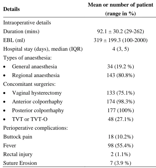
Surgeries were mostly done under regional anaesthesia with combined spinal and epidural anaesthesia. Table 3 shows the surgical procedures performed in addition to SSF and the perioperative details. All patients with uterovaginal prolapse had vaginal hysterectomies. Posterior colporrhaphy was performed in all patients but only 98% required anterior repair. Less than a third had urodynamic stress urinary incontinence requiring
Table 1: Baseline characteristics of the 177 patients who had sacrospinous ligament fixation as part of the surgery
Demographic factor Mean (range/ percentage)
Age (years) 59.6 ± 8.7 (40-79)
Parity (median; IQR) 4 (3, 6)
Menopause 148 (83.6%)
Race:
Malay 76 (42.3%)
Chinese 35 (19.8%)
Indian 62 (35.0%)
Others 4 (2.2%)
IQR: Interquartile range
continent surgery using tension free vaginal tape (TVT) or tension free vaginal tape obturator (TVT-O) kits. The mean duration of surgery was around one and a half hour. Intraoperative hemorrhage was not significant with a mean estimated blood loss of 319 ± 199.3mls with one patient requiring blood transfusion. The median duration of hospital stay was 4 days (range 1-16). Two patients (1.1%) required intraoperative bowel repair due to iatrogenic bowel injury. There were no bladder injuries or de-novo urinary symptoms.
Table 2: Preoperative staging of pelvic organ prolapse according to Baden-Walker’s classification
Types of
prolapse
Number of patients with prolapse (%)
Grade
1
Grade
2
Grade
3
Grade
4
Total
Apical prolapse •Uterovaginal
Prolapse
0 0 20
(11.3%) 113
(63.8%) 133
•Vault prolapse 0 15 (8.5%)
6
(3.4%) 23
(13.0%) 44
Cystocele 0 26
(14.7%) 46
(26%) 105
(59.3%) 177
Rectocele 0 80
(45.2%) 7
(4%) 90
(50.8%) 177
Table 3: Perioperative details of procedures performed with sacrospinous ligament fixation
Details Mean or number of patient
(range in %)
Intraoperative details
Duration (mins) 92.1 ± 30.2 (29-262)
EBL (ml) 319 ± 199.3 (100-2000)
Hospital stay (days), median (IQR) 4 (3, 5)
Types of anaesthesia:
• General anaesthesia 34 (19.2 %) • Regional anaesthesia 143 (80.8%) Concomitant surgeries:
• Vaginal hysterectomy 133 (75.1%) • Anterior colporrhaphy 174 (98.3%) • Posterior colporrhaphy 177 (100%)
• TVT or TVT-O 48 (27.1%)
Perioperative complications:
Buttock pain 18 (10.2%)
Fever 98 (55.4%)
Rectal injury 2 (1.1%)
Post-operative pyrexia was commonest affecting 55.4% (98/177) whereas buttock pain was experienced by 10.2% of the patients (18/177). Late complication of suture erosion was diagnosed in 3.9% (7/177) of the patients 6 months following surgery.
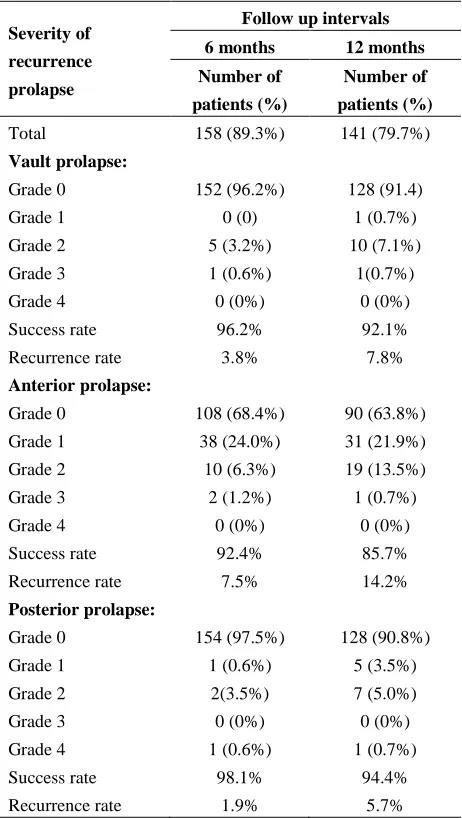
Table 4 shows stage of prolapse at 6 and 12 months’ review. From 177 women treated, 158 (89.3%) and 141 (79.7%) women came for the 6 months and 12 months follow up respectively. During the 6 months’ assessment, the success rate for all three compartments ranged from 92.4% to 98.1%. With time, the percentage reduced and ranged from 85.7% to 94.4% at 12 months. In contrast, the recurrence rate for all three compartments ranged from 1.9% to 7.5% at 6 months and 5.7% to 14.2% at 12 months. Of particular, the vault recurrence at 6 and 12 months were 3.8% and 7.8%, respectively. With regards to site-specific prolapse, the highest success and lowest recurrence rates were observed in the posterior compartment whereas the lowest success and the highest recurrence rates were noted in the anterior compartment. The success rate reduced and the recurrence rate increased across all three compartments with time. Nevertheless, none of our patients had repeated surgery during the study period.
Discussion
Huge prolapse is usually associated with an apical descent and fifty percent of anterior prolapse is caused by collapse of the apical support (10). Breaks and damages in connective tissue and neuromuscular complexes are believed to cause POP (11). De Lancey et al. has proposed three levels of pelvic organ support namely level I which suspends the apex of the vagina and the cervix from above known as the uterosacral/cardinal ligament complex. Level II attaches the mid-vagina laterally to the arcus tendineus fascia. Level III is fusion of the lower third of the vagina to the perineal body posteriorly, perineal membrane and urethra anteriorly and levator ani muscles laterally (12). Loss of level I support results in uterine and vault prolapse while loss of level II support results in cystocele and rectocele (10). SSF procedure is designed to effectively restore level I vaginal support (11). This longitudinal cohort study describes our experience in treating patients with massive uterovaginal or recurrent vault prolapse using transvaginal SSF.
Eleven percent of women had prolapse operation performed once in their lifetime and 25% of them will have recurrence with 17 to 29% requiring a repeat surgery within 5 years (5). Transvaginal SSF is the most popular (80-90%) vaginal procedures performed
Table 4: Postoperative prolapse staging, recurrence and success rate at 6 and 12 months following sacrospinous ligament fixation as part of pelvic floor reconstructive surgery
Severity of
recurrence
prolapse
Follow up intervals
6 months 12 months
Number of
patients (%)
Number of
patients (%)
Total 158 (89.3%) 141 (79.7%)
Vault prolapse:
Grade 0 152 (96.2%) 128 (91.4)
Grade 1 0 (0) 1 (0.7%)
Grade 2 5 (3.2%) 10 (7.1%)
Grade 3 1 (0.6%) 1(0.7%)
Grade 4 0 (0%) 0 (0%)
Success rate 96.2% 92.1%
Recurrence rate 3.8% 7.8%
Anterior prolapse:
Grade 0 108 (68.4%) 90 (63.8%)
Grade 1 38 (24.0%) 31 (21.9%)
Grade 2 10 (6.3%) 19 (13.5%)
Grade 3 2 (1.2%) 1 (0.7%)
Grade 4 0 (0%) 0 (0%)
Success rate 92.4% 85.7%
Recurrence rate 7.5% 14.2%
Posterior prolapse:
Grade 0 154 (97.5%) 128 (90.8%)
Grade 1 1 (0.6%) 5 (3.5%)
Grade 2 2(3.5%) 7 (5.0%)
Grade 3 0 (0%) 0 (0%)
Grade 4 1 (0.6%) 1 (0.7%)
Success rate 98.1% 94.4%
Recurrence rate 1.9% 5.7%
may simply represent a general predilection of anterior support failure following pelvic reconstructive surgery which remains unclear (14).
The rates of prolapse recurrence after SSF vary significantly in the literatures (10). Beer and Kuhn found the failure rates in various studies ranged from 3% to 37% for objective and 6% to 30% for subjective assessment respectively (15). Our objective recurrence rate was similar to others studies which was 3.8%. With time, we found the recurrence rate for both apical and posterior compartment prolapse were less than 5% and 10% at 6 and 12 months respectively. The recurrence rate was higher in the anterior compartment with 7.5% at 6 months and nearly 15% at 12 months’ review. This was in line with other studies which showed that cystocele recurrence is more common and problematic than apical recurrence (13). The risk of cystocele recurrence was also increased if it was not repaired at the time of the initial surgery (14). Another author found different results from MRI studies possibly due to small sample sizes (15). Hence, the actual impact of SSF on the position and recurrence of anterior vaginal prolapse remains uncertain (13). Nevertheless, none of our patients had repeated surgery for prolapse recurrence during the study period.
Transvaginal SSF is usually perform when the post-hysterectomy vault prolapse reaches the introitus at the end of anterior vaginal wall closure (16). Vaginal route offers greater cost effectiveness as it enables concurrent pelvic floor repair with preservation of coital function, avoidance of laparotomy and general anaesthesia, reduction of intra-operative bleeding, post-operative pain and complications, hence hospital stay (7,10,17). Other options for apical prolapse surgeries include sacral colpopexy which can be done via abdominal, laparascopic or robotic approach (11). Abdominal approach is superior as it has lower rate of vault recurrence, stress urinary incontinence and postoperative dyspareunia (3,6). However, its drawbacks include prolonged operating time, longer hospital stay and increased in surgical and anaesthetic complications (6). Laparoscopic is as effective as laparotomy sacral colpopexy with reduced blood loss and admission time (3). Robotic sacral colpopexy is less popular due to its highest inpatient costs, longer operating time and greater postoperative pain despite similar objective outcomes (11).
Various techniques and instruments namely bilateral or unilateral approach, uterine preservation, type of ligature carrier and choice of suture used in SSF have been described over the years to improve success rate. SSF can be performed either unilaterally or bilaterally
with equal outcome as there is no randomized controlled trial and most studies were limited by small sample sizes (18). Although vaginal length may be adequately restored, unilateral SSF may cause narrowing at the apex and deviation of the posterior and lateral vaginal axis from the midline by an average of 47 and 45 degrees respectively (16). Previous author reported 93% of global patients’ satisfaction with acceptable complication rates in patients who had bilateral SSF (19). However, another study found that bilateral SSF was only feasible in 73% and 56% of women with vault and severe uterovaginal prolapse respectively (20). In addition, most gynecologists favored the unilateral approach due to a shorter operating time and lesser intra-operative morbidity (19). In this study, unilateral right SSF was preferred as it was more convenient for our right-handed surgeon.
All of our patients had vaginal hysterectomy or performed concurrently prior to SSF procedure. Recently, there is a popular trend towards uterine conservation in which more women opted to retain their uterus (21). Such procedures namely vaginal sacrospinous hysteropexy which attached the uterus to the sacrospinous ligament is preferred than vaginal hysterectomy with SSF in view of equal outcome with added advantages of shorter operating time, lesser blood loss, faster recovery and fewer complications (22). To date, randomized controlled trial has shown no significant difference in terms of anatomical and functional outcome, hospital stay, complications, postoperative recovery, sexual functioning as well as recurrence and reoperation rate for both procedures (23). In current study, all patients had consented for vaginal hysterectomy with the benefits of preventing future menstrual problems and risks of cancer. Although it was believed that hysterectomy may increase the risk of vault prolapse due to its disruption of the pelvic floor supportive structures, this may not be the case for our patients as all of them had concurrent prophylactic vault suspension as recommended by previous literature (24).
direct vision with greater confidence and it is cheaper as the device is robust and reusable (20). However, its disadvantages include greater tissue dissection hence higher intraoperative blood loss, likelihood of injuries to the adjacent organs, increased intraoperative time and longer hospital stay (21). Nevertheless, the complications could be minimized if performed by experienced surgeons in pelvic reconstructive surgery. A recent prospective study showed no significant difference in surgical complications, long term objective and subjective outcome following SSF between the two devices (26).
Haemorrhage is a common intraoperative complications in various studies with transfusion rates ranging from 0.5 to 2.5% (6,7). However, our study did not show any significant bleeding with mean estimated blood loss of 319± 199.3mls. Visceral injury involving the bowel, bladder or ureter are other possible intraoperative complications but occurring less frequently. Rectal injury has been reported in 0.07% of vaginal hysterectomies, 0.4% of transvaginal SSF procedures (27) which increased to 1.9% for bilateral transvaginal SSF (19). Despite the unilateral approach used, we had higher incidence of bowel injury of 1.1%. This could be attributed by larger sample size and invasive nature of the Miya Hook technique used in our study. Hence, the challenge lies in the dissection of the very thin rectovaginal fascia particularly with the rectum bulging into the vagina (21). The risk could be prevented by infiltration of normal saline or diluted vasoconstrictor for hydro-dissection and adequate mobilization of the rectum (9). Moreover, the extent of tissue dissection could be limited by occasional digital examination of the rectum, thus avoiding injury to the rectum (27).
The most frequent post-operative complaints reported in the literature was buttock pain which occurred in about 18% of patients (20). The pain may be due to injury to the sacral nerve plexus, such as the branches of the pudendal nerve (18). Our study however, reported fever (98, 55.4%) as the commonest immediate complications despite perioperative antibiotics. This was followed by buttock pain which was self-limiting and manageable with simple analgesia, experienced by 10.2% (18) of our patients. This was in agreement with previous study which shown that the pain usually resolve spontaneously between the 6th day to 3 months post-operatively (16). In contrast, Gupta established even a lower rate of nonspecific gluteal pain in two of thirty two patients (6.25%) which subsided 6 days after surgery (10). The difference could be attributed to the difference in sample size and inter-operator’s performance.
Suture erosion may complicate vaginal reconstruction surgery particularly with the usage of braided suture. A retrospective cohort study of 92 patients who had SSF using braided polyester suture showed that suture-related complications occurred in 36% of patient with a mean time to presentation of 18.9 months (28). Seventy-four percent of them presented with vaginal bleeding and 70% required suture removal. Luck et al also reported a higher incidence of suture erosion when using braided polyester suture (31.3%) compared with polyglactin 910 (9%) for posterior compartment and anal sphincter repair. Seventy percent were symptomatic and 16% of them required additional surgical intervention (29). We used prolene (polypropylene) or ethilon (nylon) sutures for SSF which are synthetic monofilament non-absorbable sutures. Polyglactin 910 which is a synthetic braided absorbable suture was also used in our study for vaginal hysterectomy, anterior and posterior repair. We reported a lower incidence of suture erosion (3.9%) and all of them were removed in the outpatient clinic. At 6 months review, majority of them were asymptomatic with no significant recurrence of prolapse.
The main limitation in our study was its observational nature in which the data available was derived mainly from the clinical observations based on our routine clinical practice. This was a non-comparative study with a short length of postoperative follow up limited to 12 months which makes it difficult to draw precise conclusions about changes over time; hence the outcome may not exhibit the actual long-term results for SSF. Another potential source of bias may be the lack of blinding in the ‘third party’ observer to perform the prolapse grading during the postoperative review. Nevertheless, the collection of a fairly large sample sizes with only 10.7% defaulter in a longitudinal cohort subjects with reasonably homogenous demographic characteristics strengthen our study. Similar technique of SSF performed with a single surgeon input may reduce confounding variables thus allowing appropriate evaluation of SSF in the study.
counseled regarding the treatment options available, its possible risks and complications as well as the anatomical and functional consequences in the long run. Finally, it is to improve the patient’s quality of life with fast recovery and minimal complications.
Conclusion
In short, transvaginal SSF remains a high priority in our therapeutic regime given its reasonably good efficacy and safety profile. As only a few randomized controlled trials are available currently, future prospective randomized double blinded studies are needed to investigate SSF-related issues further.
References
1. Swift S, Woodman P, O’Boyle A, et al. Pelvic Organ Support Study (POSST): the distribution, clinical definition, and epidemiologic condition of pelvic organ support defects. Am J Obstet Gynecol 2005; 192(3): 795-806.
2. Olsen AL, Smith VJ, Bergstrom J, Coiling JC, Clark AL. Epidemiology of surgically managed pelvic organ prolapse and urinary incontinence. Obstet Gynecol 1997; 89(4): 501-6.
3. Maher C, Baessler K, Glazener CM, Adams EJ, Hagen S. Surgical management of pelvic organ prolapse in women: a short version Cochrane review. Neurourol Urodyn 2008; 27(1): 3-12.
4. Lo TS, Ashok K. Combined anterior trans-obturator mesh and sacrospinous ligament fixation in women with severe prolapse–a case series of 30 months follow-up. Int Urogynecol J 2011; 22(3): 299-306.
5. Randall CL, Nichols DH. Surgical treatment of vaginal inversion. Obstet Gynecol 1971; 38(3): 327-32.
6. Maher CF, Qatawneh AM, Dwyer PL, Carey MP, Cornish A, Schluter JP. Abdominal sacral colpopexy or vaginal sacrospinous colpopexy for vaginal vault prolapse: a prospective randomized study. Am J Obstet Gynecol 2004; 190(1): 20-6.
7. Petri E, Ashok K. Sacrospinous vaginal fixation-current status. Acta Obstet Gynecol Scand 2011; 90(5): 429-36.
8. Baden WF, Walker TA. Genesis of the vaginal profile: a correlate classification of vaginal
relaxation. Clin Obstet Gynecol 1972; 15(4): 1048-54.
9. Miyazaki F. Miya Hook ligature carrier for sacrospinous ligament suspension. Obstet Gynecol 1987; 70(2): 286–8.
10. Gupta P. Transvaginal sacrospinous ligament fixation for pelvic organ prolapse stage III and stage IV uterovaginal and vault prolapse. Iran J Med Sci 2015; 40(1): 58-62.
11. Barber MD, Maher C. Apical prolapse. Int Urogynecol J 2013; 24(11): 1815-33.
12. Delancey JO, Kane Low L, Miller J, Patel DA, Tumbarello JA. Graphic integration of causal factors of pelvic floor disorders: an integrated life span model. Am J Obstet Gynecol 2008; 199(6): 610.e1-5.
13. Meschia M, Bruschi F, Pifarotti P, Marchini M, Crosignani PG. The sacrospinous vaginal vault suspension: Critical analysis of outcomes. Int Urogynecol J 1999; 10(3): 155-9.
14. Paraiso MF, Ballard LA, Walters MD, Lee JC, Mitchinson AR. Pelvic support defects and visceral and sexual function in women treated with sacrospinous ligament suspension and pelvic reconstruction. Am J Obstet Gynecol 1996; 175(6): 1423–30.
15. Beer M, Kuhn A. Surgical techniques for vault prolapse: a review of the literature. Eur J Obstet Gynecol Reprod Biol 2005; 119(2): 144-55.
16. Royal College of Obstetricians & Gynaecologists, British Society of Urogynaecology. Green-top Guideline No. 46. The Management of Post Hysterectomy Vaginal Vault Prolapse; July 2015. http://www.rcog.org.uk>gtg-46. Last accessed on 15/09/2016.
17. Tseng LH, Chen I, Chang SD, Lee CL. Modern role of sacrospinous ligament fixation for pelvic organ prolapse surgery - A systemic review. Taiwan J Obstet Gynecol 2013; 52(3): 311-7.
18. Lovatsis D, Drutz HP. Safety and efficacy of sacrospinous vault suspension. Int Urogynecol J Pelvic Floor Dysfunct 2002; 13(5): 308-13.
results and quality-of-life after bilateral sacrospinous ligament fixation for genital prolapse. Eur J Obstet Gynecol Reprod Biol 2007; 132(2): 209-13.
20. Dietz V, de Jong J, Huisman M, Schraffordt Koops S, Heintz P, van der Vaart H. The effectiveness of the sacrospinous hysteropexy for the primary treatment of uterovaginal prolapse. Int Urogynecol J Pelvic Floor Dysfunct 2007; 18(11): 1271-6.
21. Wu MP, Long CY, Huang KH, Chu CC, Liang CC, Tang CH. Changing trends of surgical approaches for uterine prolapse: an 11 year population-based nationwide descriptive study. Int Urogynecol J 2012; 23(7): 865-72.
22. Frick AC, Barber MD, Paraiso ME, Ridgeway B, Jelovsek JE, Walters MD. Attitudes toward hysterectomy in women undergoing evaluation for uterovaginal prolapse. Female Pelvic Med Reconstr Surg 2013; 19(2): 103-9.
23. Detollenaere RJ, den Boon J, Stekelenburg J, et al. Treatment of uterine prolapse stage 2 or higher: a randomized multicenter trial comparing sacrospinous fixation with vaginal hysterectomy (SAVE U trial). BMC Womens Health 2011; 11:4.
24. Kantartzis KL, Turner LC, Shepherd JP, Wang L, Winger DG, Lowder JL. Apical support at the time of hysterectomy for uterovaginal prolapse. Int Urogynecol J 2015; 26(2): 207-12.
25. Ouzaid I, Ben Rhouma S, de Tayrac R, Costa P, Prudhomme M, Delmas V. Mini-invasive posterior sacrospinous ligament fixation using the CAPIO needle driver: an anatomical study. Prog Urol 2010; 20(7): 515–9.
26. Leone Roberti Maggiore U, Alessandri F, Remorgida V, Venturini PL, Ferrero S. Vaginal sacrospinous colpopexy using the Capio suture-capturing device versus traditional technique: feasibility and outcome. Arch Gynecol Obstet 2013; 287(2): 267–74.
27. Hoffman MS, Lynch C, Lockhart J, Knapp R. Injury of the rectum during vaginal surgery. Am J Obstet Gynecol 1999; 181(2): 274-7.
28. Toglia MR, Fagan MJ. Suture erosion rates and long-term surgical outcomes in patients undergoing sacrospinous ligament suspension with braided polyester suture. Am J Obstet Gynecol 2008; 198(5): 600.e1-4.
29. Luck AM, Galvin SL, Theofrastous JP. Suture erosion and wound dehiscence with permanent versus absorbable suture in reconstructive posterior vaginal surgery. Am J Obstet Gynecol 2005; 192(5): 1626-9.