International Journal of Integrated Engineering (Issue on Electrical and Electronic Engineering)
7
Azizi Miskon1*and Jatendra Uslama1 ,
1Faculty of Electrical & Electronic Engineering, UniversitiTun Hussein Onn Malaysia, 86400 Parit Raja, Batu Pahat Johor Malaysia.
*Corresponding author / email: [email protected]
Abstract
The objective of this paper is to provide the fundamental mathematical formula to predict the effect of magnetic fields (MF) on stem cell differentiation. The data were reviewed from journals related to the effects of magnetic fields on stem cell differentiation. These data were given a value for differentiation which is related to their MF strength with these conditions; MF strength that does not affect the stem cell differentiation given the value of zero, MF strength that results in stem cells death given the value of 1, and the MF strength that affects stem cells The differentiation given the value between 0.1 and 0.9. graph was plotted according to these data and the mathematical equation is designed from the graph. From this review, we suggest that the intensity of MF that can affect the stem cell differentiation is between 600µT and 9.4T in which the cell differentiation will not occur with intensity of less than 10µT and intensity of more than 12T will cause the death of stem cells. We also suggest that the limit of MF effects on stem cell differentiation lies between 10 µT and 600 µT, and the limit of MF strength that can lead to the death of stem cells lies between 9.4 T and 12 T. It can be concluded that the exposure of MF on stem cell differentiation depends not only on the MF intensity, but also on the period of exposure.
Keywords: Differentiation, Ion Resonance Frequency, Magnetic Fields, Stem Cells, Tissue Engineering.
8
1. INTRODUCTION
Stem cells are primitive cells, which are present in all organisms and have the ability to divide and give rise to more stem cells, or switch to become more specialized cells in human body like cells in brain, heart, muscle, and kidney [1]. There are two types of stem cell; embryonic stem (ES) cell and adult stem cell. ES cells are pluripotent and harvested from inner cell mass of blastocyst and possess the ability to differentiate into all of the specialized embryonic tissues [1], [2]. ES cells also may open the door to the rapidly progressing field of therapeutic cell transplantation [3]. The adult stem cells are multipotent with capacity to differentiate or transdifferentiate into cell types other than their tissue of origin [1]. Adult stem cells and progenitor cells can be found in the adult tissue. Both of these cells act as a repair system for body, replenishing the specialized cells, and maintaining the normal regenerative of organs, such as blood, skin, or intestinal tissues.
Magnetic fields (MF) produced by moving electric charge and exist all around us like earth MF and man-made MF sources. Numerous static and alternating MF arising from man-made sources have possible biological effect [4]. There are many biological functions that are modulated by extremely low frequency magnetic field (ELF-MF) [5]-[7]. However, there is not enough evidence that the ELF-MF could endanger the human health [8]. ELF-MF is MF with a range of frequency of 3 to 30Hz. Even so, MF has been shown to affect proliferation and growth factor expression in cultured cells [9]-[11] and also interfere with endorphinergic and cholinergic system [12]-[14]. Other than MF, electrical fields (EF) also have biological effects that can influence neural growth and orientation in vitro [15], and have been applied for the treatment of spinal cord injuries in recent clinical trials [16]. The response of cells to the EF was essentially passive and determined by the physical properties of the cell, however cells can also actively respond to EF [17]. Electromagnetic fields (EMF) are produced when electric current flows through an electrical conductor like power line [18]. Like MF and EF, EMF also has biological effects such as altered rate
of cell growth [5], [19], altered quantities of RNA transcript and proteins [20], altered cell surface properties [21] and effect on development [22]. However EMF-based technologies have not progressed to clinical translation and the reason for this is the scepticism due to differences in experimental exposure protocols and static MF (SMF) variation applied in experiment [23].
The objective of this paper is to design the mathematical formula to predict the effect of magnetic fields on stem cells differentiation. To the extent of our knowledge, there is no standard range of suitable magnetic fields provided which can affect the stem cells differentiation ability. Therefore, we review the data of magnetic fields and its effect on stem cells differentiation used by previous researchers. From these data, we design a mathematical equation to predict the suitable range of magnetic fields that can affect the stem cells ability to differentiate. This work will provide an essential basis prior to any future in vitro experiment, in which we can predict the strength of MF used either to trigger the cells differentiation or vice versa. Therefore, we may avoid unnecessary failure during the experimental works.
2. METHOD
International Journal of Integrated Engineering (Issue on Electrical and Electronic Engineering)
9 differentiation. The data were plotted into graph by using Microsoft Office Excel 2010 software and the result of project presented in Graphical User Interface (GUI) design by using the Microsoft Visual Studio Ultimate 2010.
Table 1: MF strengths that were used in reviewed journals and classified into their corresponding group; with group 0 for MF strength does not affect the stem cells differentiation, group 1 for MF strength that effects the stem cells differentiation, and group 2 for MF strength that kill the stem cells.
MF intensity (T) Group
10µ 0 600µ 1 800µ 1 1m 1 1.1m 1
10m 1 4.7 1 9.4 1 12 2 16 2
Table 2: Modified data of Table 1
MF strength, x (T) Value, y
10µ 0 600µ 0.1 800µ 0.2 1m 0.3 1.1m 0.4 10m 0.5 4.7 0.7 9.4 0.9 12 1 16 1
3. RESULT
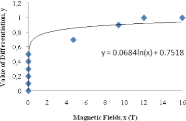
From Table 2, we design the graph using Microsoft Office Excel 2010 as shown in Fig. 1. From Table 2, we observed that the effect of MF on stem cell differentiation still occur in 600µT but the differentiation is not observed in 10µT. This may indicate that the minimum strength of MF to influence the stem cell
differentiation in frequency of 50Hz lies somewhere between 10µT and 600µT. We also observed that the effect of MF on differentiation of stem cells still occur in MF strength of 9.4T. However, such high field strength magnet like 9.4T is not easily available and such studies may not be translated into the clinical study because the current limits for magnetic field strengths approved by U.S. Food and drug Administration (FDA) is 3T [24]. This is the reason that many of the research works were carried out using the magnetic fields less than 3T. The stem cells death will occur at the exposure of 12T for more than 24h. Therefore, the minimum exposure of MF before resulting in stem cells death lies between 9.4T and 12T.
Fig. 1 Th differenti
y
Fig. 2 G
4. DISC
Cardiosp cells (C frequenc 18Hz) d and vas VEGF, experim that the of 10µT Human multipot capacity different mesench muscle, exposure (hMSC) different by enha and pero
he graph of MF iation
y = 0.0684ln(
GUI for predic
CUSSION
peres (CDps CDCs) exp cy magnetic did not affec scular marke KDR, a ment, thus thi stem cells di T [23].
n Mesenchym tential cells a y, and th tiate into hymal tissue
fat, and m e of human ) to 600µT
tiate into ad anced expres
oxisome pro
F effects on st
(x) + 0.7518
cting the MF cells
s) and card posed to e c fields (EL ct the expres ers (cTnl, N and SMA)
is may sugge id not differe
mal stem ce and possess h here are different e such as b marrow stro n mesenchym
T enables dipogenic cel ssion of lip oliferator-act
tem cells
(1)
effects on stem
iospere-deriv extremely l LF-MF) (10µ ssion of card Nkx 2.5, MH
during est an eviden entiate with M
lls (hMSC) high replicat
potentials lineages bone, cartila oma [30]. T
mal stem ce the MSC lls as indica oprotein lip tivated recep 10 m ved low µT, diac HC, the nce MF are tion to of age, The ells to ated pase ptor gamm expos differe to inv labelle oxide label using The v cells h MSC metab colony princi cells t direct vivo labelle [38], cells tissue of MF carrie The sinuso 50Hz. transc PCR) of GA both select of exp this M specif witho As fo mRNA contai homeo shown differe [42 ]. The embry Differ P19 c 50Hz, signif intens cells [26]. differe
ma [24]. In sure of MSC entiation. Th vestigate the
ed with sup (SPIO). Thi the cells, a Magnetic viability or d
has not been [31], [32], b bolism, the y formation iple function
to high MF t the iron labe
[35]-[37], g ed stem cell
for the see [36], [39], o s by stem ce F strength in
d out in orde e stem cell oidal MF o . Real-tim criptase-poly
analysis rev ATA-4 and N embryoid b ted cardiomy posing GTR MF was the fic cells, an
ut an aid of g or informat A encode re ining tra odomain, an n to be es
ent animal s
e effects of yonal carcin rentiation w cells to 1mT , however th ficant. By ex se MF with were differ The effects entiation
n addition, to MF has e his research w
effect of M per magnetic
is was done and detect i Resonance differentiatio affected by but it has an i migration c n of MSC n of MRI is and magnet elled stem ce guided loca ls to the des eding of sca
or for the en ells [37]. The n vitro expe er to verify th
is able to of 800µT w
me quant ymerase chai
vealed a rem Nkx-2.5 mRN
odies (EBs) yocytes [25]. R1 embryoni e differentia nd this expe gene transfer tion, GATA espectively anscription
nd both of sential for species [40],
MF were a noma cells was detected
T of MF w he analysis re xposing P19 the strengt rentiated int s of ELF-M
were
the imme enhanced the
was accompl MF on hMSC
c particles of by using SP ion was don Imaging (M n potential o
SPIO-labelli impact on th capacity, an C [32]-[34].
s the exposu tic force tha ells in vitro a alization of sired region affolds with ngineering o erefore, the eriment shou he equation ( differentiate with frequenc titative re in reaction markably incr NA expressio ) and purom
Hence, the c stem cells ation into ca
riment was r technology A-4 and Nk for a zinc f
factor these have cardiogenes [41], and h
also done on (P19 cells) by exposin with frequen esult was not 9 cells into th of 10mT to neuronal MF after neu
International Journal of Integrated Engineering (Issue on Electrical and Electronic Engineering)
11 morphological analysis, immunochemical analysis (MAP2 and GFAP), and developmental neuronal network activities recorded by the micro-electrode arrays (MEAs). From the results, the percentage of MAP2 positive cells and the spike frequencies had increased, but the percentage of GFAP positive cells has reduced. These results suggest that an exposure to 10mT ELF-MF would affect the characteristic of neuronal differentiation and functional neuronal properties [26]. These results also may suggest that the effect of MF on stem cell differentiation will become less significant with lower intensity of MF strength as verified by using our equation in Fig 1.
The stem cells are also able to differentiate in MF intensity of 1.1mT as demonstrated in previous work using the bone morrow stem cells (BMSC) [27]. It results in the differentiation of BMSC into osteogenesis and the increase of intracellular Ca2+ after MF stimulation. From this result, they postulated that the elevated Ca2+ is possibly the underlying biochemical mechanism which is responsible for the induction of terminal differentiation [27].
As mentioned above, most of the experiments were done to investigate the effect of MF on stem cells ability to differentiate, and were performed with the intensity less than 3T. Even so, there are also experiments done with the MF intensity above 3T. The MF intensity between 4.7T and 9.4T were found to affect the stem cells differentiation [28]. Therefore, our range for cells differentiation was limited at 9.4T.
The effects of microgravity (MG) modelled by large gradient high magnetic field (LGHMF) with intensity of 12T and 16T on hMSC led to cell death after 24 hours exposure. Almost all the cells died after 48 hours, but in the first 6 hours of osteogenic induction, it had resulted in suppression of the osteogenesis of hMSC [29]. Therefore, we assumed that 12T and 16T still affect the cell differentiation for a short period of exposure. This response may be due to the synergistic effect of high magnetic force and MG existed in their experiment modelling systems [43]-[46]. Hence, from the data reviewed, we suggest that the MF intensity
between 9.4T and 12T could lead to stem cell death.
As stated in the result section, the MF was classified into three groups according to their effects on stem cell differentiation. From these groups, we know that MF less than 600µT or more than 9.4T do not lead to stem cell differentiation. Also, the MF effect on stem cells differentiation will not occur at MF of 10µT and below. However, MF of 12T and above will result in stem cell death.
12 are necessary to ensure the safe method to differentiate stem cell into specific cell through MF exposure.
In conclusion, we suggest that the intensity of MF that can affect stem cell differentiation is between 600µT and 9.4T. Also the differentiation will not occur if the intensity is less than 10µT and with intensity of more than 12T, it will cause death of stem cells. We also suggest that the range of MF effects on stem cell differentiation lies between 10µT and 600µT. The limit of MF strength that can lead towards the death of stem cells lies between 9.4T and 12T. We conclude that the result of the exposure of MF on stem cells differentiation depends not only on the MF intensity, but also on the period of exposure.
REFERENCES
[1] R. Passier, C. Mummery,“Origin and use of embryonic and adult stem cells in
differentiation and tissue repair,”Cardiovascular Research 2003, vol. 58, pp. 324-335.
[2] C.M. Metallo, S.M. Azarin, Lin ji, et al.,“Engineering tissue from human embryonic stem cells,” J. Cell Mol Med 2008, vol. 12(3), pp. 709-729.
[3] S.G. Nir, R. David, M. Zaruba, et al.,“Human embryonic stem cells for cardiovascular repair,” Cardiovascular Research 2003, vol. 58 (2), pp. 313-323. [4] W.T. Kaune,“Introduction to
power-frequency electric and magnetic fields,” Environmental Health Perspectives Supplements 1993, vol. 101, pp. 73-81. [5] A.R. Liboff, T. Williams Jr., D.M.
Strong,et al., “Time-varying magnetic fields: effects on DNA synthesis,”Science, 1984, vol. 223, pp. 818-820.
[6] J. Jajte, M. Zmyslony, J. Palus, et al., “Protective effects of melatonin against in vitro iron ions and 7 mT, 50 Hz, magnetic fields-induced DNA damage in rat
lymphocytes,”Mutat Res 2001, vol. 483, pp. 57-64.
[7] S. Falone., A. Mirabilio, M.C. Carbone, et al., “Chronic exposure to 50Hz magnetic fields causes a significant weakening of antioxidant defence systems in aged rat brain,”Int J Biochem Cell Biol 2008, vol. 12, pp. 2762-2770.
[8] D.W. Zipse, “Health effects of extremely low frequency (50 and 60 Hertz) electric and magnetic fields,” IEEE 1991, vol. PCIC-91-09.
[9] A. Cossarizza, D. Monti, F. Bersani, et al. “Extremely low frequency pulsed electromagnetic fields increase cell proliferation in lymphocytes from young and aged subjects,”Biochem. Biophys. Res. Commun. 1989, vol. 160, pp. 692-698.
[10]R. Cadossi, F. Bersani, A. Cossarizza, et al. “Lymphocytes and low-frequency electromagnetic fields,” FASEB J. 1992, vol. 6, pp. 2667–2674.
[11]A. Cossarizza, , S. Angioni, , F. Petraglia, , et al. “Exposure to low-frequency pulsed electromagnetic fields increases interleukin-1 and interleukin-6 production by human peripheral blood mononuclear cells,” Exp. Cell Res. 1993, vol. 204, pp. 385–387.
[12]A.W. Thomas, M. Kavaliers, F.S. Prato, et al. “Pulsed magnetic field induced “analgesia” in the land snail, Cepaeanemoralis, and the effects of mu, delta, and kappa opioid receptor agonists/antagonists” Peptides 1997, vol. 18, pp. 703–709.
[13]V.V. Vorobyov, E.A. Sosunov, N.I. Kukushkin, et al.“Weak combined magnetic field affects basic and morphine-induced rat’s EEG,” Brain Res. 1998, vol. 781, pp. 182–187.
International Journal of Integrated Engineering (Issue on Electrical and Electronic Engineering)
13 rat,”Bioelectromagnetics1993, vol. 14, pp. 5–15.
[15]N.B. Patel, M.M. Poo, “Perturbation of the direction of neurite growth by pulsed and focal electric fields,” J Neurosci, 1984, vol. 4, pp. 2939-47.
[16]L.D. Duffell, “Long-term intensive electrically stimulated cycling by spinal cord-injured people: Effect on muscle properties and their relation to power output,” Muscle Nerve 2008, vol. 38, pp. 1304-11.
[17]H.M. Gerard,“The use of electric fields in tissue engineering,” Organogenesis 2004, vol. 4, pp. 11-17.
[18]R. Goodman, Y. Chizmadzhew, A. Shirley-Henderson,“Electromagnetic
fields and cells” Journal of Cellular Biochemistry 1993, vol. 51, pp. 436-441. [19]K. Takahashi, I. Kaneko, M. Date, et
al.,“Effect of pulsing electromagnetic fields on DNA synthesis in mammalian cells in culture,”Experimentia 1986, vol. 42, pp. 185-186.
[20]E. Czerska, H. Al-Baranzi, J. Casamento, et al.,“Comparison of the effect of elf fields on c-nayc oncogene expression in normal and transformed human cells,” In Transection of the Bioelectromagnetic Society, Thirteenth Annual Meeting 1991. Salt Lake City, UT, B-2-14.
[21]M.T. Marron, E.M. Goodman, P.T. Sharpe, et al.,“Low frequency electric and magnetic fields have different effects on the cell surface,” FEBS Lett 1988, vol. 230, pp. 13-16.
[22]M.R. Delgado, J. Leal, J.L. Monteagudo, et al.,“Embryological changes induced by weak, extremely low frequency electromagnetic fields,” J Anat 1982, vol. 134, pp. 533-551.
[23]G. Roberto, L. Mario, B. Lucio, et al.,“Differentiation of human adult cardiac stem cells exposed to extremely low-frequency electromagnetic fields,”
Cardiovascular Research 2009, vol. 82, pp. 411-420.
[24]S. Richard, B. Rudiger, K. Rainer, et al.,“Functional investigation on human mesenchymal stem cells exposed to magnetic fields and labeled with clinically approved iron nanoparticles,” BMC Cell Biology 2010, vol. 11, pp. 22
[25]C. Ventura, M. Maioli, Y. Asara, , et al.,“Turning on stem cell cardiogenesis with extremely low frequency magnetic fields,” FASEB J. 2004, vol. 19, pp. 155– 157.
[26]S. Atsushi, T. Yuzo, M. Hiroyuki, et al. “Developmental effects of low frequency magnetic fields on P19-Derived Neuronal Cells,” IEEE EMBS 31st Annual International Conference, September 2009. [27]W. Zhao, W. Ma, H. Wu.,“The effects of magnetic fields on the differentiation and intra-cellular free calcium of bone marrow mesenchymal stem cells,” IEEE WAC 2008.
[28]S. Magnisky, R.M. Walton, J.H. Wolfe, et al., “Magnetic resonance imaging detects differences in migration between primary and immortalized neural stem cells,” AcadRadiol 2008, vol. 15(10), pp. 1269-1281.
[29]D. Shi, R. Meng, W. Deng, et al., “Effects of Microgravity Modeled by Large Gradient High Magnetic Field on the Osteogenic Initiation of Human Mesenchymal Stem Cells,” Stem Cell Rev and Rep 2010.
[30]M.F. Pittenger, A.M. Beck S.C. Mackay, et al.,“Multilineage potential of adult human mesenchymal stem cells,” Science 1999, vol. 284, pp. 143–147.
14 [32]R. Schäfer, R. Kehlbach, M. Muller, et
al.,“Labeling of human mesenchymal stromal cells with superparamagnetic iron oxide leads to adecrease in migration capacity and colony formation ability,”Cytotherapy2009, vol. 11, pp. 68-78.
[33]E. Pawelczyk, A.S. Arbab, S. Pandit, et al.,“Expression of transferrin receptor and ferritin following ferumoxides-protamine sulfatelabeling of cells: implications for cellular magnetic resonance imaging,” NMR Biomed 2006, vol. 19, pp. 581-592. [34]R. Schäfer, R. Kehlbach, J. Wiskirchen, et
al., “Transferrin Receptor Upregulation: In Vitro Labeling of Rat Mesenchymal Stem Cells with Superparamagnetic Iron Oxide,” Radiology 2007, vol. 244, pp. 514-523.
[35]S.V. Pislaru, A. Harbuzariu, G. Agarwal, et al.,“Magnetic forces enable rapid endothelialization of synthetic vascular grafts,” Circulation 2006, vol. 114, pp. I314-I318.
[36]K. Shimizu, A. Ito, H. Honda,“Mag-seeding of rat bone marrow stromal cells into porous hydroxyapatite scaffolds for bone tissue engineering,” J BiosciBioeng 2007, vol. 104, pp. 171-177
[37]K. Shimizu, A. Ito, T. Yoshida, et al.,“Bone tissue engineering with human mesenchymal stem cell sheets constructed using magnetite nanoparticles and magnetic force,” J Biomed Mater Res B ApplBiomater 2007, vol. 82, pp. 471-480. [38]S.V. Pislaru, A. Harbuzariu, R. Gulati, et
al.,“Magnetically targeted endothelial cell localization in stented vessels,” J Am CollCardiol 2006, vol. 48, pp. 1839-1845. [39]D. Robert, D. Fayol, C.L. Visage, et
al.,“Magnetic micro-manipulations to probe the local physical properties of porous scaffolds and to confine stem cells,” Biomaterials 2010, vol. 21, pp. 1586-1595.
[40]C. Biben, R.P. Harvey,“Homeodomain factor Nkx-2.5 controls left/right asymmetric expression of bHLH gene eHand during heart development,” Genes Dev. 1997, vol. 11, pp. 1357–1369. [41]T.J. Lints, L.M. Parsons, L. Hartley, et al.,
“Nkx-2.5: a novel murine homeobox gene expressed in early heart progenitor cells and their myogenic descendants,” Development 1993, vol. 119, pp. 419– 431.
[42]D.W. Benson, G.M. Silberbach, A. Kavanaugh-McHugh, et al.,“Mutations in the cardiac transcription factor Nkx-2.5 affect diverse cardiac developmental pathways,” J. Clin. Invest. 1999, vol. 104, pp. 1567–1573
[43]Q. Airong, Z. Wei, W. Yuanyuan, et al.,“Gravitational environment produced by superconducting magnet affects osteoblast morphology and functions,”ActaAstronautica, 2008, vol. 63, pp. 929–946.
[44]P.W. Neurath,“High gradient magnetic field inhibits embryonic development of frogs,” Nature 1968, vol. 219, pp. 1358– 1359.
[45]Q. Airong, D. Shengmeng, G. Xiang, et al.,“cDNA microarray reveals the alterations of cytoskeleton-related genes in osteoblast under high magneto-gravitational
environment,”ActaBiochimica et BiophysicaSinica (Shanghai)2009, vol.
41, pp. 561–577.
[46]Q. Airong, H. Lifang, G. Xiang, et al.,“Large gradient high magnetic field affects the association of MACF1 with actin and microtubule cytoskeleton,” Bioelectromagnetics 2009, vol. 30, pp. 545–555.
International Journal of Integrated Engineering (Issue on Electrical and Electronic Engineering)
15 and vascular repair,” Experimental Hematology. 2005, vol. 33(9), pp. 980–6. [48]G. Martino, S. Pluchino,“The therapeutic
potential of neural stem cells,” Nature Reviews Neuroscience 2006, vol. 7(5), pp. 395–406.
[49]R.L. Zhang, Z.G. Zhang, M. Chopp,“Neurogenesis in the adult ischemic brain: generation, migration, survival, and restorative therapy,” Neuroscientist 2005, vol. 11(5), pp. 408– 16.
[50]S.S. Kuthiala, G.H. Lyman, O.F. Ballester, “Randomized clinical trials for hematopoietic stem cell transplantation: lessons to be learned from the European experience,” Bone Marrow Transplantation 2006, vol. 37(2), pp. 219–
21.
[51]R.A. Nash, J.D. Bowen, P.A. McSweeney, et al., “High-dose immunosuppressive therapy and autologous peripheral blood stem cell transplantation for severe multiple sclerosis,” Blood 2003, vol. 102(7), pp. 2364–72.
[52]H. Okada, I.F. Pollack, F. Lieberman, et al., “Gene therapy of malignant gliomas: a pilot study of vaccination with irradiated autologous glioma and dendritic cells admixed with IL-4 transduced fibroblasts to elicit an immune response,” Human Gene Therapy 2001, vol. 12(5), pp. 575– 95.
[53]C. Hesdorffer, J. Ayello, M. Ward, et al., “Phase I trial of retroviral-mediated transfer of the human MDR1 gene as marrow chemoprotection in patients undergoing high-dose chemotherapy and autologous stem-cell transplantation,” Journal of Clinical Oncology. 1998, vol. 16(1), pp. 165–72.
[54]E.Y. Snyder, D.L. Deitcher, C. Walsh, et al., “Multipotent neural cell lines can engraft and participate in development of
mouse cerebellum,” Cell. 1992, vol. 68(1), pp. 33–51.
[55]E.Y. Snyder, R.M. Taylor, J.H. Wolfe, “Neural progenitor cell engraftment corrects lysosomal storage throughout the MPS VII mouse brain,” Nature 1995, vol. 374(6520), pp. 367–70.
[56]J.D. Flax, S. Aurora, C. Yang, et al.,“Engraftable human neural stem cells respond to developmental cues, replace neurons, and express foreign genes,” Nature Biotechnology. 1998, vol. 16(11), pp. 1033–9.
[57]R.A. Fricker, M.K. Carpenter, C. Winkler, et al., “Site-specific migration and neuronal differentiation of human neural progenitor cells after transplantation in the adult rat brain,” Journal of Neuroscience 1999, vol. 19(14), pp. 5990–6005.
[58]J. Ourednik, V. Ourednik, W.P. Lynch, et al., “Neural stem cells display an inherent mechanism for rescuing dysfunctional neurons,” Nature Biotechnology. 2002, vol. 20(11), pp. 1103–10.
[59]H.J. Cho, N. Lee, J.Y. Lee, et al., “Role of host tissues for sustained humoral effects after endothelial progenitor cell transplantation into the ischemic heart,” J Exp Med 2007, vol. 204, pp. 3257-3269. [60]V. Hartwig, G. Giovannetti, N. Vanello, et