33
MICROENCAPSULATION TECHNIQUE FOR SELECTED PROBIOTICS AND
PREBIOTICS
Ravi D
1*, Usha G
2, Parthasarathy R
1 1Bioprocess unit, Department of Botany, Government Arts College, Coimbatore
2Final year M.Tech. Biotechnology, Department of Biotechnology, Anna University, Regional centre, Coimbatore *Corresponding author: dravi.botany@gmail.com
ABSTRACT Nowadays probiotics and prebiotics require full attention to stay viable in the food industry and the medical field. Microencapsulation is performed generally to give a protective covering for probiotics and prebiotics thereby saving from the spoilt environment. The Microencapsulation study is mainly performed in order to approve the viability of probiotics for better mode of action in food, health care and other applications. Prebiotics and sodium alginate was used as a coating material in order to microencapsulate probiotics (Lactobacillus rhamnosus and Lactobacillus plantarum). This research depicts the survival rate of encapsulated probiotics (L.plantarum and L.rhamnosus) with/without prebiotics over a period of one month.
(Key words: Microencapsulation, Probiotics, Prebiotics, Lactobacillus plantarum, Lactobacillus rhamnosus)
INTRODUCTION
Encapsulation-The future of probiotics
Entrapment of cells in a gel matrix of alginates is the most popular system of immobilization. The terms immobilization and encapsulation were used interchangeably in most reported literature. While encapsulation is the process of forming a continuous coating around an inner matrix that is wholly contained within the capsule wall as a core of encapsulated material, immobilization refers to the trapping of material within the capsule wall as a core of encapsulated material; immobilization refers to the trapping of material within or throughout a matrix. A small percentage of immobilized material may be exposed at the surface, while this is not the case for encapsulated material. Encapsulation occurs naturally when bacterial cells grow and produce Exo-polysaccharides.
The microbial cells are entrapped within their own secretions that act as a protective structure or a capsule, reducing the permeability of the material through the capsule and therefore less exposed to adverse environmental factors. Many lactic acid bacteria synthesize Exo-polysaccharides, but they produce insufficient Exo-polysaccharides to be able to encapsulate themselves fully. Microencapsulation helps to separate a core material from its environment
until it is released. It protects the unstable core from its environment, thereby improving its stability, extends the core’s shelf life and provides a sustained and controlled release.
The structure formed by the micro-encapsulation agent around the core substance is known as the wall. The properties of the wall system are designed to protect the core and to release it at controlled rates under specific conditions while allowing small molecules to pass in and out of the membrane .The capsules may range from submicron to several millimeters in size and can be of different shapes.
Microencapsulation
34
to improve the viability of probiotic organisms in frozen yoghurt during processing and storage [2]. Viability may be further improved by adjusting the concentration of sodium alginate up to 4% [3].The improvement in viability of probiotic organisms is attributed to the structure of the sodium alginate gel, which permits the diffusion of nutrients while shielding the microorganisms from molecules that may cause stress [4]. Microencapsulation has been found to reduce probiotic cell death caused by oxygen toxicity [5]. Probiotic organisms have also been found to agglomerate in the centre of beads creating anaerobic regions. An additional layer can be applied to provide probiotic organisms with a barrier from molecules that may cause stress and oxygen.
An additional layer to calcium alginate beads can be achieved by applying a polymer coating such as chitosan or poly-L-lysine (PLL) to the beads. However, the coating may affect the overall size of the bead. Large beads are undesirable in yoghurt and many other products as they may detract from the mouth feel of the product. Beads with a diameter exceeding approximately 0.1 mm (100μm) are detectable in the mouth. Therefore, beads ranging between 15 μm and 100 μm that do not compromise the viability of probiotic organisms during processing and storage are desirable.
Freeze drying – prebiotics and probiotics
Prebiotics are generally used to improve the viability of probiotic organisms during storage. It is essential that one million probiotic organisms per gram should be present in a product [4] in order to confer the health benefits of probiotic organisms. Shin and their research group [6] found that the viability of commercial Bifidobacterium spp in skim milk improved by 55.7% after 4 weeks of refrigerated storage when FOS was added. Prebiotics are also used to protect probiotic organisms during freeze drying as they provide extra solids.
Some strains of probiotic organisms are sensitive to freeze drying due to deterioration of physiological state of the cells. Cryoprotectants such as mannitol, sorbitol, sodium tripolyphosphate, xylitol, glycerol, raffinose, maltodextrin, erythritol, threitol, trehalose glucose and fructose play a role in improving the cold tolerance of the microorganisms.
Cold tolerance is improved as cryoprotectants accumulate within the cells and the osmotic
difference with the external environment is reduced. Cryoprotectants added to a cell suspension also reduced injury and death. Cryoprotectants added to reduce the mortality of probiotic organisms in freeze-dried yoghurt effectively function by promoting the formation of extracellular amorphous ice glass instead of crystalline ice [7]. Amorphous ice glass is not a destructive upon the cell membrane of some probiotic organisms. The extent to which cryoprotectant is provided may vary between cultures.
MATERIALS AND METHODS
Culture conditions
Pure lyophilized culture of L. plantarum and L.rhamnosus were obtained from National Dairy Research Institute, Karnal. Bacterial strains were cultivated for 24 hours at 35° C in 5% CO2 and 95% air atmosphere.
Microencapsulation Of Synbiotics Preparation of probiotic bacteria
The culture was grown and propagated three times successively for activation at 37°C for 24 hours. Probiotic organisms were harvested by centrifuging at 10000 rpm at 4°C for 20 minutes and the pellet was washed and suspended twice in saline solution. The final bacterial counts were adjusted to 109cells/ml and kept ready for encapsulation.
Intrusion method is the cheaper method and fastest method to encapsulate probiotics and prebiotics. Generally in intrusion method, Probiotic cultures were mixed with Alginate solution and fixed through a nozzle and injected into a hardening solution (CaCl2) and allowed to stand for one hour for
effective binding and encapsulation. Then, second coating solution of prebiotics were added and fixed through a nozzle and entrapped microcapsules forms spherical beads of synbiotics.
Microencapsulation of Probiotics and Prebiotics Probiotic – Prebiotic microcapsules were prepared by mixing 1% (v/v) of Lactobacillus plantarum and
Lactobacillus rhamnosus and the previously autoclaved (121°C, 15 min) prebiotics (fiber, 0% to 3%). The extrusion technique of microencapsulation was used [8](Krasaeekoopt et al., 2004). After washing, 1% each of Lactobacillus plantarum and
35
1% to 3% sodium alginate solution and sterilized at 121°C for 15 minutes. The cell suspension was injected through a 0.11 mm needle into sterile 0.1 M CaCl2. The beads were approximately 0.5 mm indiameter and were allowed to stand for 1 hour for gelification, rinsed and kept at 4°C for further use. Microencapsulated beads were then observed under Scanning electron microscope.
Improvement Of Viability Of Probiotics
Freeze drying of synbiotic microcapsules slurry
The freeze drying process for the encapsulated probiotics and prebiotics was performed using the freeze dryer (Lyodel, LYO1550, and India). An aqueous slurry of calcium alginate coated synbiotic microcapsules was transferred to 100 ml round bottom flask and pre-frozen to - 40°C in the pre freezer bath at atmospheric pressure for 2 h. It was allowed to freeze dry for 6 hours with reduction of pressure on the round bottom flask for 10-3 m bar and raising the shelf temperature of sample +10°C. After freeze drying, dry agglomerates of synbiotic microcapsules were obtained. The survival rate of the freeze dried probiotic, alginate and prebiotic encapsulated probiotic was estimated up to four weeks.
However, the synbiotic effects of probiotics and prebiotics are strain dependent. Only certain probiotics grow when supplemented with prebiotic
carbon source.
Yeo Siok et al reported different growth rates for 2 different strains of Lactobacillus with ten different prebiotics.
Viability checks for encapsulated synbiotic The probiotic organisms were enumerated using the method prescribed by [9]TharmarajandShah (2003).
L.plantarum and L.rhamnosus were selectively enumerated on MRS agar using anaerobic incubation at 37°C for 72 hours. Encapsulated organisms were released from Calcium alginate using sodium citrate and phosphate buffer prior to enumeration. The combined viability of freeze dried L.plantarum and
L.rhamnosus was enumerated using MRS agar[9] (Tharmaraj and Shah, 2003) by anaerobic incubation at 37°C for 48 hours.
RESULT AND DISCUSSION
Freeze Dried Microcapsules
The different combinations of prebiotic yielded various amounts of synbiotic microcapsules after freeze drying of the slurry. Higher composition of prebiotic incorporation during microencapsulation gave higher yield of synbiotic microcapsules. Initial probiotic count (cfu g-1) of freeze dried synbiotic microcapsule was varying. The viability of encapsulated probiotic cells, depend on the Physio-chemical properties of the capsules. In fact, the type and the concentration of the coating material and initial cell numbers and bacterial strains are some basic parameters which are important to maintain the probiotic viability [10]. The viability of probiotic decreased with the increase in storage time (nearly after 3 to 4 weeks). Therefore, the mortality rate was different in the microcapsules. Difference in probiotic viability in different storage times was different in a specific type of synbiotic microcapsules.
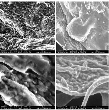
Scanning Electron Microscopic View Of Encapsulated Synbiotics
Properties of the microencapsulated bead
36
Figure 3.2.1 SEM image of Microencapsulated probiotic-prebiotic bead obtained by extrusion technique. A: Control (Lactobacillus plantarum), B: Lactobacillus plantarum coated with Borassus flabellifer and
Sodium alginate,
C: Control (Lactobacillus rhamnosus), D: Lactobacillus rhamnosus coated with Borassus flabellifer and Sodium alginate.
The Complex microstructure of the prebiotic-Ca-alginate microparticles loaded with L.plantarum and
L.rhamnosus was obtained. The inner part of the microparticles was built of a mesh-like alginate
37
or with alginate, prebiotics, increased survival in the microparticles for log4 was observed.The results indicated that microencapsulation of
L.plantarum and L.rhamnosus using alginate as encapsulating material, prebiotics and calcium chloride L. rhamnosus followed by freeze drying offered significant protection of probiotic cells.
Effect of freeze-drying on viability of encapsulated synbiotics
The viability of microencapsulated L.plantarum, L.rhamnosus after freeze drying , stored at 4°C for
Table 1: Survival rate of encapsulated probiotics (L.plantarum) with/without prebiotics over a period of 4 weeks
Probiotic cultures, (log CFU/g) Stored 0 weeks Stored 4 weeks
L.plantarum+ alginate+ prebiotics ±8.56 ±5.98
L.plantarum+ alginate ±8.23 ±6.28
Table 2: Survival rate of encapsulated probiotics (L.rhamnosus) with/without prebiotics over a period of 4 weeks
Probiotic cultures, (log CFU/g) Stored 0 weeks Stored 4 weeks
L.rhamnosus +alginate+ prebiotics ±8.58 ±7.41
L.rhamnosus+ alginate ±8.59 ±6.18
four weeks is shown in Table – 1, Table – 2. The viability of Probiotic (L.plantarum) coated with prebiotics and sodium alginate showed highest (8.56 log10 CFU/g) after encapsulated and (5.98log10
CFU/g) after storing for 4 weeks. The viability of Probiotic (L.rhamnosus) coated with prebiotics and sodium alginate showed highest (8.58 log10 CFU/g) after encapsulated and (6.18 log10
CFU/g) after storing for 4 weeks. Also researchers [11] found that a storage temperature of 4°C was effective in maintaining the viability of microencapsulated synbiotics during storage.
CONCLUSION
Microencapsulation of synbiotics thus provides a major positive outcome in food biotechnology and in medical microbiology. Future research could be concentrated on improving the viability of probiotics with further stronger coating material for encapsulation.
ACKNOWLEDGEMENT
The author and Co-authors have a great sense of gratitude to UGC, Government of India, New Delhi
for carrying out this kind of effective research in Government arts college, Coimbatore.
REFERENCES
1. Krasaekoopt W., Bhandari B., Deeth. H., (2003). ‘Evaluation of encapsulation techniques of probiotics for yoghurt: a review’, Int Diary J ,Vol 13, pp. 3-13
2. Shah, N.P. and Ravula, R.R. (2002). Microencapsulation of probiotic bacteria and their survival in frozen fermented dairy desserts. The Aust. J. Dairy Technology. 55: 139- 144.
3. Lee K., Heo T., (2000). ‘Survival of Bifidobacterium longum immobilized in calcium alginate beads in simulated gastric juices and bile salt solution’, Appl Environ Microbiol, Vol 66, spp. 869-873.
38
5. Talwalkar A., and Kailasapathy K. (2003).‘Effect of microencapsulation on oxygen toxicity in probiotic bacteria’, Aust. J. Dairy Technol, Vol 58, pp. 36-39.
6. Shin, H.S., Lee, J.H., Pestka, J.J., and Ustunol, Z. (2000). ‘Viability of bifidobacteria in commercial dairy products during refrigerated storage’, J. Food Prot, Vol 63, pp.327-331.
7. Shah, N. P. and Ravula, R., (2001)Microencapsulation of probiotic bacteria and their survival in frozen fermented dairy desserts, Aust. J. Dairy Technol., 55, 139–144.
8. Krasaekoopt W., Bhandari B., Deeth. H., (2003). ‘Evaluation of encapsulation techniques of probiotics for yoghurt: a review’, Int Diary J ,Vol 13, pp. 3-13
9. Tharmaraj, N., Shah, N.P., (2003). Selective enumeration of Lactobacillus delbrueckii ssp. bulgaricus, Streptococcus thermophilus, Lactobacillus acidophilus, Bifidobacteria,
Lactobacillus casei, Lactobacillus
rhamnosus, and Propionibacteria. Journal
of Dairy Science 86, 2288–2296.
10. Chen KN, Chen MJ, Liu JR, Lin CW, Chin HY. (2005); Optimization of incorporated prebiotics as coating materials for probiotic microencapsulation. J Food Sci. 70: M260-M266.