ANTIGEN PROCESSING PATHWAYS IN TRANSPLANTATION
by
Moin A. Saleem
A Thesis submitted to the University of London in partial fulfilment for the degree of Doctor of Philosophy with the Faculty of Medicine.
Transplantation Biology Unit
Division of Cell and Molecular Biology Institute of Child Health
ProQuest Number: 10106623
All rights reserved
INFORMATION TO ALL USERS
The quality of this reproduction is dependent upon the quality of the copy submitted.
In the unlikely event that the author did not send a complete manuscript and there are missing pages, these will be noted. Also, if material had to be removed,
a note will indicate the deletion.
uest.
ProQuest 10106623
Published by ProQuest LLC(2016). Copyright of the Dissertation is held by the Author.
All rights reserved.
This work is protected against unauthorized copying under Title 17, United States Code. Microform Edition © ProQuest LLC.
ProQuest LLC
789 East Eisenhower Parkway P.O. Box 1346
SECTION A - ABSTRACT
Antigen presentation and its regulation has been studied, with particular emphasis on the control and function of the MHC class II molecule, and on how this may affect allograft rejection.
CUT A is a specific controller of class II expression, and has recently been cloned in the human, using cell lines derived from MHC class II deficient patients. We have started to clone the gene for rat CET A, using PCR techniques, and have isolated a 433 base pair fragment. The full length CUT A sequence would be used to manipulate class II expression in experimental models, using, initially, antisense oligonucleotides.
In the interim, we have gained experience with antisense technology by targeting the IL-2 molecule. This cytokine is integral to the interaction between an APC presenting peptide on its surface, and the T-cell. We have attenuated T-cell proliferation in in vitro assays, and show evidence that this is due to specific down- regulation of IL-2 production.
In examining the intermediate steps between CUT A and class II expression, we have focused on the invariant chain. The invariant chain defines the ability of the class II molecule to present exogenous peptide. Its distribution in normal rat organs and transplanted kidneys is described. The results are unexpected, and the possible significance of the invariant chain in influencing self and non-self peptide recognition is discussed.
Additionally, we report a new group of patients with MHC class II deficiency, from ethnic groups not previously seen to be affected, and immunohistology studies on fibroblast cell lines from these patients show a complete lack of invariant chain expression.
A c k n o w le d g e m e n ts
I would like to thank my supervisors. Professor John Fabre and Dr. Kenth Gustafsson, for their invaluable help and discussion during this project.
The members of the transplantation biology team should also all take collective responsibility for attempting to transform me into a scientist. Thanks particularly to Ged Murphy, Andy Preece and Karen Strahan for dispensing on me all they know about molecular biology, to Loma Shewring for being my tissue culture guru, and to Greta Sawyer and Rebecca Schofield for helping out with the animal work. Much appreciation also to Sheng Yun for being an example of superhuman productivity in the lab, and to Rosie Dalchau for endless moral support and advice on half-timbered bam conversions, and also to Sally Cunningham and Gabi Slapak for attempting to dispel the myth that Medics can also do science. Adam Benham is also sorely missed, if only for his self-directed cries of'rubbish!' on the football pitch. Finally I would like to thank my family; my parents for successfully cloning themselves into me, and then accepting the consequences; Maryam and Safya for providing constant distraction whilst trying to write up, and my wife, Jane, for her support and trips to Sainsbury’s.
CONTENTS Page
Title page 1
SECTION A - ABSTRACT 2
Acknowledgements 3
List of Abbreviations 12
Nucleotide and Amino Acid codes 16
SECTION B - LITERATURE REVIEW B.l ANTIGEN PRESENTATION
B l.l INTRODUCTION 18
B1.2 THE MHC CLASS I PATHWAY OF ANTIGEN PRESENTATION 20
Bl.2.1 MHC class I structure 20
B .l.2.2 The nature of peptide binding to the MHC class I molecule 20
B.1.2.3 Peptide transport within the cell 22
B.1.3 THE MHC CLASS H PATHWAY OF ANTIGEN PRESENTATION 23
Bl.3.1 Structure of the class II molecule 23
Bl.3.2 Peptide and MHC class II transport 24
Bl.3.3 Characteristics of peptide binding to the MHC class II molecule 25 B .l.3.4 Presentation of endogenous peptides via the MHC class II pathway 26
B.2 CONTROL OF MHC CLASS H PRESENTATION
B.2.1.2 The discovery of CUT A 30
B.2.1.3 The functional role of CUT A 32
B.3 THE INVARIANT CHAIN
B.3.1 INTRODUCTION 35
B.3.2 SYNTHESIS AND STRUCTURE 36
B.3.2.1 The invariant chain gene 36
B.3.2.2 Regulation and induction of invariant chain synthesis 37
B.3 .2.3 Structure of the invariant chain 39
B.3.3 TRANSPORT OF THE MHC CLASS H MOLECULE THROUGH
THE CELL 41
B.3 .3 .1 Association of invariant chain with MHC class II in the ER 41 B.3.3.2 Inhibition of peptide binding to MHC class II in the ER 41 B.3.3.3 Transport of the class Il/Invariant chain complex to the endosomes 42 B.3.3.4 Dissociation of the MHC class Il/Invariant chain complex 43 B.3.4 FUNCTIONAL CONTROL OF THE INVARIANT CHAIN 45 B.3.4.1 A human class II related locus - HLA-DM 45 B.3.4.2 An alternative route for endosomal localisation 45 B.3.5 THE ROLES OF MHC CLASS H AND INVARIANT CHAIN
EXPRESSION 46
B.3.5.1 Cell surface expression of invariant chain and of MHC class II molecules 46
B.3.5.2 Consequences for antigen presentation 48
B.4 THE T-CELL AND CYTOKINE RESPONSE IN ANTIGEN PRESENTATION
B.4.1 INTRODUCTION 51
B.4.2 ACCESSORY MOLECULES INVOLVED IN T-CELL
ACTIVATION 52
B.4.3 THE B7-CD28/CTLA4 PATHWAY 54
B.4.3.1 Ligand and receptor characteristics 54
B.4.3.2 Studies on the role of B7-1 and B7-3 55
B.4.3.3 The roles of CD28 and IL-2 in the T-cell response 55
B.4.4 THE T-CELL RESPONSE 57
B.4.4.1 T-cell activation 57
B.4.4.2 The Thl/Th2 model of CD4 cell activation 57 B.4.5 CYTOKINE ACTIVATION IN ALLOGRAFT REJECTION 58
B.4.6 THE SEARCH FOR TRANSPLANTATION TOLERANCE 61
B.4.6.1 Cytokine mediated transplant tolerance 61
B.4.6.2 Anergy as a means for tolerance 62
B.5 MECHANISMS OF ALLOGRAFT REJECTION
B.5.I INTRODUCTION 65
B.5.2 THE T-CELL RESPONSE TO ALLOGRAFTS 66
B.5.3 THE ‘DIRECT’ PATHWAY OF ALLORECOGNITION 66
B.5.4.1 Processing of donor MHC molecules 74
B.5.4.2 Supporting evidence from animal models 74
B.5.4.3 In vitro evidence for indirect allorecognition in humans 76 B . 5 .4.4 The role of indirect allorecognition in acute or chronic rejection 77
B.5.5 CHRONIC REJECTION 78
B.5.5.1 A continuing problem 78
B.5.5.2 Risk factors for chronic rejection 79
B.5.5.3 Pathology of chronic rejection 80
B.5.5.4 Experimental models 81
B.5.6 THE HOST IMMUNE RESPONSE IN CHRONIC REJECTION 82
B.5.6.1 The T-cell response 82
B.5.6.2 The humoral response 83
B.5.6.3 The significance of MHC antigens in chronic rejection 84 B.5.6.4 T-cell accessory molecules in chronic rejection 85
SECTION C - DATA CHAPTERS SECTION C - CHAPTER ONE
C .l ISOLATION AND CHARACTERISATION OF A FRAGMENT OF THE RAT MHC CLASS H TRANSACTIVATOR (CHTAI GENE
C.1.1 INTRODUCTION 87
C.1.2.1 Screening of a rat spleen library 89
C. 1.2.2 Materials 89
C .l.2.3 Electrophoresis ofDNA 91
C. 1.2.4 Library titering 92
C. 1.2.5 Library screening 92
C.1.2.6 Use of the polymerase chain reaction (PCR) to isolate a fragment of
the rat CIÏTA gene 94
C. 1.2.7 Removal of rat spleen 94
C. 1.2.8 Preparation of mRNA 95
C. 1.2.9 Preparation of first strand cDNA 97
C. 1.2.10 PCR amplification 97
C. 1.2.11 Cloning of PCR product into a plasmid vector 99
C.1.2.12 Purification of plasmid DNA 101
C.1.2.13 Ethanol precipitation 103
C. 1.2.14 Sequencing reactions 104
C.1.2.15 DNA sequencing apparatus 106
C.1.3 RESULTS 107
C .l.3.1 Library titering 107
C.1.3 .2 Library screening 108
C. 1.3.3 PCR of rat cDNA using human primers 108
C.1.3.7 Sequencing of the plasmid DNA 114
C.1.3.8 Analysis of the rat CRT A gene fragment 118
C.1.4 DISCUSSION 118
SECTION C - CHAPTER TWO
C.2 THE USE OF ANTISENSE OLIGONUCLEOTIDES TO INHIBIT T-CELL PROLIFERATION IN VITRO
C.2.1 INTRODUCTION 122
C.2.2 MATERIALS AND METHODS 130
C.2.2.1 Oligonucleotide design and synthesis 130
C.2.2.2 Animals 132
C.2.2.3 Cell cultures (protocol for experiments without liposome) 132 C.2.2.4 Cell cultures (protocol for experiments with liposome) 133 C.2.2.5 Use of IL-2 dependent cells to assay IL-2 activity of culture supernatants 134
C.2.3 RESULTS 135
C.2.4 DISCUSSION 152
SECTION C - CHAPTER THREE
C.3 A REPORT AND STUDY OF MHC CLASS H DEFICIENT PATIENTS
C.3.2 MATERIALS AND METHODS 156 C.3.2.1 Lymphoproliferation to PHA, Candida and PPD 156
C.3.2.3 T-lymphocyte subsets 157
C.3.2,4 Monoclonal Antibodies used 157
C.3.2.5 Freezing of cells 158
C.3.2.6 Thawing and culture of fibroblast cell lines 159 C.3.2.7 Growth of fibroblasts on chamber slides 159 C.3.2.8 Immunoperoxidase staining of fibroblasts 160
C.3.3 RESULTS 161
C.3.3.1 Patient data 161
C.3.3.2 Immunohistology on fibroblast cell lines 165
C.3.4 DISCUSSION 171
SECTION C - CHAPTER FOUR
C.4 A SYSTEMATIC STUDY OF INVARIANT CHAIN AND MHC CLASS n EXPRESSION IN RAT TISSUES
C.4.1 INTRODUCTION 175
C.4.2 MATERIALS AND METHODS 177
C.4.2.1 Animals 177
c .4,2.4 Immunohistology 179
C.4.2.5 Kidney transplantation in rats 180
C.4.3 RESULTS 181
C.4.4 DISCUSSION
SECTION C - CHAPTER FIVE
C.5 ATTEMPTS TO DEMONSTRATE INDIRECT T-CELL ALLORECOGNITION OF DONOR MHC PEPTIDES IN TRANSPLANT PATIENTS
C.5.1 INTRODUCTION 199
C.5.2 MATERIALS AND METHODS 201
C.5.2.1 Buffers and Solutions 201
C.5.2.2 Peptides 202
C.5.2.3 Lymphocyte proliferation assay 202
C.5.2.4 Patients 203
C.5.3 RESULTS 204
C.5.4 DISCUSSION 209
SECTION D - DISCUSSION 213
LIST OF ABBREVIATIONS
a. a. amino acid
Ab Antibody
APC Antigen presentation ATP Adenine triphosphate p2m p2 microglobulin
BLS Bare lymphocyte syndrome BMT Bone marrow transplantation BSA Bovine serum albumin CD Cluster of differentiation cDNA Complementary DNA CIITA Class II transactivator
CLIP Class II loading invariant chain peptide CNS Central nervous system
con A Concanavalin A cpm counts per minute CS Chondroitin sulphate CTL Cytotoxic T-lymphocyte
CTLA-4 Cytotoxic T-lymphocyte associated antigen CTP Cytosine triphosphate
ddATP dideoxy ATP DMSO Dimethyl sulfoxide DNA Deoxyribonucleic acid DTH Delayed type hypersensitivity
E.coli Escherichia coli
ELAM-1 Endothelial leucocyte adhesion molecule-1 ELISA Enzyme linked immunosorbent assay ER Endoplasmic reticulum
FITC Fluorescein isothiocyanate
g gram or gravitational acceleration GTC guanidinium thiocyanate
GTP Guanine triphosphate HLA Human leucocyte antigen hsp Heat shock protein HSV Herpes simplex virus
ICAM-1,2 Intercellular adhesion molecule-1, 2 IDC Interstitial dendritic cell
IFN-y Interferon-y Ig Immunoglobulin li Invariant chain IL-2 Interleukin-2
IL-2R Interleukin-2 receptor kb kilobase
1 Litre
LC Langerhans cell
LFA-1 Lymphocyte function associated antigen-1 mAh Monoclonal antibody
MHC Major histocompatibility complex MLC Mixed lymphocyte culture
mRNA messenger RNA O.D. Optical density Oligo Oligodeoxynucleotide PEL Peripheral blood leucocyte
PBMC Peripheral blood mononuclear cell PBS Phosphate buffered saline
PCR Polymerase chain reaction pfu plaque forming units PHA Phytohaemagglutinin PPD Purified protein derivative RAM Rabbit anti-mouse
rIL-2 Recombinant IL-2 RNA Ribonucleic acid RPE R-Phycoerithrin
RSV Respiratory syncitial virus
s second
TAP Transporter associated with antigen processing TCR T-cell receptor
Thl/Th2 T-helper 1 / T-helper 2 TNF-a Tumour necrosis factor-a TTP Thymine triphosphate
ONE LETTER AND THREE LETTER CODE FOR AMINO ACIDS Neutral and hydrophobic;
Alanine Ala A
Valine Val V
Leucine Leu L
Isoleucine He I
Proline Pro P
Tryptophan Trp W
Phenylalanine Phe F
Methionine Met M
Neutral and Polar:
Glycine Gly G
Serine Ser S
Threonine Thr T
Tyrosine Tyr Y
Cysteine Cys C
Glutamine Gin Q
Asparagine Asn N
Basic:
Lysine Lys K
Arginine Arg R
Histidine His H
SECTION B - LITERATURE REVIEW
B .l ANTIGEN PRESENTATION
B.1.1 INTRODUCTION
Sixty years ago, an antigen was discovered by Gorer, on all the cells of a mouse, that was recognised by the immune system during rejection of a foreign tissue transplant. This work led to an explosion in the field of transplantation immunology, and Gorer’ s antigen evolved into the major histocompatibility complex (MHC) of the mouse, later renamed H-2. During the I960’ s, another class of antigens was described, that turned out to be associated with the MHC. These, class II antigens, are principally restricted to certain cells of the immune system, namely lymphocytes and reticular cells.
well as new pieces to discover. The system is not a linear one, and each fragment of new information often reveals more questions than it answers.
B.1.2 THE MHC CLASS I PATHWAY OF ANTIGEN PRESENTATION
B.1.2.1 Class I structure
MHC class I molecules consist of a polymorphic type I integral membrane glycoprotein heavy chain of about 46 kDa, noncovalently associated with a 12kDa soluble subunit, pz-microglobulin (p2m) (Bjorkman et al. 1990). High resolution x-
ray crystallographic structures have been derived for human and mouse class I molecules (Bjorkman et al. 1987; Fremont et al. 1992). The most striking aspect of these structures is that the a la 2 domain unit forms a single peptide binding site supported by a p-pleated sheet floor containing eight strands and bounded by two a-helices, one from a l and one from a2. p2m makes contact with both the
immunoglobulin-like a3 domain, and also the floor of the peptide binding region. There also exists a set of pockets, in some cases extending deep between the floor and helical walls of the binding region (Garret et al. 1989).
B l.2.2 The nature of peptide binding to the class I molecule
be found in comparable positions in most peptides, giving rise to the concept of ‘ motif amino acids important in promoting binding to a particular allele of class I. The lack of strong signals beyond 9 amino acids in these pools of eluted material fit well with the 8 and 9 residue length of specific peptides found associated with class I molecules, suggesting that length is important for tight peptide association.
Allelic polymorphism also contributes to the shape and physico-chemical character of the pockets within the binding region, and notably almost all the side chains of the polymorphic amino acids in the binding domain are orientated inward toward the region containing peptide (Bjorkman et al.l987). This suggests that the major role of polymorphism is to regulate the binding of peptides, rather than to directly affect interaction with the T-cell receptor. In fact the a la 2 domain is crucial in selective peptide binding, and mutation in this region has been shown to abolish peptide recognition by T-cells (Moots et al.l991).
The nature of peptide visualised in HLA-B27 electron density maps has permitted detailed model building of a prototype bound peptide to class I (Madden et al.l991). The consensus peptide is nine residues long, and in an extended conformation with a central kink. A critical aspect of the peptide binding is the presence of conserved hydrogen bonds to the peptide’ s NH2 and COOH termini
through conserved MHC residues. Such bonds, common to all peptides, suggest how otherwise polymorphic class I molecules can each act as effective peptide binding proteins for a wide diversity of peptide sequences.
class I bound peptides have been determined, and of those peptides whose source protein could be identified, all but one are from abundant cytoplasmic or nuclear proteins (Wei et al.l992).
B l.2.3 Peptide transport within the cell
B1.3 THE MHC CLASS H PATHWAY OF ANTIGEN PRESENTATION
B l.3.1 Structure of the class H molecule
MHC class II molecules are type I heterodimeric integral membrane proteins. Each dimer consists of one a and one p chain in noncovalent association. As with class I, the intron/exon organisation of genes encoding these chains corresponds to functional domains of the protein molecule, with the second exon containing coding information for the bulk of the positions at which extensive intraspecies polymorphism exists (Choi et al.l983).
B l.3.2 Peptide and MHC class H transport
The class II a and P subunits associate rapidly in the ER, in the presence of the invariant chain (li) to form a trimer. The role of li in class II transport and function is fully reviewed in section B.3. The class II molecule/Ii complex leaves the ER, and reaches the endosomes via the golgi apparatus, whereupon li is cleaved leaving the class II molecule free to bind with processed exogenous peptide.
B 1.3.3 Characteristics of peptide binding to the MHC class II molecule
Extensive intraspecies polymorphism is also a hallmark of MHC class II molecules, which plays a major role in determining which peptides show long-lived binding to class II molecules (Buus et al. 1987). Single residue changes in the floor of the binding site or in the helices can decrease binding of certain peptides by several orders of magnitude (Brett et al. 1989).
promiscuous peptides, capable of binding to many different class II alleles, have been identified (Sinigaglia et al. 1988). Promiscuous peptides should contain either overlapping class II binding motifs, or they should use anchors that are conserved among DR ligands, and should lack allele-specific contact sites that could prevent binding to other class II molecules. Fascinatingly, such a ‘ supermotif was recently found in the class Il-associated invariant chain peptide (CLIP), indicating that CLIP is a universal class II ligand in its interaction with the class II aP dimer (Malcherek et al. 1995; Sette et al. 1995).
Class II molecules appear to alter their structure upon peptide interaction, and there is direct evidence that peptide association is necessary for the characteristic stability of class II dimers (Sadegh Nasseri et al. 1992). Experiments also suggest that a substantial fraction of class II in the endosomal loading compartment, does not find usable peptides under normal conditions and may not reach the cell surface (Srinivasan et al. 1991). This is achieved by aggregation of the empty dimers, ultimately leading to class II destruction (Stern et al. 1992). Thus peptide binding appears to have two distinct effects; one is to contribute to dimer stability by interaction with portions of the a and P chains, and the other is to change the structure of the class II molecule to add to its intrinsic stability (Lanzavecchia et al. 1992).
B 1.3.4 Presentation of endogenous peptides via the MHC class H pathway
’ X X
antigen processing and peptide binding to class II molecules, namely a pre-golgi compartment. Perhaps the most intriguing data relates to the ability of a strictly cytoplasmic antigen, the measles virus matrix protein, to be presented by class II (Jacobson et al. 1989), and several subsequent similar reports of class II presentation of cytoplasmic proteins. The site of peptide-class II association in these cases is unresolved, although some evidence points to at least one of these antigens moving to the endosomal system for association with class II (Malnati et al. 1992). The question in that case switches to the site of peptide generation and the pathway followed by antigen or peptide to reach the endosomal compartment. This illustrates the fact that the class-specific function of MHC molecules relates primarily to their preferred site of peptide loading, which only incompletely dictates the protein sources (exogenous/endogenous) of those peptides. Clearly, however, the two systems are not interchangeable, as illustrated in the case of the severe immunodeficient phenotype seen in MHC class II deficient individuals, who possess relatively intact expression of MHC class I (section B.2.1).
B.2 CONTROL OF MHC CLASS H EXPRESSION
B2.1 MHC CLASS H DEFICIENCY
surface expression of MHC class I molecules has been described in certain cases (Amigorena et al. 1995), but the significance of this remains unclear. This confirmed the important role of MHC gene products in immune defence mechanisms, since all these patients have abnormal cellular and humoral responses to specific antigens.
Subsequent studies showed that this disorder was due to a lack of synthesis of class II molecules, and a lack of mRNA for all of the different MHC class II isotypes (LisowskaGrospierre et al. 1985). Direct transcription assays confirmed that none of the class II genes was transcribed (Reith et al. 1988). Since the MHC class II genes are not deleted, the global lack of expression of the entire class II gene family suggested a general defect in the regulation of these genes. This implicated one or several trans-acting factors (acting on the class II promoter region) would be affected, and strongly suggested that identification of the genes responsible for this disease would lead to important regulators of MHC class II expression.
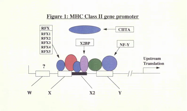
B2.1.1 The class H promoter region
Figure 1: MHC Class II ^ene promoter
CIITA
X2BP
NF-Y
RFX4
Upstream
Translation
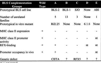
More functionally relevant information on the molecular mechanisms controlling MHC class II gene transcription has been obtained by the study of the MHC class II deficient cell lines. Cell fusion experiments between several different patient and
in vitro generated cell lines has demonstrated the existence of at least four different
complementation groups (Benichou et al. 1991), implying that mutations in at least four different genes result in the same disease phenotype (table 1). One further experimentally generated mutant cell line defines a fifth complementation group, for which no patient has yet been identified (Gladstone et al. 1978; Seidl et al. 1992). Two main groups of patients emerged, one that exhibits a defect in RFX binding, and the other showing normal RFX binding (Stimac et al. 1991). The former group displays a complete lack of occupancy of the class II promoter in vivo (Kara et al. 1991) suggesting that normal binding of RFX is required for occupancy of the X2 and Y boxes. In the second group the class II promoter region is normally occupied.
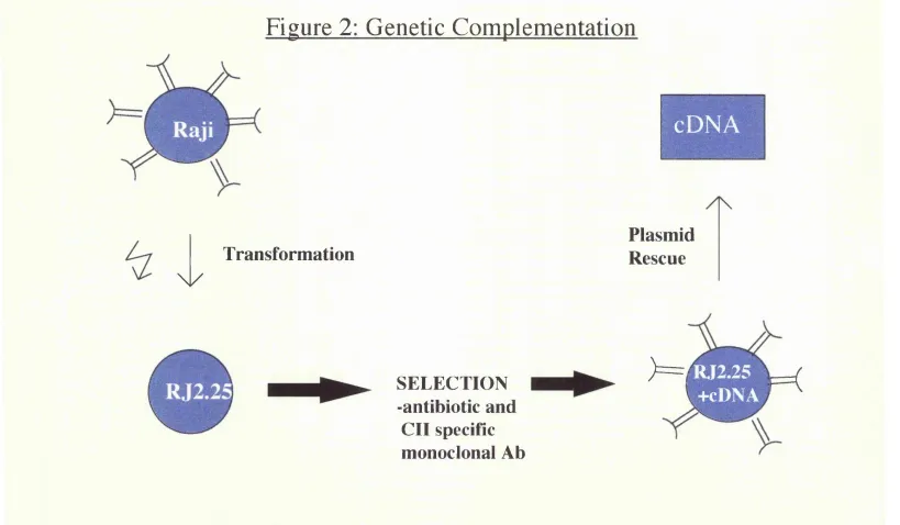
B.2.1.2 The discovery of CIITA
Figure 2: Schematic representation of complementation cloning method (Steimle
et al., 1993). Plasmid DNA prepared from the HLA class 1 positive B cell line
Raji was transfected by electroporation into the mutant (class II deficient) B cell
line RJ2.25. Class II expressing cells were selected and found to contain a
Figure 2: Genetic Complementation
Transformation
\ /
cDNA
Plasmid
Rescue
SELECTION
-antibiotic and
CII specific
monoclonal Ah
) = i
RT2.25
Table 1: PhenotvpicaK biochemical and molecular defects of MHC class II
regulatory mutants from BLS complementation groups A-E
HI
Prototypical BLS cell line BLS-2 BLS-1 SJO None ABI Number of unrelated
families
5 13 3 None 1
Prototypical in vitro mutant RJ2.25 None None 6.1.6 None
MHC class II expression 4- - -
-MHC class II promoter activity
+ - - - - nt
RFX-binding + + - - nt nt
Promoter occupancy in vivo + + - - - nt
Genetic defect CIITA ? RFX5 ? ?
nt = not tested
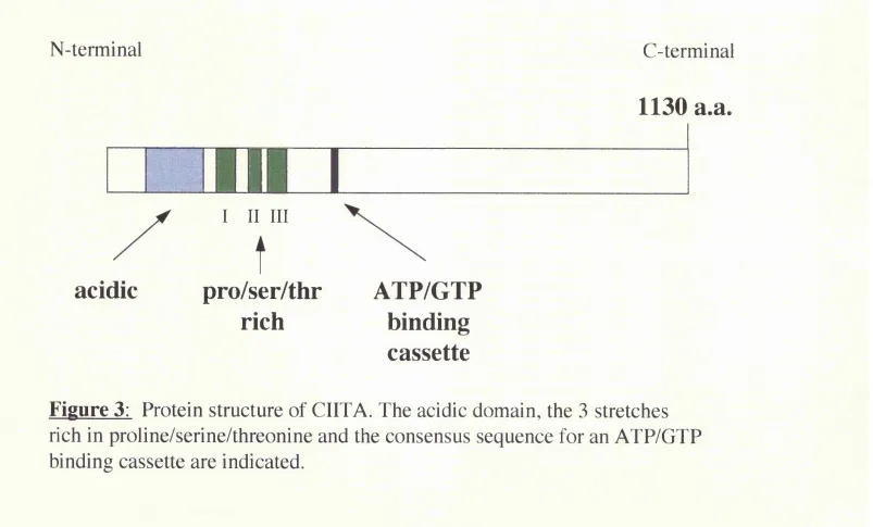
B.2.1.3 Functional role of CIITA
the X box region. CIITA itself contains a potent activation domain, supporting its role in class II transcription.
MHC class II genes are expressed constitutively in only a few cell types, but they can be induced in the majority of them, in particular by IFNy. It has been shown
that CIITA expression is controlled and induced by IFNy and that the JAKl protein tyrosine kinase activity is required to induce the expression of CIITA upon IFNy stimulation (Steimle et al. 1994; Chang et al. 1994). This indicates that CIITA is part of the signalling cascade from the IFNy receptor to the activation of class II genes. In addition, the expression of CIITA is sufficient to activate class II genes in the absence of IFNy stimulation suggesting that CIITA is the major regulatory factor for the inducible expression of class II genes.
N-terminal
C-terminal
acidic
I II III
t
pro/ser/thr
ATP/GTP
rich
binding
cassette
1130 a.a.
m.1
B.3 THE INVARIANT CHAIN
B.3.1 INTRODUCTION
The invariant chain (li) was first identified in 1978, as a common polypeptide chain in different MHC class II immunoprecipitates (Jones et al. 1978). It is so called because of its non-polymorphic nature. Class II molecules are composed of two MHC-encoded subunits, the a and p chain, which associate in the endoplasmic reticulum (ER) with li.
B.3.2 INVARIANT CHAIN SYNTHESIS AND STRUCTURE
B.3.2.1 The invariant chain gene
The genes for MHC class II are regulated in a complex manner, being constitutively expressed, inducibly expressed, or not expressed depending on the cell type. However, the regulation and tissue distribution of the invariant chain is far less clear. There are clear similarities in the promoter regions of class II and li, and also in the interferon y inducibility of the two molecules, but there is also evidence that regulation of the two is distinctive, which would have implications for the processing and presentation of peptide antigens in various cell types, under various conditions.
from the class II a and p chain genes were also found in analogous positions in the li gene suggesting a possible role in the co-regulation of expression of these genes (O'Sullivan et al. 1986). The 5’ regulatory sequences of the invariant chain gene appear to be a combination of conserved class II regulatory elements and promoter elements commonly found in other eukaryotic genes. Contained within the promoter of the li gene are sequences (X and Igammal) that are similar to the X and Y box elements of the class II gene promoter, suggesting that these sequences might be involved in its regulation and contribute to the co-expression of MHC class II and li genes (Zhu et al. 1990). This was confirmed when it was shown that sequences homologous to the class II W, X and Y elements are present in the promoter region of the li gene, and that interferon-gamma (IFN y) inducible expression of li in a cell line is regulated via these sequences (Brown et al. 1991a). Interestingly, it has very recently been shown that CIITA, a MHC class II transactivating factor which binds to the X box binding transcription factors (see section B.2 for full review), is also required for expression of the invariant chain (Chang et al. 1995), presumably binding to homologous transcription proteins that bind to the X box element on the li gene.
B.3.2.2 Regulation and induction of invariant chain synthesis
simultaneously. Also, variant lines of these cells failed to express either la or li upon IFNy stimulation. However, B lymphocyte lines exposed to IFNy enhanced the synthesis of li in the absence of la upregulation, indicating that in some cell lines li can be regulated independently of MHC class II expression (Koch et al. 1984). In human dermal fibroblasts and vascular endothelial cells, IFNy induces li simultaneously with several MHC class II antigens (Collins et al. 1984). In a human colon carcinoma cell line which constitutively expresses neither MHC class II or li, administration of TNFa or IFNy alone had no effect, but the cytokines together induced class II and li, with li mRNA detectable 10-12 hours after stimulation (Pessara et al. 1988). In a study of mouse tissue expression, upon IFNy stimulation, MHC antigen expression was dramatically increased throughout the body, with striking differences in the inducibility of certain tissues for class II and li (Momburg et al. 1986).
despite the similarities in their promoter regions, li and class II do not necessarily respond in the same way in various cell lines to cytokine induction.
B.3.2.3 Structure of the invariant chain
Figure 1: Protein structure of the human invariant chain indicating different
isoforms, formed by alternate splicing. Arrows indicate N-linked glycosylation
Structure of human invariant chain
NH2
NH2
NH2
NH2
p33
p35
p
41
p
43
TM
U
u
CLIP
COOH
COOH
COOH
COOH
29 56 81 104 2 1 6
B.3.3 MHC CLASS H TRANSPORT THROUGH THE CELL
B.3.3.1 Association of li with MHC class H in the endoplasmic reticulum
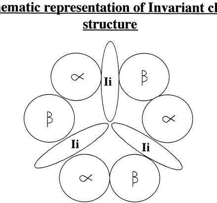
In the absence of association with class II glycoproteins, li is trimeric (Marks et al.l990). Class II molecules on the surface of cells exist as a heterodimer of a and P subunits. The membrane-distal domains of these subunits form a peptide binding site consisting of a platform of eight strands of p-pleated sheet, with two a-helices forming the boundaries. In the ER, the invariant chain associates with the newly synthesised class II dimer via its extracytoplasmic (C-terminal) region (Marks et al.l986). This complex has been demonstrated to be a nine-subunit transmembrane protein that contains three aP dimers associated with an li trimer (Roche et al. 1991b) (figure 2). The association of li with class II is rapid and efficient, due to a large molar excess if li in cells expressing class II molecules (Kvist et al.l982; Machamer et al.l995). In mice bearing a deletion of the li gene, newly synthesised MHC molecules are largely misfolded, and fail to leave the ER due to the lack of the li transport signal (Elliott et al.l994).
B.3.3.2 Inhibition of peptide binding to MHC class H in the ER
prevents premature peptide loading (Demotz.1993). A short, 25 residue, contiguous internal segment of li frequently found associated with purified class II molecules has been designated the CLIP (class II associated invariant chain peptide) region and described as a nested set of class II associated peptides. This region is critical for class II folding, ER to golgi transport and inhibition of peptide binding (Romagnoli et al.l994). The CLIP region has been mapped to li exon 3, which encodes amino acids 82-107 (Sette et al.l995). CLIPs appear to bind in the class II peptide binding groove, and overcome allele-specificity by taking advantage of one or more supermotifs (Malcherek et a l l 995). Proteolysis of the invariant chain in the complex with cathepsin B releases a p dimers that bind antigenic peptides extremely well (Roche et a l l 99lb).
B.3.3.3 Transport of the class
n/Ii
complex to the endosomestransport to the site of antigen processing and loading involves movement through early endosomes to late endosomes, the rate of which is determined by the level of li expression (Romagnoli et al.l993). In li deletion experiments, class II dimers appear misfolded and are inefficiently transported from the ER to the golgi. In addition, class II transported through the golgi accumulates an abnormally increased molecular mass associated with N-linked glycosylation (Anderson et al.l992). Thus li appears to act as a class II specific chaperone in its transport through the cell.
B.3.3.4 Dissociation of the MHC class H/lnvariant chain complex
Figure 2
B.3.4. FUNCTIONAL CONTROL OF THE INVARIANT CHAIN
B.3.4.1 A human class H related locus, HLA-DM
In 1991 a previously undiscovered class II related protein was described (Zemmour et al.l993). The genes, DMA and DMB, map between the HLA-DNA and -DOB loci, and encode a and p chains of this novel member of the immunoglobulin gene family. Recently it has been found that HLA-DM catalyses the dissociation of class II associated invariant chain peptide from MHC class II-CLIP complexes in endosomes, at an acidic pH optimum, and facilitates the binding of antigenic peptides (Denzin et a l l 995). Furthermore, X-ray crystal structure determination shows that the CLIP fragment binds to HLA-DR3 in a way almost identical to that in which antigenic peptides bind class II glycoproteins. Hence, this structure is the substrate for the loading of antigenic peptides by an exchange process catalysed by HLA-DM (Sloan et al.l995). The essential role of this molecule is illustrated by various cell lines deficient in antigen presentation, which have been shown to lack HLA-DM (Morris et a l l 994).
B.3.4.2 An alternative route for endosomal localisation
degradation and return of free class II to the cell surface. Interestingly, the efficiency of antigen presentation to T-cells is preserved in these cells, suggesting that endocytosed class II can form immunogenic complexes with newly processed antigen (Anderson et al.l993; Nijenhuis et al.l994). This process can occur physiologically as seen in class II negative cell lines, wherein li enters the endocytic route via the cell surface (Henne et al.l995), or in a B cell line in which a large population of class Il/Ii complexes are seen to reach endosomes by rapid internalisation from the cell surface (Roche et al. 1993).
B.3.5 THE ROLES OF MHC CLASS H AND INVARIANT CHAIN
EXPRESSION
B.3.5.1 Cell surface expression of li and of MHC class H molecules
molecules (Sekaly et al. 1986; Miller et al. 1986), although mice lacking li expression do show a dramatic reduction in surface class II (Bikoff et al. 1993). Confusingly, li deficient fibroblasts, although expressing an altered form of class II at the cell surface, have been shown to present antigenic peptide to T cells as efficiently as li expressing cells (Sekaly et al. 1988; Peterson et al. 1990). There is contrasting data suggesting that high li levels correlate with an increased ability of fibroblasts to present exogenous antigen (Bertolino et al. 1991). To support the former, cells expressing surface class Il/Ii complexes interact with staphylococcal toxins and stimulate polyclonal T-cells as efficiently as cells expressing only class II at the cell surface (Karp et al.l992).To support the latter, fibroblast transfectants that fail to dissociate li from class II, hence expressing mainly class Il/Ii complexes at the cell surface, are inefficient in their ability to present peptides to T-cells (Roche et al. 1992).
Conversely, human B cells expressing at least one class II isotype express normal amounts of li on the cell surface, but total class II deletion results in diminished expression of li. Also, unexpectedly, li is not synthesised on de-novo induced class II positive T-cell clones (Wilson et al. 1993b).
B.3.5.2 Consequences for antigen presentation
As alluded to above, the role for li in class II restricted antigen presentation has been controversial. There was initially indirect evidence for the importance of li, wherein fresh epidermal Langerhans cells (LC) synthesised higher amounts of li and were far more efficient at antigen presentation than cultured LC’ s (Pure et al. 1990). This evidence strengthened with the finding that antigen presentation is facilitated by the p41 form of li (Peterson et al. 1992). Furthermore the class II conformation change ascribed to li binding is retained after li dissociates. This altered conformation affects recognition by allogeneic T-cells, which preferentially recognise class II molecules that heve been associated with li (Rath et al. 1992). A dramatic finding in this respect concerns the small proportion of li that is modified by the addition of chondroitin sulphate (li-CS). This form of li is expressed at the surface of antigen presentation cells, and greatly enhances the ability of the cell to stimulate T-cells, by means of li-CS functioning as an accessory molecule, interacting with CD44 on the T-cell (Naujokas et al. 1993). Finally, li deficient mice present exogenous antigen very poorly, and are deficient in producing and negatively selecting CD4+ T-cells (Viville et al. 1993). Thus, the bulk of the evidence so far points towards a crucial role for the li/class II interaction in the ultimate presentation of immunogenic antigen, and which is also likely to play a part in selecting the particular peptide epitope presented (Bodmer et al. 1994).
B.3.5.3 The role of MHC class H distribution in antigen presentation
Class II molecules, although usually found on ‘ professional’ antigen presenting cells such as B cells and macrophages, can also be found on endothelial cells, and
constitutively on epithelial cells o f only a small num ber o f tissues (e.g. thymus,
small intestine, renal proxim al tubule). From classical teaching, the true
im m unologic periphery w as only thought to constitute satellite lym phoid tissue,
abundant in professional antigen presenting cells. It w as eventually realised,
how ever, that m acrophages and dendritic cells are also scattered th roughout
parenchym al organs, like thyroid, liver and kidney (H alloran et al. 1985; B ottazzo
et al. 1986; H art et al. 1 9 8 la ) and that perhaps, auto-im m une and allogeneic
responses could be established locally at the site o f em erging inflamm ation. It has
subsequently been dem onstrated that non-lym phoid som atic cells are also capable
o f aberrantly increasing the num bers o f M H C class II m olecules on their cell
surface (H alloran et al. 1985; B ottazzo et al. 1986), probably follow ing cytokine
exposure in their local m icroenvironm ent (H averty et al. 1989; G lim cher et
al. 1992). This leads to the hypothesis that parenchymal cells, like epithelium , might
also be able to process and present antigenic peptides to T lym phocytes (B ottazzo
et al. 1986; H averty et al. 1988). The role o f class II on epithelial cells is thus,
incom pletely understood. C hondrocytes and enterocytes for exam ple, can process
and present antigen to T cells, but o th er non-haem atopoietic cells such as
kératinocytes and pancreatic (3 cells induce antigen specific unresponsiveness in T
cells (H ines et al. 1989). Renal tubular epithelial cells, uniquely am ong the cells o f ,
transplanted organs, express M H C class II constitutively. They are speculated to
participate in the pathogenesis o f immune renal injury, and have been show n to
present both self and foreign antigen (H art et al. 1 98la), although their role in
B.3.5.4 The role of invariant chain distribution in antigen presentation
B.4 THE T-CELL AND CYTOKINE RESPONSE IN ANTIGEN
PRESENTATION
B.4.1 INTRODUCTION
B.4.2 ACCESSORY MOLECULES INVOLVED IN T-CELL ACTIVATION
Occupancy of the T-cell receptor (TCR) by processed antigen/MHC provides the first, but not totally sufficient signal for full T-cell activation. Signal two, or ‘ co stimulation’ is derived from ligand-ligand interactions between the surfaces of APC’ s and T-cells (Janeway, Jr. et al. 1994). All APC’ s (e.g. dendritic cells, activated macrophages or B-cells) are able to stimulate primed T-cells, whereas dendritic cells are the most potent APC’ s in providing these co-stimulating signals to the naive T-cell (Macatonia et al. 1993).
Figure 1: Receptor-ligand pairs between activated T-cells and APC s (Boussiotis et al., 1994)
T-cell - APC interactions
Stages of T-cell response
Functional outcome of blockade
r-Cell
LFA-l - ICAM-1 ICAM-1 - --- LFA-1 VLA-4 - ---VCAM-1
CD-2 LFA-3
CDS
CD3 ---m h c i
TCR
CD4
CD3 --- MHC 11 TCR
CD40L --- CD40 CD5 ---CD72 CD24 ---CD24 CD28 APC Adhesion Immunosuppression Antigen recognition Immunosuppression Co-stimulation ?Immunosuppression CD28 CTLA4 CTLA4 B7-2 B7-1
B.4.3 THE B7-CD28/CTLA4 PATHWAY
B.4.3.1 Ligand and receptor characteristics
To date, two CD28/CTLA4 counter-receptors have been cloned and functionally characterised, termed B7-1 and B7-2 (Hathcock et al. 1994). These molecules, which can be induced on a wide variety of APC’ s (Boussiotis et al. 1993) are members of the immunoglobulin gene superfamily (Freeman et al. 1993b). Although B7-1 and B7-2 demonstrate only 25% amino acid homology, they are both low- affinity receptors for CD28 and high affinity receptors for CTLA-4 (Linsley et al. 1993). Some investigators have shown B7-1 and B7-2 to be constitutively expressed on dendritic cells (Young et al. 1992), but other studies find B7-1 expression to be primarily induced (Hart et al. 1993)
B7-2 is also constitutively expressed on monocytes (Azuma et al. 1993) but most other APC’s require some stimuli to induce their expression.
CTLA4 than for CD28, so it is not surprising that an immunoglobulin fiision protein of CTLA4 (CTLA4 Ig) has proven to be the most effective reagent to inhibit B7 family co-stimulatory function both in vitro and in vivo (Lenschow et al. 1992).
B.4.3.2 Studies on the role of B7-1 and B7-2
Three model systems have addressed the functional role of the counter-receptors for CTLA4/CD28. The B7-1 deficient mouse provided evidence for the existence of more than one counter-receptor (Freeman et al. 1993a) wherein activated B-cells expressed the alternative receptor, B7-2, leaving the pathway intact. Secondly, B7- 1 transgenic mice (Sethna et al. 1994), constitutively expressing the B7-1 gene on mature B-cells, have greatly reduced levels of serum immunoglobulins. In addition, these mice demonstrate reduced antibody responses to T-dependent hapten-protein conjugates, which is restored by the administration of anti-B7-l mAh. This suggests that the normally regulated B7-1 response may contribute to either the initiation or the down-regulation of a T-dependent B-cell response. Finally, T-cells from CTLA4 Ig transgenic mice could not deliver B-cell help, as measured by impaired antibody function (Lane et al. 1994), implying impaired T-B-cell interactions in the presence of CTLA4 Ig.
B.4.3.3 The roles of CD28 and IL-2 in the T-cell response
et al. 1993b), up-regulation of CD40 ligand (De Boer et al. 1993), and up-regulation of CTLA4 mRNA (Lindsten et al. 1993).
B.4.4 THE T-CELL RESPONSE
B.4.4.1 T-cell activation
Occupancy of the T-cell receptor by processed antigen/MHC complex is followed by ‘ signal two’ or co-stimulation, as described above. Several groups have found that dendritic cells are the most potent APC’ s in providing these co-stimulation signals to the naive T-cell e.g.(Macatonia et al. 1993), whereas all APC’ s are able to stimulate primed T-cells. Once activated, CD4 T-cells execute a carefully orchestrated pattern of sequential de novo gene expression and cytokine production, which is essential for achieving T-cell dependent immune phenomena.
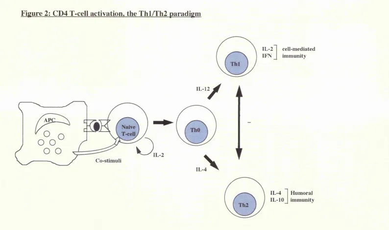
B.4.4.2 The Thl/Th2 model of CD4 cell activation
With the realisation that individual CD4 T-cell clones express phenotypically distinct cytokine profiles, a unifying concept, the Thl/Th2 paradigm, has evolved (figure 2). Activation of the T helper (Thl) phenotype is strongly associated with cell mediated immunity, whereas the Th2 phenotype is often associated with humoral immunity. Individual cytokines are viewed as regulators of Thl/Th2 cell function. Activated CD4 cells have been categorised and subclassifiied on the basis of their phenotypic pattern of cytokine production: ThO cells (IL-2, IL-4, IFNy,
lymphotoxin), Thl cells (IL-2, IFNy, lymphotoxin), and Th2 cells (IL-4, IL-5, IL- 6, IL-10) (Street et al. 1991).
presence of IL-12 or IL-4 become IFNy or IL-4 producers respectively. Exposure of the naive T-cells to ganciclovir eliminates production of both IFNy and IL-4. Thus, effector cells producing IL-4 or IFNy probably differentiate from a common IL-4 producing precursor (i.e. the ThO cell) (Kamogawa et al. 1993). That the Thl and Th2 phenotype evolve from a ThO phenotype is indirectly supported by data showing that naive T-cells which become IL-4 producers require exposure to IL-2, in addition to IL-4, in order to acquire the Th2 phenotype (Seder et al. 1994). Taken together, these studies support the concept that naive T-cells are not pre committed to a Thl or Th2 phenotype, but the phenotype varies with the cytokine micro-environment. Furthermore, Thl and Th2 cells develop from a comon precursor and may cross-regulate one another by the release of their respective cytokines (IFNy and IL-4) (Fitch et al. 1993). Hence, activation of the Thl phenotype is often accompanied by silencing of the Th2 pathway, and vice versa.
B.4.5 CYTOKINE ACTIVATION IN ALLOGRAFT REJECTION
in an autocrine fashion and leads to the paracrine activation of CDS CTL’ s. Thus the expression of IFNy and IL-2 are closely linked to the activation of cellular proinflammatory immune responses.
Figure 2: CD4 T-cell activation, the Thl/Th2 paradigm
IL-12
o
Co-stimuli
IL-2 IFN
cell-mediated immunity
IL-4
IL-4 IL-10
B.4.6 THE SEARCH FOR TRANSPLANTATION TOLERANCE
B.4.6.1 Cytokine mediated transplantation tolerance
The possibility that disruption of the cytokine network may provide a new approach to regulation of the immune response to allografts, is a considerable spur to a large body of work studying the complex cytokine interactions that occur following transplantation.
It has become clear over the past few years that blockade of the IL-2 pathway may result from (Dallman et al. 1991b), and indeed be responsible for (KupiecWeglinski et al. 1988) the development of tolerance in experimental models. This idea has been extended to suggest that a block in Thl type cells, the primary source of cytokines such as IL-2 and IFNy, is associated with the induction of tolerance (e.g. Bugeon et al. 1992). It has been shown that anergic donor-specific cells that have reduced ability to produce IL-2 may also exist in patients following transplantation. Upon stimulation with alloantigen, cells from patients who displayed hyporesponsiveness towards donor antigens were seen to express IL-2 receptor, but did not proliferate,or make IL-2 (Alard et al. 1993). This result resembles previous studies in rat models of tolerance, and strongly suggests that donor- reactive cells are present, but anergic, in hyporesponsive individuals.
established renal allografts in rats (Dallman et al. 1991a), supports a role for relative Th subset responses in graft acceptance. Since IFNy, which is produced by Thl cells, upregulates graft MHC expression (Hao et al. 1990) and augments DTK, it would be expected that a Th2 type response would suppress graft rejecting Thl type cells. This is one currently envisaged scenario for antigen-specific tolerance. The immune response to transplants is far from straightforward, and it has become clear that grafts may be rejected by either cellular or humoral means. Therefore, it may also be possible that shifting the immune response away from a cell mediated to a humoral type of response will not lead to tolerance but merely to rejection through an alternative mechanism.
B.4.6.2 Anergy as a means for tolerance
The precise nature of the second signal is unclear, and may not be provided by only a single molecule or a single pathway, and also there is heterogeneity among T- cells as to second signal requirements. The CD28/B7 pathway described seems the best candidate to date, but inhibition of accessory molecules other than CD28 has also been shown to facilitate graft survival in vivo as well as to suppress antibody responses long-term. Non-depleting anti LFA-1, anti ICAM-1 and anti VLA-4 treatments can inhibit graft rejection and may induce transplantation tolerance (Isobe et al. 1992; Paul et al. 1993). Despite the success of these anergy-inducing strategies in rodents, little success has yet been achieved in primates or humans. Therefore novel ways of inducing anergy, for example utilising the antisense oligonucleotide approach, are potentially valuable alternative approaches.
confirmed a proliferative unresponsiveness associated with reduced IL-2 production which could be overcome by the addition of IL-2.
Anergy as defined herein is a proliferative unresponsiveness, and it is confined to Thl (EL-2 producing) CD4 T-cells. While this may be indicative of an inability to mediate graft rejection, proliferation and cytolytic potential are not necessarily synonymous. Thus proliferative anergy may not equate with lack of rejection capacity in all circumstances.
In the last few years, advances have been made on several fronts in the understanding and manipulation of allograft tolerance. Under appropriate conditions, inhibition of the B7-CD28 pathway can result in antigen specific tolerance in vivo, albeit as a transient and reversible state.
B.5 MECHANISMS OF ALLOGRAFT REJECTION
B.5.1 INTRODUCTION
The notion of histocompatibility genes was introduced by Snell in 1948, by studying the genes controlling skin graft rejection in mouse inbred strains (Snell. 1948). They were initially described as transplantation antigens, long before there was any information on the antigens themselves. The discovery of the structure of the MHC molecules and the subsequent studies elucidating the function of the MHC in relation to antigen processing and presentation have made it possible to study in detail the molecular mechanisms of T-cell repertoire selection, and self restricted T-cell recognition of foreign or nominal antigen.
B.5.2 THE T-CELL RESPONSE TO ALLOGRAFTS
Understanding how T-cells recognise allo-MHC is key to understanding subsequent events leading to allograft rejection, and ultimately for the future development of specific immunotherapies. The critical role of T-cells in allograft ^ rejection was first established by the failure of neonatally thymectomised (227)and congenitally athymic nude rodents to effect rejection (Corley et al. 1977). We now know that allorecognition involves a tripartite structure consisting of T cells, MHC molecule and a peptide bound in the groove of the MHC molecule (Eckels. 1990). Recent evidence advocates the occurrence of at least two distinct, but not necessarily mutually exclusive, mechanisms of allorecognition, the ‘ direct’ and ‘indirect’ pathways (figure 1).
B.5.3 THE ‘DHŒCT’ PATHWAY OF ALLORECOGNITION
This has been the more traditional and well documented mode of allorecognition, in which T-cells recognise intact allo-MHC molecules on the surface of donor cells. It has long been recognised that the normal T-cell repertoire contains a high frequency (1-10%) of total T-cells that are capable of responding to allo-MHC molecules (Sherman et al. 1993). This translates to a precursor frequency at least
Figure 1: Direct and indirect pathways of alloantigen
presentation
DIRECT
INDIRECT
uonor
/ Donor
APC /
Donor I APC
MHC
TCR
mmi
Recipient
MHC
Donor MHC
peptide
B.5.3.1 Theories to explain direct recognition
The two fundamental questions in allorecognition are: First, why is the frequency of alloreactive T-cells so high? Secondly, how can positively selected, self MHC- restricted T-cells recognise foreign antigens as well as allo-MHC? Several hypotheses have been put forward by different groups of investigators (figure 2).
One of the earliest attempts to explain the basis for the high frequency of alloreactive cells was described by Jeme (Jeme.1971; van Boehmer et al. 1978). This theory proposed that the repertoire of T-cell receptors was evolutionarily preselected to include germline genes that had specificity for the MHC molecules of the species. Thus, for each MHC molecule in the species, there evolved a complementary receptor molecule. This readily explained why a high percentage of T-cells respond to each MHC molecule. A prediction of this theory would be that the repertoire used in recognition of antigen was separate from the allospecific repertoire. However, there is now evidence from the study of T-cell clones, that the antigen specific and alloreactive T cell repertoires may be contained within the same clones (Braciale et al. 1981; Sredni et al. 1980; Hunig et al. 1981), and that the same portion of the T-cell receptor (TCR) involved in the recognition of antigen plus MHC is involved in recognition of alloantigen (Matis et al. 1987).
to be peptides) to that selected by self-MHC, and those peptides that bind both self and allo-MHC products may bind to the allo-MHC molecule in a different orientation. This theory was based on the observation that cytotoxic T- lymphocytes (CTL) could be obtained between MHC identical strains that differed from each other in expression of minor antigens (Bevan. 1975). At the time, these antigens were believed to represent polymorphic cell surface proteins. Considering that hundreds of different proteins may be expressed by the cell and available for interaction with the MHC, the high frequency of alloreactivity was a consequence of the diversity of antigenic complexes available for recognition. In contrast to the theory of Jeme, this hypothesis permitted a high degree of TCR diversity within the alloreactive repertoire, as well as no conceptual restrictions on overlap between the allospecific and self MHC-restricted antigen specific repertoires, and has turned out in broad concept to be accurate.
Figure 2a:
MHC
class I
Donor
allogeneic
peptide
T-cell
Figure 2b:
Donor peptide
Donor
cell
Class I
molecule
High determinant
Lechler and Batchelor used results from studies of specific HLA-DR primed T-cell clones to propose that an allo-MHC can be thought of as having two functional sites (Lechler et al. 1991; Lechler et al. 1990; Lombardi et al. 1991; Lombardi et al. 1989). The first region is formed by the regions of the amino-terminal domains that contact the TCR. The second site is formed by the regions of the antigen- binding groove. Therefore, T-cell responses to to alloantigen will depend on the similarities or differences between responder and stimulator MHC sites. In the case of similarities in the region of the TCR binding site, allorecognition would be due to host T-cells exhibiting sufficient affinity to recognise novel peptides bound by the allo-MHC molecules. In the case of differences in the region of TCR binding, the actual ligand may be the MHC molecule itself, the host T-cell binding to an allo-MHC molecule to which it has by chance a ‘ better fit’ , and the bound peptide may not play a significant role. This latter model would be compatible with the high determinant density hypothesis.
B.5.3.2 Supporting evidence for these hypotheses
al. 1987), suggesting that these peptides mimic an a-helical portion of the allogeneic MHC molecule and occupy a binding site on the alloreactive TCR that is specific for the foreign MHC structure. In support of the determinant density hypothesis, ‘ empty’ HLA-A2 molecules separated on a column and renatured without peptide still stimulate allorecognition (Elliott et al. 1990). Similar conclusions were drawn by studying mutant lymphoma cell lines, RMA-S or T2, which express limited or no peptides in the context of class I expression, but nevertheless remain sensitive to lysis by some T-cell clones (Ohlen et al. 1990; Heath et al. 1991). However, the level of lysis by these clones can be increased 10- 100 fold with the addition of cleaved cytoplasmic proteins. Also, some occupied class I molecules have been shown to occur on these mutant cells, rendering such data ambiguous to interpretation (Henderson et al. 1992).
There is a large body of evidence fo r peptide dependence in allorecognition. For example, there are many examples of T-cell clones that discriminate between two MHC molecules that differ only at positions in the floor of the peptide binding groove, reflecting a specificity for bound peptide. There is also evidence of tissue specificity in recognition of a murine class II molecule, in which some cells expressing the appropriate class II molecule are unable to stimulate T-cell clones (Lombardi et al. 1989; Marrack et al. 1988). This suggests that the class II is not sufficient to account for allorecognition, a fact that can be explained by tissue differences in expression of proteins that contribute peptides for presentation with class II.
whereby foreign antigen is broken down and presented by recipient class II molecules to host T-cells, has recently come to light as a significant part of the rejection process.
B.5.4 THE ‘INDmEC T’ PATHWAY OF ALLORECOGNITION
B.5.4.1 Processing of donor MHC molecules
The basic premise for indirect allorecognition as a mechanism for initiation and/or amplification of allograft rejection is that donor alloantigens are shed from the graft, probably the cell’ s own MHC, taken up by recipient AFC’ s and presented to T-cells. At least some of the peptides eluted from cell surface class II MHC molecules represent MHC sequences, suggesting that processing of MHC by self AFC’ s may be a physiological event in vivo (Chicz et al. 1992a; Chicz et al. 1993). It has also been demonstrated that intact MHC molecules are present in the normal human circulation (van Rood et al. 1970; Charlton et al. 1970) and in renal transplant recipients (SuciuFoca et al. 1991). Frocessing of these antigens may lead to the activation of T-helper cells, which secrete cytokines and provide the necessary signals for the growth and maturation of effector CTL’ s and B cells leading to allograft rejection (Farker et al. 1992).
B.5.4.2 Supporting evidence from animal models
of donor antigens can contribute to the effector mechanism of rejection of vascularised organ allografts (Benham et al. 1995) LEW (RTl' ) rats primed for indirect allorecognition of DA (RTl. A*''* ) class I MHC molecules by immunisation with class I derived peptide reject (DAxLEW) Fi kidney grafts in an accelerated fashion.
B.5.4.3 In vitro evidence for indirect recognition in humans
B.5.4.4 The role of indirect recognition in acute or chronic rejection
It has been shown (Liu et al. 1993) by limiting dilution analysis, that the frequency of self-restricted T-cells which recognise processed allo-MHC is approximately 100-fold lower than that of T-cells recognising intact allo-MHC, and suggested that the indirect pathway of recognition may play a minor role in acute, but possibly a major role in chronic allograft rejection. Natural processing of allo-MHC during transplantation may lead to generation of multiple immunodominant peptides. Thus the actual frequency of these T-cells may be underestimated by in
vitro assays with a single peptide. The multiplicity of epitopes that may be
occurred (McKenzie et al. 1984). An additional factor which may play a critical role in the ability of the graft to present antigen once the donor APC’ s have migrated out, is the constitutive expression of MHC class II on vascular endothelium in man, but not in rodents. This may go some way towards explaining the relative difficulty in long-term graft acceptance in man, as compared to rodents.
As can be seen, the indirect pathway is implicated in the process of chronic rejection, though as yet there is no categorical evidence experimentally to verify this.
Thus the direct recognition of allo-MHC on the surface of donor cells, and the indirect recognition of processed allo-MHC presented by self APC’ s need not be mutually exclusive pathways during rejection, as each is mediated by different sets of T-cell clones. The direct pathway accounts for cytotoxic T-cell function, while the indirect pathway may account for much of Th cell function. Neither the role of indirect allorecognition in chronic rejection, nor evidence for indirect allorecognition in clinical transplant rejection have to date been demonstrated, which were the objectives in our study of human transplant recipients.
B.5.5 CHRONIC REJECTION
B.5.5.1 A continuing problem
effective immunosuppression. Concurrently, it has become clear that a significant proportion of grafts fail within the first several months or years after placement, primarily because of progressive and irreversible host immunological attack. The rate of decline has not changed over time, (Clayberger et al. 1990), for example less than half of renal allografts from cadaver donors continue to function at six years, despite 80% behaving satisfactorily at one year (Land. 1989). The rate of decline of other organ grafts are relatively similar, excepting a lower rate of chronic rejection of liver grafts, perhaps due to a putatively lower immunogenicity of this organ (Starzl et al. 1989). This has come to pass in spite of improved immunosuppression. In fact, work in our laboratory has shown that if clonal expansion of T-cells stimulated by the indirect pathway has occurred, this pathway of T-cell help for B- cells is poorly suppressed by cyclosporin (Sawyer et al. 1993), suggesting that cyclosporin may be relatively powerless in attenuating late rejection.
B.5.5.2 Risk factors for chronic rejection