Review
Nanotechnology as an alternative to reduce the spread
of COVID-19
Roberto Vazquez-Munoz 1,*, Jose L. Lopez-Ribot1,b 1,
1 Department of Biology and The South Texas Center for Emerging Infectious Diseases, The University of
Texas at San Antonio. One UTSA Circle, San Antonio, Texas 78249, US * Correspondence: [email protected]
Abstract: The current emerging COVID-19 pandemic has caused a global impact on every major aspect of our societies. It is known that SARS-Cov-2 can endure harsh environmental conditions for up to 72 h, which may contribute to its rapid spread. Therefore, effective containment strategies, such as sanitizing, are critical. Nanotechnology can represent an alternative to reduce the COVID-19 spread, particularly in critical areas, such as healthcare facilities and public places. Nanotechnology-based products are effective at inhibiting different pathogens, including viruses, regardless of their drug-resistant profile, biological structure, or physiology. Although there are several approved nanotechnology-based antiviral products, this work aims to highlight the use of nanomaterials as sanitizers for the prevention of the spread of mainly SARS-Cov-2. It has been widely demonstrated that nanomaterials are an alternative for sanitizing surfaces to inactivate the virus. Also, antimicrobial nanomaterials can reduce the risk of secondary microbial infections on COVID-19 patients, as they inhibit the bacteria and fungi that can contaminate healthcare-related facilities. Finally, cost-effective, easy-to-synthesize antiviral nanomaterials could reduce the burden of the COVID-19 on challenging environments and in developing countries.
Keywords: COVID-19, Nanotechnology, Nanomaterials, Antiviral, Sanitizers, Nanomedicine, Infectious Diseases
1. Introduction
Save from a natural disaster of cataclysmic proportions, or an all-out man-made nuclear war, infectious diseases pose the only global threat to human life on Earth. Infectious diseases have plagued humanity and represented the major cause of morbidity and mortality through the millennia, and in the process shaped human evolution. They still constitute the leading cause of premature death in the developing world. However, our modern society has become extremely complacent about the threat posed by infectious diseases, mostly due to the availability and increased access to hygiene and sanitization techniques, vaccines, and antibiotics.
Despite this unjustified level of complacency and the false sense of security, epidemiological experts and sentinel organizations around the world have provided ample warning signs, yet often ignored, of our global vulnerability against infectious diseases and their broad and borderless impact -microorganisms do not need a passport and do not understand about artificial geopolitical boundaries. This is further aggravated by globalization and climate change. In particular, the newest epidemics caused by a virus such as SARS, MERS, Ebola, and H1N1 since the beginning of the century have raised the awareness of the major threat that viral diseases still pose to humanity as a whole. However, these preoccupations were often short-lived and fast forgotten once the resulting epidemics were seemingly under control, which left us completely unprepared despite our conviction and anticipation that it was only a matter of time for the next epidemic or pandemic to come. Sure enough, we did not have to wait too much longer, and as of early 2020, humanity is confronting a pandemic in severe acute respiratory syndrome coronavirus 2 (SARS-CoV-2), which causes coronavirus disease, abbreviated as COVID-19.
2. The SARS-CoV-2 virus and the current COVID-19 pandemic
The SARS-CoV-2 is a large, enveloped, positive-stranded RNA virus, with a nucleocapsid, with a similar structure to the SARS-CoV-1; and a diameter size range from 80 to 140 nm [1]. This zoonotic coronavirus has become a major cause of emerging respiratory disease [1] and has rapidly spread around the globe, affecting billions of people, and triggering unexpected changes in the healthcare system, global economy, and interactions in the societies worldwide.
So far, there are over 8.4 million confirmed cases with COVID-19 worldwide, with an estimated death toll of over 450 thousand people [2]. The costs related to this communicable disease are expected to be in the trillions of dollars globally, but the real cost cannot be estimated [3]. Implementing strategies to reduce its spread requires the participation of the whole society, as well as the development of innovative ways of social, work, and health policies. Several measures have been implemented to control COVID-19, from campaigns aimed at improving personal hygiene practices to community approaches such as social distancing and quarantines. Among the suggested public health activities to address this pandemic are 1) efficient stay-at-home implementation, 2) fast SARS-CoV-2 testing, and 3) efficient health care response. The last point considers protecting the health care professionals by providing tools to prevent nosocomial infections, from making appropriate PPE available to ensuring sanitizing procedures.
Proper sanitizing measures are critical. A recent study revealed that SARS-CoV-2 can be detected on plastic and stainless-steel surfaces up to 72 h, whereas on copper, no viable SARS-CoV-2 was measured after 4 hours of application [4]. The inactivation of the virus by copper is not surprising, as it is known that transition metals can inactivate viral particles [5]. Thus, nanotechnology may provide an alternative for sanitizing surfaces, via antimicrobial and antiviral nanomaterials.
3. The state-of-the-art of antiviral nanomaterials: from research to the clinic.
There are different definitions of the nanomaterial concept. In general, nanomaterials can be described as single-structures which size is less than 100 nm in at least one of their three dimensions. The increasing interest in nanomaterials is due to their novel or improved physicochemical properties such as endurance, chemical reactivity, biocompatibility, conductivity, or reduced toxicity. The chemical composition of nanomaterials can be organic or inorganic, and they can be found as single structures, composites, embedded in a matrix, etc. Nowadays, nanomaterials are found in a wide range of existing products, such as in electronics, health and fitness, paints and other surface coatings, food, and clothing, among many others [6,7]. Moreover, medicine is one of the fields with a growing interest in the use of nanotechnology. Just for silver nanoparticles, the global consumption for healthcare-related nanotechnology is expected to be over 50 tons in 2020 [8].
Nowadays, nanomaterials are widely used in different healthcare-related applications, such as sanitizers, diagnosis, imaging tools, wound dressing, wearable devices, antimicrobial drugs, anticancer therapies, pharmaceuticals, drug delivery, and vaccine development, diagnosis, and even implants [9–11]. Nanomaterials have been researched due to their properties against multiple pathogenic, microorganisms [12–15] s.
or integration of viruses into the cell. Back in 2005, Lara et al showed that silver AgNPs interact with the receptors from the HIV, inhibiting its infectivity [29].
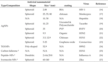
Table 1. Examples of antiviral nanomaterials that inactivate different types of viruses.
Antiviral nanomaterial
Virus Reference Type/Composition Shape Size 1 (nm) coating
AgNPs
Spheroid 2.08 BSA HIV-1 [16]
Spheroid 25, 55, 80 chitosan Monkeypox [17]
N/A 10, 50 N/A Hepatitis [18]
Spheroid 10, 25 Uncoated &
Polysaccharide Tacaribe [19]
Spheroid 35 PVP RVFV [20]
Spheroid 9.5 Organic H3N2 [21]
Spheroid 3.5, 12.9 Chitosan H1N1 [22]
Spheroid 5-25 Graphene oxide Coronavirus [23]
TiO2NPs Poly-shaped 52.9 N/A H9N2 [24]
Carbon fullerene 2 N/A N/A N/A H1N1 [25]
Peptide–NPs 3 Dendritic 2.4 & 29.8 N/A Influenza A [26]
Ivermectin-NPs 3 Spheroid 60-140 IVM Zika [27]
1 Average size. For the metallic nanoparticles, the size corresponds to their metallic core.
2 Lipidosome complex
3 Hybrid complexes
Research on nanomaterials for future antiviral treatments is still ongoing, as they have proven to effective for preventing HIV transmission [30] and pneumonia -influenza virus [31]-, in in vivo murine models. Also, AgNPs were effective at inactivating in vivo the Canine Distemper Virus in dogs [32]. Heparan Sulfate Proteoglycans (HSPG)-mimicking nanoparticles display broad-range antiviral activity, both in vivo and in vitro against herpes simplex virus (HSV), human papillomavirus, respiratory syncytial virus (RSV), dengue and lentivirus [33]. Also, some nanomaterials can be used as nanocarriers, for improving the bioavailability of current antiviral drugs [34].
Moreover, Singh et al reviewed recently different antiviral nanomedicines that are approved and under ongoing preclinical and clinical trials [35]. An example is Inflexal V©, a nanotechnology-based virosomal anti-influenza vaccine approved by the FDA [36]. Despite the current use of antiviral nanomaterials, research on other nanomaterials is still ongoing, to address their potential toxicity and side effects.
4. Current nanotechnology applications that can be used to combat COVID-19.
The typical shape of antiviral nanoparticles has an aspect ratio close to 1 (spheroids) with a usual range between 1 to 50 nm, and an average of 22 nm. Antiviral nanomaterials are typically smaller than the SARS-Cov-2 viral particle (fig. 1), therefore they may interact better with the surface proteins or even with the whole viral particle. Currently, nanomaterials are used in a wide range of commercially available applications, many of them related to healthcare. Some of the current applications that can be used against COVID-19 are described below.
The application of nanomaterials-embedded textiles has been intensely researched. These textiles may be used for PPE, such as lab coats, and facemasks, as current research shows that they improve the physicochemical properties of textiles [37], such as fire-retardant, self-cleaning, UV-protection, antimicrobial, and antiviral, among others. Several patents consider the use of fibers embedded with metallic nanoparticles, such as copper and silver nanoparticles, due to their antimicrobial and antiviral properties [9,38]. The use of nanoparticles in textiles has been increasing rapidly, with a current global consumption of around 35 tons just for silver nanoparticles [8]. Moreover, wearable smart textiles for sensing have been under study, particularly for health-related applications. One area of interest is the early detection of pathogens [39], which may include viruses, such as the SARS-Cov-2.
4.2 Surface coatings.
Nanomaterials-based coatings are currently used for several applications, and different products are now available [9]. Numerous nanomaterials, such as silver, bismuth, or titanium nanoparticles, have been developed for coating surfaces, [37,40,41]. Also, nanostructured surfaces can physically reduce the attachment of pathogens [42] and even disrupt the structure of the pathogens due to the nanoscale topography organization [43]. Nanomaterials can be embedded in paint or coatings for medical instrumentation, gloves, and other highly-touched surfaces, such as doorknobs, handrails, other medical devices, etc. to reduce the viability of viruses and other pathogens.
4.3 Sanitizers.
Currently, disinfectants with silver salts are already available [44], as silver is deemed safe for sanitizing purposes [45]. In hospitals and other healthcare-related facilities, sanitizing with nanotechnology-based products could inactivate the viruses on surfaces. Moreover, sanitizers with nanomaterials could reduce the presence of the SARS-Cov-2 on surfaces.
4.4 Other current and potential applications.
Among other uses, nanomaterials can be used to improve the function of air filters in healthcare facilities or in other places that use recirculated air. Nanotechnology-improved air filters can reduce the spread of viral particles [46,47]. Also, the use of nanotechnology for producing wound dressings has been thoroughly explored [48–50], due to their ability to protect against infections and increase the healing speed rate. Future applications may include the development of improved detection kits, as it has been one of the major needs for the current strategies to contain and follow the virus spread.
5. A silent risk: the microbial secondary infections.
The current COVID-19 pandemic has raised several health concerns beyond the disease itself. Among them, secondary infections are usually underestimated risks that may occur due to the burden on health caused by the COVID-19. Patients infected with the novel strain of coronavirus frequently show microbial dysbiosis and a decrease of probiotic bacteria [51]. Also, it is not uncommon for patients with COVID-19 to suffer from complications of acute organ injury and secondary infection [52]. Nanomaterials display a broad range of antimicrobial activity against bacteria [53,54], protozoa [55,56], and fungi [57–60]. In healthcare-related facilities, nanotechnology-based sanitizers could simultaneously reduce the presence of viruses and opportunistic microorganisms. Moreover, nanomaterials can reduce the microbial biofilms [58,59,61], leading to a reduced risk of secondary microbial infections.
In the long term, nanoantibiotics could be used to prevent or treat microbial secondary infections, particularly those that display multidrug-resistance profiles, which has been one of the rising concerns of our current crisis of communicable diseases. Nanomaterials are effective regardless of the drug-susceptibly profile of the microorganisms and may enhance the potency of the antimicrobial drugs [57,62,63].
5. Perspectives
What should be expected in the near future? Although more research is needed, antiviral nanomaterials represent an alternative to reduce the spread of COVID-19, as they have been effective against other viruses. Based on our current knowledge, nanomaterials may be potent sanitizers with broad antimicrobial activity. Nanotechnology-based sanitizers can improve safety in healthcare-related facilities and public spaces, particularly in developing countries. Moreover, their therapeutic use has proven to be effective and it will be expanded in the future, as more research becomes available. Also, as nanomaterials may enhance the antimicrobial potency of some drugs, they are an interesting alternative to control secondary infections, and for future treatments against multidrug-resistant microorganisms.
Finally, protocols that favor facile, rapid, cost-effective syntheses of antimicrobial and antiviral nanomaterials [53,64] produce potent nanomaterials that could reduce the spread of viruses and microorganisms. Affordable nanotechnology may benefit anti-COVID-19 sanitizing procedures in developing countries, where access to advanced materials is limited. Also, easy-to-synthesize nanomaterials may be used under non-favorable conditions, such as medical mobile-units, rural health care facilities, public spaces, and in medical posts for the military on the field [65,66].
Author Contributions: Both authors equally contributed to the conceptualization, writing-original draft preparation, and writing-review & editing.
Funding: RVM acknowledges support from a postdoctoral fellowship from the Mexican Council of Science and Technology (CONACYT). JLLR acknowledges support from the Margaret Batts Tobin Foundation. The authors would like to thank the graphic designer Salma Carballo, for designing the images of the SARS-Cov-2 virus and the AgNP.
Conflicts of Interest: The authors declare no conflict of interest.
References
1. Cascella, M.; Rajnik, M.; Cuomo, A.; Dulebohn, S.C.; Di Napoli, R. Features, Evaluation and Treatment
Coronavirus (COVID-19); StatPearls Publishing, 2020;
2. Coronavirus Resource Center COVID-19 Map Available online: https://coronavirus.jhu.edu/map.html
(accessed on Jun 8, 2020).
How Available online:
https://www.bloomberg.com/graphics/2020-coronavirus-pandemic-global-economic-risk/ (accessed on Apr 8, 2020).
4. van Doremalen, N.; Bushmaker, T.; Morris, D.H.; Holbrook, M.G.; Gamble, A.; Williamson, B.N.; Tamin,
A.; Harcourt, J.L.; Thornburg, N.J.; Gerber, S.I.; et al. Aerosol and Surface Stability of SARS-CoV-2 as
Compared with SARS-CoV-1. N. Engl. J. Med.2020, 382, 1564–1567, doi:10.1056/NEJMc2004973.
5. Nieto-Juarez, J.I.; Pierzchła, K.; Sienkiewicz, A.; Kohn, T. Inactivation of MS2 coliphage in Fenton and
Fenton-like systems: role of transition metals, hydrogen peroxide and sunlight. Environ. Sci. Technol.
2010, 44, 3351–3356, doi:10.1021/es903739f.
6. Vance, M.E.; Kuiken, T.; Vejerano, E.P.; McGinnis, S.P.; Hochella, M.F.; Hull, D.R. Nanotechnology in
the real world: Redeveloping the nanomaterial consumer products inventory. Beilstein J. Nanotechnol.
2015, 6, 1769–1780, doi:10.3762/bjnano.6.181.
7. Nanowerk Database Nanoparticle Database - Single-element nanoparticles Available online:
https://www.nanowerk.com/nanoparticle_database.php (accessed on Apr 20, 2020).
8. Syafiuddin, A.; Salmiati; Salim, M.R.; Beng Hong Kueh, A.; Hadibarata, T.; Nur, H. A Review of Silver
Nanoparticles: Research Trends, Global Consumption, Synthesis, Properties, and Future Challenges. J.
Chinese Chem. Soc.2017, 64, 732–756, doi:10.1002/jccs.201700067.
9. Sim, W.; Barnard, R.T.; Blaskovich, M.A.T.; Ziora, Z.M. Antimicrobial silver in medicinal and consumer
applications: A patent review of the past decade (2007–2017). Antibiotics 2018, 7, 93.
10. Dilnawaz, F.; Acharya, S.; Sahoo, S.K. Recent trends of nanomedicinal approaches in clinics. Int. J. Pharm.
2018, 538, 263–278, doi:10.1016/j.ijpharm.2018.01.016.
11. Pelaz, B.; Alexiou, C.; Alvarez-Puebla, R.A.; Alves, F.; Andrews, A.M.; Ashraf, S.; Balogh, L.P.; Ballerini,
L.; Bestetti, A.; Brendel, C.; et al. Diverse Applications of Nanomedicine. ACS Nano2017, 11, 2313–2381,
doi:10.1021/acsnano.6b06040.
12. Vazquez-Muñoz, R.; Borrego, B.; Juárez-Moreno, K.; García-García, M.; Mota Morales, J.D.;
Bogdanchikova, N.; Huerta-Saquero, A. Toxicity of silver nanoparticles in biological systems: Does the
complexity of biological systems matter? Toxicol. Lett.2017, 276, 11–20, doi:10.1016/j.toxlet.2017.05.007.
13. Romero-Urbina, D.G.; Lara, H.H.; Velázquez-Salazar, J.J.; Arellano-Jiménez, M.J.; Larios, E.; Srinivasan,
A.; Lopez-Ribot, J.L.; Yacamán, M.J. Ultrastructural changes in methicillin-resistant Staphylococcus
aureus induced by positively charged silver nanoparticles. Beilstein J. Nanotechnol.2015, 6, 2396–2405,
doi:10.3762/bjnano.6.246.
14. Baptista, P. V; Mccusker, M.P.; Carvalho, A.; Ferreira, D.A. Nano-Strategies to Fight Multidrug Resistant
Bacteria —“ A Battle of the Titans .” 2018, 9, 1–26, doi:10.3389/fmicb.2018.01441.
15. Huh, A.J.; Kwon, Y.J. “Nanoantibiotics”: A new paradigm for treating infectious diseases using
nanomaterials in the antibiotics resistant era. J. Control. Release 2011, 156, 128–145,
doi:10.1016/j.jconrel.2011.07.002.
16. Elechiguerra, J.L.; Burt, J.L.; Morones, J.R.; Camacho-Bragado, A.; Gao, X.; Lara, H.H.; Yacaman, M.J.
Interaction of silver nanoparticles with HIV-1. J. Nanobiotechnology2005, 3, 1–10,
doi:10.1186/1477-3155-3-6.
17. Rogers, J. V.; Parkinson, C. V.; Choi, Y.W.; Speshock, J.L.; Hussain, S.M. A preliminary assessment of
silver nanoparticle inhibition of monkeypox virus plaque formation. Nanoscale Res. Lett.2008, 3, 129–133,
doi:10.1007/s11671-008-9128-2.
18. Lu, L.; Sun, R.W.Y.; Chen, R.; Hui, C.K.; Ho, C.M.; Luk, J.M.; Lau, G.K.K.; Che, C.M. Silver nanoparticles
inhibit hepatitis B virus replication. Antivir. Ther.2008, 13, 252–262.
nanoparticles with Tacaribe virus. J. Nanobiotechnology2010, 8, 1–9, doi:10.1186/1477-3155-8-19.
20. Borrego, B.; Lorenzo, G.; Mota-Morales, J.D.; Almanza-Reyes, H.; Mateos, F.; López-Gil, E.; de la Losa,
N.; Burmistrov, V.A.; Pestryakov, A.N.; Brun, A.; et al. Potential application of silver nanoparticles to
control the infectivity of Rift Valley fever virus in vitro and in vivo. Nanomedicine Nanotechnology, Biol.
Med.2016, 12, 1185–1192, doi:10.1016/j.nano.2016.01.021.
21. Xiang, D.; Zheng, C.; Zheng, Y.; Li, X.; Yin, J.; O’ Conner, M.; Marappan, M.; Miao, Y.; Xiang, B.; Duan,
W.; et al. Inhibition of A/Human/Hubei/3/2005 (H3N2) influenza virus infection by silver nanoparticles
in vitro and in vivo. Int. J. Nanomedicine2013, 8, 4103, doi:10.2147/IJN.S53622.
22. Mori, Y.; Ono, T.; Miyahira, Y.; Nguyen, V.Q.; Matsui, T.; Ishihara, M. Antiviral activity of silver
nanoparticle/chitosan composites against H1N1 influenza A virus. Nanoscale Res. Lett. 2013, 8, 93,
doi:10.1186/1556-276x-8-93.
23. Chen, Y.-N.; Hsueh, Y.-H.; Hsieh, C.-T.; Tzou, D.-Y.; Chang, P.-L. Antiviral Activity of Graphene–Silver
Nanocomposites against Non-Enveloped and Enveloped Viruses. Int. J. Environ. Res. Public Health2016,
13, 430, doi:10.3390/ijerph13040430.
24. Cui, H.; Jiang, J.; Gu, W.; Sun, C.; Wu, D.; Yang, T.; Yang, G. Photocatalytic Inactivation Efficiency of
Anatase Nano-TiO2 Sol on the H9N2 Avian Influenza Virus. Photochem. Photobiol.2010, 86, 1135–1139,
doi:10.1111/j.1751-1097.2010.00763.x.
25. Ji, H.; Yang, Z.; Jiang, W.; Geng, C.; Gong, M.; Xiao, H.; Wang, Z.; Cheng, L. Antiviral activity of nano
carbon fullerene lipidosome against influenza virus in vitro. J. Huazhong Univ. Sci. Technol.2008, 28, 243–
246, doi:10.1007/s11596-008-0303-6.
26. Lauster, D.; Glanz, M.; Bardua, M.; Ludwig, K.; Hellmund, M.; Hoffmann, U.; Hamann, A.; Böttcher, C.;
Haag, R.; Hackenberger, C.P.R.; et al. Multivalent Peptide–Nanoparticle Conjugates for Influenza-Virus
Inhibition. Angew. Chemie - Int. Ed.2017, 56, 5931–5936, doi:10.1002/anie.201702005.
27. Surnar, B.; Kamran, M.Z.; Shah, A.S.; Basu, U.; Kolishetti, N.; Deo, S.; Jayaweera, D.T.; Daunert, S.; Dhar,
S.; Macdonald, J.T. Orally Administrable Therapeutic Synthetic Nanoparticle for Zika Virus. 2019,
doi:10.1021/acsnano.9b02807.
28. Cojocaru, F.D.; Botezat, D.; Gardikiotis, I.; Uritu, C.M.; Dodi, G.; Trandafir, L.; Rezus, C.; Rezus, E.;
Tamba, B.I.; Mihai, C.T. Nanomaterials designed for antiviral drug delivery transport across biological
barriers. Pharmaceutics 2020, 12, 171.
29. Lara, H.H.; Ayala-Nuñez, N. V.; Ixtepan-Turrent, L.; Rodriguez-Padilla, C. Mode of antiviral action of
silver nanoparticles against HIV-1. J. Nanobiotechnology2010, 8, 1–10, doi:10.1186/1477-3155-8-1.
30. Kovarova, M.; Council, O.D.; Date, A.A.; Long, J.M.; Nochii, T.; Belshan, M.; Shibata, A.; Vincent, H.;
Baker, C.E.; Thayer, W.O.; et al. Nanoformulations of Rilpivirine for Topical Pericoital and Systemic
Coitus-Independent Administration Efficiently Prevent HIV Transmission. PLoS Pathog. 2015, 11,
e1005075, doi:10.1371/journal.ppat.1005075.
31. Donovan, B.W.; Reuter, J.D.; Cao, Z.; Myc, A.; Johnson, K.J.; Baker, J.R. Prevention of Murine Influenza
a Virus Pneumonitis by Surfactant Nano-Emulsions. Antivir. Chem. Chemother. 2000, 11, 41–49,
doi:10.1177/095632020001100104.
32. Bogdanchikova, N.; Vazquez-Muñoz, R.; Huerta-Saquero, A.; Pena Jasso, A.; Aguilar Uzcanga, G.; Picos
Díaz, P.L.; Pestryakov, A.; Burmistrov, V.; Martynyuk, O.; Luna Vázquez Gómez, R.; et al. Silver
nanoparticles composition for treatment of distemper in dogs. Int. J. Nanotechnol. 2016, 13, 227,
doi:10.1504/IJNT.2016.074536.
33. Cagno, V.; Andreozzi, P.; D’Alicarnasso, M.; Silva, P.J.; Mueller, M.; Galloux, M.; Goffic, R. Le; Jones,
inhibition mechanism. Nat. Mater.2018, 17, 195–203, doi:10.1038/NMAT5053.
34. Donalisio, M.; Leone, F.; Civra, A.; Spagnolo, R.; Ozer, O.; Lembo, D.; Cavalli, R. Acyclovir-Loaded
Chitosan Nanospheres from Nano-Emulsion Templating for the Topical Treatment of Herpesviruses
Infections. Pharmaceutics2018, 10, 46, doi:10.3390/pharmaceutics10020046.
35. Singh, L.; Kruger, H.G.; Maguire, G.E.M.; Govender, T.; Parboosing, R. The role of nanotechnology in
the treatment of viral infections. Ther. Adv. Infect. Dis.2017, 4, 105–131, doi:10.1177/2049936117713593.
36. Herzog, C.; Hartmann, K.; Künzi, V.; Kürsteiner, O.; Mischler, R.; Lazar, H.; Glück, R. Eleven years of
Inflexal® V-a virosomal adjuvanted influenza vaccine. Vaccine 2009, 27, 4381–4387.
37. Brabazon, D.; Pellicer, E.; Zivic, F.; Sort, J.; Baró, M.D.; Grujovic, N.; Choy, K.L. Commercialization of
nanotechnologies-A case study approach; Brabazon, D., Pellicer, E., Zivic, F., Sort, J., Dolors Baró, M.,
Grujovic, N., Choy, K.-L., Eds.; Springer International Publishing: Cham, 2017; ISBN 9783319569796.
38. Suryaprabha, T.; Sethuraman, M.G. Fabrication of copper-based superhydrophobic self-cleaning
antibacterial coating over cotton fabric. Cellulose2017, 24, 395–407, doi:10.1007/s10570-016-1110-z.
39. Libertino, S.; Plutino, M.R.; Rosace, G. Design and development of wearable sensing nanomaterials for
smart textiles. In Proceedings of the AIP Conference Proceedings; 2018; Vol. 1990, p. 020016.
40. Hebalkar, N.Y.; Acharya, S.; Rao, T.N. Preparation of bi-functional silica particles for antibacterial and
self cleaning surfaces. J. Colloid Interface Sci.2011, 364, 24–30, doi:10.1016/j.jcis.2011.07.087.
41. Hasan, J.; Crawford, R.J.; Ivanova, E.P. Antibacterial surfaces: The quest for a new generation of
biomaterials. Trends Biotechnol.2013, 31, 295–304, doi:10.1016/j.tibtech.2013.01.017.
42. Zhao, L.; Chu, P.K.; Zhang, Y.; Wu, Z. Antibacterial coatings on titanium implants. J. Biomed. Mater. Res.
Part B Appl. Biomater.2009, 91B, 470–480, doi:10.1002/jbm.b.31463.
43. Elbourne, A.; Crawford, R.J.; Ivanova, E.P. Nano-structured antimicrobial surfaces: From nature to
synthetic analogues. J. Colloid Interface Sci.2017, 508, 603–616, doi:10.1016/j.jcis.2017.07.021.
44. Ku, T.S.N.; Walraven, C.J.; Lee, S.A. Candida auris: Disinfectants and Implications for Infection Control.
Front. Microbiol.2018, 9, doi:10.3389/fmicb.2018.00726.
45. Culver, A.; Geiger, C.; Simon, D. Safer Products and Practices for Disinfecting and Sanitizing Surfaces; San
Francisco, 2014;
46. Vaze, N.; Pyrgiotakis, G.; McDevitt, J.; Mena, L.; Melo, A.; Bedugnis, A.; Kobzik, L.; Eleftheriadou, M.;
Demokritou, P. Inactivation of common hospital acquired pathogens on surfaces and in air utilizing
engineered water nanostructures (EWNS) based nano-sanitizers. Nanomedicine Nanotechnology, Biol. Med.
2019, 18, 234–242, doi:10.1016/j.nano.2019.03.003.
47. Joe, Y.H.; Park, D.H.; Hwang, J. Evaluation of Ag nanoparticle coated air filter against aerosolized virus:
Anti-viral efficiency with dust loading. J. Hazard. Mater. 2016, 301, 547–553,
doi:10.1016/j.jhazmat.2015.09.017.
48. Ali, A.; Ahmed, S. A review on chitosan and its nanocomposites in drug delivery. Int. J. Biol. Macromol.
2018, 109, 273–286, doi:10.1016/j.ijbiomac.2017.12.078.
49. Mishra, M.; Kumar, H.; Tripathi, K. Diabetic Delayed Wound Healing and the Role of Silver. Dig. J. Nano
2008, 3, 49–54, doi:10.1201/9781420015133.ch5.
50. Zivic, F.; Grujovic, N.; Mitrovic, S.; Ahad, I.U.; Brabazon, D. Characteristics and applications of silver
nanoparticles. In Commercialization of Nanotechnologies-A Case Study Approach; Springer International
Publishing, 2017; pp. 227–273 ISBN 9783319569796.
51. Xu, K.; Cai, H.; Shen, Y.; Ni, Q.; Chen, Y.; Hu, S.; Li, J.; Wang, H.; Yu, L.; Huang, H.; et al. Management
of corona virus disease-19 (COVID-19): the Zhejiang experience. Zhejiang Da Xue Xue Bao. Yi Xue Ban
52. Luo, X.; Xia, H.; Yang, W.; Wang, B.; Guo, T.; Xiong, J.; Jiang, Z.; Liu, Y.; Yan, X.; Zhou, W.; et al.
Characteristics of patients with COVID-19 during epidemic ongoing outbreak in Wuhan, China. medRxiv
2020, 2020.03.19.20033175, doi:10.1101/2020.03.19.20033175.
53. Vazquez-Muñoz, R.; Arellano-Jimenez, M.J.; Lopez, F.D.; Lopez-Ribot, J.L. Protocol optimization for a
fast, simple and economical chemical reduction synthesis of antimicrobial silver nanoparticles in
non-specialized facilities. BMC Res. Notes2019, 12, 773, doi:10.1186/s13104-019-4813-z.
54. Vega-Jiménez, A.L.; Almaguer-Flores, A.; Flores-Castaneda, M.; Camps, E.; Uribe-Ramirez, M.;
Aztatzi-Aguilar, O.G.; De Vizcaya-Ruiz, A. Bismuth subsalicylate nanoparticles with anaerobic antibacterial
activity for dental applications. Nanotechnology2017, 28, 435101, doi:10.1088/1361-6528/aa8838.
55. Kurvet, I.; Juganson, K.; Vija, H.; Sihtmäe, M.; Blinova, I.; Syvertsen-Wiig, G.; Kahru, A.; Kurvet, I.;
Juganson, K.; Vija, H.; et al. Toxicity of Nine (Doped) Rare Earth Metal Oxides and Respective Individual
Metals to Aquatic Microorganisms Vibrio fischeri and Tetrahymena thermophila. Materials (Basel).2017,
10, 754, doi:10.3390/ma10070754.
56. Gaafar, M.R.; Mady, R.F.; Diab, R.G.; Shalaby, T.I. Chitosan and silver nanoparticles: Promising
anti-toxoplasma agents. Exp. Parasitol.2014, 143, 30–38, doi:10.1016/j.exppara.2014.05.005.
57. Lara, H.H.; Guisbiers, G.; Mendoza, J.; Mimun, L.C.; Vincent, B.A.; Lopez-Ribot, J.L.; Nash, K.L.
Synergistic antifungal effect of chitosan-stabilized selenium nanoparticles synthesized by pulsed laser
ablation in liquids against Candida albicans biofilms. Int. J. Nanomedicine 2018, 13, 2697–2708,
doi:10.2147/IJN.S151285.
58. Montes, M.; Pierce, C.G.; Lopez-Ribot, J.L.; Bhalla, A.S.; Guo, R. Properties of silver and copper
nanoparticle containing aqueous suspensions and evaluation of their in vitro activity against candida
albicans and staphylococcus aureus biofilms. J. Nano Res. 2016, 37, 109–121,
doi:10.4028/www.scientific.net/JNanoR.37.109.
59. Lara, H.H.; Ixtepan-Turrent, L.; Jose Yacaman, M.; Lopez-Ribot, J. Inhibition of Candida auris Biofilm
Formation on Medical and Environmental Surfaces by Silver Nanoparticles. ACS Appl. Mater. Interfaces
2020, acsami.9b20708, doi:10.1021/acsami.9b20708.
60. Vazquez-Muñoz, R.; Avalos-Borja, M.; Castro-Longoria, E. Ultrastructural Analysis of Candida albicans
When Exposed to Silver Nanoparticles. PLoS One2014, 9, e108876, doi:10.1371/journal.pone.0108876.
61. Vazquez-Munoz, R.; Lopez, F.D.; Lopez-Ribot, J. Silver nanoantibiotics display strong antifungal activity
against the emergent multidrug-resistant yeast Candida auris under both planktonic and biofilm
growing conditions. bioRxiv2020, 2020.05.31.126433, doi:10.1101/2020.05.31.126433.
62. Vazquez-Muñoz, R.; Meza-Villezcas, A.; Fournier, P.G.J.; Soria-Castro, E.; Juarez-Moreno, K.;
Gallego-Hernández, A.L.; Bogdanchikova, N.; Vazquez-Duhalt, R.; Huerta-Saquero, A. Enhancement of
antibiotics antimicrobial activity due to the silver nanoparticles impact on the cell membrane. PLoS One
2019, 14, e0224904, doi:10.1371/journal.pone.0224904.
63. Romero-Urbina, D.G.; Lara, H.H.; Velázquez-Salazar, J.J.; Arellano-Jiménez, M.J.; Larios, E.; Srinivasan,
A.; Lopez-Ribot, J.L.; Yacamán, M.J. Ultrastructural changes in methicillin-resistant Staphylococcus
aureus induced by positively charged silver nanoparticles. Beilstein J. Nanotechnol.2015, 6, 2396–2405,
doi:10.3762/bjnano.6.246.
64. Vazquez-Munoz, R.; Arellano-Jimenez, M.J.; Lopez-Ribot, J.L. Fast, facile synthesis method for
BAL-mediated PVP-bismuth nanoparticles. MethodsX2020, 7, 100894, doi:10.1016/j.mex.2020.100894.
65. Ho, Z.J.; Hwang, Y.F.; Lee, J.M. Emerging and re-emerging infectious diseases: challenges and
opportunities for militaries. Mil. Med. Res.2014, 1, 21, doi:10.1186/2054-9369-1-21.