RVC OPEN ACCESS REPOSITORY – COPYRIGHT NOTICE
This is the peer-reviewed, manuscript version of the following article:
Meeson, R. Making internal fixation work with limited bone stock. In Practice 2017;39:98-106.
The final version is available online at BMJ: http://dx.doi.org/10.1136/inp.j595.
The full details of the published version of the article are as follows:
TITLE: Making internal fixation work with limited bone stock
AUTHORS: R Meeson
JOURNAL TITLE: Veterinary Record
PUBLISHER: BMJ Publishing Group
PUBLICATION DATE: March 2017
Making internal fixation work with limited bone stock
1
2
Richard Meeson (rmeeson@rvc.ac.uk)
3
4
Department of Comparative Biomedical Sciences, RVC, University of London, NW1
5
Introduction
7
Fractures are common in small animal practice and there are many options for
8
managing them. It is important that the fracture is evaluated and a plan made as to the
9
most appropriate method to treat it (Shales 2008). The most popular method for
10
managing many fractures, especially diaphyseal ones, is by using plates and screws
11
as they provide rigid fixation, usually with reliable healing. Additionally, when compared
12
to external skeletal fixation, there is typically less postoperative management required.
13
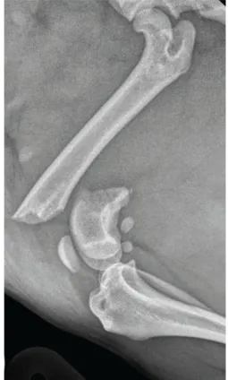
However, some fractures are comminuted, or sufficiently close to a joint
(juxta-14
articular), that they limit the amount of bone available to achieve a standard stable
15
plate and screw fixation (Fig 1).
16
17
Creating a stable internal fixation: three bicortical screws doctrine
18
Various factors should be considered when choosing the size of implant, such as type
19
and location of the fracture, age, activity, size of bone, weight of animal, and condition
20
of the soft tissue, (Table 1). However, based on evaluation of over 1000 bone plate
21
cases, the most important factor was patient weight (Brinker 1977), and hence the AO
22
plate sizing chart, which is based on weight, is the starting point for plate size selection
23
(Johnson and others 2005, Piermattei and others, 2006).
24
Once a plate size has been identified, an overlay templating method using an acetate
25
or digital software determines whether and how the implant may fit. Conventional
26
wisdom is at least three or four bicortical screws (six to eight cortices) should be placed
27
in each fracture fragment (Johnson and others 2005, Piermattei and others, 2006).
28
Interestingly, the original evidence for this is impossible to find and appears to be
29
based on experience and logic. From a mechanical point of view, one screw alone, will
30
of the screw, and therefore will not provide fracture stability. Two screws (monocortical
32
or bicortical), in each main fragment is therefore the minimum for stability.
33
Unfortunately, such a construction will fail if one screw breaks or if the interface
34
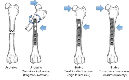
between bone cortex and screw is threatened due to bone resorption. Thus, for safety
35
reasons a minimum of three screws in both the proximal and the distal fragment is
36
recommended (Fig 2). Short fracture fragments can make this requirement difficult to
37
achieve, but not necessarily impossible.
38
39
Double Plating
40
Double plating can be extremely useful for achieving a rigid fixation and increased
41
numbers of cortices within a fracture fragment. Beneficially, this is achieved using the
42
standard inventory of stock plates and screws, and does not necessarily require
43
additional locking instrumentation, or specialised plates and implants. A good rule of
44
thumb is at least one of the plates needs to ideally have two bicortical, or one bicortical
45
and one monocortical (preferably locked – see later) screws placed. Double plating
46
can be ‘parallel’ (Fig 3) or bi-axial, often referred to as ‘orthogonal’ if placed at
right-47
angles (Figs 4 & 5 & Table 2).
48
A warning, however, is that this approach comes with two potential downsides. The
49
first is that, in using more screws to increase the stability of the fracture repair, the
50
repair will become significantly stiffer which, if excessive, could theoretically slow or
51
retard the healing process. I have, on rare occasions, had to remove one of a pair of
52
plates due to these concerns. Secondly, in placing further implants on the bone, there
53
can be more disruption to the soft tissues and the blood supply to the bone, potentially
54
reducing the ability of the fracture to heal at the expense of using internal fixation.
55
osteosynthesis can be used to reduce the impact, but discussion of these is beyond
57
the scope of this article.
58
Bi-axial double plating, most commonly placed orthogonally, frequently results in one
59
of the plates being predominantly edge loaded (bending forces are applied against the
60
width, not the depth of the plate, thereby significantly increasing its resistance to
61
bending). Theoretically, the use of bi-axial orthogonal double plating can provide a
62
much stiffer construct than a single plate especially in resistance to torsion and bending.
63
Therefore, when double plating, it is important to consider the sizes of the plates used.
64
More often than not, one and sometimes both may be downsized to avoid excessively
65
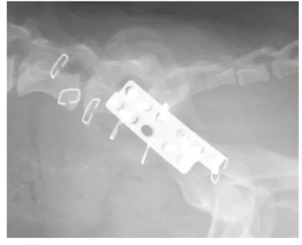
stiff repairs and to increase the numbers of screws available, such as in figure 3, where
66
a 2.7mm plate was appropriate for the dog’s weight, however wouldn’t allow minimum
67
numbers of bicortical screws. As an alternative, two 2.0 plates were placed instead,
68
allowing increased numbers of cortices to be achieved. Downsizing one or both plates
69
can also reduce the increased plate profile from a second plate, making it easier to
70
close the soft-tissues over the top.
71
72
Plates with increased screw hole density - VCP
73
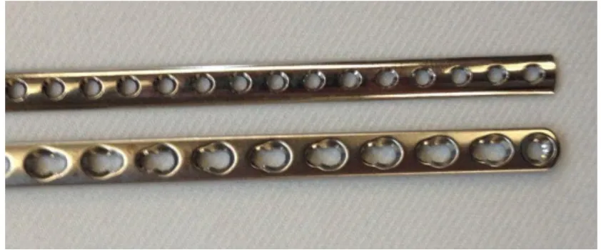
The Veterinary Cuttable Plate (VCP) has relatively higher numbers of screws per unit
74
length of plate when compared to the equivalent DCP (Fig 6). However, a single 2.0/2.7
75
VCP for instance, is significantly weaker to bending than a 2.7 DCP/LCP, having
76
approximately 1/3 the stiffness, but by stacking two of them on top of each other the
77
composite stiffness can be as much as doubled (Frutcher and Holmberg 1991).
78
Factors affecting the stiffness achieved through stacking include the size of plate and
79
the length of the upper plate in relation to the lower plate of the stack. A further
80
disadvantage of the VCP is its inability to provide fragment compression as it does not
81
83
Locking Plates
84
Locking plates are of great interest to the veterinary orthopaedic community, and do
85
have certain advantages over conventional non-locking plates as reviewed by Arthurs
86
2015. The main difference between locking plates and locking plates is
non-87
locking plate stability is dependent upon friction at the plate to bone and screw to bone
88
interfaces. Non-locking plates can fail by screw toggling (screw head moving within the
89
screw hole), which leads to screw loosening and loss of plate-bone fixation (Smith and
90
others 2007). Therefore, non-locking systems rely on each individual screw’s
91
resistance to pullout; hence the more screws placed, the more cortices and the more
92
stable the fixation.
93
A locking screw on the other hand, relies on friction at the threaded screw-plate
94
interface i.e. its locking mechanism. This means that the construct does not rely on
95
friction between the plate and the bone, or the screw and the bone, and hence should
96
be more stable with fewer cortices or poorer quality bone. These plates are extensively
97
used in osteoporotic fractures in people for this very reason. The down side of these
98
systems is nearly all them have a fixed angle of the screws, by virtue of their being
99
locked. This can mean that it may not be possible to aim two bicortical locked screws
100
within the bone fragment (Fig 7).
101
Alternatives include placing a monocortical locked screw (see next section for more
102
detail), or to use a locking system that can be easily contoured to allow placement of
103
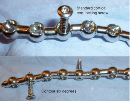
a locked screw into the bone segment (OrthoMed SOP (Fig 8), Vetisco Evolox). The
104
OrthoMed SOP (String of Pearls), is popular, as it allows six degrees of contouring,
105
and makes use of standard AO locking cortical screws (Fig 8). The use of
non-106
locking AO style screws, is both its strength by minimising investment in inventory, but
107
with other locking screws (Fig 9), and are therefore more prone to implant failure
109
through screw breakage. Further systems, now available allow the placement of
110
screws at different angles within the hole and still achieve a ‘locked screw’. These
111
newer variable angled locked screw systems (Securos PAX, Freelance VetLox),
112
however, have not been extensively evaluated yet (Arthurs 2015).
113
114
Creating a hybrid fixation
115
Adding a locked screw to a conventional fixations to create a ‘hybrid fixation’ can be
116
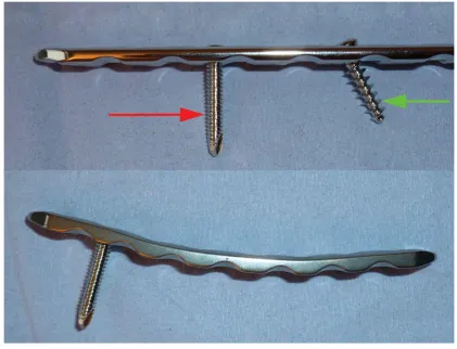
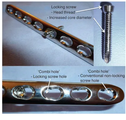
very useful. Plating systems such as the DePuy Synthes Locking Compression Plates
117
(LCP), have ‘combi holes’. These plate holes combine the old Dynamic Compression
118
Plate (DCP), style hole with a locking screw hole. One end of the plate hole allows for
119
placement of a standard non-locking cortical or cancellous screw and can be used in
120
either compression or neutral fashion. The other end has a thread cut into it, allowing
121
it to accept a specially designed locking screw (Fig 9). This means that each combi
122
hole can be used in one of two modes: either in a ‘Locking mode’ – with special locking
123
screws, nor in a non-locking ‘conventional DCP mode’ with standard cancellous or
124
cortical screws.
125
A veterinary mechanical study showed that adding a single locked screw to an
126
otherwise non-locking construct will increase its resistance to torsion (Gordon 2009),
127
and may be clinically useful (Fig 10). The use of locking screws also has advantages
128
in poor quality bone, or when insufficient cortices are available. Therefore if there is
129
only room for two bicortical screws, it is advisable to place at least one as a locked
130
screw. There are important rules when mixing locking and non-locking screws in any
131
one bone segment, so called ‘hybrid usage’; it is essential to place the non-locking
132
screws first and the plate must also be adequately contoured so there is contact
133
If contouring is suboptimal, the non-locking screws may distort the fracture alignment.
135
Once the locking screws are placed, locked screws can follow. Placing
non-136
locking screws after locked screws in any one fracture segment, will lead to the
137
different types of fixation method working against each other, as the locking screw will
138
prevent the non-locking screw from creating contact and friction between the plate and
139
the bone. Therefore, rather than acting synergistically, the repair may fail.
140
If a monocortical screw is required, then a locking screw is preferable to a non-locking
141
monocortical screw (Fig 11). Locking monocortical screws are mechanically more
142
reliable than non-locking as they have two points of fixation; the near cortex of the
143
bone and the plate itself, and therefore they resist load to failure better than standard
144
monocortical cortex screws in bone. Monocortical locked screws are supposed to
145
provide sufficient stability and load transfer, despite only loading the near cortex. This
146
latter concept has been questioned in small animals due to the presence of
147
comparatively very thin cortices and therefore, bicortical screw fixation, or double plate
148
fixation is probably safer if achievable.
149
The minimum number of locked or combination or locked and non-locked screws is
150
unknown. The author would tentatively suggest aiming for an absolute bare minimum
151
of four cortices IF at least one cortex had a locked screw and one or more bicortical
152
screw(s) were present, in a reconstructed fracture. Extremely careful post-operative
153
care would be necessary, and other considerations such as location, bone quality,
154
other injuries, age, activity and quality of repair would need to be considered.
155
Otherwise, a suggested minimum would be five cortices with at least a single
156
monocortical or bicortical locked screw.
157
Veterinary Anatomical Plates
158
There is an increasing diversity of veterinary designed plates on the market, from a
159
distal metaphyseal antebrachial fracture. The ‘T’ plate, (Fig 11) being wider at one end,
161
with screws orientated along the wide portion of the plate, allows increased screw
162
purchase in a short wide fracture fragment, such as the distal radial epiphysis. These
163
T plates are also useful for short ilial fractures just cranial to the acetabulum, “cotyloid
164
fractures”. Historically the plate has been quite short, however longer plates with a T
165
shaped head are now available. ‘Veterinary T’- and ‘L-plates’ for use in veterinary
166
practice are available in different sizes (ranging from 2 mm to 3.5 mm plates).
167
Other useful plates include the hockey-stick or supracondylar plate ‘J plate’ (Fig 12),
168
which is very useful for achieving a rigid plate fixation where there is limited bone for
169
screw purchase due to the curvature of the femoral condyle in supracondylar fractures.
170
Acetabular plates (Fig 13) are useful for acetabular fractures but have also been used
171
for femoral trochlea ridge fractures. Double hook plates can be used in proximal
172
femoral fractures as well as for intertrochanteric osteotomies. These can be
173
manufactured for cats using a VCP and pin cutters to fashion two hooks to fold over
174
and insert into the proximal aspect of the greater trochanter.
175
Other procedure specific plates can also be useful. For instance, the Tibial Plateau
176
Levelling Osteotomy (TPLO) Plate for cruciate instability, is very well adapted to short
177
proximal tibial fractures, especially the DePuy Synthes TPLO plate that has fixed
178
angled locked screws proximally, specifically orientated not to breach the articular joint
179
surface or to impinge on each other (Fig 14).
180
181
182
Plates with Six Degrees of Freedom – Reconstruction, Malleable and
183
Contourable plates
184
(six degrees) contouring by increased malleability and plate design (Fig 15). This
186
means it is possible to contour the plate to obtain more screws in a smaller, or unusual
187
shaped bone fragment, however these plates are inherently weaker to allow contouring,
188
Therefore, compared to the same size DCP, the reconstruction plate is more likely to
189
fail.
190
Locking plates with three degrees of contouring freedom also exist. They combine the
191
increased contouring potential with the advantages of locking screws, but have the
192
disadvantage of usually being ‘weaker plates’. Systems available include the Depuy
193
Synthes UniLock plate, Veterinary Instrumentation Cuttable Malleable Locking Plate,
194
and Vetisco Evolox..The OrthoMed SOP (Fig 8), also allows six degrees of freedom
195
with locking screws, but has been biomechanically shown not to be mechanically
196
inferior to the equivalent DCP (Arthurs 2015).
197
198
Creating a plate rod
199
Adding an intra-medullary (IM) pin to a plate fixation is a useful and popular technique
200
(Hulse 1997, Reems and others 2003). An IM pin helps to distract the fracture and
201
maintain alignment during surgery. If the pin can be placed from the shorter fragment
202
into the longer fragment, such as in a proximal femoral fracture with a pin placed from
203
proximal to distal, it will improve the stability of the construct. However, if the IM pin
204
can only be placed from the longer fragment into the shorter fragment, such as the
205
case with distal femoral condylar fracture, there may be no meaningful increase in
206
stability provided, although, it may help in initial reduction by re-aligning and distracting
207
the fragments. A pin size of 40% of the canal diameter is usually recommending and
208
taken from the pre-operative radiographs, potentially from the contralateral limb,
209
measured on the radiographic projection that the screws are placed from and to i.e.
210
Choosing a pin of 40% the diameter allows the placement of screws past the pin whilst
212
still providing a mechanical advantage. In the example shown, the medullary canal
213
isthmus measured 5.3mm on the lateral radiographic view (not shown) and a 2mm pin
214
was selected to give 38% fill (Fig 17). If locking screws are used, then monocortical
215
screws may be necessary as placing locking screws past the pin can be impossible at
216
times.
217
218
Additional implants to reconstruct the bone and improve stability
219
Other small implants, such as additional small K-wires are useful for fracture reduction
220
and alignment but will not add much to the mechanical strength and therefore shouldn’t
221
be relied upon to shore-up a tenuous plate-screw fixation. Compression from a lag
222
screw is extremely beneficial as it creates absolute stability for bone healing, and the
223
compression also results in impaction of fragments with a marked increase in frictional
224
resistance to motion. What this means is that it greatly reduces the forces born by
225
implants. An option if a fracture component is completely reconstructable is to lag two
226
segments together to in effect make a single larger fragment, which then provides more
227
bone for screw purchase in the newly formed larger fragment.
228
229
Human Anatomical Plates
230
In recent years, aided by the development of locking technology there has been an
231
explosion in human site-specific anatomical pre-contoured, shape specific plates.
232
Some of these can be made use of in veterinary orthopaedics and offer the advantage
233
of the ability to use a mixture of locking and conventional screws in addition to offering
234
varied screw positions and plate shapes. Most of these plates are derived from the
235
veterinary LCP screws and instrumentation, or compatible style veterinary offerings.
237
The human distal radial plates probably are the most useful for veterinary patients (Fig
238
17), and I have used these in a range of fractures including cat pelvic fractures,
239
complex ulna fractures and humeral Y fractures, where bone stock is limited (Fig 18).
240
Some have contouring planes so that corners can be bent over relatively easily without
241
deforming the screw holes. Furthermore some plates have locking screw holes
242
intentionally angled to ensure maximum purchase and to avoid physes or articular
243
surfaces. The main consideration is most of these human plates were not designed for
244
weight bearing application as bipedal humans will not weight bear on forelimb/upper
245
limb plates. As such the plates are relatively thin and should be used with due
246
consideration in veterinary small animal orthopaedic applications where weight bearing
247
may be intended.
248
249
Fixation combinations
250
Combining the different fixation options outlined above can have excellent results (Fig
251
19). However, if after considering all internal fixation options, it is not possible to
252
provide two bicortical screws in a single plate, or one bicortical and one locked
253
monocortical screw then other fixation systems such as external skeletal fixators may
254
be necessary. The circular external skeletal fixator has been shown to be particularly
255
useful in this context, as well as circular-linear hybrids containing a single ring allowing
256
several pins to be placed in a short segment of bone and then connected to a linear
257
fixator along the longer bone fragment.
258
Summary
259
Plates and screws are an excellent means to stabilise many fractures however for
260
a stable and reliable fixation. There are many ways to achieve this, each with relative
262
advantages and disadvantages, and some lend themselves well to a particular fracture
263
location or configuration (Table 3). Some approaches are straightforward, while others
264
are more costly and some require more advanced planning. In any case, consideration
265
of double plating, locking implants, anatomical plates, human orthopaedic plates,
266
plate-rods, malleable plates, or combinations should allow the veterinary orthopaedic
267
surgeon to achieve a stable, reliable fixation, even when it appears unachievable on
268
first inspection (Fig 20).
269
270
Tables:
272
Table 1: Factors Influencing your Choice of Implants
273
General Animal Factors
274
Age (young, adult, geriatric), weight relative to bone size (overweight, breed
275
conformation), systemic illness, nutritional state, patient activity
276
Veterinary Factors
277
Implants and equipment available, expertise and experience available, time
278
and availability for follow-up
279
Fracture factors
280
Complexity of fracture, location of fracture, soft-tissues available (for closure
281
and blood supply), open or closed, bone loss
282
283
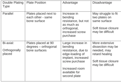
Table 2: Types of Double Plating
285
Double Plating Type
Plate Position Advantage Disadvantage
Parallel Plates placed next to each other - same bone surface
Increase in bending
resistance, but not as much as
orthogonal, increased screw purchase
May struggle to fit two plates on same surface
Soft tissue closure may be difficult
Bi-axial:
Orthogonally placed
Plates placed at 90 degrees – orthogonal bone surfaces
Large increase in bending
resistance, due to edge-loading of implant, increased screw purchase Increased room available for second plate More extensive dissection may be needed, may retard healing
Soft tissue closure may be difficult
Table 3: Common Juxta-articular Fractures
288
Common juxta-articular fractures and ideas for management
289
Femoral Supracondylar Fractures
290
These are challenging usually due to caudal curve of the femoral condyle. It often helps
291
to place one or two temporary or permanent crossed K-wires to aid initial stability. An
292
arthrotomy into the proximal stifle joint also helps ensure good exposure. The femoral
293
condylar veterinary plate ‘Hockey-Stick’ ‘J plate’ is particularly good here (Fig 13), to
294
ensure at least 3 bicortical screws, however care needs to be taken to avoid the
295
proximal section of the plate diverging away from the femoral diaphysis when
296
concentrating on plating over the condyle distally.
297
Distal radius and Ulna
298
Most commonly seen in toy breeds, options include a straight plate if you can achieve
299
2 bicortical screws distally ± IM pin in the ulna for additional stability. Veterinary or
300
human T plates make use of the distal widening of the radius and allow two bicortical
301
screws in the short distal fragment (Fig 12). Again ulna IM pin can help with stability.
302
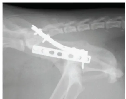
Proximal Femur
303
The best option here is to take time to accurately contour a plate along and over the
304
top of the greater trochanter (Fig 17). The greater trochanter offers a large block of
305
bone stock and screws can be angled in to this to achieve purchase. A plate bending
306
press if usually necessary to get sufficient bend on the proximal aspect of the plate. A
307
screw can be angled up the femoral neck to increase purchase. A forked plate is
308
another option and can be manufactured from a VCP in cats. Additional intra-medullary
309
pins in the femur can also be beneficial.
310
These can be particularly challenging. It is important to avoid the tarso-crural joints
312
surface, and orthogonal plating may help, however assessment of fracture healing due
313
to the metalwork obscuring the fracture on radiographs is a significant problem and
314
care should be taken with soft-tissue closure. It is also worth considering placing locked
315
screws if available (Fig 10).
316
Proximal Tibia
317
The TPLO plate is essentially a plate designed to stabilize a short proximal tibial
318
fragment and works well here. T plates can also be used, but be aware that there are
319
strong rotation forces acting in these region, potentially rotating the proximal femur
320
caudally. Additional placement of a pin and tension band may be advisable.
321
322
References
324
325
ARTHURS G. (2015) Advances in internal Fixation locking plates. In practice 37
:13-326
20
327
328
BRINKER WD, FLO GL, LAMMERDING JJ. & BLOOMBERG MS. (1977). Guideline
329
for selecting proper implant size for treatment of fractures in the dog and cat J. Am.
330
Anim. Hosp. Assoc. 13:476–477.
331
332
FRUTCHER AM, HOLMBERG DL (1991). Mechanical analysis of the veterinary
333
cuttable plate. Vet Comp Orthop Traumatol. 4:116–119.
334
335
GEMMILL, T. (2007) Advances in the management of diaphyseal fractures. In Practice
336
29, 584-593
337
338
GORDON S MOENS NM, RUNCIMAN J & MONTEITH G. (2009). The Effect of the
339
Combination of Locking Screws and Non-Locking Screws on the Torsional Properties
340
of a Locking-Plate Construct. Veterinary and Comparative Orthopaedics and
341
Traumatology 23(1):7-13
342
343
HULSE, D. A., HYMAN, W., NORI, M. & SLATER, M. (1997) Reduction in plate strain
344
346
JOHNSON, A. L., HOULTON, J. E. F. & VANNINI, R. (2005) AO Principles of Fracture
347
Management in the Dog and Cat. Davos, AO Publishing
348
349
PIERMATTEI, D., FLO, G. & DeCAMP, C. (2006) Brinker, Piermattei, and Flo’s
350
Handbook of Small Animal Orthopedics and Fracture Repair, 4th edn. Philadelphia,
351
Saunders Elsevier
352
353
REEMS, M. R., BEALE, B. S. & HULSE, D. A. (2003) Use of a plate–rod construct and
354
principles of biological osteosynthesis for repair of diaphyseal fractures in dogs and
355
cats: 47 cases (1994-2001). Journal of the American Veterinary Medical Association
356
223, 330-335
357
358
SHALES, C. (2008) Fracture management in small animal practice 1. Triage and
359
stabilisation. In Practice 30, 314-320
360
361
SMITH, W. R., ZIRAN, B. H., ANGLEN, J. O. & STAHEL, P. F. (2007) Locking plates:
362
tips and tricks. American Journal of Bone and Joint Surgery 89, 2298-2307
363
364
Figure Legends
366
Figure 1: Distal femoral fracture with limited bone stock in distal fragment
367
368
Figure 2: Three screw doctrine: One bicortical screw per segment allows rotation. Two
370
bicortical screws prevents rotation but remains at high risk of failure. Three bicortical
371
screws are therefore the recommended minimum.
372
373
Figure 3: Parallel double plated ilial fracture. Based on the dog’s weight a 2.7mm plate
375
would have been selected however there was only room for two bicortical screws. By
376
placing two 2.0mm plates (DePuy Synthes DCP), five bicortical screws were placed in
377
the shorter fragment.
378
379
Figure 4: Orthogonal double plated feline ilial fracture, allowed 4 bicortical screws to
381
be placed (DePuy Synthes DCP laterally, DePuy Synthes 1.5/2.0 VCP stacked
382
dorsally)
383
384
Figure 5: (a) Short comminuted calcaneal fracture. (b) The fracture was double plated,
386
which allowed for placement of four bicortical screws into the calcaneus
387
388
389
Figure 6: The 2/2.7 mm veterinary cuttable plate (top) has more screw holes per unit
391
length than the 2.7 mm locking compression plate (bottom), or a dynamic compression
392
plate
393
394
395
Figure 7: Locking Compression Plate (LCP, DePuy Synthes) allows for placement of
397
fixed angle locking screw, which requires plate contouring to orientate screw position,
398
as well as non-locking screws which can be angled within the screw hole.
399
400
Figure 8: String-of-Pearls plate (SOP, OrthoMed), allows for contouring in 3 planes,
402
and uses non-locking cortical screws as part of its locking mechanism.
403
404
Figure 9: A locking compression plate (LCP) has ‘combi-holes’ allowing placement of
406
a locking or non-locking screw. LCP locking screws have a thread on the head to
407
engage in the plate hole, and also have an increased core diameter to make the screw
408
stronger, thus reducing the chance of failure
409
410
411
Figure 10: (a) Orthogonal view radiographs of double spiral tibial fracture with a short
413
distal fragment. (b) Postoperative orthogonal radiographs show locking screws marked
414
*. Only two screws were placed in the distal segment (circled); however, one was
415
placed as a locking screw (*) increasing the stability of the fixation
416
417
Figure 11: (left) Distal radial fracture in a toy breed dog, stabilised with a veterinary T
419
plate employing two distal screws. (right) Other designs of veterinary T plates with
420
three distal screw holes are also available
421
422
Figure 12: (left) ‘Hockey-stick’ plate which allows three bicortical screws to be screwed
424
into the curved distal condyle. (right) This type of plate was used to stabilise a
425
supracondylar femoral fracture
426
427
428
Figure 13: A mid-acetabular fracture in a cat which was stabilised with an anatomical
430
acetabular plate. An additional ilial fracture was plated with a seven-hole dynamic
431
compression plate. A sacroiliac luxation was also present and was stabilised with a 2.7
432
mm screw.
433
434
435
Figure 14: Broad locking tibial plateau levelling osteotomy plate. This plate is useful for
437
proximal tibial fractures due to the proximal locking screws being clustered in a small
438
space and orientated to avoid each other
439
440
Figure 15: Reconstruction plates have increased malleability to allow six degrees of
442
freedom, which is useful to achieve increased numbers of screws in some short bone
443
fragments. However, the plates are weaker than the equivalent-sized straight dynamic
444
compression plate
445
446
Figure 16: Proximal comminuted femoral fracture in a cat. A plate has been contoured
448
over the greater trochanter to make use of the proximal bone stock (DePuy Synthes
449
2.4mm LCP). Further, an intra-medullary pin (2mm) has been added to increase
450
stability.
451
452
Figure 17: Human anatomical plates - 2.4mm Distal Radial Plates (DePuy Synthes
454
2.4mm Distal Radius Plates). These plates have 'combi holes' allowing flexible usage.
455
They come in a range of shapes, and have contouring planes, to allow plate contouring
456
without damaging the screw holes. They are thinner and relatively weaker than the
457
equivalent LCP/DCP stock plate.
458
459
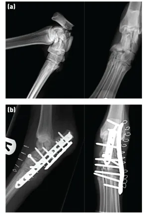
Figure 18: Veterinary use of Human 2.4 Distal Radial Plates (DePuy Synthes). a)
461
Comminuted canine olecranon fracture was stabilised by placement of a lag screw to
462
reconstruct the main fragment, and then a radial L-plate was placed laterally to achieve
463
2 bicortial screws in the fragment. A second caudal plate (double orthogonal plating),
464
was also placed due to the dog being known to be highly active. b) Distal humeral
465
bicondylar 'Y' fracture with very short lateral condylar fragment. A human radial L plate
466
was also used here, this time with 3 screws in the distal segment, all placed as locking
467
screws, combined with a standard 2.7 LCP plate on the medial aspect.
468
469
Figure 19: Comminuted articular distal radial fracture in a lurcher was repaired using
471
multiple techniques. The distal fragments were stabilised with a lag screw to reduce
472
and stabilise the articular surface. K wires were placed to temporarily position the distal
473
fragment to the radial diaphysis which was stabilised with a veterinary T plate (DePuy
474
Synthes 2.7mm), placing 2 bicortical screws in the newly formed single distal fragment.
475
The lag screw was then removed and replaced through a medial plate (orthogonal
476
double plating) (DePuy Synthes 2.7mm LCP), which allowed an additional
477
monocortical locked screw to be placed.
478
Figure 20: Suggested algorithm for dealing with limited bone stock with internal fixation.
480
Preferred methods bold arrows, suitable methods thin arrows, and possible methods
481
dashed arrows.
482