TUBERCULOSIS: A GLOBAL THREAT
Singla Rajeev*1, Bhat Varadaraj G1
1
Department of Pharmaceutical Chemistry, MCOPS, Manipal University,Manipal-576104,India
*Corresponding author:
Email id: [email protected] , [email protected] Contact no. : 91-9916824228, 91-9896134234
Summary
Tuberculosis (TB) is a major cause of illness and death worldwide, especially in Asia and Africa. Globally, 9.2 million new cases and 1.7 million deaths from TB occurred in 2006, of which 0.7 million cases and 0.2 million deaths were in HIV-positive people.Still powerful new anti-TB drugs with new mechanisms of action have not been developed in the last over thirty years. It is expected that development of the new effective anti-TB drug will bring us various outcomes such as shortening the total duration treatment, improvement of the treatment completion ratio, prevention and treatment of the multiple drug resistant tuberculosis (MDR-TB) and reducing the total medical expenditure.
Keywords: Tuberculosis, Mycobacterium tuberculosis, XDR-TB, WHO
Introduction
Each year, 8.74 million develop tuberculosis and nearly 2 million die. This means that someone somewhere contracts TB every four seconds and one of them dies every 10 seconds.[3] The global community woke up to this disease when, in 1993, the WHO declared TB as a global emergency. The epidemiology of TB can be considered the model of web causation of disease with the agent, host and environment playing their respective parts. Traditionally, there have been three approaches to the epidemiology of tuberculosis, namely: a. The Etiologic Approach (analytic epidemiology): mainly dealing with the risk factors associated with the agent – M tuberculosis;
b. The Descriptive Approach: dealing with the traditional incidence and prevalence of tubercular infection; and
c. The Predictive Approach: dealing with what happens next – forecasting the tubercular epidemic.
The reason for the increase in the number of tuberculosis cases are probably due to growing epidemic of HIV infection, malnutrition leading to reduced immunity, use of indiscriminate/inadequate chemotherapy and multidrug resistance due to partial adherence to chemotherapy.
THE ETIOLOGY OF TUBERCULAR IFECTIO
Mycobacterium tuberculosis is the etiologic agent for a contagious disease, Tuberculosis in humans, an air borne disease. Although the disease most often affects the lungs but can also affects the CNS , the lymphatic system, circulatory system, the genitourinary system, bones, joints and the skin. When persons with pulmonary TB cough or sneeze, they produce tiny droplet nuclei that contain Tb bacteria, which can remain suspended in the air for prolonged period of time. Anyone who breathes air that contain these droplet nuclei can become infected with TB. Mycobacterium tuberculosis survives and persists for prolonged periods within its host in an asymptomatic, latent state and can reactivate years later if the host’s immune system weakens. The dormant bacilli synthesize and accumulate triacylglycerol, reputed to be an energy source during latency. Among the phospholipases, phospholipase C plays an important role in the pathogenesis.
Mycobacteria are slow growing,Acid fast Gram positive, non-motile, pleomorphic rods, obligate aerobes.The taxonomic history of the genus mycobacterium is intricate and difficult to disentangle from that of related taxa, notably Corynebacterium, &ocardia and Rhodococcus. M. tuberculosis divides every 15 to 20 hours, which is extremely slow compared to other bacteria, which tend to have division times measured in minutes (for example, E.Coli can divide roughly every 20 minutes). It is a small bacillus that can withstand weak disinfectants and can survive in a dry state for weeks. Its unusual cell wall, rich in lipids (e.g., mycolic acid), is likely responsible for this resistance and is a key virulence factor.
Intercalated within this environment are the phthiocerol dimycocerosates, cord factor or dimycolyltrehalose, sulfolipids, phosphatidylinositol mannosides and the related lipomannan and lipoarabinomannan, etc., agents responsible for much of the pathogenesis of tuberculosis. Interest in the biosynthesis of the cell-wall core, regarded, unlike the ancillary lipids, as essential to bacterial viability and integrity, is now driven by the pressing need for alternative drugs to counteract drug-resistant tuberculosis. In a manner analogous to the roles of lipid I and II in peptidglycan formation, synthesis of the entire arabinogalactan is initiated by transferring activated sugars to decaprenyl-phosphate, giving rise to the linker disaccharide, followed by stepwise elongation of the galactan, and the arabinan, apparently one sugar at a time.
STRUCTURE OF MYCOBACTERIAL GEOME:
The complete genome sequence of the best-characterized strain of Mycobacterium tuberculosis, H37Rv, has been determined and analysed in order to improve our understanding of the biology of this slow-growing pathogen and to help the conception of new prophylactic and therapeutic interventions. The genome comprises 4,411,529 base pairs, contains around 4,000 genes, and has a very high guanine + cytosine content ranges from 60 to 70 mol% that is reflected in the biased amino-acid content of the proteins. M. tuberculosis
differs radically from other bacteria in that a very large portion of its coding capacity is devoted to the production of enzymes involved in lipogenesis and lipolysis, and to two new families of glycine-rich proteins with a repetitive structure that may represent a source of antigenic variation. Mycobacteria have only one or two copies of the genes encoding the ribosomal RNAs(in contrast, E.coli has seven copies) and these genes are arranged in the typical rRNA operon.[4]
SELF-PROTECTIO AGAIST ATTEMPTS BY THE HOST AT
ITRACELLULAR KILLIG:
Tubercle bacilli invade their hosts, and thus the host attempts to kill them in the ways it would any invading organism. However, M. tuberculosis divide inside host macrophages, cells which are adapted to kill invading microbes. In this section, we review how the metabolic and biochemical process of these mycobacteria allow them to survive once they enter host cells.
The consequences of phagosome- lysosome fusion- which include production of hydrolytic enzyme and lowering of intracellular pH- plus the production of peroxide by macrophages are important in killing invading microbes, and probably also have role in killing M. tuberculosis. A wide range of microbes elaborate the enzymes catalase and superoxide dismutase to destroy the harmful peroxide and other toxic oxygen metabolites. Therefore, either the production of toxic oxygen metabolites by the host is not alone sufficient to kill mycobacteria or else pathogenic mycobacteria have ways other than producing the above enzymes of evading toxic oxygen metabolites.[6]
MULTIDRUG RESISTAT TUBERCULOSIS(MDRTB):
Tuberculosis therapy involve an initial intensive two month regime comprising multiple antibiotics- Isoniazid(INH), rifampicin(RMP),pyrazinamide(PZA) and ethambutol(EMB) or streptomycin(SM). For next four months, only INH or RMP are administered to eliminate any persisting tubercle bacilli. MDRTB refers to simultaneous resistance to at least INH and RMP(with or without resistance to other drugs). Globally, about three per cent of all newly diagnosed patients have MDR-TB. The proportion is higher in patients who have previously received antituberculosis treatment reflecting the failure of programmes designed to ensure complete cure of patients with tuberculosis. While host genetic factors may probably contribute, incomplete and inadequate treatment is the most important factor leading to the development of MDR-TB. The definitive diagnosis of MDR-TB is difficult in resource poor low income countries because of non-availability of reliable laboratory facilities. Efficiently run tuberculosis control programmes based on directly observed treatment, short-course (DOTS) policy is essential for preventing the emergence of MDR-TB. Isoniazid, the most powerful mycobactericidal drug available, ensures early sputum conversion and helps in decreasing the transmission of TB. Rifampicin, by its mycobactericidal and sterilizing activities is crucial for preventing relapses. Thus, isoniazid and rifampicin are keystone drugs in the management of TB. While resistance to either isoniazid or rifampicin may be managed with other first-line drugs, resistance to both isoniazid and rifampicin (MDR-TB) demands treatment with second-line drugs. These drugs have limited sterilising capacity and are not suitable for shortcourse treatment. Thus, patients with MDR-TB require prolonged treatment with drugs that are less effective and more toxic. Therefore, it is necessary to distinguish MDR-TB from mere drug-resistant tuberculosis by performing mycobacterial culture and sensitivity testing because the therapeutic implications are different.
EXTESIVELY DRUG RESISTAT TUBERCULOSIS: (XDR-TB):
Extensive Drug Resistant TB (also referred to as Extreme Drug Resistance) is type of TB which shows resistance to at least isoniazid and rifampin among first-line anti-TB drugs, resistance to any fluoroquinolone, and resistance to at least one second-line injectable drug (amikacin, capreomycin, or kanamycin).The description of XDR-TB was first used earlier in 2006, following a joint survey by WHO and the US Centers for Disease Control and Prevention (CDC).Resistance to anti-TB drugs in populations is a phenomenon that occurs primarily due to poorly managed TB care. Problems include incorrect drug prescribing practices by providers, poor quality drugs or erratic supply of drugs, and also patient non-adherence. XDR-TB poses a grave public health threat, especially in populations with high rates of HIV and where there are few health care resources. Recommendations outlined in the WHO Guidelines for the Programmatic Management of Drug Resistant Tuberculosis include:
• strengthen basic TB care to prevent the emergence of drug-resistance
• ensure prompt diagnosis and treatment of drug resistant cases to cure existing cases and prevent further transmission
• increase collaboration between HIV and TB control programme to provide necessary prevention and care to co-infected patients
CURRET THERAPY:
For treating uncomplicated tuberculosis, daily INH,PZA and RMP for two months followed by 4 months of daily RMP and INH is effective. In this regimen, the medicines are self administered(Refer Table 1). To ensure compliance and to help prevent the development of drug resistance strains of the tubercle bacillus, Directly Observed Preventive Therapy (DOPT) should be used. Preventive therapy is recommended for patients infected with HIV and house hold members of patients with tuberculosis. MDRTB is extremely difficult to treat; mortality may be as high as 80%. Recommended therapy include ethambutol plus RMP for 6 months followed by PZA plus RMP for 9 months, or PZA plus ciprofloxacin for 6 to 12 months; the effectiveness of these treatment is still being investigated.
Table 1
Recommended Drugs for the Initial Treatment of Tuberculosis in Children and Adults
Daily Dose\ * Twice Weekly Dose
Drug Children Adults
Maximal Daily Dose in Children and Adults
Children Adults
Isoniazid 10 to 20 mg/kg PO or IM
5 mg/kg PO or IM
300 mg 20 to 40 mg/kg Max. 900 mg
15 mg/kg Max. 900 mg
Rifampin 10 to 20 mg/kg PO
10 mg/kg PO
600 mg 10 to 20 mg/kg Max. 600 mg
10 mg/kg Max. 600 mg
Pyrazinamide 15 to 30 mg/kg PO
15 to 30 mg/kg PO
2 g 50 to 70 mg/kg
50 to 70 mg/kg
Streptomycin 20 to 40 mg/kg IM
15 mg/kg ** IM
1 g ** 25 to 30 mg/kg IM
25 to 30 mg/kg IM
Ethambutol 15 to 25 mg/kg PO
15 to 25 mg/kg PO
2.5 g 50 mg/kg 50 mg/kg
Definition of abbreviations: PO = perorally; IM = intramuscularly.
* Doses based on weight should be adjusted as weight changes.
ISOIAZID(IH):
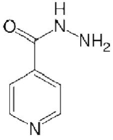
Figure 1 Isoniazid
Isoniazid(also called isonicotinyl hydrazide or INH) (see Figure 1) is a first line drug for prophylaxis and treatment of tuberculosis. Isoniazid is never used on its own to treat active tuberculosis because resistance quickly develops. Only Patients with a recently positive purified protein derivative(PPD) skin test and normal chest radiograph findings are given a 6- to 9- month course of INH.
INH has been commercially prepared from p-pyridinecarboxylic acid or its esters and hydrazine hydrate or from p-cyanopyridine and hydrazine hydrate. p- Cyanopyridine was converted to corresponding amide, followed by reaction with hydrazine hydrate to synthesize INH.
Pathophysiology:
The M.tuberculosis KatG gene encodes a dual function enzyme called catalase peroxidase, which confers sensitivity in M.tuberculosis to INH. INH is a prodrug that require activation by the mycobacterial catalase peroxidase enzyme (KatG) to an active form, which then exerts a lethal effects on intracellular targets. INH enters the organism by diffusion and by oxygen dependent active transport. The drug has been reported to affect to virtually every aspect of
M.tuberculosis metabolism.
Mechanism of action:
INH, highly active against the MTB complex(M. tuberculosis, M. bovis , M. africanum & M. microti) has very low MICs(0.02 to 0.06 µg/ml) against these pathogens. It inhibits the synthesis of mycolic acids(Long chain α-branched β-hydroxylated fatty acids) in
Toxic effects of INH also result from inhibition of lactate dehydrogenase, an enzyme that converts lactate to pyruvate, and from inhibition of cytochrome P450. Pharmacogenetic studies suggest that patients with certain cytochrome P450 genotypes may be more predisposed to hepatotoxicity during INH therapy for latent tuberculosis.
INH undergoes &-acetylation in the liver to a variety of products that include acetylhydrazine, a potent hepatotoxin. These metabolites are excreted in the urine. With long-term administration at therapeutic doses, INH can cause clinically significant and even fatal hepatic injury in 1% of patients and elevated liver enzyme levels in 10-20% of patients.
In vitro studies of a variety of animal cell lines demonstrated that INH toxicity results from the induction of apoptosis with associated disruption of mitochondrial membrane potential and DNA strand breaks.
Precautionary measurement to reduce Toxicity:
Eat the following foods with caution:
cheeses, including American, Blue, Boursault, Brick, Brie, Camembert, Cheddar, Emmenthaler, Gruyere, Mozzarella, Parmesan, Romano, Roquefort, Stilton, and Swiss;
sour cream and yogurt;
beef or chicken liver, fish, meats prepared with tenderizer, bologna, pepperoni, salami, summer sausage, game meat, meat extracts, caviar, dried fish, herring, shrimp paste, and
tuna;
avocados, bananas, figs, raisins, and sauerkraut;
soy sauce, miso soup, bean curd, and fava beans;
yeast extracts;
ginseng;
chocolate;
caffeine (coffee, tea, cola, etc.); and
beer (alcoholic and nonalcoholic), red wine (especially Chianti), sherry, vermouth, and other distilled spirits.
Mechanism of Resistance:
Resistance to isoniazid (INH) in Mycobacterium tuberculosis is attributed to mutations in several genes. The katG gene,which encodes catalase-peroxidase, is the gene most commonly altered, with the majority of mutations occurring at codon 315. Mutations in the promoter regions of inhA and oxyR-ahpC genes have been identified in INH resistant strains but not INH-susceptible strains. Four independent mutations were also reported to be found in the
An increased amount of NADH may prevent formation of isonicotinic acyl NADH or may promote displacement of the isonicotinic acyl NADH from InhA. While our studies have identified this mechanism in M. smegmatis, results reported in early literature lead us to believe that it can occur in Mycobacterium tuberculosis.
RIFAMPICI(RMP):
Figure 2 Rifampicin
Rifampicin or Rifampin(See Figure 2) is a bactericidal antibiotic drug of a rifamycin group. Rifamycins(e.g. rifampicin, rifabutin & rifapentine), group of antibiotics characterized by a natural ansa structure(chromophoric naphthohydroquinone group spanned by a long aliphatic bridge), are potent inhibitors of prokaryotic DNA dependent RNA polymerase. It is a semisynthetic derivative of Rifamycin B obtained from Streptomyces mediterranei. Rifampicin is prepared by mild oxidation of a Mannich base of rifamycin SV(obtained by fermentation), followed by mild reduction to obtain 3-formylrifamycin SV, which is then reacted with 1-amino-4-methylpiperizine to form rifampicin.
The bactericidal action covers all subpopulations of TB bacilli, but acts best on slowly or intermittently(spurters) dividing bacilli as well as on many atypical mycobacteria. Both extra and intracellular organisms are affected. It has good sterilizing and resistance preventive actions.
Rifampin diffuses well to most body tissues and fluids, including the cerebrospinal fluid (CSF), where concentrations are increased if the meninges are inflamed; concentrations in the liver, gallbladder, bile, and urine are higher than those found in the blood. Therapeutic concentrations are achieved in the saliva, reaching 20% of serum concentrations. Rifampin crosses the placenta, with fetal serum concentrations at birth found to be approximately 33% of the maternal serum concentration; it penetrates into aqueous humor and is distributed into breast milk. Because it is lipid-soluble, rifampin may reach and kill susceptible intracellular, as well as extracellular, bacteria and Mycobacteria species.
Mechanism of action:
Mechanism of resistance:
Genetic studies conducted in the early 1980s recorded that E. coli strains resistant to RMP had missense mutations and short deletions in the rpoB gene. Further studies explains 17 mutational alterations affecting 14 amino acid residues in the E. coli RNA polymerase β subunit that mediate RMP resistance in this organism. Virtually all the mutations were missense mutations, and amino acid substitutions at one of two positions(residues 526 & 531) were found in 80% of the resistant strains. Interestingly, no synonymous (silent) nucleotide substitutions were identified in the 122 isolates sequenced for the 411-bp region. For resistant strains bearing many of these rpoB mutations, MICs in excess of 8.0 µg of RMP per ml have recently been reported.
Toxicity:
RMP causes cholestasis at both the sinusoids and canaliculi of the liver because of the defect in uptake by hepatocytes and defect in excretion, respectively. RMP may produce liver dysfunction. Hepatitis occurs in 1% or less of patients, and usually in the patient with pre-existing liver disease.
Hypersensitivity reactions may occur, usually characterized by a “flu” type syndrome.
Nephrotoxicity appears to be related to a hypersensitivity reaction and usually occours after intermittent or interrupted therapy. It has been suggested that some of the adverse effects associated with RMP may be attributed to its metabolite desacetylrifampicin.
RMP does not bind to mammalian nuclear RNA polymerase and therefore does not affect the RNA synthesis in human beings. RMP, however, may affect mammalian mitochondrial RNA synthesis at a concentration that is 100 times higher that that which affects bacterial RNA synthesis.
ETHAMBUTOL (EMB):
Figure 3 Ethambutol
Ethambutol [(S,S')-2,2'-(ethylenediimino)di-1-butanol; EMB],(See Figure 3) a synthetic compound with profound bacteriostatic antimycobacterial activity, is a first line anti-tubercular drug that is most commonly used in combination with other drug as recommended by World Health Organisation DOTS/ DOTS plus regimen in the treatment of tuberculosis. It is also used as part of a combination regimen in the therapy of Mycobacterium avium
Mechanism of action:
Ethambutol diffuses into actively growing M. tuberculosis such as tubercle bacilli.
There is evidence that the drug exerts its bacteriostatic activity by virtue of inhibition of arabinosyl transferase III, an enzyme that polymerizes arabinose into arabinan and then arabinogalactan, a mycobacterial cell wall constituent. Mycolic acid are linked through their carboxy groups to the end terminal 5’- hydroxyl groups of D-arabinofuranose residues of arabinogalactan forming the complex. Disruption of the arabinogalactan synthesis inhibits the formation of this complex and may lead to increased permeability of the cell wall.
Ethambutol appears to inhibit the synthesis of one or more metabolites, thus causing impairment of cell metabolism, arrest of multiplication, and cell death. No cross resistance with other available antimicrobial agents has been demonstrated.
Mechanism of resistance:
The genes embAB of M. avium encode the drug target for ethambutol, the arabinosyl transferase III responsible for the polymerization of arabinose into the arabinan of arabinogalactan and overproduction of this ethambutol-sensitive target leads to ethambutol resistance. EMB-resistant organisms had mutations located in codon 306 of embB that resulted in the replacement of the wild-type Met residue with Ile or Val. Automated sequence analysis of the 5' region (1,892 bp) of embB in an additional 69 resistant and 30 EMB-susceptible M. tuberculosis isolates from diverse geographic localities and representing 70 distinct IS6110 fingerprints confirmed the unique association of substitutions in amino acid residue 306 of EmbB with EMB resistance. Organisms with MICs exceeding 5 microgram/mL are generally considered resistant to ethambutol. M. tuberculosis resistance to ethambutol is due to random spontaneous genetic mutations, occurring at a rate of approximately 1 in 10 organisms; mutations most commonly result in increased production of the enzyme arabinosyl transferase, which overwhelms the inhibitory effects of ethambutol.
Primary (pretreatment) resistance rates of M. tuberculosis to ethambutol vary widely. Although a report from New York City cited primary resistance as high as 14 percent, a global survey performed in 35 countries between 1994 to 1997 found a primary ethambutol resistance rate of 1 percent.[61]
Toxicity:
PYRAZIAMIDE(PZA):
Figure 4 Pyrazinamide
Pyrazinamide(See Figure 4), the pyrazine analogue of nicotinamide, is a first line defense for short-course tuberculosis. Pyrazinamide is only used in combination with other drugs such as isoniazid and rifampicin in the treatment of Mycobacterium tuberculosis. It is never used on its own. It has no other medical uses. In particular, it is not used to treat other mycobacteria;
Mycobacterium bovis and Mycobacterium leprae are innately resistant to pyrazinamide. Pyrazinamide is used in the first two months of treatment to reduce the duration of treatment required. Regimens not containing pyrazinamide must be taken for nine months or more. It shows no significant bactericidal effect and is considered a “ Sterilizing drug”.
Pyrazinamide in conjunction with rifampin is a preferred treatment for latent tuberculosis.
Pyrazinamide can be synthesized with a significant yield by reacting chloropyrazine with an alkali metal fluoride to form 2-fluoropyrazine, by reaction with an alkali metal cyanide, it is converted to 2-cyanopyrazine, which on reaction with concentrated H2SO4 gives pyrazinamide.
Mechanism of action:
In vitro and in vivo studies have demonstrated that pyrazinamide is only active at a slightly acidic pH (pH 5.5). Pyrazinamide is a prodrug that stops the growth of Mycobacterium tuberculosis. M. tuberculosis has the enzyme pyrazinamidase which is only active in acidic conditions. Pyrazinamidase converts pyrazinamide to the active form, pyrazinoic acid. Pyrazinoic acid is thought to inhibit the enzyme fatty acid synthetase I, which is required by the bacterium to synthesise fatty acids although this has been disputed.
Mechanism of Resistance:
The gene pncA encoding the M. tuberculosis Pyrazinamidase has been sequenced , and mutations in pncA were found in small number of PZA-resistant M. tuberculosis strains. Mutations in pncA have been identified in PZA-resistant strains, and transformation of these strains with a functional pncA gene restored pyrazinamidase activity and PZA susceptiblity.
Toxicity:
Fever, porphyria and dysuria have rarely been reported.
The principal adverse effect is a hepatic reaction. Hepatotoxicity appears to be dose related, and may appear at any time during therapy. GI disturbances including nausea, vomiting and anorexia have also been reported.
- Hematologic and Lymphatic
Thrombocytopenia and sideroblastic anemia with erythroid hyperplasia, vacuolation of erythrocytes and increased serum iron concentration have occurred rarely with this drug. Adverse effects on blood clotting mechanisms have also been rarely reported.
- Other
Mild arthralgia and myalgia have been reported frequently. Hypersensitivity reactions including rashes, urticaria, and pruritus have been reported. Fever, acne, photosensitivity, porphyria, dysuria and interstitial nephritis have been reported rarely.
STREPTOMYCI(STR):
Figure 5 Streptomycin
STR(See Figure 5) is an aminocyclitol glycoside antibiotic derived from actinobacterium
Streptomyces griseus. It is made up of three components, streptidine, streptose & N-methyl-L-glucosamine. It was the first really effective drug for tuberculosis.
Mechanism of action:
Streptomycin is a bactericidal antibiotic. It kills sensitive microbes by hurting cell membranes and inhibiting protein synthesis. Specifically, it binds to the 16S RNA of the bacterial ribosome, interfering with the binding of formyl-methionyl-tRNA to the 30S subunit. This prevents initiation of protein synthesis and leads to death of microbial cells. Humans have structurally different ribosomes from bacteria, thereby allowing the selectivity of this antibiotic for bacteria. Streptomycin cannot be given orally, but must be administered by regular intramuscular injection.
Mechanism of Resistance:
The practical implication of the observation of one 16S rRNA gene copy in the slowly growing mycobacteria is that single nucleotide changes can result in antibiotic resistance(dominant behavior).
Kapur et al. characterized five STR-resistant M. tuberculosis strains by automated DNA sequencing of the region of rpsL that contains the mutations of interest. Two of the five strains had missense mutations in codon 43 (AAG3AGG; Lys3Arg). More recent analysis of a sample of greater than 125 STR-resistant strains recovered from patients from global sources has found missense mutations in codon 43 or 88 in approximately 30% of the organisms.
Toxicity:
STR damages eighth nerve damage, more commonly the vestibular branch. Patients can also suffer from renal insufficieny.
General Precautions to Consider Cross-sensitivity and/or related
problems
for
the
First
Line
Drugs
Patients hypersensitive to ethionamide, niacin (nicotinic acid), other rifamycins (rifabutin and rifapentine), or other medications chemically related to rifampin, isoniazid, pyrazinamide, or ethambutol may be hypersensitive to this medication also.
Pregnancy/Reproduction
ote: Tuberculosis during pregnancy should be managed on a case-by-case basis because of the complexity of management decisions. Pyridoxine is indicated for all pregnant women
receiving isoniazid.
Pregnancy—
Rifampin
Rifampin crosses the placenta. It has rarely caused postnatal hemorrhages in the mother and infant when administered during the last few weeks of pregnancy; vitamin K may be indicated. Neonates should be carefully observed for evidence of adverse effects.
Imperfect osteogenesis and embryotoxicity were reported in rabbits given up to 20 times the usual daily human dose. Studies in rodents have shown that rifampin given in doses of 150 to 250 mg per kg of body weight (mg/kg) a day causes congenital malformations, primarily cleft
palate and spina bifida.
Isoniazid
Studies in rats and rabbits have shown that isoniazid may be embryocidal. However, isoniazid has not been shown to be teratogenic in mice, rats, or rabbits.
Pyrazinamide
Adequate and well-controlled studies in humans have not been done; the risk of
teratogenicity has not been determined.
Animal reproduction studies have not been conducted with pyrazinamide.
Ethambutol
Ethambutol crosses the placenta, resulting in fetal plasma concentrations that are approximately 30% of maternal plasma concentrations. However, problems in humans have
not been documented.
Studies in mice given high doses of ethambutol have shown that ethambutol causes a low incidence of cleft palate, exencephaly, and vertebral column abnormalities. In addition, studies in rats given high doses of ethambutol have shown that ethambutol causes minor abnormalities of the cervical vertebrae. Studies in rabbits given high doses of ethambutol have shown that ethambutol may cause monophthalmia, limb reduction defects, hare lip and
cleft palate.
Breast-feeding
ote: Women who are receiving isoniazid and are breast-feeding should receive pyridoxine.
Rifampin, isoniazid, pyrazinamide, and ethambutol are distributed into breast milk. However,
problems in humans have not been documented.
Pediatrics
Ethambutol may cause reversible optic neuritis; therefore, patients should be monitored regularly for visual acuity, visual fields, and red-green color discrimination. Because cooperation is essential for performance of these tests, use of ethambutol in young children whose visual acuity cannot be monitored requires careful consideration of risks and benefits.
Geriatrics
Reserve second- line drugs for the treatment of multidrug- resistant
tuberculosis(MDR-TB) should be used in specialized centres adhering to
WHO standards for Tb control. These are as follows:
AMIKACI:
Figure 6 Amikacin
Multi-drug resistant tuberculosis has emerged as a significant problem with the of bacterial resistance to this very powerful antibiotic, its use is tightly regulated. resurfacing of tuberculosis and thus the need to use the second line drugs with the resultant increased incidence of adverse effects. Amikacin(See Figure 6) is an aminoglycoside antibiotic used to treat different types of bacterial infections. It is also potentially active against M. tuberculosis
bacilli. Although they are not usually considered as antitubercular drugs, one may be the component of four to six drug regimen to treat suspected or known MDR-TB. It is a semisynthetic derivative of Kanamycin to which it resembles in pharmacokinetics, dose and toxicity. The outstanding feature of amikacin is its resistance to bacterial aminoglycoside inactivating enzymes. This is accomplished by the L-hydroxyaminobuteroyl amide (L-HABA) moiety attached to N-3 which inhibits acetylation, phosphorylation and adenylation in the distant amino sugar ring (C-2,C-3,C-4). To prevent the development
Mechanism of action:
Aminoglycosides like amikacin, capreomycin, kanamycin etc. work by binding to the bacterial 30S ribosomal subunit(some work by binding to the 50S subunit), inhibiting the translocation of the peptidyl-tRNA from the A-site to the P-site and also causing misreading of mRNA, leaving the bacterium unable to synthesize proteins vital to its growth. They kill bacteria by inhibiting protein synthesis as they bind to the 16S rRNA and by disrupting the integrity of bacterial cell membrane. However, their exact mechanism of action is not fully known.Depending on their concentration they act as bacteriostatic or bacteriocidial agents.
Aminoglycosides competitively displace cell biofilm-associated Mg2+ and Ca2+ that link the polysaccharides of adjacent lipopolysaccharide molecules. "The result is shedding of cell membrane blebs, with formation of transient holes in the cell wall and disruption of the normal permeability of the cell wall. This action alone may be sufficient to kill most susceptible Gram-negative bacteria before the aminoglycoside has a chance to reach the 30S ribosome".
Recent experimental studies show that the initial site of action is the outer bacterial membrane. The cationic antibiotic molecules create fissures in the outer cell membrane, resulting in leakage of intracellular contents and enhanced antibiotic uptake. This rapid action at the outer membrane probably accounts for most of the bactericidal activity. Energy is needed for aminoglycoside uptake into the bacterial cell. Anaerobes have less energy available for this uptake, so aminoglycosides are less active against anaerobes.
Mechanism of Resistance:
An A1400G mutation of the rrs gene was identified in Mycobacterium tuberculosis (MTB) strain ATCC 35827 and in 13 MTB clinical isolates resistant to amikacin-kanamycin (MICs, >128 mg/ml). High-level cross resistance may result from such a mutation since MTB has a single copy of the rrs gene. Another mechanism(s) may account for high-level amikacin-kanamycin resistance in two mutants and lower levels of resistance in four clinical isolates, all lacking the A1400G mutation.
Toxicity:
Nephrotoxicity occurs mostly in chronic cases of Amikacin consumption. Creatinine levels should be monitored every 2-3 days as an indicator of impending renal toxicity. Nephrotoxicity occurs in as many as 10% to 25% of patients receiving aminoglycosides. Aminoglycosides may also cause irreversible ototoxicity (ear loss).
CAPREOMYCI:
Figure 7 Capreomycin
Capreomycin(See Figure 7) is a peptide antibiotic considered with aminoglycoside antibiotics. Capreomycin is lesser active for Mycobacterium avium when compared with amikacin or kanamycin.[83]
Mechanism of action:
2′-O-C1920 in helix 69 of 23S rRNA. Loss of these previously unidentified rRNA methylations confers resistance to capreomycin and viomycin. Many bacterial genera including enterobacteria lack a tlyA gene and the ensuing methylations and are less susceptible than mycobacteria to capreomycin and viomycin. When the ribosomal subunits associate during translation, the two tlyA-encoded methylations are brought into close proximity at interbridge B2a. The location of these methylations indicates the binding site and inhibitory mechanism of capreomycin and viomycin at the ribosome subunit interface.[84]
Mechanism of resistance:
Resistance is associated with ribosomal changes in the 16S rRNA; there is possible cross-resistance with streptomycin (STR),but this is not always complete. For example, kanamycin (KAN), amikacin (AMI) and CAP were still efficacious in vitro when resistance to STR had developed. In addition, CAP was still efficacious in vitro in several strains resistant to STR, AMI and KAN. It has been shown that one mechanism of resistance to CAP is via inactivation of a ribosomal methylase TlyA. Interestingly, many bacteria lack TlyA and may be naturally resistant to CAP through this mechanism. No cross-resistance has been observed between CAP and isoniazid (INH), aminosalicylic acid, cycloserine (CYS), ethionamide (ETA), or ethambutol (ETH).[85]
Toxicity:
Auditory side effects, kidney tubulopathy, leukocytosis and leucopenia neuromuscular blockade and rarely but thrombocytopenia.
CYCLOSERIE (DCS):
Figure 8 Cycloserine
Cycloserine (See Figure 8) is an antibiotic active as an antitubercular agent. It is a second line drug used only in case of MDR-TB, XDR-TB or when one or more first line drug cannot be used. Although in principle active against other bacteria as well, cycloserine is not commonly used in the treatment of infections other than tuberculosis.
Mechanism of action:
The antibacterial activity of D-cycloserine has been correalted with the inhibition of enzymes necessary for the peptidoglycan synthesis. The primary site is donor site of alanine. D-Alanine ligase and that the acceptor site of this enzyme and that of alanine racemase are secondary site of action.[87] D-cycloserine inhibits the formation of the cell membrane more strongly than protein synthesis, whereas L-cycloserine inhibits both processes equally; also that the action of L-cycloserine on the sensitive bacteria is probably caused by the suppression of the effect of some transaminase.[88] Cycloserine is believed to be partial agonist at the glycine recognition site on the N-methyl-D-aspartate receptor.
Mechanism of resistance:
DCS inhibits cell wall synthesis by competing with D-Alanine for the enzymes D-alanyl-D-alanine synthetase (Ddl) and D-D-alanyl-D-alanine racemase (AlrA) and also inhibiting the synthesis of these proteins. Over expression of alrA cause DCS resistance. A G→T transversion in the
alrA promoter may lead to the overexpression of alr. [89]
Toxicity: The toxicity of cycloserine is closely related to excessive blood level caused by high dosage or inadequate renal clearance. Milder clinical manifestation include somnolence, tremor, dysarthria,vertigo, confusion, nervousness and irritability. More severe psychiatric reaction include psychosis with suicidal tendencies , paranoia, catanoia or depression.[90]
ETHIOAMIDE:
Figure 9 Ethionamide
Ethionamide(2-ethylthioisonicotinamide)(See Figure 9) is an bacteriostatic antibiotic used as a second line drug for the treatment of tuberculosis especially in MDR-TB.
Mechanism of action: Its mechanism of action is similar to that of isoniazid. Ethionamide is a prodrug which is converted via oxidation by catalase-peroxidase to an active acylating agent, ethionamide sulfoxide, which in turn inactivates the inhA enoyl reductase enzyme. In the case of ethionamide, it has been proposed that the ethionamide sulfoxide acylates cys 243 in inhA
protein.[92]
Toxicity: Severe allergic reactions (rash; hives; difficulty breathing; tightness in the chest; swelling of the mouth, face, lips, or tongue); change in sense of smell; depression; easy bruising or bleeding; low blood sugar (eg, increased heartbeat, headache, chills, sweating, tremor, increased hunger, changes in vision, nervousness, weakness, dizziness, drowsiness, fainting); tingling of hands or feet; vision changes (eg, loss of vision); yellowing of the skin or eyes.
KAAMYCI:
Figure 10 Kanamycin
Kanamycin(See Figure 10) is an aminoglycoside antibiotic isolated from Streptomyces kanamyceticus . It kills sensitive bacteria by stopping the production of essential proteins needed by the bacteria to survive. Mechanism of action and resistance is same as of amikacin.
Toxicity:
Serious side effects like change in hearing (either hearing loss or ringing in the ears) , toxicity to the kidneys and allergic reaction to the drug .[94]
OFLOXACI:
Figure 11 Ofloxacin
Ofloxacin (See Figure 11) is a fluorinated carboxy quinolone exhibited a marked bactericidal effect is a racemic mixture of the chiral compound. It is a second line antitubercular drug and are used either for the drug resistant TB or when first line drug cannot be used due to their side effects/toxicity.[95]
nickel (Raney nickel is produced by mixing 14 ml of 50% aqueous Raney nickel catalyst slurred in 100ml of absolute alcohol) in the presence of hydrogen resulting in an addition-elimination sequence to yield 7,8- dihydro-2,3-dihydro-3-methyl-4H-1,4-benzoxazine. This is followed by cycloacylation to give 7,8-dihydro-2,3- dihydro-3-methyl-4-methylenemalonate-1,4-benzoxazine and then condensation by heating at 145oC in polyphosphoric acid to yield an ethyl ester (9,10-difluoro- 3-methyl-7-oxo-2,3-dihydro-7H-pyrido-[1,2,3-de]- 1,4-benzoxazine carboxylic acid ethyl ester). Removal of the carboxylic ethyl group yields 9, 10-difluoro-3-methyl-7-oxo-2, 3-dihydro-7H-pyrido-[1, 2, 3-de]- 1, 4-benzoxazine carboxylic acid and finally the condensation with Nmethylpiperazine to give ofloxacin[96]
Mechanism of action:
It inhibit the enzyme bacterial DNA gyrase, which nicks double stranded DNA, introducing negative supercoils and then reseals the nicked ends. This is necessary to prevent excessive positive supercoiling of the strands when they separate to permit replication or transcription. The DNA gyrase consist of two A and two B subunits; A subunits carries out nicking of DNA, B subunit introduces negative supercoils and then A subunit reseals the strands. It binds to A subunit with high affinity and interfere with its strand cutting and resealing function. In place of DNA gyrase or topoisomerase IV, the mammalian cells possess an enzyme topoisomerase II(that also remove positive supercoils) which have low affinity for Fluoroquinolone drugs- hence the low toxicity to host cells.5 The bactericidal effect ofthe drug is mediated through the stabilization of a cleavable complex via a cooperative drug binding process to a partially denatured DNA pocketcreated by DNA gyrase. The drug binds to supercoiled DNA in a manner similar to that to which it binds to the enzyme-DNA complex.
Mechanism of Resistance:
Spontaneous single step mutations to high-levelresistance to oxolinic acid map in the gyrA locus in E. coli and can produce cross resistance to the fluoroquinolones . Purified enzyme that contains A subunits isolated from such a mutant is manyfold more resistant to inhibition by oxolinic acid.79 Two mechanism described till today is alterations in the targets of quinolones,and decreased accumulation due to impermeability of the membraneand/or an overexpression of efflux pump systems. Recently, mobileelements have also been described, carrying the qnr gene, whichconfers resistance to quinolones.[97]
LEVOFLOXACI:
Levofloxacin (See Figure 12), an oral chiral fluoroquinolone agent, is an optical S-(-) isomer of ofloxacin. Invitro it is generally twice as potent as ofloxacin. It is a second generation drug preferred only in case of MDR-TB or XDR-TB.
Mechanism of action and Resistance is same as of ofloxacin.
Toxicity of Ofloxacin and Levofloxacin:
Joint/muscle/tendon pain or swelling (tendonitis, tendon rupture), sunburn (sun sensitivity). Chest pain, change in the amount of urine, dark urine, easy bruising/bleeding, fainting, fast/irregular heartbeat, mental/mood changes (e.g., suicidal thought or severe depression), persistent nausea/vomiting, persistent sore throat or fever, seizures, unusual fatigue, yellowing eyes and skin.
p-AMIO SALICYLIC ACID(PAS):
Figure 13 p-Aminosalicylic acid
PAS(See Figure 13) is a bacteriostatic antibiotic used especially in the treatment of MDR-TB. It can be synthesized from the carboxylation of the 3-aminophenol.
Mechanism of action: (Refer Figure 14)Its main mechanism is competitive inhibition of mycobacterial dihydropteroate synthase so inhibiting the synthesis of dihydropteroate. Secondly, aminosalicylic acid may inhibit the synthesis of the cell wall component, mycobactin, thus reducing iron uptake by M. tuberculosis.
Mechanism of resistance:
The aminobenzoic acid antagonizes competitively the inhibitory effect of para-aminosalicylic acid on the growth of mycobacterium tuberculosis. Increased destruction of the drug would appear to be eliminated as mechanism of resistance to para-aminosalicylic acid since complete para-aminosalicylic acid activity was demonstrated in culture of resistant strains.
Toxicity:
Gastrointestinal
1. Diarrhea , Nausea, vomiting and abdominal pain also seen.
Other toxicities
1. Hypersensitivity reactions 2. Hepatitis and Loeffler’s syndrome reported rarely. 3. Malabsorption of vitamin B
12, folic acid, iron and lipids. 4. Crystalluria
5. Goiter and hypothyroidism
DRUGS I THE CLIICAL STAGE:
- Capreomycin for inhalation
Capreomycin is used for the treatment of multidrug-resistant tuberculosis (MDR-TB), but it is limited therapeutically by its severe side effects. Capreomycin particles were manufactured by spray drying and characterized in terms of size and drug content. Pharmacokinetic parameters were determined by noncompartmental methods with healthy guinea pigs after administration of capreomycin particles by insufflation. The efficacy of the particles was evaluated by histopathological analysis and in terms of wet organ weight and bacterial burden in TB-infected animals. Lungs of animals receiving a 14.5-mg/kg dose of capreomycin particles showed significantly lower wet weights and smaller bacterial burdens than those of animals receiving any other treatment. These results were supported by histopathological analysis. The feasibility of inhaling capreomycin in a novel powder form, with the ultimate objective of the treatment of MDR-TB, is demonstrated by pharmacokinetic and pharmacodynamic studies with guinea pigs. If applied to humans with MDR-TB, such a therapeutic approach might simplify drug delivery by eliminating injections and might reduce adverse effects through lowering the dose.[98]
- Diamine SQ-109
SQ109 is a novel [1,2]-diamine-based ethambutol (EMB) analog developed from high-throughput combinatorial screening. The present study aimed at characterizing its pharmacodynamics and pharmacokinetics.The antimicrobial activity of SQ109 was confirmed in vitro (Mycobacterium tuberculosis-infected murine macrophages) and in vivo
and lung comparable to that of EMB administered at 100 mg kg−1day−1, but was less potent than INH at 25 mg kg−1day−1. Monitoring of SQ109 levels in mouse tissues on days 1, 14 and 28 following 28-day oral administration (10 mg kg−1day−1) revealed that lungs and spleen contained the highest concentration of SQ109, at least 10 times above its MIC.Pharmacokinetic profiles of SQ109 in mice following a single administration showed its
Cmax as 1038 (intravenous (i.v.)) and 135 ng ml−1 (p.o.), with an oral Tmax of 0.31 h. The elimination t1/2 of SQ109 was 3.5 (i.v.) and 5.2 h (p.o.). The oral bioavailability was 4%. However, SQ109 displayed a large volume of distribution into various tissues. The highest concentration of SQ109 was present in lung (>MIC), which was at least 120-fold (p.o.) and 180-fold (i.v.) higher than that in plasma. The next ranked tissues were spleen and kidney. SQ109 levels in most tissues after a single administration were significantly higher than that in blood. High tissue concentrations of SQ109 persisted for the observation period (10 h).This study demonstrated that SQ109 displays promising in vitro and in vivo antitubercular activity with favorable targeted tissue distribution properties.[99]
- Diarylquinoline TMC207:
Mechanism:- TMC207, or J, is among the Diarylquinoline (DARQ) class of compounds, which have a very specific and previously unknown anti-mycobacterial mechanism. It is postulated that J inhibits the proton pump of the M. tuberculosis adenosine triphosphate (ATP) synthase that is the main source of fuel for M. tuberculosis.[107]
- Gatifloxacin:
Mechanism :- An in vitro study looking at the bactericidal action of gatifloxacin (G) by itself and in combination with isoniazid (H) or rifampin (R) showed that G added limited bactericidal activity for the first two days, but not thereafter. The hypothesis is that G is active against the occasionally dividing bacteria, but that in the static, persisting bacilli it does not contribute any sterilizing activity to that of the other drugs [107]
- Linezolid
Linezolid is a synthetic antibiotic of the oxazolidinone class used for the treatment of tuberculosis in comination with other drugs. Linezolid works on the initiation of protein synthesis. (This is in contrast to most other protein synthesis inhibitors, which inhibit
elongation.)It does this by stopping the 30S and 50S subunits of the ribosome from binding together. Linezolid binds on the 23S portion of the 50S subunit close to the peptidyl transferase and chloramphenicol binding sites. This then stops the interaction with the 30S subunit. Resistance to linezolid usually develops as the result of a point mutation known as
G2576T, in which a guanine base is replaced with thymine in base pair 2576 of the genes coding for 23S ribosomal RNA.Linezolid is toxic to mitochondria (probably because of the similarity between mitochondrial and bacterial ribosomes). Signs of mitochondrial toxicity include lactic acidosis and peripheral neuropathy. Painful sensory neuropathy.[100]
- Metronidazole for lateral infection
antituberculous activity under hypoxic conditions in vitro but lacks activity against murine TB. Among the M. tuberculosis genes identified as being essential for bacillary survival during hypoxia were Rv3134c, Rv0823c, Rv1129, Rv1894c, and Rv0020c. Rv3134c is directly upstream of both genes of the 2-component response regulator, dosR/dosS, raising the possibility that its disruption might have polar effects on genes downstream of this operon, which is required for M.bovis survival under hypoxic conditions.
- Moxifloxacin:
Mechanism:- Moxifloxacin is a broad-spectrum antibiotic, active against gram-negative, gram-positive, and anaerobic bacteria. It has a mechanism that is distinct from H in that it affects bacteria by binding to the DNA gyrase and topisomerase IV, which are involved in bacterial replication. Compared to some other fluoroquinolones, it has a lower minimum inhibitory concentration (MIC) and a half-life that is equivalent to other fluoroquinolones. Unlike some other effective fluoroquinolones, M is not phototoxic (Ji 1998).[107]
- itroimidazolpyran PA-824:
Mechanism:- PA-824 kills M. tuberculosis bacilli by inhibiting the synthesis of protein and cell wall lipids (Stover 2000). It has specific bactericidal effect against M. tuberculosis complex. In mice, it has a minimum bactericidal dose (MBD) of 100 mg/kg/day. When used at MBD by itself, PA-824 56 was bactericidal during initial therapy phase at a level comparable to an equipotent dose of isoniazid in humans (Tyagi 2005). It was even shown to be effective against MDR strains and TB bacilli grown under oxygen depletion showing potential bactericidal impact in latent state TB bacilli (Lenaerts 2005). Its sterilization effects rival those of R and H. PA-824 at a single oral dose of 25 and 100 mg/kg reached high levels in the lungs and spleen. Regular dosing over 14 days showed PA-824 was at higher levels in target tissues than in plasma and that the plasma concentration is dose dependent (Global Alliance 2004).[107]
- itrodihydro-imidazooxazole derivative OPC-67683
OPC-67683, a nitro-dihydro-imidazooxazole derivative that was screened to help combat the unmet needs in TB treatment. The compound is a mycolic acid biosynthesis inhibitor found to be free of mutagenicity and to possess highly potent activity against TB, including MDR-TB, as shown by its exceptionally low minimum inhibitory concentration (MIC) range of 0.006–0.024 µg/ml in vitro and highly effective therapeutic activity at low doses in vivo. Additionally, the results of the post-antibiotic effect of OPC-67683 on intracellular
combination with drugs, including anti-retrovirals, that induce or are metabolized by cytochrome P450 enzymes.[101]
- Pyrrole LL-3858
LL-3858 is a pyrrole derivative being developed by Lupin, Ltd. Little published information is available about this compound. Its mechanism of action is unknown. Its MIC in vitro against M. tuberculosis has been reported to be 0.12 to 0.25 µg/mL, and it demonstrated synergistic activity with rifampicin in vitro. As of the last public report, this compound is in phase I of clinical development in India.[106]
- Rifapentine
It is an cyclopentyl rifamycin derivative antibiotic drug and has a similar profile of microbiological activity to rifampicin. It inhibits DNA-dependent RNA polymerase in susceptible strains of Mycobacterium tuberculosis but not in mammalian cells. At therapeutic levels, rifapentine exhibits bactericidal activity against both intracellular and extracellular M. tuberculosis organisms. Both rifapentine and the 25-desacetyl metabolite accumulate in human monocyte-derived macrophages with intracellular/extracellular ratios of approximately 24:1 and 7:1, respectively. It is mostly used for the treatment of pulmonary tuberculosis.Hyperuricemia was the most reported side effect of this drug. It is contraindicated in patients with history of hypersenstivity to any of the rifamycins.
- Vitamin D
Mycobacterium tuberculosis. 1,25-dihydroxycholecalciferol, has significant immunomodulatory effects leading to (a) shift in cytokine profile of T-helper (Th1 to Th2) and (b) reduced antigen presentation, reduced production of Th1-promoting cytokines, reduced expression of co-stimulatory molecules in the antigen-presenting cell. In addition, it was demonstrated that the addition of vitamin D3 derivatives inhibits the differentiation of IFN-gamma-producing Th1 cells while it augments the differentiation of IL-4- or IL-10-producing Th2 cells.
DRUGS I THE PRECLIICAL STAGE:
- Dipiperidine SQ-609
Dipiperidine SQ-609 is a novel compound structurally unrelated to existing anti-TB drugs. It kills M. tuberculosis by interfering with cell wall biosynthesis (precise mechanism unknown). Antimicrobial activity has been demonstrated in vivo in mouse models.
- Gyrase inhibitor
ABT-719 (A-86719.1) is the first compound of a new class of novel DNA gyrase inhibitors, the 2-pyridones, with potent antibacterial activity against gram-positive, gram-negative, and anaerobic organisms. DNA gyrase is an ATP-dependent enzyme that acts by creating a transient double-stranded DNA break. It is unique in catalyzing the negative supercoiling of DNA and is essential for efficient DNA replication, transcription, and recombination. DNA gyrase is a tetrameric A2B2 protein. The A subunit carries the breakage-reunion active site, whereas the B subunit promotes ATP hydrolysis. The M. tuberculosis genome analysis has identified a gyrB-gyrA contig in which gyrA and gyrB encode the A and B subunits, respectively. Newer fluoroquinolones, including moxifloxacin and gatifloxacin, exhibit potent activity against M. tuberculosis, and show potential to shorten the duration for TB treatment.
- on- fluorinated quinolones
Recently, a series of 8-methoxy non-fluorinated quinolones (NFQs) have been developed by Procter & Gamble. NFQs lack a 6-fluorine in their quinolone nucleus differentiating them from fluorinated quinolones such as gatifloxacin and moxifloxacin. NFQs target a broad spectrum of bacteria and they seem to act preferentially through inhibition of DNA gyrase (Barry et al., 2001; Jones et al., 2002). NFQs are currently being tested against M. tuberculosis.
- Oxazolidinones
- Synthase inhibitor FAS20013
FAS20013 is a novel compound identified by Fasgen. It belongs to the class of β-sulphonylcarboxamides. Fasgen claims that “FAS20013 will kill more organisms in a 4-hour exposure than isoniazid or rifampicin can during a 12- to 14-day exposure. The compound is very effective in killing MDR-TB organisms that are resistant to multiple drugs currently in use. A series of recent laboratory experiments indicates the superior effect of FAS20013 compared to current drugs in terms of its ability to "sterilize" TB lesions and kill latent TB. Therapeutic evaluation of FAS20013 has repeatedly shown its effectiveness in mice, and appears to have no serious side effects. The compound is up to 100% bioavailable when administered orally. To date no doselimiting toxicity has been encountered, even when doses 10 times the effective dose were administered.” The compound is thought to act through inhibition of ATP synthase. However, available scientific publications assessing the efficacy of this compound are of poor quality.
- Translocase inhibitor
Translocase I inhibitor compounds derived from capuramycin demonstrated rapid bactericidal activity against several different mycobacterial species. SQ641 was the most active of the compounds, with a MIC of 0.12 to 8 µg/ml, a postantibiotic effect of 55 h, and interesting synergistic effects with other antitubercular drugs. Capuramycin (CM), a naturally occurring nucleoside antibiotic produced by Streptomyces griseus, inhibits bacterial translocase I, an enzyme essential in peptidoglycan (PG) biosynthesis of all bacteria. CM, however, has a narrow spectrum of activity and works best on mycobacteria. All the CM compounds were bactericidal and killed M. tuberculosis much faster than other first-line anti-TB drugs, and a single exposure to SQ641 caused a long-lived effect on the recovery of M. tuberculosis bacilli compared with exposure to INH. These striking features, if effective in animal models of TB, could reduce the time frame for effective anti-TB chemotherapy. Koga et al. demonstrated in vivo efficacies of CM analogues delivered intranasally against M. tuberculosis and M. intracellulare in mouse models of infection. In their current form, however, none of the translocase I inhibitors are absorbed well during oral administration (unpublished results). We are evaluating alternative treatment routes and drug delivery vehicles in experimental animal models of TB to harness the exceptional in vitro activities of SQ641.
CURRET RESEARCH WORK:
a) β- Sulfonylcarboxamides
result of inhibition of a potentially unique pathway/target involved in central energy metabolism.
b) Cell wall biosynthesis inhibitors
The mycobacterial arabinogalactan linkage disaccharide [alpha-L-Rha-(1-->3)-alpha-D-GlcNAc] provides a basis for the design of new antitubercular drugs, since it supports a key skeletal structure in the bacterial cell wall. A series of analogues of the linker was synthesized by coupling appropriate thiorhamnosyl donors modified at their 4-positions, with an N-acetyl glucosamine acceptor. In a cell-free enzyme inhibition assay, three analogues inhibited the activity of the galactosyltransferase that adds a Galf residue to the linkage disaccharide. Although the compounds were modest inhibitors, these data confirm the viability of this approach to anti-mycobacterial agents. It is especially significant that the three effective compounds are modified at the site of the acceptor atom in the natural substrate.118 A series of arabino glycosyl triazoles with varying hydrophobic groups were synthesised as putative mimics of decaprenolphosphoarabinose (DPA) as potential inhibitors of mycobacterial cell wall biosynthesis. Biological testing against Mycobacterium bovis BCG revealed low to moderate anti-mycobacterial activity, with strong dependence on the identity of the hydrophobic side chain.
c) Dihydrolipoamide acyltransferase inhibitors
Antibiotics are typically more effective against replicating rather than nonreplicating bacteria. However, a major need in global health is to eradicate persistent or nonreplicating subpopulations of bacteria such as Mycobacterium tuberculosis (Mtb). Hence, identifying chemical inhibitors that selectively kill bacteria that are not replicating is of practical importance. For this inhibitors of dihydrolipoamide acyltransferase (DlaT), an enzyme required by Mtb to cause tuberculosis in guinea pigs and used by the bacterium to resist nitric oxide-derived reactive nitrogen intermediates, a stress encountered in the host were screened.. Chemical screening for inhibitors of Mtb DlaT identified select rhodanines as compounds that almost exclusively kill nonreplicating mycobacteria in synergy with products of host immunity, such as nitric oxide and hypoxia, and are effective on bacteria within macrophages, a cellular reservoir for latent Mtb. Compounds that kill nonreplicating pathogens in cooperation with host immunity could complement the conventional chemotherapy of infectious disease.
d) Diphenyl ether based inhibitors of Fabl (inhA)
been compiled into the single consensus score. As the scores ranged from 47.27% to 65.81%, these bioactive compounds appear to have a novel mechanism of action against M. tuberculosis, and their structural features should be studied further for their potential use as new antitubercular drugs.
Structure-based design studies resulted in the discovery of alkyl substituted diphenyl ether inhibitors of InhA, the enoyl reductase from Mycobacterium tuberculosis. Compounds such as 5-hexyl-2-phenoxyphenol are nM inhibitors of InhA and inhibit the growth of both sensitive and isoniazid-resistant strains of Mycobacterium tuberculosis with MIC90 values of 1–2 µg/mL. However, despite their promising in vitro activity, these compounds have Clog P
values of over 5. In efforts to reduce the lipophilicity of the compounds, and potentially enhance compound bioavailability, a series of B ring analogues of 5-hexyl-2-phenoxyphenol were synthesized that contained either heterocylic nitrogen rings or phenyl rings having amino, nitro, amide, or piperazine functionalities.[122]
e) Energy metabolism
The antituberculosis activity of the front-line drug pyrazinamide (PZA), a weak acid (pyrazinoic acid) precursor, can be enhanced by inhibitors of energy metabolism and anaerobiosis. The susceptibility of M. tuberculosis to benzoic acid (BA) esters and amides was determined alone and in the presence of inhibitors of energy metabolism such as N,N'-dicyclohexylcarbodiimide (DCCD) and azide and also under anaerobic conditions in the form of MIC and drug exposure followed by colony count. Some BA esters such as propyl hydroxybenzoic acid and 4-dodecyloxylbenzoic acid had significant activity whereas amides of BA had no activity. As for PZA, inhibitors of energy metabolism DCCD and azide enhanced the antituberculosis activity of weak acids under normal atmospheric oxygen tension. However, unlike PZA, weak acids did not show antituberculosis activity and the inhibitors of energy metabolism did not enhance the weak acid activity under anaerobic conditions. The enhancement of weak acid activity by inhibitors of energy metabolism for M. tuberculosis was not seen in other bacterial species such as Helicobacter pylori. These results suggest that while the antituberculosis activity of weak acids can be enhanced by inhibitors of energy metabolism as for PZA, weak acids act differently from PZA in that they were inactive against M. tuberculosis under anaerobic conditions. The significance of these findings is discussed in the context of the unique physiology of M. tuberculosis and the development of new tuberculosis drugs.
f) FtsZ inhibitors
of bacteria in broth cultures, indicating that FtsZ antagonists may serve as chemical leads for the development of new broad-spectrum antibacterial agents. Results illustrate the utility of small-molecule chemical probes to study FtsZ polymerization dynamics and the feasibility of FtsZ as a novel therapeutic target.
g) Indole derivatives
A series of hydrazones of indole-3-carboxaldehyde, benzo[b]thiophene-3- carboxaldehyde, and thiophene-3-carboxaldehyde were synthesized. The compounds have been evaluated for antitubercular activity against Mycobacterium tuberculosis H37RV. Several compounds showed inhibitory activity against M. tuberculosis in the preliminary screening at a concentration of 6.25 µg/mL; subsequent dose response studies indicated that the most active compounds had IC50s of 5.96 & 5.4 µg/mL. Comparative structure-analysis of a series of novel hydrazones suggests that, &-methyl-&’-(2-methyl-1H-indol-3-ylmethylene)-& -phenylhydrazine and &-(2,5-dichlorophenyl)-&’-(1H-indol-3-ylmethylene)hydrazine may represent prototypical structures for the development of a new series of novel antitubercular agents.
h) InhA inhibitors
InhA, the enoyl acyl carrier protein reductase (ENR) from Mycobacterium tuberculosis, is one of the key enzymes involved in the type II fatty acid biosynthesis pathway of M. tuberculosis. Through high-throughput screening, discovery of a series of arylamides as a novel class of potent InhA inhibitors had been done. These direct InhA inhibitors require no mycobacterial enzymatic activation and thus circumvent the resistance mechanism to antitubercular prodrugs such as INH and ETA that is most commonly observed in drug-resistant clinical isolates. The crystal structure of InhA complexed with one representative inhibitor reveals the binding mode of the inhibitor within the InhA active site. Further optimization through a microtiter synthesis strategy followed by in situ activity screening led to the discovery of a potent InhA inhibitor with in vitro IC50 = 90 nM, representing a 34-fold potency improvement over the lead compound.
i) Malate synthase inhibitors
As the isocitrate lyase (ICL), the malate synthase is an enzyme of the glyoxylate shunt, a metabolic pathway of M. tuberculosis that appears to be upregulated during the chronic stage of infection. Since malate synthase and ICL are part of the same metabolic pathway, inactivation of malate synthase is expected to result in survival defects phenotypically similar to that observed in icl mutants. Identification of inhibitors for ICL has proven lengthy and laborious due to the conformation of the enzyme active side. Therefore, efforts are currently being focused on identifying inhibitors of the second identified component of the glyoxylate shunt, the malate synthase.
j) Mycobacterial sulfation pathway inhibitors