University of Pennsylvania
ScholarlyCommons
Publicly Accessible Penn Dissertations
1-1-2014
High Fat Diet Affects The Dopamine Reward
System: Importance Of Sex And Critical
Developmental Periods
Jesse Lea Carlin
University of Pennsylvania, jessecarlin13@gmail.com
Follow this and additional works at:
http://repository.upenn.edu/edissertations
Part of the
Neuroscience and Neurobiology Commons
, and the
Pharmacology Commons
This paper is posted at ScholarlyCommons.http://repository.upenn.edu/edissertations/1226
Recommended Citation
Carlin, Jesse Lea, "High Fat Diet Affects The Dopamine Reward System: Importance Of Sex And Critical Developmental Periods" (2014).Publicly Accessible Penn Dissertations. 1226.
High Fat Diet Affects The Dopamine Reward System: Importance Of Sex
And Critical Developmental Periods
Abstract
Obesity is a costly and growing health concern for the modern world and puts individuals at increased risk for chronic illnesses. Although obesity is associated with many detrimental peripheral effects, food over
consumption is centrally mediated. The hypothalamus and brainstem regions control homeostatic food intake, while hedonic food intake is mainly controlled by the central reward system. Palatable foods are rewarding and over ride the homeostatic system and cause over consumption. Foods high in sugar and fat acutely activate dopamine neurons in the classical reward pathway consisting of the ventral tegmental area projecting to the nucleus accumbens and prefrontal cortex. Chronic intake of palatable foods is associated with neuroadaptations that can lead to behavioral changes and further over consumption. This dissertation characterizes the behavioral, transcriptional, and circuitry changes in the central dopamine system after chronic high fat diet and high fat withdrawal in male and female mice. Four models of diet-induced obesity were examined. Chapters 2 and 3 examined diet induced obesity models and standard chow intervention in different age groups in order to reveal a developmental period sensitive to programming effects. We
discovered that both age and sex were critical factors in the development and the reversal of neuroadaptations seen in obesity. Early life nutrition is particularly important to the developing brain and overnutrition during this time period leads to epigenetic changes that may contribute to the neuroadaptations that persist after intervention. In chapter 4, we examined exercise as an intervention to prevent the neuroadaptations and neuroinflammation associated with high fat diet intake. We discovered that although exercise had a beneficial effect of weight gain, it was not able to reverse reward dysfunction or neuroinflammation in all cases of high fat intake. It is possible that the neuroadaptations that occur after high fat consumption contribute to the
difficulty individuals have with weight loss. Understanding how age and sex impact brain and behavior in the high fat withdrawal stage will have implication for obesity management and interventions. We know now that the brain of an individual is markedly different than the brain of a lean individual and these differences can predispose one to overconsumption during dietary intervention. Since both pharmacological and behavioral therapies are often combined with diet replacement, understanding the brain during this switch and factors that can raise adherence rates will help the success of therapies in the future.
Degree Type
Dissertation
Degree Name
Doctor of Philosophy (PhD)
Graduate Group
Pharmacology
First Advisor
Keywords
Dopamine, epigenetics, exercise, High-Fat, Obesity, Reward
Subject Categories
HIGH FAT DIET AFFECTS THE DOPAMINE REWARD SYSTEM: IMPORTANCE OF SEX AND
CRITICAL DEVELOPMENTAL PERIODS
Jesse Lea Carlin
A DISSERTATION
in
Pharmacology
Presented to the Faculties of the University of Pennsylvania
in
Partial Fulfillment of the Requirements for the
Degree of Doctor of Philosophy
2014
Supervisor of Dissertation
____________________
Teresa M Reyes, Ph.D
Assistant Professor of Pharmacology
Graduate Group Chairperson
_______________________
Julie A Blendy, Ph.D
Professor of Pharmacology
Dissertation Committee
Tracy Bale, Ph.D; Professor of Neuroscience
Julie Blendy, Ph.D; Professor of Pharmacology
Irwin Lucki, Ph.D; Professor of Psychology in Psychiatry
HIGH FAT DIET AFFECTS THE DOPAMINE REWARD SYSTEM: IMPORTANCE OF SEX AND
CRITICAL DEVELOPMENTAL PERIODS.
COPYRIGHT
2014
ACKNOWLEDGMENT
The research in this thesis is the outcome of a collaboration of the efforts of people much smarter than me. Without their patience, guidance, and intelligence this project would have never made it to completion. I am much indebted to all your support over the last few years.
First and foremost, thank you to my thesis mentor, Dr. Teresa Reyes. You have been a wonderful inspiration and I have learned so much under your guidance. I hope I have made you proud and I look forward to a successful career in science because of all you have taught me.
A very special thank you to the Reyes Lab: Dr. Nicola Grissom, Sarah Mckee, Dr. Lori Christ, Robert George, and the undergrad gang. The support you gave me was second only to the wonderful distractions and conversations you provided. You kept me
grounded and focused and I am so thankful for your help.
Thank you to my thesis committee: Dr. Tracy Bale, Dr. Julie Blendy, Dr. Irwin Lucki, and Dr. Kendra Bence. I truly appreciate your input from the beginning of this project and all your guidance and critiques provided throughout its development.
Thank you to my family, especially Mom and Dad. I’ve depended on your love and support my entire life, even if I only realized the magnitude in the last few years or so. Everything I accomplish is a reflection of your love and sacrifice. You not only stocked my fridge throughout graduate school, but also gave me the confidence and push to finish it. I love you!
Thank you Bobby. You filled my every day with love and happiness and made the journey easy. I love you more than anything. Thank you for the room to grow and thrive over the last few years, for all the understanding, and for all the best weekends in
between. You are the light at the end of the tunnel and I can’t wait for our next adventure to begin.
And a big thank you to the Pharmacology Graduate Group, especially Sarah Squire, the Biomedical Graduate School, and my fellow Pharm Students! Good luck in the future!
ABSTRACT
HIGH FAT DIET AFFECTS THE DOPAMINE REWARD SYSTEM: IMPORTANCE OF
SEX AND CRITICAL DEVELOPMENTAL PERIODS
Jesse Lea Carlin
Teresa M Reyes, Ph.D.
Obesity is a costly and growing health concern for the modern world and puts
individuals at increased risk for chronic illnesses. Although obesity is associated with
many detrimental peripheral effects, food over consumption is centrally mediated. The
hypothalamus and brainstem regions control homeostatic food intake, while hedonic
food intake is mainly controlled by the central reward system. Palatable foods are
rewarding and over ride the homeostatic system and cause over consumption. Foods
high in sugar and fat acutely activate dopamine neurons in the classical reward pathway
consisting of the ventral tegmental area projecting to the nucleus accumbens and
prefrontal cortex. Chronic intake of palatable foods is associated with neuroadaptations
that can lead to behavioral changes and further over consumption. This dissertation
characterizes the behavioral, transcriptional, and circuitry changes in the central
dopamine system after chronic high fat diet and high fat withdrawal in male and female
mice. Four models of diet-induced obesity were examined. Chapters 2 and 3 examined
diet induced obesity models and standard chow intervention in different age groups in
order to reveal a developmental period sensitive to programming effects. We discovered
that both age and sex were critical factors in the development and the reversal of
neuroadaptations seen in obesity. Early life nutrition is particularly important to the
developing brain and overnutrition during this time period leads to epigenetic changes
we examined exercise as an intervention to prevent the neuroadaptations and
neuroinflammation associated with high fat diet intake. We discovered that although
exercise had a beneficial effect of weight gain, it was not able to reverse reward
dysfunction or neuroinflammation in all cases of high fat intake. It is possible that the
neuroadaptations that occur after high fat consumption contribute to the difficulty
individuals have with weight loss. Understanding how age and sex impact brain and
behavior in the high fat withdrawal stage will have implication for obesity management
and interventions. We know now that the brain of an individual is markedly different than
the brain of a lean individual and these differences can predispose one to
overconsumption during dietary intervention. Since both pharmacological and behavioral
therapies are often combined with diet replacement, understanding the brain during this
switch and factors that can raise adherence rates will help the success of therapies in
TABLE OF CONTENTS
ACKNOWLEDGMENT ... III
ABSTRACT ... IV
TABLE OF CONTENTS ... VI
LIST OF TABLES ... VIII
LIST OF ILLUSTRATIONS ... IX
CHAPTER 1: GENERAL INTRODUCTION ... 1
Central Acting Interventions for Obesity ... 2
The Obese Brain ... 3
Obesity Interventions ... 5
Homeostatic Regulation of Food Intake ... 8
The Dopamine Reward Pathway ... 10
Hedonic Eating ... 12
Neuroadaptations in DA system after Chronic HFD ... 15
Neuroadaptations in Leptin, Insulin, Ghrelin in the VTA ... 16
The Neurobiological Overlaps of Addiction and Obesity Pathways ... 18
Sex Differences in the Dopamine Reward System and Obesity ... 22
Consequences of Early Life Nutrition on Obesity and Brain Development ... 24
The Basics of Epigenetics ... 28
Epigenetics and the Early Life Environment ... 31
Reward System and Epigenetic Effects of Exercise on the Brain ... 33
Conclusion ... 37
CHAPTER 2: REVERSAL OF THE DOPAMINE SYSTEM DYSFUNCTION IN
RESPONSE TO HIGH FAT DIET ... 39
Introduction ... 39
Methods and Procedures ... 41
Results ... 45
Discussion ... 49
Table and Figure Legends ... 55
Tables and Figures ... 58
CHAPTER 3: RESPONSE OF THE DOPAMINE REWARD SYSTEM TO HIGH FAT
DIET AND DIET WITHDRAWAL DEPENDS ON AGE OF ONSET AND SEX ... 66
Introduction ... 67
Materials and Methods ... 70
Results ... 73
Discussion ... 82
Figure and Table Legends ... 89
Tables and Figures ... 92
CHAPTER 4: VOLUNTARY WHEEL ACCESS AND HIGHLY PALATABLE DIET
REGULATE GENE EXPRESSION AND BEHAVIOR IN THE CENTRAL REWARD
SYSTEM ... 102
Introduction ... 102
Materials and Methods ... 104
Results ... 108
Discussion ... 112
Figure Legends ... 120
Figures and Tables ... 124
CHAPTER 5: GENERAL DISCUSSION ... 132
Implications and Conclusions ... 157
BIBLIOGRAPHY ... 161
LIST OF TABLES
Chapter 2
Table 1. Gene Expression Summary and Statistics in Males………...60
Table 2. Gene Expression Summary and Statistics in Females………..61
Chapter 3
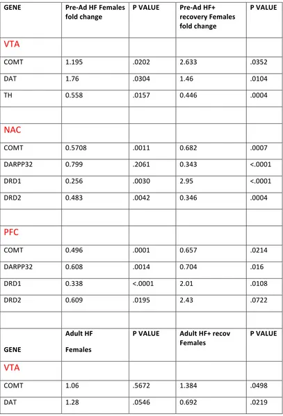
Table 1. Gene Expression Summary and Statistics in Males……….92
Table 2. Gene Expression Summary and Statistics in Females………94
Chapter 4
Table 1 Neuro-inflammatory gene expression in the prefrontal cortex………130
LIST OF ILLUSTRATIONS
Chapter 2
Figure 1. Sucrose preference but not saccharin preference is altered after high-fat diet (HFD) exposure and returns to control levels after HFD recovery in males and
females ………..60
Figure 2. Chronic high-fat diet (HFD) and recovery after HFD alters dopamine related gene expression in males and females. ……….61
Figure 3. Chronic high fat diet (HFD) reduced TH protein levels in the ventral tegmental area in both males and females. ………..62
Figure 4. Decrease in Dopamine levels in PFC and NAC after HFD from birth and mixed recovery after HFD removal. ……….63
Figure 5. Changes in DNA methylation status of DAT promoter parallel changes in gene expression in the VTA………64
Supplemental Figure 1. Body weights increase after 12 weeks HF diet and do not normalize after 4wk recovery period………65
Supplemental Figure 2. Corticosterone levels in males were not different after 12 weeks HF diet or after HF + recovery………...66
Chapter 3
Figure 1. 12 week exposure to high fat diet causes increase in body and fat pad weights and these increases are not normalized by a 4 week standard chow
replacement………..96
Figure 2. Sucrose preference is decreased after high-fat diet (HFD) exposure in Pre-Adolescent HF males and females and recovers after standard chow
replacement………..97
Figure 3. Chronic HFD causes decrease intake of palatable food during one-hour challenge. Standard chow replacement during experiment or 4 weeks caused an increase in palatable food intake………98
Figure 4. Chronic high-fat diet (HFD) alters dopamine related gene expression in males. Pre-Adolescent HF males were less able to normalize gene expression in examined reward regions of the brain………..99
Figure 6. Dopamine levels are increased after chronic HFD in Pre-Adolescent HF males. Dopamine levels in the NAc do not normalize after 4wk recovery………..101
Chapter 4
Figure 1. Two weeks of voluntary wheel running decreased dopamine receptor D1 and mu opioid receptor gene expression and altered DNA methylation at promoter
regions……….…..124
Figure 2. Animals with access to HF + exercise did not gain as much weight as sedentary animals on HF, but were still heavier than controls………..125
Figure 3. Animals given access to running wheel and/or high fat diet did not show alterations in anxiety behavior. However, both exercised and/or high fat diet access reduced natural reward intake………..126
Figure 4. Gene expression of dopamine receptors and DNA methyltransferase are increased by high fat diet and not reversed by exercise in reward regions of the
brain………127
Figure 5. Gene expression of neuro-inflammatory related genes were increased by high fat diet and normalized by exercise in the prefrontal cortex……….128
Figure 6. Neuro-inflammatory related genes were increased by high fat diet and further increased by the combination of high fat diet and exercise in the
CHAPTER 1: General Introduction
The current statistics on obesity in the U.S are staggering with 34.4% of adult
males and 36.1% of adult females considered obese in 2010 (Ogen et al., 2012). The
obesity trend in adolescents is just as dire; 23.9 million children ages 5-19 are
overweight or obese and the epidemic continues to grow (Go et al., 2013). Both genetic
and environmental factors contribute to the development of obesity. Population studies
have revealed specific gene mutations to cause monogenic forms of human obesity
(Farooqi et al., 2005), and monozygotic twin studies reveal adiposity levels to be strongly
heritable (Stunkard et al., 1986; Maes et al., 1997). However, those single gene
mutations are very rare and human genetics are very stable. Therefore, it is unlikely that
the current obesity epidemic is explained by mutations or gene x environment
interactions alone. The most drastic change for humans has occurred in the
environment. The broad availability of energy dense foods and the decrease in daily
activity has most Americans living in an environment that drives obesity and makes
weight loss extremely difficult. The obesogenic environment affects the individual as
early as the fetal stage. Over nutrition or poor nutrition during early life development can
put one on a trajectory toward obesity in adulthood. Body weight and eating patterns
early in life are extremely important in setting the risk for development of obesity and the
ability to lose weight. In fact, it has recently been shown that establishment of obesity at
age 5 predicts obesity at age 18 (Cunningham et al., 2014). Sex is another factor that
plays a role in an individual’s obesity risk. Women tend to have higher rates of obesity
compared to males (Go et al., 2013). Women have higher overall body fat content as
well as additional distribution to the breast, buttocks, and lower body (Martin et al.,
have different behaviors toward palatable foods that could put them at increased risk for
overconsumption. Women rate food images as more pleasant, they have more negative
feelings in the hunger state, and have a higher fMRI response to food cues compared to
males (Geliebter et al., 2013). In an effort to better treat and/or prevent obesity, an
understanding of how sex and early life experiences interact to affect obesity risk is
required.
Central Acting Interventions for Obesity
Approaches to the treatment of obesity include dietary intervention, exercising,
pharmacological treatments, and gastric bypass surgery. Success rates at weight loss
vary enormously between individuals and, in most cases, lost weight is gained back.
Although obesity is associated with peripheral effects, such as increased adiposity, high
blood pressure, and inflammation, food consumption is controlled centrally. In fact, many
of the single gene mutations known to cause obesity do so by failing to produce satiety.
The most studied Mendelian inherited obesity genes (leptin, leptin receptor,
pro-opiomelanocortin, melanocortin 3 receptor, melanocortin 4 receptor) lie within the central
leptin-melanocortin signaling pathway of the hypothalamus and result in human obesity
when loss of function occurs from mutation (Montague et al., 1997; Clement et al., 1998;
Farooqi et al., 2007; Krude et al., 1998; Yeo et al., 1998). Looking at obesity as a
central food intake issue is an important direction for the field and has led to the
development of centrally acting drugs to combat adiposity. Clinical drug trials for weight
loss have targeted the brain in order to attempt to decrease food intake. Most
medications prescribed for obesity regulate satiety through an effect on serotonergic,
noradrenergic or dopaminergic receptor systems (Bray et al., 2000, Clapham et al.,
and, thus decreased food-seeking behavior. Four centrally acting noradrenergic agents
(phentermine, diethylpropion, phendimetrazine, benzphetamine) are currently
FDA-approved for management of obesity (Yanovski et al., 2014). These agents reduce
appetite by activation of adrenergic and dopaminergic receptors. However, even in the
most successful trials, high rates of drop out and weight gain occur at follow-up time
points (Ioannides-Demos et al., 2012). There are still many obstacles, including
non-CNS effects that make it so difficult to lose weight and need to be overcome during
obesity intervention. Age of onset of obesity is one factor not taken into consideration
when analyzing clinical weight loss trial data. It is true that weight loss becomes more
challenging as one ages, and adults who were obese as children find it even more
difficult to lose weight (Cockrell & Skinner, 2008). Identifying the factors that make
childhood obesity more resistant to weight loss may lead to changes in
pharmacotherapy for this population in the future.
The Obese Brain
Obesity is associated with excessive overeating and preference for palatable, high-fat
foods even when body energy stores are high (Sclafani, 2001; Gaillard et al.,
2008). There seems to be a mismatch between the caloric needs of the body and what
the brain tells the body to consume. Since the brain controls food intake, much research
has been done to see how the obese brain has changed in comparison to its lean
counterpart. Imaging studies in obese patients have given us insight into the regions
possibly involved in the loss of homeostatic food consumption. Combined evidence
points to regions involved mainly in reward processing, taste, and attention that are
altered in obese patients. Imaging studies in obese patients have detected increased
of highly palatable foods (Rothemond et al., 2007; Scheinle et al., 2009, Sharmueller et
al., 2012). Imaging studies have also revealed diminished functional connectivity
between these regions important for reward evaluation (Stoeckel et al., 2009; Kulleman
et al., 2011). These changes are thought to lead to deficiencies in evaluating food
reward value and contribute to the drive to overeat. In fact, behavioral studies have
shown obese individuals to direct their attention to food related stimuli more so than lean
individuals (Nijs et al., 2010). This shift in attention and preference may also increase the
vulnerability to further over consume food. The shift in attention toward a learned reward
is not specific to obesity in general, but has also been seen to occur in addicted
individuals and drug cues. Obese patients seem to have an increased central response
to food cues, but how does the brain react during actual food consumption?
Interestingly, studies have revealed a decrease in the hedonic response to food intake.
Over weight individuals have reduced activation of the striatum in response to ingesting
a palatable solution (Stice et al., 2010). Although obese individuals seem to react
strongly to food cues, the hedonic response to intake is diminished. Hedonic drive is
mediated by the dopamine reward system and is acutely activated by rewards like
palatable foods, sex, and drugs of abuse. A decrease in dopamine receptors have been
reported in the striatum of obese patients and this has implications for appetite control as
well as other reward behaviors (Wang et al., 2001, Steele et al., 2010). Some, but not
all, differences in the obese brain reverse after individuals lose weight. Imaging studies
have also looked at the post-obese population and have shown insula activation remains
elevated after weight loss (DelParigi et al., 2004). On the other hand, experiments have
also shown normalization of dopamine receptors after bariatric surgery (Steele et al.,
2010). The neurobiological changes that persist after intervention may contribute to the
some kinds obesity interventions can reverse or prevent the brain changes seen in
obesity better than other types on intervention.
The previous studies support the theory of reward dysfunction in obesity. Obese
individuals seem to be more sensitive to food cues; however, reward activation is
diminished during food intake. This would suggest that there is an increased drive to
obtain food. However, because patients are less sensitive to the rewarding effects of
food, they would therefore consume more in an effort to obtain sufficient reward levels.
This has been demonstrated in progressive ratio studies where obese individuals give a
higher reinforcing value to food (Epstein et al., 2007; Temple et al., 2008). Future studies
are warranted to see if a higher reinforcing value plus diminished reward activation are
behind the overconsumption of palatable foods in obese individuals and if there are sub
populations of obese patients that may benefit from targeting overconsumption
behaviors.
Obesity Interventions
Successful weight loss is extremely difficult for the obese population and is
influenced by many factors, such as age of onset and sex. Caloric restriction or “dieting”
is very effective for weight loss, but has proven to be very difficult to adhere to in the
general population. One reason is caloric restriction induces biological changes that
make adherence difficult. The over abundance of highly rewarding, highly palatable
foods makes relapse even easier. Dieting produces changes in appetite, notably an
increase in hunger (Pasman et al., 1997) and persistent thoughts about eating (Green et
al., 2005). Other approaches to the treatment of obesity include, exercise,
pharmacological treatments, and gastric bypass surgery. Pharmacological interventions
Pharmaceutical trials focus exclusively on reduction of body mass and normalization of
markers of obesity and do not usually assess the state of mind or drive to eat. Gastric
bypass surgery is a last line of defense treatment for extreme obesity and is very
successful in long-term body weight reduction. Adaptive responses that increase
appetite after weight loss do not seem to occur after weight loss induced by bariatric
surgeries as they do with typical dieting interventions (Berthoud et al., 2012). Bariatric
surgery patients have a diminished desire to eat (Shultes et al., 2010) and exhibit
normalization of dopamine receptor D2 (DR2) levels after surgery (Steele et al., 2010;
Dunn et al., 2010). However, dieting is still the most common way to attempt weight loss
in the obese population. How the dopamine reward system changes after high fat
replacement needs to be studied in order to better understand the behavioral changes
that either promote or hinder adherence to a diet. Characterizing how dopamine circuitry
changes when high fat diet is switched to a low fat diet may help us discover why dieting
is so difficult for obese individuals. Additionally, characterizing important factors, such as
age and sex that impact the response to high fat withdrawal will be important for
analyzing clinical weight loss trial results in the future.
Many factors, such as stress, can make caloric restriction difficult and cause
return to high fat food consumption. High-fat diet removal elicits palatable food cravings
and exposure to foods cues and/or stressful experiences can cause relapse (Nair et al.,
2009). Many kinds of stress, such as physical, psychosocial, and pharmacological
stressors can stimulate palatable food seeking and return to unhealthy eating habits
(Grilo et al., 1989; Ghitza et al., 2006; Nair et al., 2011). This is comparable to what is
seen in drug addiction. After extinction of the drug reward, different types of stressors
can reinstate drug seeking (Hyman et al., 2006). Many researchers have noted
documented that removal of a high fat/high sugar diet causes withdrawal symptoms
similar to opiate withdrawal (Avena et al., 2008a). The mechanisms behind
stress-induced reinstatement of palatable food seeking are still being examined. Nair et al.,
(2011) have discovered that blocking dopamine receptors in the prefrontal cortex can
inhibit stress-induced reinstatement of food seeking. Ghitza et al. (2006) discovered a
similar role for CFR1 receptors in the amygdala. By blocking activation of CRF1, food
seeking is not reinstated by a pharmacological stressor. Food cues also have the ability,
like stress, to trigger relapse to palatable food seeking. The reinstatement by food cues
is likely to be mediated by additional mechanisms and brain regions than reinstatement
by stressors. Priming with drugs that increase dopamine, such as cocaine or
amphetamine works to reinstate palatable food seeking as well as priming with food or a
cue that predicts palatable food (Ghitza et al., 2007; Keiflin et al., 2008; Odum &
Shahan, 2004). One factor to consider when interpreting these results is many of these
studies were performed in lean rodent models. There are many neurobiological changes
that occur after diet induced obesity and research on palatable food reinstatement after
acute high fat withdrawal or long term high fat withdrawal need to be examined.
We have seen that stressors can be a trigger to cause increased intake of
palatable foods. However, can removal of the palatable food be a stressor in itself?
Little is known about the consumption of palatable foods following withdrawal from
chronic HFD and the reward-relevant neurobehavioral changes that may occur. We
know that food restriction in lean animals causes stress and increases administration of
drugs of abuse like cocaine and amphetamine (Carroll and Meisch, 1980; Stuber et al.,
2002). Other studies have confirmed a strong relationship with food restriction and the
increase in the rewarding properties of drugs such as amphetamine (Takahashi et al.,
that food restriction in lean animals is similar to withdrawal of a high fat diet in obese
animals, given what is known about the differences in reward relevant circuitry in the
brains of lean and obese animals. High fat diet withdrawal has also been shown to
increase responding for sucrose in obesity prone rats (Pickering et al., 2009). This
suggests that the propensity to relapse may be stronger in some individuals than others.
Further, we know from Zorilla lab and colleagues (Cottone et al., 2008a; 2008b; 2009)
that intermittent access to a high fat diet causes anxiety behavior and increases
responding to palatable food reward in rodents. Others have found that an acute
withdrawal (only 24 hours) causes anxiety behavior in an open field test (Teegarden et
al., 2007). Withdrawal from a high fat diet can increase basal corticosterone levels
(Sharma et al., 2013) but has no effect on stress induced cort levels (Pickering et al.,
2009) and changes in CRF levels in the amygdala (Teegarden et al., 2007). Removal of
other rewarding substances, such as sucrose, can also be a stressor and cause an
increase in anxiety-like behaviors (Avena et al., 2008b). Body weight loss goes hand in
hand with high fat diet removal. It is possible that body weight loss can cause an
activation of the body’s stress system as well (Bailey et al., 2004). Looking at a time
point where high adiposity is still present but the diet has been withdrawn can be a
helpful way to control for the stress of high body weight loss. Although changes in CRF1
levels were not seen in the amygdala after high fat withdrawal in a diet induced obese
model (Sharma et al., 2013), it is still possible that activation of stress circuitry after high
fat diet removal can contribute to reward behavior observed after high fat diet removal.
The hypothalamic and mesolimbic dopamine systems participate in the
integration and control of food intake. Dopamine acting in the hypothalamus is important
for basal food intake and influences feeding frequency and volume (Meguid, et al.,
2000). For example, dopamine is released in the lateral hypothalamic area in response
to feeding and normalizes after meal termination (Yang et al., 1996). Dopamine release
in the hypothalamus leads to activation of neuropeptides involved in both food intake
stimulation (e.g. melanin concentrating hormone (MCH), neuropeptide Y (NPY), Agouti
related peptide (AgRP), galanin, and orexin) and food intake inhibition (e.g.,
a-melanocyte-stimulating hormone (MSH), cocaine and amphetamine-regulated transcript
(CART), and corticotrophin-releasing factor (CRF)) depending on the complex metabolic
conditions occurring in the body. The basic circuitry of the hypothalamus and brain stem
is under multiple points of regulation and has the ability to sense macronutrients,
hormones, gut signals, and energy stores to control energy metabolism and food intake
behavior in order to maintain homeostasis. There are two main sources of circulating
nutrients: food intake, and production of glucose and lipids by the liver. Increased
availability of macronutrients such as glucose and lipids activates sensing pathways in
the brain, either directly through metabolic signals (e.g., fatty acids), or indirectly,
through stimulation of insulin and leptin biosynthesis and secretion. Leptin is
synthesized by white adipose tissue, and its level increases in proportion to fat mass.
Among its many actions, high levels of leptin potently suppress food intake and stimulate
metabolic processes to dissipate excessive energy stores. The activation of brain
efferent pathways in turn suppresses food intake and hepatic output of glucose and
lipids. (Schwartz et al., 1996; Obici et al., 2002; Loftus et al. 2000; and reviewed in Lam
et al., 2005). In obesity, however, this feeding circuit is altered and palatable foods are
overconsumption of palatable foods and obesity can eliminate the ability of
intracerebroventricular (icv) administration of insulin to reduce food intake (Posey et al.,
2009). The hypothalamus also becomes insensitive to leptin signals and there is a
decrease in AGRP signaling to the mesolimbic pathway (Varela & Horvath, 2012). How
the high fat diet impairs the negative feedback system is unclear. One hypothesis is that
high fat diet causes neuroinflammation in the hypothalamus that impairs insulin
sensitivity and the signal to stop eating (DeSouza et al., 2005). Palatable foods that drive
obesity are often consumed after energy needs are met. It is thought that other brain
regions, such as the mesolimbic dopamine area, control the intake of energy dense
foods and this is one area pharmacotherapy can target in obese patients in the future.
The Dopamine Reward Pathway
The modern environment is characterized by the availability of highly palatable,
energy dense foods as well as powerful food cues. High fat and high sugar foods
activate the mesolimbic dopamine system to drive food intake even after energy
requirements have already been met (Berthoud et al., 2007; Stratford & Kelly, 1999).
Dopamine neurons in the mesolimbic pathway originate in the ventral tegmental area
and have been implicated in the reinforcing and motivational aspects of food intake
(Ikemoto & Panksep, 1996; Brennan et al., 2001; Ishiwari et al., 2004). Dopamine
neurons are found in the VTA and project to the NAC to form the classic reward pathway
studied in drug addiction. The dopamine neurons in the VTA also innervate several
regions of the prefrontal cortex (PFC), central amygdala, basolateral amygdala (BLA)
and hippocampus. All the reward related regions are inter-connected and synapse back
on each other in complex ways. The VTA–NAc circuit is crucial for the recognition and
tyrosine, through the conversion of l-tyrosine to l-dihydroxyphenylalanine (l-DOPA) by
the rate-limiting enzyme tyrosine hydroxylase (TH). DOPA is subsequently converted to
DA by the enzyme l-aromatic amino acid decarboxylase. DA is then transported from the
cytoplasm to storage vesicles via the synaptic vesicular monoamine transporter (VMAT).
After DA is released into the synapse, the plasma membrane DA transporter (DAT) can
transports DA in and out of the terminal depending the concentration gradient and other
factors (Amara and Kuhar, 1993). The conversion of DA to HVA occurs extra-neuronally
by the enzyme catechol-O-methyltransferase (COMT) (Feldman et al., 1997).
Receptors that translate dopamine release into a signal are found in projecting
regions NAc, PFC, amygdala, and other regions. DA receptors are classified into two
subfamilies, D1- and D2-like receptors (Brown and Makman, 1972; Kebabian and Calne,
1979), based on their pharmacological and functional properties. The D1-like receptors
are dopamine receptor D1 (DR1) and D5 (DR5) (Sunahara et al., 1990 and Sunahara et
al., 1991), whereas the D2-like receptors are dopamine receptor D2 (DR2), D3 (DR3),
and D4 (DR4) (Sokoloff et al., 1990) based on their respective gene sequences.
Dopamine receptors D1 and D5 are coupled to adenylyl cyclase through G protein Gs,
while dopamine D2, D3, and D4 receptors inhibit adenylyl cyclase by coupling to Gi/o.
The D1-like and D2-like receptors are both present in all dopamine-containing regions of
the rodent brain (Meador-Woodruff, 1994). Higher levels of DR2 mRNA are detected in
the substantia nigra and VTA. DR3 display a much more restricted pattern of distribution
and are mainly found in limbic regions (Levesque et al., 1992). D1R knock out (KO)
mice show normal appearance and no obvious neurological defects, but exhibit growth
retardation and low survival after weaning (Drago et al., 1994). Midbrain levels of
dopamine are elevated in D1R KO mice, suggesting a compensatory increase in
al., 1998). D2R KO mice exhibit significantly lower levels of locomotor activity than
controls (Palmer et al., 2003) but total dopamine levels are not altered in D2R KO mice,
suggesting D2R does not exert strong tonic control of dopamine activity (Benoit-Marand
et al., 2001). On the post-synaptic side, the dopamine signal is transduced after
activation of DR1 by the actions of Protein phosphatase 1 regulatory subunit 1B
(DARPP-32). DARPP-32 is phosphorylated by cAMP-dependent protein kinase (PKA)
and cyclin-dependent kinase 5 (Cdk5) on threonine residues, thr34 and thr75, resulting
in inhibition of protein phosphatase-1. Additionally, DARPP32 dephosphorylation at
serine residue 97 has also been observed and this modification appears to be important
in both addiction and food intake behaviors (Stipanovich et al., 2008). Dopamine
signaling elicited by drugs of abuse or palatable foods also increases the activity of
ΔFOSB, cyclic AMP-responsive element binding protein (CREB), and cyclin-dependent
kinase 5 (CDK5) pathways in medium spiny neurons (Joressen et al., 2007; Carlezon et
al., 1998; Bibb et al., 2001). These proteins play a prominent role in the synaptic
plasticity seen after chronic drug intake and could possibly regulate plasticity after other
rewarding substances, such as palatable food.
Hedonic Eating
Energy dense foods are widely available and there are many aspects of modern
society tempting us to eat. Wanting’ and ‘liking’ reactions are generated by neural
systems after the intake of sweet, palatable foods. Drive to eat palatable foods is still
present even when the individual is sated. For example, in humans, pleasure ratings for
chocolate never hit zero even after consumption of two whole bars (Lemmons et al.,
2009; Small et al, 2001). The “always room for dessert” phenomenon is also seen in
“sweetness” never completely diminish (Berridge et al., 1991). This suggests the ability
of certain foods to override homeostatic stop signals and drive over consumption. The
“wanting” and “liking” of palatable foods is regulated by dopamine and opioid
neurotransmitters in the reward regions. Palatable food activates “hedonic hot spots” in
the reward system, specifically the nucleus accumbens, ventral pallidum, amygdala,
obitofrontal cortex, anterior cingulate cortex and the anterior insula cortex (de Araujo et
al., 2003; Aldridge & Berridge, 2010; Kringelbach et al., 2004; Small et al., 2003).
Consumption of palatable foods increases dopamine concentrations in the NAc
(Bassarero & DiChiara, 1999). Dopamine in this region is thought to coordinate the
action of food intake, increase arousal, and conditioned learning in order to obtain the
food reward. Increases in dopamine, as seen in dopamine transporter (DAT) knockout
mice, produce elevated “wanting” for sweet foods (Pencina et al., 2003). Palatable food
is a powerful motivating force. Rats prefer palatable solutions to cocaine infusions
(Lenior et al., 2007), and will voluntarily expose themselves to a noxious environment or
pain to obtain high fat food over standard chow (Cabanac and Johnson, 1983; Foo and
Mason, 2005). In fact, DA is necessary for the motivation to consume food (Ungerstedt ,
1971; Zhou & Palmiter, 1995) and DA-deficient mice from tyrosine hydroxylase gene
knockout are hypophagic and eventually die of starvation unless DA is restored to the
striatum (Szczypka et al., 2001). Dopamine also controls food choice. Dopamine
agonists have been shown to enhance preference for and the motivation to obtain fatty
foods in rodents (Thanos et al., 2011, Cooper et al., 2006). These taken together show
that palatable foods activate the dopamine reward system acutely and cause
overconsumption of calories. How exactly palatable food elicits the release of dopamine
is currently unclear. It has been demonstrated, however, that the reward effect is
Further investigation is needed to see what role post-ingestive factors, such as fat
sensing in the gut, have in reward signaling in the brain.
Interestingly, mutant mice lacking dopamine are still able register the hedonic
impact and show preferences, and learning to work for a palatable sweet reward. This
suggests other neurotransmitters have a role in maintaining hedonic intake of food. In
fact, a number of investigators found opioid or cannabinoid receptor activation in the
nucleus accumbens to stimulate appetite by enhancing the ‘liking’ for the perceived
palatability of food (Bodnar et al., 2005; Cooper, 2004; Kelly et al., 2002; Zhang &
Kelley, 2000). Opioids in the nucleus accumbens shell are involved with the “wanting”
aspect of palatable food intake through the modulating effect they have on dopamine
release. Activation of mu opioid receptors indirectly dis-inhibit dopamine neurons and
thereby increase dopamine release. For example, DAMGO, a mu-opioid receptor
agonist, more than doubled the amount of food intake when administered centrally to
rats (Pencina & Berridge, 2005). Moreover, anandamide, an endocannabinoid that likely
acts in the brain by stimulating the CB1 type of cannabinoid receptor, has been shown to
act in the nucleus accumbens to magnify the impact of sucrose taste (Mahler et al.,
2007). There has also been evidence for additional hedonic neurotransmitters acting in
the nucleus accumbens to enhance “liking” of palatable foods. The ventral
pallidum/nucleus accumbens region receives orexin inputs from the hypothalamus
(Nixon & Smale, 2007) and orexin in this region can enhance ‘liking’ for sweet rewards
(Harris et al., 2005). While more studies are needed, it is possible that dopamine
dysfunction in the reward system seen in obesity can allow these other
neurotransmitters to have a larger role and promote over consumption of palatable
Neuroadaptations in DA system after Chronic HFD
Research in both human and animals suggests that long-term intake of palatable
foods leads to neuroadaptations in the dopamine reward system that could have
implications for obesity treatments and other reward behaviors. Several lines of evidence
support the hypothesis of dysregulated dopamine function in obesity. Chronic
consumption of a high fat diet is associated with a state of reward hypofunction and
imaging studies reveal blunted activation of brain reward regions during consumption of
palatable foods in obese individuals (Stice et al., 2008). Other human studies have
shown a down regulation of dopamine D2 receptor in obese individuals (Wang et al.,
2001). This is consistent with obesity being overrepresented in Taq1AA1 allele carriers
that affects dopamine D2 receptor (DR2) expression (Blum et al., 1996, Nobel et al.,
1991). Down regulation of DR2 could potentially be involved in the increased craving
and increased reward seeking that is associated with the obese population (Blum et al.,
2008; Downs et al., 2009). Down regulation of DR2 has previously been associated with
many other disorders that are involved with dysregulation of motivation such as
addiction, and attention deficit hyperactivity disorder (Volkow et al., 2011a; Volkow et al.,
2011b).
Obesity prone rats have shown increases in extracellular dopamine in the
striatum (Narayanaswami et al., 2005). However, when animals become obese we see a
decrease in dopaminergic tone. Animal models of diet-induced obesity (DIO) exhibit
lower basal extracellular dopamine levels in the nucleus accumbens (NAc) and the
ventral tegmental area (VTA) (Geiger et al., 2007; Geiger et al., 2009; Cone et al., 2010;
Rada et al., 2010). Diet induced obesity models have lower dopamine turnover (Davis et
(Speed et al., 2011) compared to standard chow controls in the nucleus accumbens.
Genes involved in dopamine uptake and metabolism in addition to dopamine receptors
DR1 and DR2 have been reported to decrease expression in obese rodent models (Alsio
et al., 2010, Huang et al., 2005). Protein levels of both DAT and DR2 protein levels are
also diminished after high fat diet intake (Huang et al., 2006; South & Huang, 2008;
Johnson & Kenny, 2010). Decreases in sucrose preference (Vucetic et al., 2012, Carlin
et al., 2013), a decrease in response for a sucrose pellet (Davis et al., 2008) and a
decrease response to food reward (Corwin et al., 2011; Cottone et al., 2008; Johnson
and Kenny, 2010) are associated with the dopaminergic protein changes after high fat
intake. Changes in DR2 expression may impact the animal’s willingness to work for a
goal (Trifilieff et al., 2013) and an animal’s sensitivity to dopamine agonists. For
example, rats eating high fat chow are more sensitive to quinpirole-induced yawning (D2
and D3 receptor-mediated; Baladi and France, 2010).
Neuroadaptations in Leptin, Insulin, Ghrelin in the VTA
The previous body of literature supports altered dopamine function in obesity and
suggests a decreased sensitivity to reward in the obese population. Whether the high fat
diet is directly affecting dopamine metabolism and release at the molecular level or other
adjacent neurotransmitter systems are indirectly changing VTA activity is currently
unclear. It is possible that hormones involved with energy homeostasis may play a role
in natural reward intake and changes in signaling may disrupt dopamine and lead to
hedonic overeating. It is known that insulin, leptin, and ghrelin can decrease food reward
behaviors and modulate the function of neurotransmitter systems that mediate food
reward. Insulin receptors are expressed in brain regions that are rich in DA neurons and
(Figlewicz et al., 2003). This suggests an interaction between energy sensing and DA
systems occurring in the reward regions. Manipulations of insulin concentrations
significantly affect DA synthesis, turnover, and signaling in reward regions (Kwok and
Juorio, 1986; Lim et al., 1994; Lozovsky et al., 1981; Saller, 1984). For example, acute
injection of insulin into the brain enhances expression and activity of dopamine
transporter (DAT) (Carvelli et al., 2002; Figlewicz et al., 1994). High fat diet consumption
causes both peripheral and central insulin resistance over time. Because insulin
regulates DA systems, it is likely that decreases in insulin signaling could contribute to
the changes seen in dopamine circuitry and reward behaviors in obesity (Daws et al.,
2011; Niswender et al., 2011). Leptin levels are associated with total adipose fat stores
and may directly or indirectly regulate dopamine dysfunction in obesity. Leptin receptors
are also found in the VTA and are downregulated in DIO animals (Figlewicz et al., 2003;
Fulton et al., 2006; Blendy et al., 2005). Leptin has been shown to modulate response
for rewarding brain stimulation, and modulate conditioning for high fat food (Fulton et al.,
2000 and Fulton et al., 2004). Leptin knockout mice (ob/ob mice) have a significant
impairment of DA release and tyrosine hydroxylase capacity in the NAc (Fulton et al.,
2006).
Ghrelin is a gut-derived hormone that is associated with food-deprivation and
ghrelin receptors are also distributed in the ventral tegmental area and co-express with
tyrosine hydroxylase (Mitchell et al., 2001). Ghrelin is involved in appetitive actions and
the food seeking response (Naleid et al., 2005) possibly through the action of increasing
VTA action potentials and the release of dopamine in the nucleus accumbens (Abizaid et
al., 2006; Abizaid, 2009). It is thought that ghrelin signaling is altered in obese
individuals and affects dopamine dependent behaviors such as novelty seeking (Savage
downstream signaling mechanism in the VTA for these energy related hormones is
PI3Kinase. Studies have identified that insulin and leptin increase PI3kinase activity in
the VTA (Figlewicz et al., 2007) and increase JAK-STAT phosphorylation, which is
necessary for the decrease in feeding response (Morton et al., 2009). One potential
cellular target for PI3Kinase is the DAT. It has been shown that insulin regulates the
expression and activity of DAT through PI3Kinase (Garcia et al., 2005; Vaughan et al.,
1997) and diabetic models lacking insulin show DAT deficits (Figlewicz et al., 1994;
Sevak et al., 2008). This implies that insulin resistance in obesity may alter DAT
expression and activity and therefore alter DA signaling and reward seeking behavior.
Energy related hormones are perfectly poised to regulate reward behavior and palatable
food intake. If overweight individuals are less sensitive to homeostatic signals and
therefore less sensitive to natural rewards they may consume more in an effort to obtain
a larger positive effect. This is similar to the phenomenon of “tolerance” in addicted
individuals. Moreover, altered sensitivity to natural rewards may also mean altered
sensitivity to drugs of abuse that activate the same reward pathways.
The Neurobiological Overlaps of Addiction and Obesity Pathways
Dopamine is the key neurotransmitter involved in drug abuse as well as food
intake. It is important to understand factors, such as obesity, that might alter sensitivity to
the behavioral effects of drugs acting directly or indirectly at DA receptors. The repeated
stimulation of dopamine reward pathways leads to adaptations in neurotransmitters and
reward circuitry that may lead to increases in compulsive behaviors affecting both food
and drug intake (Volkow & Li, 2004). Human studies reveal that obese individuals
display decreased propensity to use recreational drugs and a decreased prevalence of
Additionally, obese individuals have been shown to self-administer less nicotine and
demonstrated diminished hedonic drive toward nicotine cigarettes than their lean
counterparts (Blendy et al., 2005). In one demographic study being “obese” or extremely
obese” has been associated with an increase risk of alcohol abuse disorder as well as
other mood and anxiety disorders (Petry et al., 2008). Research in both fields confirms
the mechanisms behind addiction and overconsumption of palatable food converge on
the same molecular targets in the reward system. One neuroadaptation found in both
obesity and addiction is the decreased expression of DR2. Positron emission
tomography has revealed decreased DR2 in the striatum of addicted individuals that
persists well after drug withdrawal (reviewed in Volkow et al., 2009). This finding has
also been replicated in preclinical rodent studies given chronic exposure to drugs of
abuse (Thanos et al., 2007; Nader et al., 2006; Volkow et al., 2001). Down regulation of
both DR2 and DR1 expressing neurons in the striatum that mediate the direct striatal
pathway enhances the sensitivity to drugs of abuse (Fergusen et al., 2010; Hideka et al.,
2010). Dysregulated dopamine signaling in this region is likely to contribute to the
compulsive and impulsive drug intake seen in addiction (Goldstein et al., 2002).
High impulsivity may underlie the inability of obese individuals to resist excessive
eating. Both addicted and obese individuals show impairment in PFC and OFC regions
of the brain that control reward and impulsivity (Volkow et al., 2001; Volkow et al., 1993;
Volkow et al., 2008). Moreover, decreased D2R striatal expression and signaling has
been detected among obese individuals (Geiger et al. 2009; Wang et al., 2001, de
Weijer et al. 2012) and in rodents given a high fat diet (Johnson & Kenny, 2010). Other
studies have shown overeating palatable foods can produce behavioral and
neurochemical signs that resemble dependence in laboratory animal models such as
2011 and Volkow et al., 2011c). When rewarding substances target the same
downstream signaling mechanisms they have the ability to cross sensitize or cause
tolerance towards one another. For example, eating high fat chow increases sensitivity
of drugs acting at both D3 and D2 receptors (Baladi et al., 201, Corwin et al., 2011).
High fat diet intake also leads to increased sensitivity to cocaine (Baladi et al., 2012;
Shumsky et al., 1997) and methamphetamine (McGuire et al., 2011).
Addictive drugs act on known receptors to increase dopamine release or
dopamine levels in the nucleus accumbens. It is not yet fully elucidated how palatable
foods increase dopamine in nucleus accumbens, but there are some downstream
molecular targets that are affected in both palatable food consumption and addiction that
could give us some hints. Dopamine signaling in the accumbens modulates the activity
of ΔFOSB, cyclic AMP-responsive element binding protein (CREB), DARPP32 and
CDK5 signaling pathways in medium spiny neurons, and influences the rewarding
properties of both food and addictive drugs (Kenny, 2011). One example of this is
ΔFOSB. ΔFOSB is increased in the striatum of rodents given a high fat or high sucrose
diet and is associated with the motivation to obtain palatable food (Christiansen et al.,
2008; Teegarden & Bale, 2007). Cocaine and other drugs of abuse increase the
expression of ΔFOSB throughout the striatum, more specifically in the dopamine
receptor D1 and dynorphin-expressing medium spiny neurons in the direct pathway
(Muller et al., 2005). The overlap of addiction and food intake pathways can also be
seen with peptides involved in homeostatic feeding and is reviewed extensively in
(Volkow et al., 2013). Orexigenic peptides ghrelin, orexin, melanocortin, and
neuropeptide Y are found to affect reward regions and modulate reward behavior such
as alcohol intake (Leggio et al., 2011; Gilpin et al., 2012), conditioned reward learning for
preference (Harris et al., 2007; Proudnikov et al., 2008 ). Anorexigenic peptides also act
in reward regions of the VTA, NAc, and amygdala to modulate reward behavior.
Glucagon-like peptide-1 (GLP-1), cholecystokinin (CCK), peptide YY (PYY), galanin,
cocaine-and amphetamine-regulated transcript (CART), corticotropin-releasing hormone
(CRH), and oxytocin have been shown to affect behaviors towards amphetamines
(Erreger et al., 2012; Vaccarino et al., 1994; Crawley et al., 1985; Beinfeld et al., 2003),
alcohol (Barson et al., 2011; Le Strat et al., 2012), and cocaine (Cippitelli et al., 2012;
Upadhya et al., 2012). The regulation of homeostatic food intake, palatable foods, and
other natural rewards all converge on the dopamine neurons in the ventral tegmental
area. The dopamine reward system is hijacked by unnatural rewards (drugs of abuse)
and neuroadaptations that occur in addiction to drugs of abuse are similar to those seen
in obesity. The combined results suggest that both obese and drug-addicted individuals
suffer from overlapping impairments in dopaminergic pathways that lead to
overconsumption and impulsivity.
Another neurotransmitter system that is involved in both palatable food intake
and reward is the central opioid system. Both neurotransmitter systems are activated in
concert and are dependent on each other. For example, opioids increases intake of
palatable foods in sated animals and this depends on the expression of dopamine
receptor D2 (Hayward et al., 2007). Further, opioid receptors are distributed throughout
regions important for food and drug reward (Olszewski, et al., 2007) and mu opioid
receptor activation increases dopamine release (Bontempi et al., 1997; Horner et al.,
2012; Steiner et al., 1999). Mu opioid receptor (MOR) expression is altered by high fat
diet at different time periods. Chronic high fat diet in adulthood decreases MOR
expression (Vucetic et al., 2011; Ong et al., 2013; Blendy et al., 2005). Conversely, high
(Vucetic et al., 2010; Ong et al., 2011; Grissom et al., 2013; Carlin et al., 2013b). It is
yet to be discovered how high fat diet regulated the expression of MOR, however,
studies out of our lab point to possible recruitment of epigenetic markers (Vucetic et al
2010; Grissom et al., 2013). It seems that changes in opioid signaling could play a role in
the reward dysfunction of obesity either by amplifying the effects of dopamine
dysregulation or altering the palatability of food. Endogenous opioids code the
palatability of food in the brain and neuroadaptations in this system could have
implications in overconsumption of food in obesity. If MOR is down regulated in obesity,
it could be a homeostatic mechanism to reduce the drive to eat and the dopamine
dysfunction just happens to over ride that effect. Conversely, down regulation of MOR
can add to the hypo reward seen in obesity which could further drive palatable food
intake in order to normalize reward levels in the brain. Further experiments are needed
to answer how endogenous opioids are altered alongside dopamine and if these
neuroadaptations reverse after standard chow replacement.
Sex Differences in the Dopamine Reward System and Obesity
Sex differences are found in both the central dopamine system and in the
presentation of obesity. The overlapping dopamine pathways seen in obesity and drug
addiction are also highly influenced by sex and sex hormones. We see that women are
more susceptible to developing eating disorders and obesity compared to men (Hoek et
al., 2003; Berghöfer et al., 2008; Keel et al., 2007). Women also have lower success
rates at dieting then men. This could be due to their more intense interaction with
palatable foods. Women tend to show increased brain activity in the prefrontal cortex
and OFC after ingestion of palatable liquids compared to men and this has been
al., 2002). This difference in inhibitory control could underlie the lower success rates
when losing weight with dieting (Appelhans et al., 2011). Studies have shown sex
hormones directly influence food intake, body weight, and fat distribution. Preclinical
studies in rodents have shown an opposite effect of what we see in humans. Male mice
are more vulnerable to HFD-induced weight gain (Martin et al., 2007) and this sex
difference can be attributed to the positive metabolic effects of estrogen in females
(Heine et al., 2000). Further studies are needed to see why this protective effect is
absent in the human population, which is clearly a more complicated issue than what is
observed in rodent. Sex hormones also have an effect in the brain and can modulate
central leptin signaling. Female rats are more sensitive to the anorexic effects of leptin
than male rats (Clegg et al., 2003). Given the gender differences in obesity and the high
rate of failure in weight loss therapeutics, examining overconsumption and dopamine
circuitry in females would be an extremely important contribution to the field.
Sex differences are also present in the incidence rates of psychiatric disorders.
For example, major depression, which involves the disruption in the dopamine reward
system, is twice as common in females then in males (Marcus et al., 2005). Women
also experience addiction to drugs of abuse differently than men. Female drug users
tend to be more sensitive to cocaine-conditioned stimuli and report more intense
cravings (Elman et al., 2001; Robbins et al., 1999). Sex differences persist when
examining the central reward system in rodents. Female rats develop cocaine CPP after
fewer conditioning sessions and with lower doses compared to males (Parylak et al.,
2008). This suggests that females are more sensitive to the rewarding effects of drugs
acting the dopamine system and this could be behind the sex differences seen in
addiction incidence rates (Zakharova et al., 2009). Estradiol has the ability to modulate
the nucleus accumbens (Becker, 1999) and in cell culture (Alyea et al., 2008). Female
gonadal steroids inhibit DA neurotransmitter turnover in prefrontal regions and ventral
tegmental area (Handa et al., 1997) and regulate the reuptake of DA in regions involved
in satiety and motivation depending on estrus cycle (Thompson et al., 1997).
Experiments looking at sex differences have added richness to the addiction field.
Studies in the field of neuroscience of obesity often do not include females in the
analysis. This is potentially excluding 50% of the population that will receive the
pharmacotherapies that come out of these studies. Examining how neuroadaptations to
chronic high fat diet and their reversibility are influenced by sex is an important step in
developing proper drug targets to combat obesity.
Consequences of Early Life Nutrition on Obesity and Brain Development
Age is another critical factor to consider when studying obesity and the brain.
There are important times during development in which the brain seems to be
particularly sensitive to the environment. Changes in the environment during critical time
periods can alter the trajectory of development and affect the health of the adult in the
future. We see this sensitive time period come into play when setting adulthood body
weight. Obese children are more likely to become obese adults (Serdula et al., 1993)
and obese children will find it more difficult to lose weight when they become adults
(Cockrell-Skinner et al., 2010). It seems this critical time period is set before age five in
humans. A recent study showed that obesity by age 5 could predict future obesity at age
18 (Cunningham et al., 2014). At first glance, childhood obesity is similar to adult
obesity in that it is caused by increase food intake, sedentary lifestyle, complex
et al., 2002). However, childhood obesity can be more detrimental to future health and
weight loss goals in the future.
Both genetic makeup and early-life environmental factors play a significant role
determining body weight and adiposity. The early life environment begins in utero and is
affected by maternal nutrition and health during pregnancy. It seems that the risk for
obesity can begin at conception and depend on the in utero nutritional and hormonal
environment. Both maternal overnutrition and maternal under nutrition has been
associated with obesity later in life (Whitiker & Dietz, 1998; Boney et al., 2005; Ravelli et
al., 1976). The in utero environment has the ability to program adulthood appetite,
neuroendocrine functioning, and metabolism. Modeling maternal obesity in animals has
yielded a wealth of data on altered genes and behaviors that can contribute to behaviors
in offspring. The brain is developing throughout gestation; the dopamine reward system
circuitry and its control on food intake are not spared from maternal programming.
Maternal high fat diet alters hypothalamic neurons that express appetite-regulating
neuropeptides, leading to hyperphagia (Plagemann et al., 1999; Muhlhausler et al.,
2006) and exacerbated preference for fatty foods in offspring (Vucetic et al., 2010; Bayol
et al., 2007). Naef and colleagues (2013a) have shown offspring exposed to high fat diet
in utero demonstrate reduced anticipatory response to a food reward. Additionally, these
offspring had increased responding for high fat food (Naef et al., 2011). It seems
maternal high-fat diet not only increases offspring’s risk for obesity, but it also leads to
alterations in dopaminergic circuitry. Our lab documented changes in DAT, dopamine
receptors DR1 and DR2, DARPP-32, and TH, and mu opioid receptor (MOR) mRNA in
reward regions in offspring from a high fat fed dam. These mRNA changes were
associated with alterations in DNA methylation at the promoter region of these genes
maternal diet during pregnancy with offspring behavior. These studies show that
gestation is a particularly sensitive time period in programming feeding behavior and
dopamine circuitry. The brain continues to develop after gestation in humans and even
more so in rodents. Another study done by Naef, et al (2010) targeted the last week of
gestation as well as the 3 week weaning period. Maternal high fat diet during the later
gestation period and weaning led to offspring with weaker amphetamine-induced
extracellular NAc DA levels, higher NAc DAT activity, and reduced D2 receptor mRNA in
the VTA. This study demonstrates that post-natal time periods are also sensitive to
nutritional insults. The early post-natal period is very important for brain development
and sensitive to many types of environmental insults, not just nutrition, which can be
associated with dysfunction in adulthood. Disruption of early life environment in humans
can lead to increased risk of depression risk (reviewed in Hein et al., 2010, Chapman et
al., 2008), inflammation (Danese et al., 2008), glucocorticoid resistance (Carpenter et
al., 2004), reduced cortical volume (van Harmelen et al., 2010) and reduced
hippocampal volume (Vythilingam et al., 2002) in the adult population. These studies
point to the existence of important sensitive time windows in development that can set
an individual on a trajectory to dysfunction in adulthood. How early life nutrition interacts
with the brain during these critical development periods and leads to adult reward
dysfunction and obesity needs to be further explored.
The brain continues to develop from postnatal day one through adolescence. If
the environment is particularly damaging during that period, programming of adulthood
diseases can occur. “Metabolic imprinting” is a term used to describe how alterations in
developing young can predispose individuals to obesity and its associated illnesses. We
already examined how gestation was critical for the development of feeding and reward