R E S E A R C H P A P E R
Multiple effects of curcumin on promoting expression of the exon
7-containing
SMN2
transcript
Dairong Feng1,2,3•Yi Cheng1,5•Yan Meng1,4•Liping Zou4•Shangzhi Huang1• Jiuyong Xie2,3
Received: 4 May 2015 / Accepted: 14 August 2015 / Published online: 19 September 2015
ÓSpringer-Verlag Berlin Heidelberg 2015
Abstract Survival of motor neuron 2(SMN2) is a modifier gene for spinal muscular atrophy (SMA), a neurodegener-ative disease caused by insufficient SMN protein mostly due toSMN1defect.SMN2is nearly identical toSMN1but unfortunately only able to produce a small amount of SMN protein due to exon 7 skipping. The exon 7-containing
SMN2transcript (SMN2_E7?) can be increased by a dietary compound, curcumin, but the involved molecular changes are not clear. Here we have found that in fibroblast cells of a SMA type II patient, curcumin enhanced the inclusion of
SMN2exon 7. Examination of the potential splicing factors showed that curcumin specifically increased the protein and transcript levels of SRSF1. The increased SRSF1 protein
was mainly nuclear and hyperphosphorylated. Interestingly, the curcumin effects on the SMN2 andSRSF1 transcripts were inhibited by a protein deacetylase inhibitor, tricho-statin A. Moreover, in support of its role in the SMN2
splicing, knocking down SRSF1 reduced the inclusion of
SMN2 exon 7. Thus, curcumin appears to have multiple effects on theSMN2 transcript and its splicing regulators, including the change of alternative splicing and transcript/ protein level as well as phosphorylation. Protein deacety-lases and phosphatases are likely involved in these effects. Interestingly, the effects all seem to favor production of the
SMN2_E7?transcript in SMA patient cells.
Keywords CurcuminSMN2SplicingSRSF1 Phosphorylation
Introduction
Spinal muscular atrophy (SMA) is an autosomal recessive neuromuscular disorder with an estimated incidence of 1 in 6000 live births, representing the primary genetic cause of infant mortality (Monani2005; Burghes and Beattie2009). The disease is characterized by degeneration of a-motor neurons in the anterior horn of the spinal cord and by consequent skeletal muscle atrophy (Monani 2005; Burghes and Beattie 2009). More than 96 % of SMA patients have insufficient amount of the survival of motor neuron (SMN) protein due to the homozygous deletion of theSMN1gene (Coovert et al.1997; Lefebvre et al.1995). Interestingly, a paralogous human geneSMN2encodes the same but only a small amount of the SMN protein (Le-febvre et al.1995; Kashima et al.2007), due to its various extents of exon 7 skipping in different cells/tissues (Burnett et al. 2009; Lorson et al. 1998). Of the many SMN-Electronic supplementary material The online version of this
article (doi:10.1007/s12263-015-0486-y) contains supplementary material, which is available to authorized users.
& Shangzhi Huang
& Jiuyong Xie [email protected]
1 Department of Medical Genetics, Institute of Basic Medical Sciences, Chinese Academy of Medical Sciences and Peking Union Medical College, Beijing 100005, China
2 Department of Physiology and Pathophysiology, College of
Medicine, Faculty of Health Sciences, University of Manitoba, Winnipeg, MB R3E 0J9, Canada
3 Department of Biochemistry and Medical Genetics, College of Medicine, Faculty of Health Sciences, University of Manitoba, Winnipeg, MB R3E 0J9, Canada
4 Department of Pediatrics, Chinese PLA General Hospital,
Beijing 100853, China
5 Present Address: Department of Diagnostic Ultrasound, Beijing Anzhen Hospital, Capital Medical University, Beijing 100029, China
deficient tissues (Zhang et al.2008), spinal cord is the most affected in terms of function and survival (Burghes and Beattie2009; Chen et al. 2008).
A C6-to-T transition in exon 7 of the SMN2 gene is shown to contribute to the skipping, by disrupting the binding of a splicing activator SRSF1 (serine-/arginine-rich splicing factor 1) (Cartegni and Krainer2002; Cartegni et al.
2006) and (or) promoting the binding of a splicing repressor hnRNP A1 (heterogeneous nuclear ribonucleoprotein A1) (Kashima et al. 2007; Kashima and Manley 2003), in splicing reporter assays. Another splicing repressor of the
SMN2exon 7 is SAM68 (Src-associated substrate in Mitosis of 68 kDa) (Pedrotti et al. 2010). There are likely more factors involved in the regulation, as suggested by the effects of anti-sense oligonucleotides targeting other regions of the
SMN2pre-mRNA (Singh et al.2006; Hua et al.2010). Though the expression of SMN2 is not sufficient to compensate for the homozygous loss ofSMN1 (Lefebvre et al.1995), multiple copies ofSMN2 increase SMN pro-tein level and inversely correlate with disease severity in SMA patients and transgenic mice (Hsieh-Li et al. 2000; Monani et al. 2000; Wirth et al. 2006; Swoboda et al.
2005). Thus, as a modifier gene for SMA, the alternative splicing ofSMN2 exon 7 provides a promising target for SMA therapy (Hua et al.2010).
Curcumin is a dietary polyphenol compound enriched in the turmeric root. It has been used in clinical trials of numerous human diseases (Gupta et al. 2013; Darvesh et al. 2012), likely involving its regulation of multiple targets including histone acetyl-transferase (Shishodia
2013; Shishodia et al. 2007). It has also been reported to increase the SMN2_E7? transcript and SMN protein in fibroblast cells from a patient with SMA type I (Sakla and Lorson2008); however, the underlying molecular changes have been unclear. For the treatment of human diseases (Gupta et al.2012), it is necessary to identify its potential targets and effects.
In this study, we report that curcumin increases expression of theSMN2_E7?transcript and SMN protein in fibroblast cells from a SMA patient with multiple effects: enhancing
SMN2exon 7 inclusion, increasing transcript/protein level, and phosphorylation of the splicing activator SRSF1. These effects likely involve deacetylases and phosphatases.
Results
Curcumin increases the SMN protein and the proportion ofSMN2_E71transcript in fibroblast cells form a patient with SMA type II
To investigate potential curcumin effect on the expression of SMN protein and the usage of SMN2 exon 7, a SMA
dermal fibroblast cell line (BJ301J) was established by using the skin biopsy of a patient (SMA type II, 6-month, male), who is deficient in both copies of theSMN1gene but contains three copies of SMN2 (Figs. S1 and S2). In our initial tests, 15–25lM of curcumin increased the
SMN2_E7? transcript, but only 25lM was sufficient to upregulate the SMN protein level. We thus used 25lM of curcumin on these cells in the following experiments.
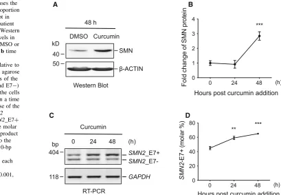
At 25lM, curcumin efficiently increased the SMN pro-tein level by 1.8-folds after 48 h of treatment in the BJ301J cells (Fig.1a, b, p\0.001). Consistent with the protein upregulation, the percentage of the SMN2_E7? transcript was significantly increased from 45.0 % (±2.2 %,n=3, calculated at molar level, same as in the following) at 0 h to 59.1 % (±2.3 %,n=3,p\0.01) at 24 h, and further to 65.1 % (±0.9 %, n =3, p\0.001) at 48 h (Fig.1c, d) upon curcumin treatment.
Curcumin-induced increase in the SMN2_E71
variant is inhibited by trichostatin A, a deacetylase inhibitor
Curcumin inhibits the histone acetyl-transferase (HAT) and phosphatases 2A and 5 (Balasubramanyam et al.2004; Han et al.2012). To examine whether protein deacetylation or phosphorylation is involved in the increase inSMN2_E7? by curcumin, the BJ301J cells were pretreated for 2 h with trichostatin A (TSA, 1 lM), an inhibitor of deacetylase of histone and non-histone proteins (Dokmanovic et al.2007; Glozak and Seto 2007), or okadaic acid (OA, 10 nM), an inhibitor of the protein Ser/Thr phosphatases PP1 and PP2A (Bialojan and Takai1988), respectively, followed by curcumin treatment for 24 h (Fig.2a). TSA but not OA pretreatment prevented the increase in the percentage of the
SMN2_E7? transcripts by curcumin in RT-PCR analysis (lane 3 vs. lane 2). Thus, curcumin-induced increase in the
SMN2_E7?variant is specifically inhibited by the protein deacetylase inhibitor TSA, suggesting that a deacetylation step is required for the regulation.
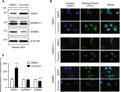
SRSF1 is specifically increased at both protein and transcript levels by curcumin
reduced by 19.8 % (±1.2 %, n=3, p\0.001), respec-tively (Fig.3c). HnRNP A1 did not change significantly in either protein level or transcript level (Fig.3). Thus, SRSF1 and SAM68 are specifically regulated by curcumin at both protein and transcript levels.
Curcumin increases the hyperphosphorylated isoform of SRSF1 in the nucleus
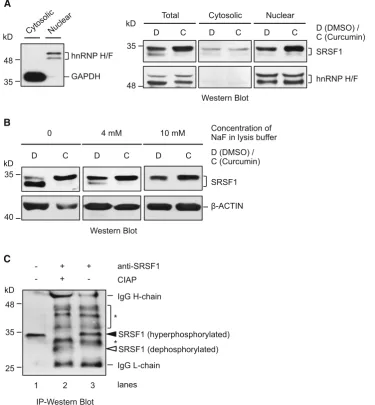
To further characterize the changes of the splicing factors regulated by curcumin, we examined the phosphorylation status of nuclear SRSF1, which is required for SRSF1 nuclear/speckle localization and recruitment to nascent transcripts (Ngo et al. 2005). For this purpose, we used cleanly separated cytoplasmic and nuclear proteins, as indicated by the exclusive presence of GAPDH and hnRNP F/H in the respective fractions (Fig.4a, left panel). Anal-ysis of these fractions indicated that the increase in total SRSF1 protein was mainly in the nucleus (right panel). Moreover, of the two bands below 35 kD recognized by the SRSF1 antibody (clone 96), it was the upper band at about 34 kD that became much stronger in the nucleus upon curcumin treatment. Thus, curcumin increases the level of a specific nuclear SRSF1 protein isoform.
The lower band of SRSF1 at about 32 kD is clearly visible in the presence of 4 mM of NaF in the protein lysates (Fig.4a); however, it was not detectable when a
higher concentration of NaF (10 mM) was used (Fig. 4b), suggesting that it is a less phosphorylated isoform. To explore the phosphorylation status of the two SRSF1 iso-forms upon curcumin treatment, we immunoprecipitated (IP) the SRSF1 protein from the cytoplasmic fraction, followed by treatment with calf intestinal alkaline phos-phatase (CIAP) (Fig. 4c). Upon CIAP treatment, the* 34-kD SRSF1 disappeared, compared to the untreated cell lysate or SRSF1 precipitate in western blot of the IP samples (lane 2 vs. lanes 1 and 3). Simultaneously, an extra much lower band appeared at about 30 kD in the same CIAP-treated sample (lane 2). Thus, it is likely that the
*34-kD SRSF1 increased by curcumin is a hyperphos-phorylated isoform.
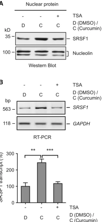
Curcumin-induced increase in SRSF1 is inhibited by trichostatin A
To see whether TSA also has effect on the curcumin reg-ulation of SRSF1, we examined its protein and transcript levels in the BJ301J cells pretreated with deacetylase inhibitor TSA (2 h) followed by treatment with curcumin (24 h). SRSF1 protein was again increased in the nuclear fraction by curcumin addition (Fig.5a). By contrast, cur-cumin-induced increase in the nuclear SRSF1 protein was reduced by TSA pretreatment (Fig.5a). Accompanying the protein change, upregulation of the SRSF1 transcript A
D B
C
0 24 48
0 20 40 60 80
SM
N
2
-E
7
+
(mo
la
r %
)
(h)
Hours post curcumin addition
** ***
0 24 48
0 1 2 3 4
Hours post curcumin addition
F
o
ld
c
h
ange of
S
M
N
pr
ot
ei
n
(h) ***
SMN
β-ACTIN Curcumin
48 h
Western Blot 40
50 kD
DMSO
RT-PCR
GAPDH SMN2_E7+
SMN2_E7- 0 24 48 (h)
Curcumin
404 bp
118
Fig. 1 Curcumin increases the SMN protein and the proportion ofSMN2_E7?transcript in fibroblast cells form a patient with SMA type II.aA Western blot of SMN protein levels in the cells treated with DMSO or curcumin for 48 h, andbtime course of the changes
(244.6±21.9 %, n =3, p\0.01, Fig.5b) by curcumin in comparison with DMSO was abolished upon pretreat-ment by TSA (117.0±11.4 %, n =3, p\0.001, Fig.5b). Therefore, a protein deacetylation step is likely also required for the curcumin-induced increase in SRSF1 transcript/protein as well.
SRSF1 knockdown reduces the inclusion ofSMN2 exon 7
To determine the role of SRSF1 inSMN2exon 7 usage and in the curcumin effect, we carried out lentiviral vector-mediated shRNA knockdown of SRSF1 in the BJ301J cells (Fig.6a). In mock-treated or scrambled shRNA-expressing cells, the molar percentage of theSMN2_E7?variant was increased from about 43.7–56.1 % by curcumin (Fig.6b, upper panel, lanes 1–4). Upon SRSF1 knockdown, the
SMN2_E7? transcript level was reduced significantly to 32.5 % (±1.4 %, n=3, p\0.01, lane 5 vs. lanes 1 and 3). Treatment with curcumin was still able to increase the
level but merely to 44.0 % (±1.9 %,n =3, lane 6), sig-nificantly lower than that in control samples (p\0.001, lanes 2 and 4). In contrast, hnRNP A1 knockdown did not change the basal level of the SMN2 splice variants (lanes 7–8). Thus, SRSF1 knockdown reduces the percentage of exon 7 inclusion in the SMA patient cells, supporting SRSF1 as a weak splicing activator of SMN2 exon 7. However, it is not essential for the curcumin enhancement of exon 7 inclusion, suggesting the existence of more factors involved in the curcumin effect.
Taken together, these data indicate that the dietary compound curcumin has multiple effects onSMN2 and its splicing regulators to promote the expression of the
SMN2_E7? transcripts in SMA fibroblast cells. These effects likely involve protein deacetylation and/or phosphorylation.
Discussion
SMA patient fibroblast cells from a type I patient have been used to investigate the regulation of SMN2 pre-mRNA splicing by small compounds (Sakla and Lorson 2008; Dayangac-Erden et al.2011). In this study, the BJ301J cells were derived from a patient (male, 9 months old) with SMA type II. They are homozygously deleted of theSMN1
but have three copies of theSMN2gene (Fig. S1), with one more copy ofSMN2than the previously reported GM3813 cells (from a SMA type I, 3-year old male) (Sakla and Lorson2008; Dayangac-Erden et al.2011). In both types of cells, the fold increases in the percentages ofSMN2_E7? transcripts are similar (about 1.5-fold) upon 24-h treatment with 25 lM of curcumin. This similar change implies that the curcumin targets in promoting the production of full-length SMN2 transcript might be the same in different sources and types of SMA cells.
Curcumin has pleiotropic effects on a number of targets in cells (Shishodia 2013; Shishodia et al. 2007), particu-larly the histone acetyltransferase p300-/CREB-binding protein (p300/CBP) (Balasubramanyam et al.2004). Of the different effects by curcumin, changes in splicing and transcript levels (Figs. 1,2,3) could be contributed by its inhibition of p300/CBP and protein/histone acetylation (Balasubramanyam et al. 2004). This is supported by the inhibition of these curcumin effects by the histone deacetylase (HDAC) inhibitor TSA (Figs. 2, 5). Particu-larly for SRSF1, we have shown that curcumin inhibits the histone acetylation/transcription of factors involved in the nonsense-mediated decay (NMD) pathway, consequently increasing the level of NMD-targeted SRSF1 variant transcripts in cells (Feng et al.2015). The HDAC inhibitor may also increase the processivity of RNA Polymerase II and reduce co-transcriptional association of splicing A
B
lanes SMN2_E7+
GAPDH SMN2_E7- Curcumin OA
- + + + DMSO TSA
1 2 3 4
RT-PCR
270 404 bp
118
DMSO DMSO TSA OA 0
20 40 60 80
SM
N
2
-E
7+
(
m
ol
ar
%
)
- + + + Curcumin
1 2 3 4 lanes
** *
regulators with certain alternative exons (Hnilicova et al.
2011), which may counteract the curcumin-enhanced exon 7 usage.
The curcumin effect on SMN2 splicing and SRSF1 phosphorylation (Figs.1,2,4) may also involve the inhi-bition of phosphatases such as PP1 and PP2A. PP1 binds to the beta-4 sheet of the SRSF1 RNA recognition motif (RRM1) domain, thus dephosphorylating the splicing fac-tor (Novoyatleva et al. 2008). PP1 inhibition promotes
SMN2exon 7 inclusion in fibroblast cells of SMA patients and spinal cord of SMA mice (Novoyatleva et al.2008). Both PP1 and PP2A can be inhibited by okadaic acid (OA). PP2A activity can also be inhibited by curcumin (Han et al.
2012). However, in our experiments, OA pretreatment did not change the curcumin effect on exon 7 inclusion in cells (Fig.2). A reasonable explanation is that curcumin and OA have overlapping effects on the phosphatases and thus the
phosphorylation of the splicing factors for SMN2 exon 7 usage.
In the immunoprecipitation experiment (Fig.4c), we were not able to efficiently immunoprecipitate the nuclear SRSF1 protein, likely due to occupation of its RRM1 domain by phosphatases (Novoyatleva et al.2008), which is part of the N-terminal antigen targeted by the SRSF1 antibody.
In summary, we have observed multiple effects of cur-cumin on the expression of genes involved in the produc-tion of the full-length SMN2 protein. These effects on gene transcription/splicing and protein phosphorylation could be contributed by the inhibitory effect of curcumin on the histone acetyl-transferase p300/CBP and protein phos-phatases. These two targets are likely important for the overall beneficial effects for producing the full-length SMN2 protein.
A
C
DMSO
Curcumin
SRSF1
DMSO
Curcumin
SAM68
DMSO
Curcumin
hnRNP
A1
Nucleus (DAPI)
Splicing Factors
(FITC) Merge
B
hnRNP A1
SAM68
β-ACTIN DMSO Curcumin
SRSF1 35
35 kD
70
48
Western Blot
SRSF1 hnRNPA1 SAM68 0
100 200 300
R
e
la
ti
ve
l
e
ve
l o
f t
ra
n
scr
ip
ts (
%
)
DMSO Curcumin
**
***
Fig. 3 SRSF1 is specifically increased by curcumin.aRepresentative western blots of total proteins of splicing factors involved in the regulation of the SMN2 exon 7 alternative splicing upon 24-h treatment of BJ301J cells with DMSO or curcumin.b-ACTIN protein-loading control.bImmunostaining of splicing factors (green) in the BJ301J cells upon 24-h treatment with DMSO or curcumin. Nuclei
Materials and methods
Cell culture and treatment
Human fibroblast cells (BJ301J) were derived from a type II SMA patient (male, 9 months old) with three copies of the SMN2 gene and maintained in Dulbecco’s modified Eagle’s medium supplemented with 10 % fetal
bovine serum, 2 mM glutamine, and 1 % penicillin– streptomycin solution (Invitrogen) at 37°C in a humid-ified atmosphere containing 5 % CO2. Cells were treated with DMSO or 25lM of curcumin (Sigma-Aldrich) for 0, 24, or 48 h. In the assay of inhibitors on transcript of
SMN2 or SRSF1, cells were pretreated with each inhi-bitor for 2 h followed by 24-h addition of DMSO or 25lM of curcumin.
A
C
hnRNP H/F
GAPDH 48
35
kD D C D C D C
Total Cytosolic Nuclear
D (DMSO) / C (Curcumin)
hnRNP H/F SRSF1
Western Blot 35
48 kD
kD
D C D C D C
0 4 mM 10 mM
D (DMSO) / C (Curcumin)
SRSF1 35
40
β-ACTIN Concentration of NaF in lysis buffer B
IgG H-chain
SRSF1 (hyperphosphorylated)
IgG L-chain - + +
- + -
anti-SRSF1
CIAP
IP-Western Blot 48
35
25
lanes kD
2 3 1
SRSF1 (dephosphorylated)
*
*
Western Blot
Fig. 4 Curcumin increases the hyperphosphorylated isoform of SRSF1 in the nucleus.aLeft panelWestern blotting analysis of the fractionation of cytosol and nuclei of the BJ301J cells, GAPDH and hnRNP H/F are as the specific markers for cytosolic and nuclear fractions, respectively.Right panelWestern blotting analysis of the expression of SRSF1 in total lysates (T), cytosolic (C) and nuclear (N) fractions of the BJ301J cells upon 24-h treatment with DMSO or 25lM of curcumin.hnRNP H/Fprotein loading control. Two bands are shown in all of the lysate samples, both of them can be recognized by SRSF1 antibody (clone 96).bWestern blotting analysis of SRSF1
Knockdown of splicing factors using lentiviral vector-mediated transduction
Lentiviral particles were prepared using the shRNA plas-mids pGIPZ-shSRSF1 (Open Biosystems, RMM4431-99938975, 50-TCG AGA TCG AGA TCT TCC A-30), pGIPZ-shhnRNPA1 (50-GTG TAA AGC ATT CCA ACG A-30), and pGIPZ-scrambled (50-TAG TGA AGC CAC AGA ATA T-30) according to our previous procedures (Liu et al.2012; Yu et al.2009). Cells were treated with DMSO
or curcumin 7 days after transduction. The silencing effects were confirmed by RT-PCR and immunoblotting.
Reverse transcription polymerase chain reaction
Cytoplasmic RNA was fractionated according to our pre-vious procedure (Feng et al. 2015; Ma et al. 2007) and extracted using the GenElute Mammalian Total RNA Miniprep Kit (Sigma-Aldrich). One microgram of cyto-plasmic RNA was included in a 10ll reverse transcription reaction. PCR reactions were carried out for 26–30 cycles. The sequences (50–30) of the primer pairs were as follows, with the forward primer listed first followed by the reverse primer for each gene.SMN2: AAG ACT GGG ACC AGG A
B
D C C
TSA D (DMSO) / C (Curcumin)
SRSF1
Nucleolin Nuclear protein
- - +
Western Blot 35
100 kD
563 bp
118
D C C
TSA
D (DMSO) / C (Curcumin)
SRSF1
GAPDH - - +
RT-PCR
- - + 0
100 200 300
SR
SF
1
t
ra
n
s
c
ri
p
t (
%
)
D C C D (DMSO) / C (Curcumin) TSA ***
**
Fig. 5 Curcumin-induced increase in SRSF1 is inhibited by tricho-statin A (TSA).aWestern blots of SRSF1 protein in the nuclei of the BJ301J cells upon 2 h of pretreatment with or without TSA, followed by 24-h addition of DMSO or curcumin. Nucleolin nuclear pro-tein loading control.bUpper panel RT-PCR analysis of theSRSF1 transcripts in the cytoplasm of the BJ301J fibroblast cells upon 2 h of pretreatment with or without TSA, followed by 24-h addition of DMSO or curcumin.GAPDHRNA loading control.Lower panelA bar graphof theSRSF1transcript changes (mean±SD,n=3) as in theupper panel.SRSF1transcript was normalized to that ofGAPDH and then to that of the sample treated with DMSO but not TSA (-), which is set as 100 %. **p\0.01, ***p\0.001, comparison between the indicated samples
A
B
hnRNP A1
β-ACTIN SRSF1
Western Blot
shRNA
35
35 kD
48
SMN2_E7+
SMN2_E7-lanes
D (DMSO) / C (Curcumin)
1 2 3 4 SRSF1
D C Mock
D C
hnRNP A1
D C Scrambled
D C
5 6 7 8 GAPDH
RT-PCR
shRNA
404 bp
118
Mock Scrambled SRSF1 hnRNPA1 0
20 40 60 80
SM
N
2
-7
+ (mo
la
r %
)
shRNA
1 2 3 4 5 6 7 8 lanes D C D C D C D C D (DMSO) /
C (Curcumin)
AAA GC, TAT CTT CTA TAA CGC TTC ACA TTC CAG; SRSF1: CCT CCA GAC ATC CGA ACC AAG, TGC TAC GGC TTC TGC TAC GAC;hnRNP A1: GTC TAA GTC AGA GTC TCC TAA AGA GCC, TCT CAT TAC CAC ACA GTC CGT G;SAM68: GCT GAC GGC AGA AAT TGA GAA G, TTG ACA GGT ATC AGC ACT CGC TC;GAPDH: GTC AAC GGA TTT GGT CGT ATT G, AAC CAT GTA GTT GAG GTC AAT GAA G. PCR products were resolved in 2–3 % agarose gels containing 0.5lg/ml ethidium bromide. The gels presented in figures are inverted digital images. The abundance of the
SMN2_E7?splice variants is expressed as molar percent-ages relative to the total of theSMN2 variants (E7?and E7-).
Western blotting
Cells were rinsed three times with ice-cold PBS, harvested using cell scrapers, pelleted by centrifugation at 14,000 rpm for 30 s at 4°C, and lysed in RIPA buffer (containing 2 mM PMSF, 2 mM Na3VO4and 10 mM NaF) (Feng et al. 2015). Protein was quantified using the Bradford method, and samples were run on 10 or 12 % Tris–glycine acrylamide gels and then transferred to polyvinylidene fluoride membranes. The membranes were blocked in 5 % dry milk and probed with the following mouse antibodies, which were all purchased from Santa-Cruz Biotechnology unless otherwise indicated: anti-SMN (H-7, 1:400), anti-SRSF1 (clone 96, 1:500), anti-hnRNP A1 (9H10, 1:1000), anti-SAM68 (7-1, 1:250), anti-nucle-olin (H-6, 1:1000), anti-b-actin (C4, 1:1000), and anti-GAPDH (Sigma, 1G5, 1:2000). After incubation with peroxidase-conjugated goat anti-mouse immunoglobulin M or G secondary antibodies (Sigma-Aldrich, 1:2000), pro-teins were visualized using enhanced chemiluminescence (GE Healthcare). Densitometry of the resulting bands was analyzed by Image J (developed by the U.S. National Institutes of Health and available athttp://rsb.info.nih.gov/ ij/).
Immunostaining
BJ301J fibroblast cells were plated over the slides in six-well plates, treated with DMSO or curcumin for 24 h, rinsed twice in ice-cold phosphate-buffered saline (PBS) with 1 % BSA, fixed with 4 % paraformaldehyde (PFA) for 15 min and then permeabilized with 0.2 % Triton X-100 for 10 min at room temperature. The fixed cells were incubated overnight at 4°C with mouse monoclonal antibodies anti-SRSF1, anti-hnRNP A1, and anti-Sam68, respectively. The primary antibodies were diluted at 1:100 in TBS (20 mM TrisCl, 500 mM NaCl) containing 1 % BSA. Cells were rinsed twice with TBS and incubated with
goat anti-mouse fluorescent secondary antibody (conju-gated with FITC, 1:1000) in the dark for 1 h at room temperature. Cell nuclei were counterstained with DAPI (1:6000). The stained cells were mounted with mounting media (Sigma-Aldrich). Images were taken at 1009 mag-nification with an Olympus microscope.
Fractionation of nuclear and cytoplasmic proteins
BJ301J fibroblast cells were rinsed with ice-cold PBS three times in the dishes then harvested in 1 ml of ice-cold PBS using rubber scrapers. Cell pellets were collected into 1.5-ml tubes by centrifuging at 14,000 rpm for 30 s and then resuspended in ice-cold NP-40 buffer supplemented with 2 mM PMSF, 2 mM Na3VO4 and 10 mM NaF. After centrifugation at 14,000 rpm for 2 min, the supernatant was used as cytoplasmic fraction by additional centrifu-gation at 14,000 rpm through 24 % (w/v) sucrose cushion, the nuclear pellets were washed twice using 1 ml of ice-cold NP-40 buffer followed by resuspension in RIPA buffer supplemented with 2 mM PMSF, 2 mM Na3VO4 and 10 mM NaF, then used as nuclear fraction after sonication.
Immunoprecipitaion and phosphatase assay
BJ301J fibroblast cells were rinsed with cold PBS for three times. The cytoplasmic and nuclear lysates were prepared according to the protocol described in the section of ‘‘Cytoplasm and nuclei fractionation.’’ Protein G beads were washed with cold PBS for five times, then packed with 2lg of anti-SRSF1 antibody and incubated at 4°C under rotary agitation for 4 h. After washing with cold PBS, the packed beads were incubated overnight with lysates at 4°C. When the incubation time was over, the supernatant was removed by centrifugation, and the beads were washed in lysis buffer three times for further analysis. The precipitations were suspended in 19reaction buffer with 10 units of CIAP and incubated at 37°C for 60 min. After that, the suspensions were mixed with 69 SDS loading buffer, heated at 95°C for 5 min to denature the proteins, and separated them from the protein G beads, and the supernatants were used for western blotting analysis.
Statistical analysis
Data were analyzed by two-tailed Student’s t test. A
p value\0.05 was considered significant.
proofreading the manuscript. This work was supported by a Manitoba Research Chair Fund and in part by a Canadian Institutes of Health Research (CIHR) Operating Grant FRN_106608 to JX, by FRN_2006BIA05A07 and 2006BIA05A08 from the National Key Technology R&D Program of China to SH.
Compliance with ethical standards
Conflict of interest All of the authors declare no conflict of interests.
Ethical standards All procedures performed in this study involving the SMA patient were in accordance with the ethical standards of the institutional and/or national research committee and with the 1964 Declaration of Helsinki and its later amendments or comparable ethical standards. Informed consent was obtained from the SMA patient included in the study.
References
Balasubramanyam K et al (2004) Curcumin, a novel p300/CREB-binding protein-specific inhibitor of acetyltransferase, represses the acetylation of histone/nonhistone proteins and histone acetyltransferase-dependent chromatin transcription. J Biol Chem 279(49):51163–51171
Bialojan C, Takai A (1988) Inhibitory effect of a marine-sponge toxin, okadaic acid, on protein phosphatases. Specificity and kinetics. Biochem J 256(1):283–290
Burghes AH, Beattie CE (2009) Spinal muscular atrophy: why do low levels of survival motor neuron protein make motor neurons sick? Nat Rev Neurosci 10(8):597–609
Burnett BG et al (2009) Regulation of SMN protein stability. Mol Cell Biol 29(5):1107–1115
Cartegni L, Krainer AR (2002) Disruption of an SF2/ASF-dependent exonic splicing enhancer in SMN2 causes spinal muscular atrophy in the absence of SMN1. Nat Genet 30(4):377–384 Cartegni L et al (2006) Determinants of exon 7 splicing in the spinal
muscular atrophy genes, SMN1 and SMN2. Am J Hum Genet 78(1):63–77
Chen HH et al (2008) The RNA binding protein hnRNP Q modulates the utilization of exon 7 in the survival motor neuron 2 (SMN2) gene. Mol Cell Biol 28(22):6929–6938
Coovert DD et al (1997) The survival motor neuron protein in spinal muscular atrophy. Hum Mol Genet 6(8):1205–1214
Darvesh AS et al (2012) Curcumin and neurodegenerative diseases: a perspective. Expert Opin Investig Drugs 21(8):1123–1140 Dayangac-Erden D et al (2011) Carboxylic acid derivatives of histone
deacetylase inhibitors induce full length SMN2 transcripts: a promising target for spinal muscular atrophy therapeutics. Arch Med Sci 7(2):230–234
Dokmanovic M, Clarke C, Marks PA (2007) Histone deacetylase inhibitors: overview and perspectives. Mol Cancer Res 5(10): 981–989
Feng D et al (2015) Increase of a group of PTC(?) transcripts by curcumin through inhibition of the NMD pathway. Biochim Biophys Acta 1849(8):1104–1115
Glozak MA, Seto E (2007) Histone deacetylases and cancer. Oncogene 26(37):5420–5432
Gupta SC et al (2012) Discovery of curcumin, a component of golden spice, and its miraculous biological activities. Clin Exp Pharmacol Physiol 39(3):283–299
Gupta SC, Patchva S, Aggarwal BB (2013) Therapeutic roles of curcumin: lessons learned from clinical trials. AAPS J 15(1):195–218
Han X et al (2012) Curcumin inhibits protein phosphatases 2A and 5, leading to activation of mitogen-activated protein kinases and death in tumor cells. Carcinogenesis 33(4):868–875
Hnilicova J et al (2011) Histone deacetylase activity modulates alternative splicing. PLoS One 6(2):e16727
Hsieh-Li HM et al (2000) A mouse model for spinal muscular atrophy. Nat Genet 24(1):66–70
Hua Y et al (2010) Antisense correction of SMN2 splicing in the CNS rescues necrosis in a type III SMA mouse model. Genes Dev 24(15):1634–1644
Kashima T, Manley JL (2003) A negative element in SMN2 exon 7 inhibits splicing in spinal muscular atrophy. Nat Genet 34(4):460–463
Kashima T et al (2007) hnRNP A1 functions with specificity in repression of SMN2 exon 7 splicing. Hum Mol Genet 16(24):3149–3159
Lefebvre S et al (1995) Identification and characterization of a spinal muscular atrophy-determining gene. Cell 80(1):155–165 Liu G et al (2012) A conserved serine of heterogeneous nuclear
ribonucleoprotein L (hnRNP L) mediates depolarization-regu-lated alternative splicing of potassium channels. J Biol Chem 287(27):22709–22716
Lorson CL et al (1998) SMN oligomerization defect correlates with spinal muscular atrophy severity. Nat Genet 19(1):63–66 Ma S et al (2007) Relocalization of the polypyrimidine tract-binding
protein during PKA-induced neurite growth. Biochim Biophys Acta 1773(6):912–923
Monani UR (2005) Spinal muscular atrophy: a deficiency in a ubiquitous protein; a motor neuron-specific disease. Neuron 48(6):885–896
Monani UR et al (2000) The human centromeric survival motor neuron gene (SMN2) rescues embryonic lethality in Smn(-/-) mice and results in a mouse with spinal muscular atrophy. Hum Mol Genet 9(3):333–339
Ngo JC et al (2005) Interplay between SRPK and Clk/Sty kinases in phosphorylation of the splicing factor ASF/SF2 is regulated by a docking motif in ASF/SF2. Mol Cell 20(1):77–89
Novoyatleva T et al (2008) Protein phosphatase 1 binds to the RNA recognition motif of several splicing factors and regulates alternative pre-mRNA processing. Hum Mol Genet 17(1):52–70 Pedrotti S et al (2010) The splicing regulator Sam68 binds to a novel exonic splicing silencer and functions in SMN2 alternative splicing in spinal muscular atrophy. EMBO J 29(7):1235–1247 Sakla MS, Lorson CL (2008) Induction of full-length survival motor neuron by polyphenol botanical compounds. Hum Genet 122(6):635–643
Shishodia S (2013) Molecular mechanisms of curcumin action: gene expression. BioFactors 39(1):37–55
Shishodia S, Singh T, Chaturvedi MM (2007) Modulation of transcrip-tion factors by curcumin. Adv Exp Med Biol 595:127–148 Singh NK et al (2006) Splicing of a critical exon of human Survival
Motor Neuron is regulated by a unique silencer element located in the last intron. Mol Cell Biol 26(4):1333–1346
Swoboda KJ et al (2005) Natural history of denervation in SMA: relation to age, SMN2 copy number, and function. Ann Neurol 57(5):704–712
Wirth B et al (2006) Mildly affected patients with spinal muscular atrophy are partially protected by an increased SMN2 copy number. Hum Genet 119(4):422–428
Yu J et al (2009) The heterogeneous nuclear ribonucleoprotein L is an essential component in the Ca2?/calmodulin-dependent protein kinase IV-regulated alternative splicing through cytidine– adenosine repeats. J Biol Chem 284(3):1505–1513