A Mouse Model for Breast Cancer Induced by
Amplification and Overexpression of the
neu
Promoter and Transgene
Edward J. Weinstein, Daniel I. Kitsberg*, and Philip Leder
Department of Genetics, Harvard Medical School, Howard Hughes Medical
Institute, Boston, Massachusetts U.S.A.
Communicated by P. Leder. Accepted October 8, 1999.
Introduction
The neu oncogene was first isolated from DNA of adrenal neuroblastomas of neonatal rats (1). The human homologue of this gene was later identified (2–4) and named c-erbB-2in reference to its homology to the c-erb-B proto-oncogene. Since its discovery, c-erbB-2 amplification and over-expression have been associated with a
number of human malignancies, including breast, colon, lung and ovarian (5–7). Amplifi-cation of this gene in breast cancer is seen in 17.5% to 34% of tumors examined, with copy numbers ranging from 4 to 50 (8,9). Amplifica-tion of c-erbB-2correlates with a higher risk for metastatic disease (10,11), as well as shortened disease-free and overall survival periods (12). This is most likely due to the fact that amplifi-cation is closely correlated to over-expression of c-erbB-2(13,14).
The transforming potential of erbB-2/neu
has been demonstrated in a number of ways. In
Abstract
Background:ErbB-2 is a critical oncogenic marker in human breast cancer. Its appearance correlates with poor prognosis and it is, therefore, an impor-tant target for physiologic investigation and thera-peutic intervention. With this in mind, we have cre-ated and characterized two mouse breast cancer models that express rat wild type neu, the homo-logue of ErbB-2,and rat mutant neuunder the control of the normal mouse neupromoter. These models in which the copy number of the neu gene is moder-ately amplified should more closely parallel the ex-pression pattern of ErbB-2 seen in some cases of hu-man breast cancer.
Materials and Methods: Transgenic mouse models were constructed by injecting one of the two pronu-clei of a fertilized FVB/n egg and implanting it into a pseudopregnant Swiss /Webster mouse. Tissue
ex-pression was analyzed through the use of reverse transcription polymerase chain reaction and mam-mary histopathology examined by fixing, staining and mounting of the entire gland.
Results: In the former wild type model, we show that low level, long term expression of neuleads to abnormal lobuloalveolar development in virginal glands and incomplete regression in multiparous glands. Malignant foci form following multiple rounds of pregnancy and regression. In the latter model, a similarly directed transgene carrying the constitutively activated, mutant form of the rat neu gene, a stronger but similar phenotype is displayed.
Conclusion: Evidently minor perturbations in am-plified neu expression are sufficient to alter mam-mary development and induce malignant transfor-mation.
Address correspondence and reprint requests to: Dr. Philip Leder, Department Of Genetics, Harvard Medical School, Boston, MA 02115. Phone: 617-432-7667; Fax: 617-432-7944; E-mail: leder@rascal.med.harvard.edu *Present address: Alomone Labs, POB 4287, Jerusalem, 91042 Israel.
vitro transformation assays have been utilized to show that high levels of expression of
c-erbB-2 mediate transformation in NIH3T3 cells (15), as determined by tumor formation after injection into athymic mice and in an-chorage-independent growth assays. The level of expression has been shown to be critical for transformation. Mouse mammary tumor virus (MMTV)– based expression vectors with neu
were proven to achieve transformation in the NIH3T3 cell line; whereas, SV40–driven neu,at a 20-fold lower level of expression, failed to do so (16,17). The effect of over-expression in murine mammary epithelia was analyzed through construction of MMTV/neutransgenic mice (18). Multiparous female mice with the oncogenic rat neu(MMTV/neu*) develop mam-mary adenocarcinomas at an average age of three months. MMTV promoter–based overex-pression of the proto-oncogenic form of rat neu
(MMTV/neu) also results in focal breast tumors but with a longer latency (19).
In order to further understanding of cellular
neu’s in vivo regulation, the 5' upstream region of the transcriptional start site of the mouse gene was cloned and characterized (20). Chloram-phenicol acetyltransferase (CAT) assays were used to determine functional variation in tran-scriptional activity among various lengths of the upstream region in NIH3T3 cells. One such con-struct containing 1.2 kb 5' flanking sequence was found to have CAT levels at thirteen fold higher than that of the full length 4.5 kb promoter.
Most mouse models developed to study the effects of over-expression of an oncogene in the mammary gland utilize extremely strong pro-moters that allow for high levels of transcrip-tional activity (21). Although these animals have proven useful in the study of signal trans-duction in the transformed tissue, the promot-ers they utilize are—of necessity—nonphysio-logic. Not only are artificially elevated levels of oncogene expression allowed by these promot-ers, but, they are also not necessarily spatially nor temporally regulated in a physiologic man-ner. Although most human breast cancers are not associated with current pregnancy, most of the promoters that are widely used in mouse models of mammary gland tumorigenesis re-quire the induction of pregnancy to reach high levels of transcriptional activity (22,23). Neuis expressed in the virgin, pregnant, lactating and regressing mammary gland of the mouse (24), making it difficult for transgene promoters, such as those of MMTV or the whey acidic
pro-tein (WAP), to accurately model the conse-quence of amplification of the neugene through-out the course of murine development. We de-sired to create a model that involves over-expression of neuin a manner more consistent with that seen in human breast cancer. By con-structing transgenic mice containing the first 1.2 kb upstream sequence of the mouse neu
gene fused to either the cellular or transformed (Val664Glu) rat neu transgene, we designed a system that attempts to mimic cases of human breast cancer involving amplification of the
erbB-2locus in expression and regulation.
Materials and Methods
Cloning the Murine neuPromoter
A 544 base pair fragment of the neupromoter was amplified by polymerase chain reaction (PCR) from mouse genomic DNA using the primers 5'-GATATCCCAGAGAGTCTT-3' and 5'-TCAGGC-TGGACCAGGCTGCG-3', and ligated into pCR (Invitrogen, Carlsbad, CA). This was then used as a probe to screen a 129 mouse genomic library (Genome Systems, St. Louis, MO). The clones were digested with Nco Iand Xba Iin order to iso-late the 1.2 kb fragment and blunt end cloned into the pSV2IgH/neuor pSV2IgH/neu*vector from which the IG promoter/enhancer had been ex-cised by Hind IIIdigest.
Mammary Gland Whole Mounts
The inguinal mammary gland fat pad was ex-cised with scissors and forceps and spread on a glass slide. It was allowed to air dry for 1 min be-fore overnight incubation in Tellyesniczky’s fixa-tive (70% ethanol:formaldehyde:glacial acetic acid at 20:1:1 ratio) at room temperature. Tissue was rinsed with running water for 1 hr followed by three 6-hour incubations in acetone. Mam-mary glands were then rehydrated by successive washings in 100%, 95% and 70% ethanol, fol-lowed by 30 min in running water. Tissues were then stained overnight in Carmine Red Stain [1 gram carmine red dye (Sigma, St. Louis, MO), 2.5 g potassium alum in 500 ml water]. After washing tissues in running water for 2 hr they were dehydrated in successive washes of 50%, 70%, 95% and 100% ethanol, followed by two overnight incubations in xylene.
Histologic Evaluation
Scien-tific, Inc., Lodi, CA). 4 m sections were cut and stained with hematoxylin and eosin by the Transgenic Core Pathology Laboratory at the University of California at Davis, CA.
Transgene Injection
The pNcN plasmid was linearized with Pvu I.
Approximately 10 g of DNA was run on a 1% agarose gel. The appropriate sized band was ex-cised and the DNA was purified on a Qiagen col-umn. The DNA was diluted in injection buffer (0.15 M KCl, 5 mM NaCl, 10 mM PIPES) at a concentration of 1–10 ng/l. The DNA was then injected into one of the two pronuclei of a fertil-ized FVB/n egg and implanted into the oviduct of a pseudopregnant Swiss/Webster mouse.
Identification of Transgenic Animals
DNA was prepared from tail samples by incu-bating tails overnight at 50°C in 500 l Tail Buffer (17 mM Tris [pH 7.5], 17 mM EDTA, 170 mM NaCl, 0.85% SDS and 0.2% proteinase K added immediately prior to use). After proteins were pelleted in 250 l 6M NaCl and cen-trifuged, DNA was precipitated in 95% ethanol and resuspended in 100 l water. The DNA was electrophoresed through a 0.8% agarose gel af-ter complete digestion with BamHI restriction enzyme. The gel was blotted overnight in 0.4 N sodium hydroxide onto a Genescreen Plus membrane (NEN Research Products, Boston, MA). The filter was then rinsed for 10 min in 2 SSC (0.30 M sodium chloride, 0.030 M sodium citrate) and crosslinked by ultraviolet light. Filters were prehybridized for at least 1 hr at 42°C in hybridization solution (50% for-mamide, 5 SSC, 5% dextran sulfate, 20 mM sodium phosphate, 1 Denhardt’s reagent, 0.5% sodium dodecyl sulfate and 20 g/ml sonicated herring sperm DNA) before probe was added. An 800-basepair Pst I fragment of SV40 polyA vector (18) was labeled with [∝32P]dCTP using random priming (Strata-gene, La Jolla, CA) and hybridized to the mem-brane overnight at 42°C. Filters were washed at room temperature twice for 15 min in 2 SSC/ 0.1% SDS and once for 10 min in 0.1 SSC/ 0.1% SDS before exposing to film.
Tumor Lysates and Western Blot Analysis
Frozen tumor was homogenized in 2 ml NP-40 lysis buffer [50 mM Tris (pH 7.5), 150 mM NaCl, 1% NP-40, 2 mM EDTA, 1 mM sodium
ortho-vanadate, 10 mM NaF plus one Complete tablet (Roche Diagnostics, Chicago IL)] for 3 min on ice and incubated at 4°C for 30 min. The lysates were spun at maximum speed in a microcen-trifuge at 4°C for 20 min and the supernatant was transferred into a clean Eppendorf tube. Equal concentrations of protein, as determined using the BioRad dye-binding microassay with bovine serum album as a standard, were elec-trophoresed through 8% SDS-PAGE gels. Bound proteins were then transferred to a PVDF membrane (Millipore, Bedford MA) and blocked overnight in blocking buffer (1 TBS with 0.1% Tween-20 and 5% nonfat dried milk). Immunodetection was performed using anti-neu
Ab-3 (Oncogene Research Products, Cambridge, MA) at a dilution of 1:200. Signal development was performed using an enhanced chemilumi-nescence method (Amersham, Piscataway, NJ).
Transfections into MCF-7 Cells
CalPhos Maximizer (Clontech, Palo Alto, CA) was used to transfect MCF-7 cells with DNA at a concentration of 5.0 g of the construct of in-terest and 0.5 g of marker in 35 mm plates (Corning, Acton, MA). Cells were incubated 4.5 hours at 37°C and 5% CO2 in DMEM
(Gibco-BRL, Gaithersburg, MD) supplemented with 10% FBS (Sigma), 2% L-glutamate (Gibco-BRL) and 1% penicillin/streptomycin (Gibco-BRL). The cells were washed twice with 1 PBS and incubated overnight in sup-plemented DMEM, and expanded into 15 cm plates and allowed to grow for 24 hr. Geneticin (Gibco-BRL) was added to DMEM at a concen-tration of 800 g/ml. After allowing 3 days in selection media for non-transfected cells to die, clones were picked and expanded.
RT-PCR/Southern
de-naturation, 94°C for 1 min; annealing, 52°C for 1 min; and extension, 68°C for 2 min. All PCR reactions underwent 30 cycles, unless other-wise noted. Amplified products were elec-trophoresed through 1% agarose gels and transferred overnight in 0.4 N NaOH to Gene-screen Plus membrane (NEN Research Prod-ucts). The filters were then prehybridized and probed as described above. All cDNAs were in-dividually tested in parallel reactions lacking reverse transcriptase to confirm a lack of conta-minating genomic DNA.
Results
Expression in Mammary Epithelial Cells
We first sought to determine if a fragment of the neupromoter would be active in mammary epithelial cells. The 1.2 kb region 5' of the neu transcriptional start site was previously shown to induce high levels of CAT activity when transfected into NIH3T3 cells (20). This 1.2 kb promoter was isolated from a mouse 129 ge-nomic library and blunt end cloned into the
pSv2IgH/neu vector (25) from which the im-munoglobulin (IG) promoter/enhancer had been excised. The human mammary epithelial cell line MCF-7 was then transfected with this
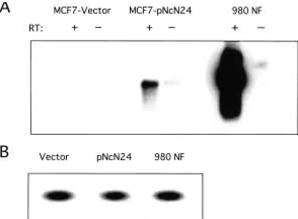
construct, termed pNcN, or vector alone, and multiple clonal populations were picked. Re-verse transcription polymerase chain reaction (RT-PCR) was then performed using primers for the SV40 polyA region. The RT-PCR was also performed on a cell line, termed 980NF, established from a mammary gland tumor ex-cised from an MMTV/neu* transgenic mouse (18), which served as a positive control. Multi-ple clones of cells transfected with the neu pro-moter/neutransgene construct do transcribe the transgene from the murine promoter (Fig. 1A), showing that the promoter is active in mam-mary cells. RT-PCR was performed using hu-man -actin primers to control for loading (Fig. 1B). No bands were identified in the cell line with vector alone nor in any of the control re-actions lacking reverse transcriptase.
Construction of Transgenic Mice
We next created transgenic mice overexpress-ing the neu promoter/neu transgene construct. Recombinant plasmid was microinjected into the male pronucleus of a one cell mouse em-bryo and three transgenic founder animals were created. One founder died at approxi-mately 1 month of age and one was incapable of passing the transgene, but the third founder
Fig. 1. Analysis of transgenic neupromoter activity in MCF-7 cells. (A) RT-PCR of MCF-7 cells transfected with pNcN construct or vector alone. The mammary cell line 980NF was derived from an MMTV-neu*–induced tumor and serves as
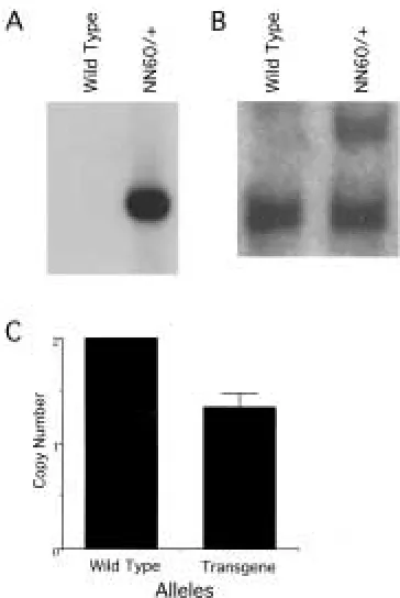
did survive to adulthood and passed the trans-gene to progeny in a Mendelian fashion as de-termined by Southern blot analysis with an SV40 polyA probe (Fig. 2A). This line, named NN60, has been fully characterized and will be described herein.
In order to determine the plasmid insertion number, restriction enzyme digests were per-formed on genomic DNA from wild type and transgenic mice (Fig. 2B). When digested with
BamHI, DNA from transgenic animals showed an additional band when hybridized with a probe for neu. This probe recognizes both the endogenous murine gene as well as the trans-genic rat neu.When quantified by phosphoim-ager densitometry analysis, it can be seen that the NN60 line of mice contains only a single additional copy of the murine neu promoter, followed by the rat transgene (Fig. 2C).
Expression of the Transgene in the Mouse
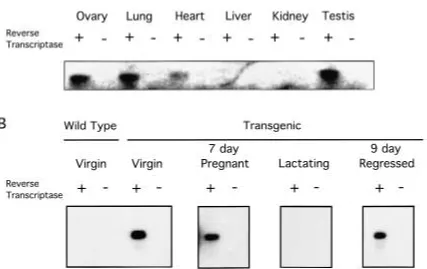
Endogenous neu expression varies, depending on the organism and the stage of development investigated. Inconsistencies can be found throughout the literature, presumably due to the method of detection utilized (26). We wanted to determine which organs were ex-pressing the transgene and to confirm that the promoter was indeed active in the mammary gland. Through use of RT-PCR with primers to the SV40 polyA region, we found that the transgenic promoter was active in the ovary, lung, heart and testis (Fig. 3A). The liver and kidney, however, failed to express the trans-gene. In all cases, a control RT-PCR was per-formed with water substituted for the reverse transcriptase enzyme to ensure that amplifica-tion was occurring from cDNA and not ge-nomic DNA. All of the control lanes were neg-ative.
The same primer pairs were used to eluci-date the transgene activity in the various stages of mammary gland development (Fig. 3B). Amplification of the targeted cDNA was clearly seen in the lanes representing the virgin, 7-day pregnant and 9-day regressing mammary gland. The transcript of the transgene did not appear to be present in the lactating animal. A wild type virgin female was used as a control for specificity of the primers. The lane in Figure 3B representing this animal contains no signal, nor do the lanes representing RT-PCR reactions with water substituted for reverse transcrip-tase.
Effects of an Additional Copy of neu in the Mammary Gland
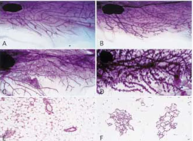
We decided to study the consequence of trans-gene expression in the mammary gland by ex-amining ductal morphogenesis. Our expres-sion analysis showed that the transgene was produced in the mammary gland even before the animal was mated. Therefore, we sought to determine if there were any long-term devel-opmental abnormalities associated with low level transgene expression in the virgin mam-mary glands. Mammam-mary gland whole mounts stained with carmine red alum were prepared in order to study the growth of the ductal tree (27).
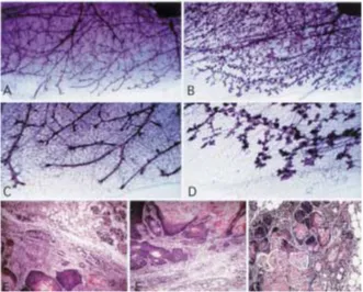
At the onset of puberty, in the first 3–4 weeks of development, end buds appear at the mammary ductal tips in female wild type mice. Fig. 2. Southern blot analysis of NN60
trans-genic mice. Genomic tail DNA was digested with
Terminal end buds drive morphogenesis within the gland by supplying differentiated ductal and myoepithelial cells during elongation of the ducts (28). The pattern of growth into the pe-riphery continues in the virgin until the ducts reach the limits of the mammary fat pad, at which time the end buds regress. With the ex-ception of some activity associated with the es-trous cycle, the gland remains mitotically quies-cent until pregnancy occurs. A clear zone of the fat pad remains between the ducts of the mature virgin, which will eventually be occupied by lobules during pregnancy (29). This quiescent nature of the adult virgin mammary gland was not seen in the transgenic animal. Although ad-ditional ductal branching did not seem to occur, epithelial cells were seen to fill the interductal spaces (Figs. 4A and 4B). This was reminiscent of a gland in early stages of pregnancy. With continuing age, lobuloalveolar structures were formed and multiplied and some degree of
duc-tal enlargement occurred (Figs. 4C and 4D). Precocious lobular development and alveolar hyperplasia were confirmed by hematoxylin and eosin staining of the virgin mammary gland sections. A majority of the alveoli showed evi-dence of secretory activity and small droplets of fat could be seen throughout the cytoplasm of the epithelial cells (Figs. 4E and 4F). In addi-tion, the epithelial secretions showed a punctate pattern of yellow staining, indicative of lacto-ferrin, a protein product seen in lactating or re-gressing glands (30,31).
A more severe phenotype of this mouse could be seen in the mammary gland after two rounds of pregnancy and regression. In a wild type mammary gland there was a brief phase of secretory engorgement after weaning, followed by absorption of the milk proteins and collapse of the alveoli in a wild type mammary gland (32; Figs. 5A and 5C). In contrast, the trans-genic mammary gland showed a failure to fully Fig. 3. Expression of the pNcN transgene in
transgenic mice. (A) RT-PCR was performed on ovary, lung, heart, liver and kidney of a virgin fe-male and testis of a fe-male NN60 transgenic mouse using SV40 polyA primers. (B) RT-PCR with SV40 polyA primers used on female mammary gland
involute (Figs. 5B and 5D), with lobuloalveo-lar structures branching off of the primary and secondary ducts persisting months after wean-ing. This occurred after the first pregnancy, but became increasingly more pronounced after multiple rounds of lactation and weaning. The ducts also appeared to be slightly dilated. How-ever, as in the case of the virgin transgenic an-imals, the overall ductal structure was largely preserved.
Even after continuous mating for over a year and a half, these transgenic animals carry-ing the neu promoter/neu transgene failed to develop palpable mammary tumors. Upon dis-section, however, small foci could be seen throughout the fourth inguinal and third tho-racic glands. Many of these lesions were kera-toacanthomas, composed largely of squamous epithelium producing large quantities of lami-nar keratin (Figs. 5E and 5F). Small areas of glandular differentiation were seen at the mar-gins of some mammary glands (Fig. 5E), which were delineated by fibrous connective tissue. In some cases, there were nests of dysplastic
squamous cells producing extensive laminar keratin (Fig. 5G) and displaying high mitotic rates. All of these tumors appeared to be sur-rounded by dense fibrous stroma and confined to the mammary gland tissue. No primary tu-mors were found in any other tissue and metastases were never noted.
Effect of Expression of Oncogenic neuwhen Transcribed from the neuPromoter
In addition to the construction of the NN60 transgenic line, we also created a transgenic mouse over-expressing the oncogenic form of
neu (Val664Glu) transcribed by the murine neu
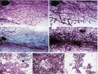
promoter. This mouse, termed TNT for “trans-forming neu transgene,” contained the same transgene that was found in the NN60 mouse, except for the point mutation in the transmem-brane domain, which rendered the protein con-stitutively active. We had multiple lines of this mouse with the same phenotype, which was ex-tremely similar to that of the NN60 animals. In the adult virgin mammary gland, there was pre-Fig. 4. Whole mounts and hematoxylin/eosin
stained sections of wild type and NN60 trans-genic virgin mammary glands.Virgin mammary gland from 9-month-old (A) wild type female and (B) NN60 transgenic littermate. Whole mounts were spread on slides, fixed and stained with carmine red in order to elucidate ductal structure.
cocious lobuloalveolar development throughout the tissue, as well as dilation of the primary and secondary ducts (Figs. 6A and 6B) with some re-gions showing more extensive proliferation. Similar to the virgin gland containing the neu
promoter and the cellular neu transgene, the overall architecture of the ducts appeared to be intact.
The phenotype of the multiparous animal was even more striking. After three rounds of pregnancy, the mammary gland of the neu pro-moter/transforming neu transgene animal showed an inability to involute when com-pared with the wild type control (Figs. 6C and 6D). Although some residual acinar structure usually remained in an FVB/n female after many pregnancies, the level seen in these transgenics was extraordinarily high. Hema-toxylin and eosin stained slides revealed that the mammary gland appeared to be in a stage of early regression (Fig. 6G), presenting with copious amounts of both lipid secretions and
lactoferrin. In addition, when the fourth in-guinal mammary gland was examined by whole mount, one could see an average of four to five small tumor foci spread throughout the gland. Several multiparous females also de-veloped palpable tumors, with an average age of onset of 14.5 months. Hematoxylin and eosin stained slides of these tumor sections re-vealed tumors with multiple morphologies. Several tumors were composed of solid nests of cells (Fig. 6E), that, as in the case with the nodules from the mammary gland of the
neu promoter/cellular neu transgene animal, appeared to be producing noticeable quantities of laminar keratin. Masses consisting of dys-plastic glands and squamous epithelium were also noted. A tumor not usually noted in MMTV/neu mice was also seen, in which nests of micropapillary neoplasm were formed and surrounded by a dense connective tissue (Fig. 6F), with necrosis occurring within the centers.
Fig. 5. Multiparous mammary glands of wild type and NN60 transgenic mice. Mammary gland whole mounts taken from wild type (A and C) and transgenic (B and D) animals 17 days after weaning of their second litter. At the lower portion of 10 magnification (A and B), proliferation of lobu-loalveoar structures can be seen throughout the
Expression of Transforming neuin Mammary Gland Tumors
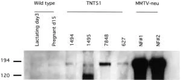
The phenotype of the TNT transgenic mice was considerably more subtle than that of the MMTV/activated neu line, which developed mammary gland tumors in multiparous animals within 3 to 4 months of life. Since the transgene was the same in both mouse models, we hy-pothesized that differences may exist in expres-sion levels. Lysates were prepared from several tumors of the TNT mice and analyzed by west-ern blot with anti-ErbB-2 antibodies (Fig. 7). Levels of the transgene varied among TNT tumor samples, but all tumors showed elevated levels of expression when compared with wild type mammary tissue. In addition, all of the lysates examined showed considerably lower expres-sion levels than those of the MMTV/neutumors.
Discussion
The correlation between over-expression and amplification of the c-erbB-2 gene and human
cancer is persuasive and well documented (33,34). Equilibrium between monomeric and dimeric erbB-2 protein may play a role in ex-plaining the importance of gene over-expres-sion in carcinomas. As gene amplification and over-expression occurs, the ratio of protein shifts to the dimerized state, which leads to higher levels of receptor phosphorylation and activation of cellular substrates. It therefore fol-lows, and has been shown in vitro (16,17), that high levels of expression of neuwill transform cells in culture; whereas, lower levels tran-scribed from weaker promoters may not. Al-though not able to transform cells over a short period of time, we hypothesized that a weak promoter driving low levels of neuthroughout the life of the mouse would have a pathologi-cal effect on the mammary gland. To create a mouse model for human breast cancer, we am-plified neu by adding a transgene of the neu
promoter directing neucDNA expression. This promoter should have expression levels more closely related to those seen in patients than Fig. 6. Mammary gland whole mounts and
tu-mor histopathology of the TNT transgenic mouse. (A) One year virgin mammary whole mount compared with (B), its TNT transgenic vir-gin littermate. Both mammary glands display nor-mal ductal structure; whereas, the transgenic shows aberrant proliferation of lobuloeolar structures along the ducts. (C) Wild type adult multiparous mammary gland has undergone complete
those achieved in past models. Furthermore, the neupromoter should regulate temporal and spatial transcription of neuin a way that phys-iologically parallels cases of human cancer in-volving neuamplification and overexpression. Examination of expression levels in MCF-7 cells, a human mammary epithelial cell line, helped to fortify this belief. The promoter was found to be transcriptionally active, but at a lower level than that observed with the MMTV promoter.
The transgenic mouse line created with a single inserted copy of this construct, called NN60, does show elevated levels of neuin the mammary gland. The extremely sensitive method of RT-PCR is needed to show tran-scription of the transgene. Since this protocol is not quantitative, we cannot compare the level of transgene in the various stages of mammary gland development nor compare it to the levels of endogenous neu. It is clear, however, that RNA from the transgene is present in the vir-gin, pregnant and regressed mammary gland, while absent or below the level of detection in the lactating gland. It should be noted that an identical pattern of expression was seen by others when examining endogenous neu
through use of RT-PCR (24). In addition, the promoter proved to be active in the ovary, testis, lung, and heart of the adult mouse, while inactive in the liver and kidney. Most of these findings confirm previous expression studies (26). Of notable exception was the study that showed neuexpression in the adult rat kidney (35), leading one to expect expres-sion in the mouse as well. In addition,
al-though there have been no rodent studies doc-umenting the presence of neuin the testis, one report of immunostaining for erbB-2 indicated that the testes were negative in adult human tissue (36). Whether these are species-specific differences or the result of upstream enhancer elements missing from the transgenic promoter remains to be evaluated. Perhaps a more inter-esting facet concerning tissue expression is that not all tissues over-expressing neu show a pathological phenotype. The transgenic ani-mals have a normal lifespan and, although there are clearly phenotypes in the female mammary gland, organs such as the heart, ovary and lung appear normal. It is not clear, however, whether this difference in organ re-sponse to the transgene is due to levels of ex-pression within the organ or the need for addi-tional, tissue-specific factors to influence the activity of neu.
may be useful in the study of the effect of other oncogenes on the virgin gland.
The proliferative phenotype is enhanced once the mouse has undergone multiple rounds of pregnancy and regression. Rather than reverting to a near virgin state, the gland continues to produce milk proteins and lipids long after weaning. These results present an in-teresting contrast to those obtained from the MMTV/neu transgenic mouse (19), wherein mammary gland function and development was normal until the appearance of focal tu-mors. This may be due to the fact that neu, un-der the direction of the transgenic neu pro-moter, appears to be expressed evenly throughout the gland, as can be seen by ob-serving its proliferative effects in a mammary gland whole mount. The MMTV promoter, however, tends to be active in some regions of the mammary gland, while inactive in others. This model also raises interesting questions concerning the protective effect of pregnancy against breast cancer. It has long been estab-lished that a woman’s risk of developing breast cancer decreases if she has undergone full-term pregnancy before the age of twenty (37). This model suggests, however, that in cases where the neu gene is amplified, there is a prolifera-tive effect associated with pregnancy, as can be seen in the whole mounts of the multiparous mammary glands.
Oncogenic forms of neutransgenes have fa-cilitated the study of the transforming potential of neuin mice, although their physiologic rele-vance has been questionable. An activating point mutation in the transmembrane ErbB-2,
similar to the rat Val664Glu has never been identified in human breast cancer patients. Re-cent work, however, revealed somatic muta-tions within the transgene of MMTV/neumice over-expressing the non-transforming proto-oncogene, which lead to elevated levels of ErbB-2 tyrosine phosphorylation (38). Our mice carrying the neu promoter/transforming
neutransgene recapitulate the phenotype seen in the NN60 line and develop palpable mam-mary gland tumors at approximately 14.5 months of age. These tumors not only have a much longer latency than those of the MMTV/activated neu mice, but show signifi-cantly lower levels of transgenic protein.
The mouse models described here show that even a small perturbation in neu expres-sion can lead to a clear phenotype in the mam-mary gland. This reinforces the strong
correla-tion between neuamplification and breast can-cer. In addition, these models demonstrate the usefulness of the neupromoter in the study of oncogenes in the virgin mammary gland.
Acknowledgments
We would like to thank A. Harrington for mi-croinjection and significant technical assistance and H. Chen and J. Pinkas for extensive review of this manuscript. We also thank M. Bedford, H. Chen, K Fitzgerald, and J. Pinkas for tech-nical assistance and advice on experimental ap-proaches.
References
11. Shih C, Padhy LC, Murray M, Weinberg RA. (1981) Transforming genes of carcinomas and neuroblastomas introduced into mouse fibro-blasts. Nature290:261–264.
12. King CR, Kraus MH, Aaronson SA. (1985) Am-plification of a novel v-erbB-related gene in a hu-man mammary carcinoma. Science229:974–976.
13. Coussens L, Yang-Feng TL, Liao YC, et al. (1985) Tyrosine kinase receptor with extensive homology to EGF receptor shares chromosomal location with neu oncogene. Science 230: 1132– 1139.
14. Semba K, Kamata N, Toyoshima K, Yamamoto T. (1985) A v-erbB-related protooncogene, c-erbB-2,
is distinct from the c-erbB-1/epidermal growth factor-receptor gene and is amplified in a human salivary gland adenocarcinoma. Proc. Natl. Acad. Sci. U.S.A.82:6497–6501.
15. Zeillinger R, Kury F, Czerwenka K, et al. (1989) HER-2 amplification, steroid receptors and epi-dermal growth factor receptor in primary breast cancer. Oncogene4:109–114.
16. Prigent SA, Lemoine NR. (1992) The type 1 (EGFR-related) family of growth factor recep-tors and their ligands. Prog. Growth Factor Res. 4:
1–24.
17. Slamon DJ, Godolphin W, Jones LA, et al. (1989) Studies of the HER-2/neuproto-oncogene in hu-man breast and ovarian cancer. Science244:707– 712.
18. Parkes HC, Lillycrop K, Howell A, Craig RK. (1990) C-erbB2 mRNA expression in human breast tumours: comparison with c-erbB2 DNA amplification and correlation with prognosis. Br. J. Cancer61:39–45.
10. Tiwari RK, Borgen PI, Wong GY, Cordon CC, Osborne MP. (1992) HER-2/neu amplification and overexpression in primary human breast cancer is associated with early metastasis. Anti-cancer Res.12:419–425.
11. Makar A, Desmedt EJ, DePotter CT, Vanderhey-den JD, Schatteman ES. (1990) Neuoncogene in breast cancer and its possible association with the risk of distant metastases. Acta Oncologica29:
931–934.
12. Slamon DJ, Clark GM, Wong SG, Levin WJ, Ull-rich A, McGuire WL. (1987) Human breast can-cer: correlation of relapse and survival with am-plification of the HER-2/neu oncogene. Science
235:177–182.
13. Persons DL, Borelli KA, Hsu PH. (1997) Quanti-tation of HER-2/neuand c-mycgene amplification in breast carcinoma using fluorescence in situ hybridization. Mod. Pathol.10:720–727.
14. Berger MS, Locher GW, Saurer S, et al. (1988) Correlation of c-erbB-2 gene amplification and protein expression in human breast carcinoma with nodal status and nuclear grading. Cancer Res.48:1238–1243.
15. Hudziak RM, Schlessinger J, Ullrich A. (1987) Increased expression of the putative growth fac-tor recepfac-tor p185HER2 causes transformation and tumorigenesis of NIH 3T3 cells. Proc. Natl. Acad. Sci. U.S.A.84:7159–7163.
16. Hung MC, Schechter AL, Chavray PY, Stern DF, Weinberg RA. (1986) Molecular cloning of the
neu gene: absence of gross structural alteration in oncogenic alleles. Proc. Natl. Acad. Sci. U.S.A.
83: 261–264.
17. Di Marco E, Pierce JH, Knicley CL, Di Fiore PP. (1990) Transformation of NIH 3T3 cells by over-expression of the normal coding sequence of the rat neugene. Mol. Cell Biol.10:3247–3252. 18. Muller WJ, Sinn E, Pattengale PK, Wallace R,
Leder P. (1988) Single-step induction of mam-mary adenocarcinoma in transgenic mice bear-ing the activated c-neu oncogene. Cell 54: 105– 115.
19. Guy CT, Webster MA, Schaller M, Parsons TJ, Cardiff RD, Muller WJ. (1992) Expression of the
neu protooncogene in the mammary epithelium of transgenic mice induces metastatic disease.
Proc. Natl. Acad. Sci. U.S.A.89:10578–10582. 20. White MR, Hung MC. (1992) Cloning and
char-acterization of the mouse neupromoter. Oncogene
7:677–683.
21. Cardiff RD, Muller WJ. (1993) Transgenic mouse models of mammary tumorigenesis. Cancer Surv.
16:97–113.
22. Pittius CW, Sankaran L, Topper YJ, Henning-hausen L. (1988) Comparison of the regulation of the whey acidic protein gene with that of a hybrid gene containing the whey acidic protein gene promoter in transgenic mice. Mol. Endocrinol.
2:1027–1032.
23. Knepper JE, Medina D, Butel JS. (1986) Differ-ential expression of endogenouse mouse mam-mary tumor virus genes during development of the BALB/c mammary gland. J. Virol. 59: 518– 521.
24. Schroeder JA, Lee DC. (1998) Dynamic expres-sion and activation of ERBB receptors in the de-veloping mouse mammary gland. Cell Growth Dif-fer.9:451–464.
25. Flanagan JG, Leder P. (1988) neuProtooncogene fused to an immunoglobulin heavy chain gene requires immunoglobulin light chain for cell surface expression and oncogenic transforma-tion. Proc. Natl. Acad. Sci. U.S.A.85: 8057–8061. 26. Dougall WC, Qian X, Peterson NC, Miller MJ,
Samanta A, Green MI. (1994) The neuoncogene: signal transduction pathways, transformation mechanisms and evolving therapies. Oncogene9:
2109–2123.
27. Sympson CJ, Talhouk RS, Alexander CM, et al. (1994) Targeted expression of stromelysin-1 in mammary gland provides evidence for a role of proteinases in branching morphogenesis and the requirement for an intact basement mem-brane for tissue-specific gene expression. J. Cell Biol.125:681–693.
28. Daniel CW, Silberstein GB. (1987) In: Neville MC, Daniels CW (eds.) The Mammary Gland Devel-opment, Regulation, and Function. Plenum Press, New York, N.Y., pp. 3–36.
29. Borellini F, Oka T. (1989) Growth control and differentiation in mammary epithelial cells. En-viron. Health Perspect.80:85–99.
30. Lee M, Kim H, Jeon D, et al. (1996) Iron metab-olism-related genes and mitochondrial genes are induced during involution of mouse mam-mary gland. Biochem. Biophys. Res. Commun. 224:
164–168.
31. Teng C, Pentecost BT, Chen YH, Newbold RR, Eddy EM, McLachlan JA. (1989) Lactotransfer-rin gene expression in the mouse uterus and mammary gland. Endocrinology 124:992–999. 32. Strange R, Li F, Saurer S, Burkhardt A, Fris RR.
(1992) Apoptotic cell death and tissue remodel-ling during mouse mammary gland involution.
Development115:49–58.
33. Ross JS, Fletcher JA. (1998) The HER-2/neu
oncogene in breast cancer: prognostic factor, predictive factor, and target for therapy. Stem Cells
16:413–428.
34. Revillion F, Bonneterre J, Peyrat JP. (1998)
ERBB2 oncogene in human breast cancer and its clinical significance. Eur. J. Cancer 34: 791– 808.
35. Kokai Y, Cohen JA, Drebin JA, Greene MI. (1987) Stage- and tissue-specific expression of the neu oncogene in rat development. Proc. Natl. Acad. Sci. U.S.A.84:8498–8501.
normal human adult and fetal tissues. Oncogene
5:953–962.
37. MacMahon B, Cole P, Lin TM, et al. (1970) Age at first birth and breast cancer risk. Bull. World Health Organ.43: 209–221.
38. Siegel PM, Dankort DL, Hardy WR, Muller WJ. (1994) Novel activating mutations in the
neu proto-oncogene involved in induction of mammary tumors. Mol. Cell Biol. 14: