R E S E A R C H A R T I C L E
Open Access
Panoramic radiography and cone-beam computed
tomography findings in preoperative examination
of impacted mandibular third molars
Ilkay Peker
1*, Cigdem Sarikir
1, Meryem Toraman Alkurt
1and Zeynep Fatma Zor
2Abstract
Background:Preoperative radiographic examination of impacted mandibular third molars (IMTM) is essential to prevent inferior alveolar nerve injury during extraction. The purpose of this study was to evaluate the correlation between cone-beam computed tomography (CBCT) and digital panoramic radiography (DPR) findings in preoperative examination of IMTM.
Methods:This retrospective study included 298 teeth in 191 individuals. The relationship between the inferior alveolar canal (IAC) and the IMTM (buccal, lingual, interradicular or inferior), the position of the IMTM with respect to the IAC (contact, no contact), the morphologic shape of the mandible in the IMTM region (round, lingual extended, lingual concave), the type of IMTM (vertical, horizontal or angular) and the number of roots of the IMTM were evaluated on CBCT images. DPR images were evaluated for the number of roots of the IMTM and for the most common
radiographic findings indicating a relationship between the IAC and the IMTM (darkening of the roots, diversion of the IAC, narrowing of the IAC and interruption of the white line). Data were statistically analyzed with Cramer V coefficient, Kappa statistic, chi-square and Fisher’s exact test.
Results:There was a significant difference in number of roots detected on DPR versus CBCT images. There was a significant association between the type of IMTM and the morphologic shape of the mandible on CBCT images. Darkening of the roots and interruption of the white line on DPR images were significantly associated with the presence of contact between the IMTM and the IAC on CBCT images.
Conclusions:Panoramic radiography is inadequate, whereas CBCT is useful to detect multiple roots of IMTM. When darkening of the roots and interruption of the white line are observed on panoramic images, there is increased likelihood of contact between the IMTM and the IAC. CBCT is required in these cases.
Keywords:Cone-beam computed tomography, Digital panoramic radiography, Impacted mandibular third molars, Inferior alveolar canal
Background
Extraction of impacted mandibular third molars (IMTM) is a routine procedure in oral surgery, with several pos-sible postoperative complications [1]. The most common complications are injury to the inferior alveolar nerve (IAN) or to the lingual nerve, dysesthesia and lingual fracture of the mandible [2-4]. The incidence of tempor-ary IAN injury related to extraction of IMTM varies
from 0.4% to 9.4% [5-8]. In contrast, the rate of perman-ent IAN injury is reported to be less than 1% [9]. Factors that increase the risk of nerve damage include close proximity between the third molars and the inferior alveolar canal (IAC) and the presence of direct contact between the tooth roots and the IAN [10-12]. Some authors have reported that the most important factor for IAN injury is the anatomical relationship between the impacted third molar and the IAC [13,14]. Add-itionally, it has been reported that factors such as sur-geons’ experience, operative procedures, institutional * Correspondence:drilkaypeker@gmail.com
1
Department of Dentomaxillofacial Radiology, Gazi University Faculty of Dentistry, 82. Sok No: 4 06510, Emek-Ankara, Turkey
Full list of author information is available at the end of the article
settings, and anatomical and radiographic factors can affect the likelihood of IAC damage [8,15].
Accurate assessment of the relationship between the IMTM and the IAC before surgery is necessary to avoid IAN injury [16]. Panoramic radiography is frequently used as the standard diagnostic imaging method for this purpose in clinical practice [17]. In many cases, pano-ramic images are sufficient for preoperative assessment of IMTM; however, this technique cannot provide any information about the buccolingual direction [18]. As-sessment of the buccolingual direction is very important for cases in which the IMTM and the IAC are in close proximity [18,19]. Three-dimensional (3D) imaging with conventional computed tomography and cone-beam com-puted tomography (CBCT) is recommended in these cases to detect the exact relationship [18].
CBCT was developed for dentomaxillofacial imaging because it produces a lower radiation dose with high spatial resolution, is affordable and requires less space than conventional computed tomography [17-20]. Previ-ous studies have reported that CBCT is more accurate than conventional methods such as panoramic radiog-raphy for determining the relationship between impacted third molars and the IAC [17,21-24].
This study provides information to assist clinicians in deciding when CBCT is required in the preoperative examination of IMTM. The study’s purpose was to de-scribe and evaluate the correlation between CBCT and digital panoramic radiography (DPR) findings in detect-ing the number of roots of IMTM and the relationship between panoramic signs and the presence of contact between the IMTM and the IAC.
Methods
Patients
This retrospective study was approved by the Ethical Board of the Institutional Ethics Committee of Ankara University Faculty of Dentistry (Ankara, Turkey). Informed consent was obtained from all volunteers. This study included 298 teeth in 191 individuals applied to our clinic between January 2011 and October 2013. The patients under-went preoperative radiographic examination to evalu-ate the relationship between IMTM and the IAC. IMTM associated with any pathology such as cysts or tumors were excluded from the study.
Imaging
DPR images were obtained using a Veraviewpocs 2D unit (J. Morita Mfg. Corp., Kyoto, Japan), operating at 60–90 kVp, 1–10 mA, with a 0.5 mm focal spot and an exposure time of 7.4 seconds. CBCT images were obtained using a Promax 3D unit (Planmeca Oy, Helsinki, Finland), operat-ing at 84 kVp, 9–14 mA, with a 0.16 mm voxel size, an exposure time of 6 seconds and a field of view of 8 cm.
Image evaluation
DPR and CBCT images were evaluated independently by two oral radiologists with at least 2 years of experience on the computer monitor (21 inch LCD monitor with 1280 × 1024 resolution) in a quiet room with subdued ambient lighting. The observers were allowed to ma-nipulate the contrast and brightness features and to use the software’s zoom tool. Because this was a retrospect-ive study and the patients had been scanned with stand-ard settings, the dataset could not be reoriented as suggested by Lübbers et al., who reported that oblique plane scanning, which needs a small number of slices with a relatively small volume, can be helpful if the IAC is difficult to visualize [19,25]. After 30 days, all images were re-evaluated by the same two observers.
The number of tooth roots (1, 2, 3 or 4) was evaluated on DPR and CBCT images. Roots were considered to be separate when the furcation was located in the cervical or middle third of the roots [22]. The relationship be-tween the IMTM and the IAC was evaluated on pano-ramic images according to criteria established by Rood and Shehab [26]. In the present study, the most common radiographic findings (darkening of the roots, diversion of the mandibular canal, narrowing of the mandibular canal and interruption of the white line) were examined as previously described by Szalma et al. [27].
IMTM were classified into three types based on their orientation on cross-sectional slices of CBCT images: type A, vertical (impacted teeth oriented in an upright position, 90° to the mandible); type B, horizontal (impacted teeth oriented parallel to the mandible); and type C, angular (teeth angled in a forward/backward position or < 90° to the mandible) [21]. The buccolingual relationship between the IMTM and the IAC was classified as buccal, lingual, interradicular or inferior [23]. The position of the IAC with respect to the third molar was classified as contact (no bone between the IAC and the third molar) or no contact (bone between the IAC and the third molar). The morphologic shape of the bone in the third molar region was classified as: type 1, round (round on both buccal and lingual sides); type 2, lingual extended (slightly straight on the buccal side with a bony extension on the lingual side); and type 3, lingual concave (lingual concave on the lingual side and round on the buccal side) [21].
Data analysis
Correlation between DPR and CBCT images was evalu-ated by chi-square and Fisher’s exact tests, with a signifi-cance level of p < 0.05.
Results
The study comprised 123 women (64.4%) and 68 men (35.6%) between 19 and 61 years of age (mean 30.1 years). Intraobserver agreement was excellent for all variables and for both imaging methods, according to Cramer V co-efficient (Observer 1: 0.904 for DPR and 0.964 for CBCT; Observer 2: 0.946 for DPR and 0.962 for CBCT). Interob-server agreement was moderate for the number of roots on DPR images (0.632) and was excellent (0.888) for the other variables, according to Cohen’s Kappa coefficient.
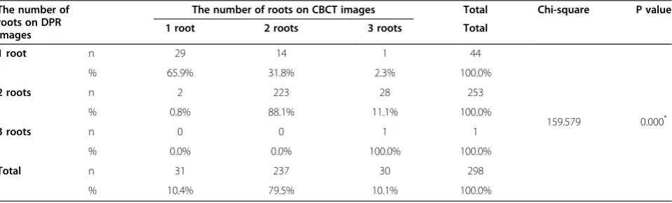
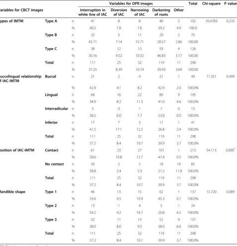
There was a significant difference in the number of third molar tooth roots detected on DPR versus CBCT images (p < 0.05; Table 1). There was a significant associ-ation between darkening of the roots and interruption of the white line on DPR images and the presence of con-tact between the IMTM and the IAC on CBCT images (p < 0.05). No significant association was found between other variables on DPR images and the position of the IAC-IMTM on CBCT images (p > 0.05; Table 2). There was a significant association (p < 0.05) between type of IMTM and the morphologic shape of the mandible on CBCT images (Table 3). Angular IMTM were most often found in patients with round and lingual extended mandi-bles, while vertical IMTM were most often found in lin-gual concave mandibles. The IAC was most often located on the lingual side of the IMTM, and there was often con-tact between the IMTM and the IAC. However, no statisti-cally significant difference was found (p > 0.05) between the position of the IAC-IMTM and the buccolingual rela-tionship of the IAC-IMTM on CBCT images (Table 4).
Discussion
IAN injury is a serious complication during extraction of mandibular third molars. Risk factors for injury include
surgeon’s experience, age and sex of the patient, opera-tive tissue damage, postoperaopera-tive edema and surgical procedures [10]. It has been reported that the most im-portant factor for IAN injury is the anatomical relation-ship between the impacted third molar and the IAC [13,14]. However, other authors have emphasized that multiple factors, including surgeon’s experience, surgical technique, institutional setting, and anatomical and radiographic factors are associated with an increased risk of IAC damage [8,15].
Accurate preoperative evaluation is necessary for suc-cessful surgery because the oral surgeon must know the angle and/or type of impacted third molar to select a suitable procedure and to prevent IAN injury and perforation and fracture of the mandible [10]. Panoramic radiography is a standard diagnostic tool for initial assessment of the relationship between the IMTM and the IAC. Because this method produces two-dimensional images, it cannot provide information in axial, coronal and sagittal planes [10]. CBCT is a more reliable im-aging method in the preoperative assessment of man-dibular third molars [10,15,17,19,21,24,27]. In this study, the correlations between preoperative DPR and CBCT findings and intra- and interobserver agreement were investigated.
The number of mandibular third molar roots visible on DPR versus CBCT images has been investigated in relatively few studies [22,28]. These studies reported that panoramic radiography has limited accuracy in deter-mining the number of roots and that CBCT is more reliable for this purpose [22,28]. In the present study, intraobserver agreement was excellent for all variables on both DPR and CBCT images, whereas interobserver agreement was moderate for detection of number of roots on DPR images. Also, there was a statistically sig-nificant difference in number of roots detected on DPR versus CBCT images. The results demonstrated that DPR images were inadequate to detect multiple roots
Table 1 Relationship between DPR and CBCT findings for the number of roots
The number of roots on DPR images
The number of roots on CBCT images Total Chi-square P value
1 root 2 roots 3 roots Total
1 root n 29 14 1 44
159.579 0.000*
% 65.9% 31.8% 2.3% 100.0%
2 roots n 2 223 28 253
% 0.8% 88.1% 11.1% 100.0%
3 roots n 0 0 1 1
% 0.0% 0.0% 100.0% 100.0%
Total n 31 237 30 298
% 10.4% 79.5% 10.1% 100.0%
DPR: Digital panoramic radiography. CBCT: Cone-beam computed tomography. *
of IMTM. This result is in agreement with previous studies [22,28].
Several studies have reported that the risk of IAN injury increases when specific findings are observed on panoramic images taken to determine the relationship between third molars and the IAC [4,24,29-33]. These findings include darkening of the roots and interruption
of the white line of the IAC [24,29-35]. Eyrich et al. reported that narrowing of the IAC increased the risk of IAN impairment [8]. The probability of contact between third molar roots and the IAC was higher in cases with the abovementioned signs on panoramic images [24,29-36]. Gomes et al. reported no statistically significant association between the presence of panoramic radiographic signs and Table 2 Relationship of the variables between DPR and CBCT images
Variables for DPR images Total Chi-square P value
Variables for CBCT images Interruption in white line of IAC
Diversion of IAC
Narrowing of IAC
Darkening of roots
Other
Types of IMTM Type A n 41 8 8 40 5 102 10.4783 0.233
% 40.2 7.8 7.8 39.2 4.9 100.0
Type B n 32 5 11 20 2 70
% 45.71 7.14 15.71 28.57 2.86 100.00
Type C n 38 12 13 59 4 126
% 30.16 9.52 10.32 46.83 3.17 100.00
Total n 111 25 32 119 11 298
% 37.25 8.39 10.74 39.93 3.69 100.00
Buccolingual relationship of IAC-IMTM
Buccal n 21 2 4 21 1 49 11.351 0.499
% 42.9 4.1 8.2 42.9 2.0 100.0%
Lingual n 68 16 22 80 9 195
% 34.9 8.2 11.3 41.0 4.6 100.0%
Interradicular n 5 0 1 7 0 13
% 38.5 0.0 7.7 53.8 0.0 100.0%
Inferior n 17 7 5 11 1 41
% 41.5 17.1 12.2 26.8 2.4 100.0%
Total n 111 25 32 119 11 298
% 37.2 8.4 10.7 39.9 3.7 100.0%
Position of IAC-IMTM Contact n 61 23 27 101 1 213 54.113 0.000*
% 28.6 10.8 12.7 47.4 0.5 100.0%
No contact n 50 2 5 18 10 85
% 58.8 2.4 5.9 21.2 11.8 100.0%
Total n 111 25 32 119 11 298
% 37.2 8.4 10.7 39.9 3.7 100.0%
Mandible shape Type 1 n 46 13 15 62 1 137 13.720 0.089
% 33.6 9.5 10.9 45.3 0.7 100.0%
Type 2 n 13 1 4 5 1 24
% 54.2 4.2 16.7 20.8 4.2 100.0%
Type 3 n 52 11 13 52 9 137
% 38.0 8.0 9.5 38.0 6.6 100.0%
Total n 111 25 32 119 11 298
% 37.2 8.4 10.7 39.9 3.7 100.0%
DPR: Digital panoramic radiography. CBCT: Cone-beam computed tomography. IAC: Inferior alveolar canal.
IMTM: Impacted mandibular third molar. *
IAN paresthesia after third molar extraction [29]. However, Ghaeminia et al. found that there was a significant associ-ation between panoramic radiographic signs and IAN exposure [36], a finding that has been supported by several authors [24,27,35,36]. These authors agreed that CBCT is useful for the assessment of IMTM in the buccolingual direction. In the present study, contact between the third molar roots and the IAC was most often detected on CBCT images in cases with interruption of the white line of the IAC and darkening of the roots on DPR images, a finding in agreement with several previ-ous studies [12,24,27,29-32,34].
Oral surgeons must know the type and/or angle of the impacted third molar before surgery to prevent perforation and fracture of the mandible, and to select appropriate operation procedures [34]. Previous studies have classified the IMTM as vertical, horizontal or angular, based on its orientation to the mandible [21,32]. Tantanapornkul et al. reported that the horizontal type was the most frequent (52%), followed by angular (32%) and vertical (16%) [32]. Momin et al. reported similar results, with 42% horizontal, 37% angular and 21% vertical [21]. Msagati et al. and Syed et al. found that the mesioangular type was the most common (76% in Msagati’s study and 50.75% in Syed’s study) [37,38]. Lübbers et al. reported that mesially
angulated (40.2%) and vertical (29%) were the most com-mon types [19]. In the present study, the most frequent type was found to be angular (42.28%), followed by vertical (34.24%) and horizontal (23.48%). This finding was in agreement with the results of Lübbers et al. [19]. Differences between studies may arise from different study samples.
The shape of the mandible is an important factor in determining the use of elevators during surgery to avoid direct or indirect pressure on the IAN and perforation or fracture of the bone [21,39]. Two-dimensional images cannot provide information about bone morphology. Preoperative palpation of the related region and 3D im-aging are necessary to determine the shape of the man-dible [39-41] and to provide important information for the oral surgeon during elevation. The shape of the pos-terior mandible has been described in the literature by different classifications for various purposes. The poster-ior mandible has been categorized as convex, parallel or undercut for implant placement [39-41]. Watanabe et al. classified the mandible as round, lingual concave or buc-cal concave, and reported that the round shape (61%) was the most common [39]. In contrast, Lin et al. found that the least common type was round (21%) in the posterior mandible [41]. Momin et al. classified the Table 3 Relationship between type of IMTM and morphologic shape of the mandible on CBCT images
Shape of the mandible Types of IMTM Chi-square P value
Vertical Horizontal Angular Total
Round n 35 34 68 137
11.061 0.026*
% 25.5% 24.8% 49.6% 100.0%
Lingual extended n 7 7 10 24
% 29.2% 29.2% 41.7% 100.0%
Lingual concave n 60 29 48 137
% 43.8% 21.2% 35.0% 100.0%
Total n 102 70 126 298
% 34.2% 23.5% 42.3% 100.0%
*
Statistically significant difference p < 0.05. IMTM: Impacted mandibular third molar.
Table 4 Relationship between the position of the IAC-IMTM and the buccolingual position of the IAC-IMTM on CBCT images
Position of IAC-IMTM
Buccolingual relationship IAC-IMTM Chi-square P value
Buccal Lingual Interradicular Inferior Total
Contact n 32 144 12 25 213
6.437 0.092
% 15.0% 67.6% 5.6% 11.7% 100.0%
No contact n 17 51 1 16 85
% 20.0% 60.0% 1.2% 18.8% 100.0%
Total n 49 195 13 41 298
% 16.4% 65.4% 4.4% 13.8% 100.0%
IAC: Inferior alveolar canal.
mandible as round, lingual extended or lingual concave to assess bone morphology in the third molar region for preoperative planning. They reported that the round shape (49%) was the most common, followed by lin-gual concave (32%) and linlin-gual extended (18%) [21]. The authors also investigated the correlation between impaction type and mandibular shape and reported that there was no significant association between these variables. In this study, mandibular shape was classi-fied into three types: round, lingual extended and lin-gual concave. The prevalence of round and linlin-gual concave types was equal (approximately 46%) and the lingual extended was least common (approximately 8%). This finding is in agreement with the results of Momin et al. [21]. Different mandibular shape preva-lence in different studies can arise from racial features. There was a significant difference in impaction type according to mandible shape in this study, in contrast to the findings of Momin et al. [21].
Surgeon’s knowledge about the location of the IAN is very important in the preoperative evaluation of im-pacted third molars to direct the elevator and luxate the involved tooth. IAN injuries commonly occur during third molar removal because of compression and trac-tion on the nerve through movements of the tooth roots [6]. IAN injury may occur during elevation of mesioan-gular impacted third molars because the roots may move downward and may compress the nerve [23]. Also, move-ments of the third molar root in the buccolingual direc-tion can cause compression of the IAN. The surgical approach is generally started on the buccal side of the im-pacted third molar in cases in which the surgeon lacks information about the buccolingual course of the IAN before surgery. However, the IAN may experience un-desirable forces if it is positioned lingually, and IAN injury has been reported in such cases [17,23]. CBCT images allow the clinician to perform comprehensive treatment planning and surgical method selection during preopera-tive assessment [23]. Previous studies have reported that the IAC is most frequently positioned on the lingual side of impacted third molars and that contact between the IAC and the impacted teeth was generally observed in those cases [10,17,23,34]. In the present study, the IAC was most frequently located on the lingual side of the IMTM and they were commonly in contact.
Conclusions
This study revealed that the number of roots cannot be determined accurately on panoramic radiography im-ages. CBCT is useful to detect multiple roots of IMTM. When darkening of the roots and interruption of the white line were observed on panoramic images, the prob-ability of contact between the IMTM and the IAC in-creased. CBCT is required in these cases. These findings
are in agreement with previous studies. A significant asso-ciation was found between mandible shape and third molar impaction type in this study, a finding that differs from previous studies. Further studies are necessary to de-termine the association between mandible shape and type of impaction for mandibular third molars. Conclusion: CBCT is required in the preoperative assessment of IMTM when darkening of the roots and interruption of the white line are observed on panoramic images.
Abbreviations
DPR:Digital panoramic radiography; CBCT: Cone-beam computed tomography; IAC: Inferior alveolar canal; IAN: Inferior alveolar nerve; IMTM: impacted mandibular third molar; 3D: Three-dimensional.
Competing interests
The authors declare that they have no competing interests.
Authors’contributions
IP formulated the conception and design of the study, participated in data acquisition and interpretation of data analysis and prepared the manuscript. CS contributed to data acquisition and interpretation. MTA supervised interpretation, edited, wrote and gave final approval to the manuscript. ZFZ contributed to data acquisition and interpretation. All authors read and approved the final manuscript.
Acknowledgments
The authors would like to thank to Edanz Editing Service for language editing and Dr. Tülin Otbiçer (Specialist in Measurement of Evaluation in Education, Ankara, Turkey) for data analysis.
Author details
1Department of Dentomaxillofacial Radiology, Gazi University Faculty of
Dentistry, 82. Sok No: 4 06510, Emek-Ankara, Turkey.2Department of Oral and Maxillofacial Surgery, Gazi University Faculty of Dentistry, Ankara, Turkey.
Received: 11 April 2014 Accepted: 9 June 2014 Published: 14 June 2014
References
1. Libersa P, Savignat M, Tonnel A:Neurosensory disturbances of the inferior alveolar nerve: a retrospective study of complaints in a 10-year period.
J Oral Maxillofac Surg2007,65:1486–1489.
2. Better H, Abromowitz I, Shlomi B, Kahn A, Levy Y, Shaham A, Chashu G:The presurgical workup before third molar surgery: how much is enough? Sensory nerve impairment following mandibular third molar surgery.
J Oral Maxillofac Surg2004,62:689–692.
3. Roeder F, Wachtlin D, Schulze R:Necessity of 3D visualization for the removal of lower wisdom teeth: required sample size to prove non-inferiority of panoramic radiography compared to CBCT.Clin Oral Investig2012,
16:699–706.
4. Hillerup S:Iatrogenic injury to oral branches of the trigeminal nerve: records of 449 cases.Clin Oral Investig2007,11:133–142.
5. Blondeau F, Daniel NG:Extraction of IMTM: postoperative complications and their risk factors.J Can Den Assoc2007,73:325.
6. Rood JP:Permanent damage to inferior alveolar and lingual nerves during the removal of IMTM. Comparison of two methods of bone removal.Br Dent J1992,172:108–110.
7. Gülicher D, Gerlach KL:Sensory impairment of the lingual and inferior alveolar nerves following removal of IMTM.Int J Oral Maxillofac Surg2001,
30:306–312.
8. Eyrich G, Seifert B, Matthews F, Matthiessen U, Heusser CK, Kruse AL, Obwegeser JA, Lübbers HT:3-Dimensional imaging for lower third molars: is there an implication for surgical removal?J Oral Maxillofac Surg2011,
69:1867–1872.
10. Xu GZ, Yang C, Fan XD, Yu CQ, Cai XY, Wang Y, He D:Anatomic relationship between impacted third mandibular molar and the mandibular canal as the risk factor of inferior alveolar nerve injury.
Br J Oral Maxillofac Surg2013,51:e215–e219.
11. Kipp DP, Goldstein BH, Weiss WW Jr:Dysesthesia after mandibular third molar surgery: a retrospective study and analysis of 1,377 surgical procedures.JADA1980,100:185–192.
12. Monaco G, Montevecchi M, Bonetti GA, Gatto MR, Checchi L:Reliability of panoramic radiography in evaluating the topographic relation nship between the mandibular canal and impacted third molars.JADA2004,
135:312–318.
13. Nakayama K, Nonoyama M, Takaki Y, Kagawa T, Yuasa K, Izumi K, Ozeki S, Ikebe T:Assessment of the relationship between IMTM and inferior alveolar nerve with dental 3-dimensional computed tomography.
J Oral Maxillofac Surg2009,67:2587–2591.
14. Sedaghatfar M, August MA, Dodson TB:Panoramic radiographic findings as predictors of inferior alveolar nerve exposure following third molar extraction.J Oral Maxillofac Surg2005,63:3–7.
15. Lübbers HT, Matthews F, Damerau G, Kruse AL, Obwegeser JA, Grätz KW, Eyrich GK:No plane is the best one-the volume is!Oral Surg Oral Med Oral Pathol Oral Radiol2012,113:421.
16. Flygare L, Ohman A:Preoperative imaging procedures for lower wisdom teeth removal.Clin Oral Investig2008,12:291–302.
17. Ghaeminia H, Meijer GJ, Soehardi A, Borstlap WA, Mulder J, Bergé SJ:
Position of the impacted third molar in relation to the mandibular canal. Diagnostic accuracy of cone beam computed tomography compared with panoramic radiography.Int J Oral Maxillofac Surg2009,38:964–971. 18. Feifel H, Riediger D, Gustorf-Aeckerle R:High resolution computed
tomography of the inferior alveolar and lingual nerves.Neuroradiol 1994,36:236–238.
19. Lübbers HT, Matthews F, Damerau G, Kruse AL, Obwegeser JA, Gratz KW, Eyrich GK:Anatomy of impacted lower third molars evaluated by computerized tomography: is there an indication for 3-dimensional imaging.Oral Surg Oral Med Oral Pathol Oral Radiol Endod2011,111:547–550. 20. Honey OB, Scarfe WC, Hilgers MJ, Klueber K, Silveira AM, Haskell BS, Farman AG:
Accuracy of cone-beam computed tomography imaging of the temporomandibular joint: comparisons with panoramic radiology and linear tomography.Am J Orthod Dentofacial Orthop2007,132:429–438. 21. Momin MA, Matsumoto K, Ejima K, Asaumi R, Kawai T, Arai Y, Honda K,
Yosue T:Correlation of mandibular impacted tooth and bone morphology determined by cone beam computed topography on a premise of third molar operation.Surg Radiol Anat2013,35:311–318. 22. Suomalainen A, Ventä I, Mattila M, Turtola L, Vehmas T, Peltola JS:Reliability
of CBCT and other radiographic methods in preoperative evaluation of lower third molars.Oral Surg Oral Med Oral Pathol Oral Radiol Endod2010,
109:276–284.
23. Ghaeminia H, Meijer GJ, Soehardi A, Borstlap WA, Mulder J, Vlijmen OJ, Bergé SJ, Maal TJ:The use of cone beam CT for the removal of wisdom teeth changes the surgical approach compared with panoramic radiography: a pilot study.Int J Oral Maxillofac Surg2011,40:834–839. 24. Neves FS, Souza TC, Almeida SM, Haiter-Neto DQ, Freitas FN, Bóscolo FN:
Correlation of panoramic radiography and cone beam CT findings in the assessment of the relationship between IMTM and the mandibular canal.
Dentomaxillofac Radiol2012,41:553–557.
25. Lübbers HT, Kruse AL, Obwegeser JA, Gratz KW, Eyrich G:Oblique high resolution tomography: The ideal plane for visualization of the gonial section of the mandibular canal and its related structures?J Healthcare Engineering2012,3:87–104.
26. Rood JP, Shehab BA:The radiological prediction of inferior alveolar nerve injury during third molar surgery.Br J Oral Maxillofac Surg1990,28:20–25. 27. Selvi F, Dodson TB, Nattestad A, Robertson K, Tolstunov L:Factors that are associated with injury to the inferior alveolar nerve in high-risk patients after removal of third molars.Br J Oral Maxillofac Surg2013,51:868–873. 28. Guerrero ME, Botetano R, Beltran J, Horner K, Jacobs R:Can preoperative imaging help to predict postoperative outcome after wisdom tooth removal? A randomized controlled trial using panoramic radiography versus cone-beam CT.Clin Oral Investig2014,18:335–342.
29. Gomes AC, Vasconcelos BC, Silva ED, Caldas Ade F Jr, Pita Neto IC:
Sensitivity and specificity of pantomography to predict inferior alveolar nerve damage during extraction of impacted lower third molars.
J Oral Maxillofac Surg2008,66:256–259.
30. Khan I, Halli R, Gadre P, Gadre KS:Correlation of panoramic radiographs and spiral CT scan in the preoperative assessment of intimacy of the inferior alveolar canal to IMTM.J Craniofac Surg2011,22:566–570. 31. Jerjes W, El-Maaytah M, Swinson B, Upile T, Thompson G, Gittelmon S,
Baldwin D, Hadi H, Vourvachis M, Abizadeh N, Al Khawalde M, Hopper C:
Inferior alveolar nerve injury and surgical difficulty prediction in third molar surgery: the role of dental panoramic tomography.J Clin Dent 2006,17:122–130.
32. Tantanapornkul W, Okouchi K, Fujiwara Y, Yamashiro M, Maruoka Y, Ohbayashi N, Kurabayashi TA:A comparative study of cone-beam computed tomography and conventional panoramic radiography in assessingthe topographic relationship between the mandibular canal and impacted third molars.Oral Surg Oral Med Oral Pathol Oral Radiol Endod2007,
103:253–259.
33. Nakagawa Y, Ishii H, Nomura Y, Watanabe NY, Hoshiba D, Kobayashi K, Ishibashi K:Third molar position: reliability of panoramic radiography.
J Craniofac Surg2007,65:1303–1308.
34. Jung YH, Nah KS, Cho BH:Correlation of panoramic radiographs and cone beam computed tomography in the assessment of a superimposed relationship between the mandibular canal and impacted third molars.
Imaging Sci Dent2012,42:121–127.
35. Jhamb A, Dolas RS, Pandilwar PK, Mohanty S:Comparative efficacy of spiral computed tomography and orthopanthomography in preoperative detection of relation of inferior alveolar neurovascular bundle to the impacted mandibular third molar.J Oral Maxillofac Surg 2009,67:58–66.
36. Szalma J, Lempel E, Jeges S, Olasz L:Darkening of third molar roots: panoramic radiographic associations with inferior alveolar nerve exposure.J Oral Maxillofac Surg2011,69:1544–1549.
37. Msagati F, Simon EN, Owibingire S:Pattern of occurrence and treatment of impacted teeth at the Muhimbili National Hospital, Dar es Salaam, Tanzania.BMC Oral Health2013,13:37.
38. Syed KB, Zaheer KB, Ibrahim M, Bagi MA, Assiri MA:Prevalence of impacted molar teeth among Saudi population in asir region, Saudi Arabia - a retrospective study of 3 years.J Int Oral Health2013,5:43–47. 39. Watanabe H, Mohammad Abdul M, Kurabayashi T, Aoki H:Mandible size
and morphology determined with CT on a premise of dental implant operation.Surg Radiol Anat2010,32:343–349.
40. Quirynen M, Mraiwa N, van Steenberghe D, Jacobs R:Morphology and dimension of the mandibular jaw bone in the interforaminal region in patients requiring implants in the distal areas.Clin Oral Imp Res2003,
14:280–285.
41. Lin MH, Mau LP, Cochran DL, Shieh YS, Huang PH, Huang RY:Risk assessment of inferior alveolar nerve injury for immediate implant placement in the posterior mandible: A virtual implant placement study.
J Dent2014,5712:349–347.
doi:10.1186/1472-6831-14-71
Cite this article as:Pekeret al.:Panoramic radiography and cone-beam computed tomography findings in preoperative examination of impacted mandibular third molars.BMC Oral Health201414:71.
Submit your next manuscript to BioMed Central and take full advantage of:
• Convenient online submission
• Thorough peer review
• No space constraints or color figure charges
• Immediate publication on acceptance
• Inclusion in PubMed, CAS, Scopus and Google Scholar
• Research which is freely available for redistribution