R E S E A R C H A R T I C L E
Open Access
Mutations in
dock1
disrupt early Schwann
cell development
Rebecca L. Cunningham
1, Amy L. Herbert
1, Breanne L. Harty
1,2, Sarah D. Ackerman
1,3and Kelly R. Monk
1,2*Abstract
Background:In the peripheral nervous system (PNS), specialized glial cells called Schwann cells produce myelin, a lipid-rich insulating sheath that surrounds axons and promotes rapid action potential propagation. During development, Schwann cells must undergo extensive cytoskeletal rearrangements in order to become mature, myelinating Schwann cells. The intracellular mechanisms that drive Schwann cell development, myelination, and accompanying cell shape changes are poorly understood.
Methods:Through a forward genetic screen in zebrafish, we identified a mutation in the atypical guanine nucleotide exchange factor,dock1, that results in decreased myelination of peripheral axons. Rescue experiments and complementation tests with newly engineered alleles confirmed that mutations indock1cause defects in myelination of the PNS. Whole mountin situhybridization, transmission electron microscopy, and live imaging were used to fully define mutant phenotypes.
Results:We show that Schwann cells indock1mutants can appropriately migrate and are not decreased in number, but exhibit delayed radial sorting and decreased myelination during early stages of development.
Conclusions:Together, our results demonstrate that mutations indock1result in defects in Schwann cell development and myelination. Specifically, loss ofdock1delays radial sorting and myelination of peripheral axons in zebrafish.
Keywords:dock1, Schwann cell development, Myelination, Zebrafish
Background
Myelin, a lipid-rich multi-membrane structure, is an innovation of jawed vertebrates that enables the efficient conduction of action potentials. Schwann cells are the myelinating glia of the peripheral nervous system (PNS), and one Schwann cell myelinates one axonal segment. Schwann cells are derived from the neural crest and undergo a distinct series of developmental stages [1, 2]. These developmental stages of Schwann cells require migration as well as unique and substantial changes in cell shape. Schwann cell precursors (SCPs) migrate great distances longitudinally down peripheral nerves. SCPs develop into immature Schwann cells, which undergo a unique process called radial sorting in which Schwann cells extend processes into axon bundles and select an
axon to myelinate [3]. Prior to myelination, Schwann cells wrap themselves 1–1.5 times around a selected axon segment in what is termed the pro-myelinating state. A mature Schwann cell extends and wraps its membrane to form a myelin sheath around an axonal segment. Cytoskeletal dynamics are needed to facilitate these different stages of Schwann cell development and extensive changes in cell shape, but the intracellular intermediates between extracellular signals and the re-modeling of the Schwann cell cytoskeleton are not well defined.
The Rho-GTPase Rac1 is well known for its role in
fa-cilitating cell shape changes through regulating
polymerization of the actin cytoskeleton and mediates Schwann cell development [4]. In Schwann cells, diffe-rential levels of Rac1 direct when a Schwann cell stops migrating and begins radial sorting and myelination [5]. Schwann cell-specific ablation of Rac1 in a mouse model causes delays in radial sorting and myelination, as well as aberrant Schwann cell process extension [5–7].
* Correspondence:monk@ohsu.edu
1
Department of Developmental Biology, Washington University School of Medicine, St. Louis, MO 63110, USA
2Vollum Institute, Oregon Health and Science University, Portland, OR 97239,
USA
Full list of author information is available at the end of the article
Furthermore, Rac1 can function downstream ofβ1-integrin in Schwann cells performing radial sorting [5]; however, the intracellular mechanisms that influence the temporal and spatial activation of Rac1 following extracellular signaling during Schwann cell development are not well understood.
Guanine nucleotide exchange factors (GEFs) have the ability to temporally and spatially regulate the activation of RhoGTPases, such as Rac1, because many GEFs can regulate the same RhoGTPase [8]. Roles of specific GEFs during distinct stages of Schwann cell development are beginning to be understood and help to broaden our knowledge of how extracellular signals are translated to intracellular signals in order to facilitate alterations in Schwann cell shape and movement [9–12]. In addition to canonical GEFs, atypical GEFs also have the ability to activate RhoGTPases. One such family of atypical GEFs, the Dock1-related GEFs, is composed of 11 family mem-bers, including Dock1 (also known as Dock180). Dock1 is highly evolutionarily conserved across species and can specifically bind and activate Rac1 [13–15]. In vitro and in vivo studies in various model organisms have shown that Dock1 influences a variety of cytoskeletal-related cell processes such as phagocytosis and cell migration [16–19]. Thus, Dock1 represents an ideal intracellular candidate to study for a role in cell shape regulation.
Although Dock1 has been studied in several biological contexts and is expressed in Schwann cells [20], a role for Dock1 has not yet been described in Schwann cell myelination. The ability of Dock1 to initiate changes in cell shape to facilitate phagocytosis and cell migration makes Dock1 an attractive candidate to investigate for a role in regulated cell shape changes throughout the de-velopment of Schwann cells, particularly during stages of radial sorting and myelination, when Rac1 levels most influence Schwann cell biology [4]. Two other members of the Dock1 family, Dock7, which activates the RhoGT-Pase Cdc42 [20], and Dock8 [21], which can activate Cdc42 and Rac1, have been shown to influence SCP mi-gration through in vitro and in vivo knockdown experi-ments. Therefore, other members of the Dock1 family may also be key intracellular signals regulating the timing of Schwann cell development.
In this study, we utilized zebrafish to study Schwann cell myelination [22], and we identify and characterize Dock1 as a regulator of early Schwann cell myelination. Although previous morpholino experiments in zebrafish
have implicated dock1 in myoblast development and
vasculature morphogenesis [23–25], a role for Dock1 in Schwann cell development has not been examined. In a screen for genetic regulators of myelination, we identi-fied an early stop codon in dock1that causes decreased expression of a mature myelin marker,myelin basic protein (mbp), in the PNS. Transmission electron microscopy (TEM) revealed that fewer axons are myelinated in mutants
during early stages of myelin development, while axon number is not affected. We determined that SCP cell num-ber and migration is not affected indock1mutants. Instead, radial sorting is delayed and early markers of myelination are reduced. These data suggest that Dock1 may contribute to the timely process extension of Schwann cells required for radial sorting and myelination.
Methods and materials
Zebrafish lines and rearing conditions
Zebrafish were reared in accordance with the Washington University IRB and animal protocols and were raised in the Washington University Zebrafish Consortium ( http://zebra-fish.wustl.edu/husbandry.htm). Zebrafish were crossed as either pairs or harems, and embryos were subsequently raised at 28.5 °C in egg water (5 mM NaCl, 0.17 mM KCl, 0.33 mM CaCl2, 0.33 mM MgSO4). Larvae were staged at
hours post fertilization (hpf) and days post fertilization (dpf). The following mutant and transgenic strains were utilized in this study: dock1stl145, dock1stl365, dock1stl366,
Tg(sox10(4.9):nls-eos) [26], Tg(foxd3:gfp) [27], and
Tg(kdlr:mcherry) [28]. Homozygousdockstl145fish are viable
as adults, therefore maternal zygotic (MZ) dock1stl145 ani-mals were generated by crossing a dockstl145/stl145 female with adock1stl145/+male.
Genotyping
To identify adult and larval zebrafish for either rearing or phenotypic analyses, the following primers were used to amplify a region of interest by PCR: stl145 F: 5’-CATA
GGCGTTCTTCACTGAG-3′ and R: 5’-CGTATTTCC
CACTAAACAGC-3′, stl365F: 5’-GCAGCCACTTTAAA
GCTTCCCG-3′ and R: 5’-GCTGCTTACCTTGCCCT
TGTC-3′,andstl366F: 5’-CCAGTGCCTCACTTCATAT
CTCC-3′ and R: 5’ CTCTTAGTCTCACGCAACACT
CATG-3′. After PCR, a restriction enzyme digest assay was performed and the resulting fragments were analyzed on a 3% agarose gel. Thestl145C-to-T mutation disrupts a BstNI site so that the wild-type PCR product is cleaved into 48 and 527 base pair (bp) products, and the mutant PCR product is 575 bp. The stl365allele contains a one bp insertion that disrupts an EcoRV binding site so that the wild-type PCR product is cleaved into 86 and 159 bp products, and the mutant PCR product is 245 bp. The
stl366allele contains a 13-bp deletion that disrupts a
Hpy-CH4III site so that the wild-type PCR product is cleaved into 323 bp and 165 bp products, and the mutant PCR product is 488 bp.
Zebrafish mutant strain generation
dock1stl145was identified in a forward genetic screen
Genome Technology Access Center (GTAC) at Wash-ington University. The wild-type to mutant allele ratio was determined using a bioinformatics pipeline gener-ated in-house, and a SNP subtraction analysis suggested
that dock1 was most likely the gene of interest [30].
dock1 was confirmed as the gene responsible for the
stl145 mutant phenotype through rescue experiments
and complementation tests using two other dock1
mutant alleles, dock1stl365 and dock1stl366, which were generated by TALENs. The TALEN targeter tool (https://tale-nt.cac.cornell.edu/) and GoldyTALEN kit
[31] were utilized to build each TALEN in a pCS2+
backbone. The repeat variable domains chosen for each
stl365 TALEN arm and stl366TALEN arm were: stl365
left arm: NN HD NG HD NI HD HD NG NN NI HD
NN HD NI NN NI NN NI NN NI;stl365right arm: HD
NG NG NG NN NI NN NG NG NN NI HD HD HD
NG NN NI NN NG;stl366left arm: NN NG NG NI NG
NI NG NG HD NI NG HD NG NN NI NI NN NN NI
NN;stl366right arm: NN HD NG NG NI NI NI HD NI
NG NI HD NG NN NI HD HD HD NN HD. The TALEN constructs were transcribed with the mMES-SAGE mMACHINE SP6 ULTRA Kit (Ambion) and equal concentrations (~ 50 pg) of left and right arm mRNA were injected into 1-cell stage wild-type embryos. Lesions that were successfully transmitted to the F0 germline were identified by restriction enzyme digest analysis as described above. Mutant bands were gel ex-tracted using a QIAquick Gel Extraction kit (Qiagen) and then Sanger sequenced to identify the lesion.
Posterior lateral line nerve (PLLn) dissection and RNA isolation
Posterior lateral line nerves (PLLn) were dissected from 6-month-old adult zebrafish. Animals were euthanized in ice water until gill motion ceased for 5 min, followed by transection of the hindbrain. Using angled forceps, the skin was pulled back from behind the operculum on both sides of the animal to expose the PLLn. Small spring-loaded dissection scissors were used to cut the PLLn near the operculum and then forceps were used to gently remove the nerve by slowly pulling the nerve toward the anterior of the fish. Both nerves were transferred to microcentrifuge tubes sitting on dry ice and then flash fro-zen in liquid nitrogen and stored at −80 °C. To isolate
RNA, 40 nerves were pooled from 20 different
6-month-old adult zebrafish and total RNA was obtained using standard TRIzol (Life Technologies) RNA extraction, with the exception of the homogenization method. Nerves were pooled into a total of 500 μl TRIzol and then tho-roughly homogenized following these steps in succession: vortexing for 30 s, disruption with a plastic-tipped electric homogenizer for 1 min, and passaged through a syringe
and successively smaller needles (22.5 and 27 gauge), 10 times each.
RT-PCR
To make cDNA, 1μg of total RNA was reverse transcribed using the High Capacity cDNA Reverse Transcription Kit (Applied Biosystems) using random hexamers as per manufacturer instructions. RT-PCR for dock1 was per-formed on adult PLLn cDNA. The following primers were
used: F: 5’-CGGAGTGGCCGTCTACAACTATG-3′
(bor-dering exons 1 and 2) and R: 5’-CAAGCCGGAAACAC
ACCCTTC-3′ (bordering exons 3 and 4). Milli-Q water was used as a substrate for a control RT-PCR.
Rescue experiment
Full-length zebrafishdock1 was cloned into pCS2+ using Gibson Assembly.dock1was amplified in two pieces from zebrafish cDNA using a Phusion mastermix (NEB) and the following primers: part 1: F: 5’-TCTTTTTGCAGGAT
CCCATAGAGAAGCGAGAAAAAGTGTG-3′and R: 5’
-CTCCATGATGATCTGCACGTG-3′and part 2: F: 5’-T
CAGCGACATACTGGAGGTGC-3′ and R: 5’-TAATA
CGACTCACTATAGTTGAGGTGTCAGCTGCTTTTCC
G-3′. Gibson Assembly was then performed using an
in-house Gibson reaction mixture (gifted by the
Solnica-Krezel lab, Washington University in St. Louis). Briefly, the fragments were gel extracted and purified using the QIAquick Gel Purification Kit (Qiagen). 30 ng of pCS2 +, linearized with Clal and Xbal, were combined with 5-fold excess of thedock1PCR fragments and 15μl of the Gibson Assembly enzyme-reagent mixture. The mixture was incubated at 50 °C for 1 h and then 10μl were trans-formed into DH5 alpha cells and plated on ampicillin plates. Subsequent colonies were grown, miniprepped with a Qiagen Kit, and Sanger sequenced. Synthetic mRNA for injection was generated by linearizingdock1in pCS2+ with Not1 and then transcribing with the mMESSAGE mMA-CHINE SP6 ULTRA Kit (Ambion). Approximately 120 pg
ofdock1mRNA in 2 nl was injected into 1-cell stage
em-bryos generated from a dock1stl145 heterozygous in-cross. In situ hybridization for mbpwas performed and scoring of expression was performed blinded to genotype.
Whole mount in situ hybridization and qualitative scoring Whole mount in situ hybridization (WISH) was performed as described [32,33] on larvae treated with 0.003% phenyl-thiourea from 24 h post fertilization (hpf) to inhibit pig-mentation until fixation in 4% paraformaldehyde. The previously characterized riboprobes used in this study were:
sox10[34],krox20[35], andmbp[36]. All phenotypes were
“strongly reduced”= patches of mbpexpression or no ex-pression, similar to scoring as performed previously [37]. “Strong” mbp expression was assigned a value of 3, “ re-duced,”a value of 2, and“strongly reduced,”a value of 1 to code each phenotype as a number for a Chi-squared anaylsis.
Transmission Electron microscopy and quantifications TEM was performed on 3 dpf, 5 dpf, and 21 dpf cross-sections of the PLLn according to standard proto-cols [38, 39]. Larvae were cut between body segments 5 and 6 and juvenile 21 dpf fish were cut immediately pos-terior to the heart. A Jeol JEM-1400 (Jeol USA) electron microscope and AMT V601 digital camera were used to image samples. Quantification of percent myelinated axons, sorted axons, total axon number, and number of Schwann cell nuclei was performed on the entire cross section of the PLLn. The scorer was blinded to genotype, and quantification was performed manually as described previously [37].
Lifeact microinjections and live imaging
One-cell stage zebrafish embryos were injected with ~ 15–20 ng of sox10:Lifeact-RFP (a gift from the Lyons lab, University of Edinburgh) and 25 ng of transposase mRNA. 1 dpf larvae were then screened for expression
of sox10:Lifeact-RFP in Schwann cells at 24 hpf. For
live-imaging, larvae were anesthetized in Tricaine and embedded in 0.8% agarose on a 35 mm glass bottom dish filled with 0.2% Tricaine and covered with a 22 × 22 mm2 coverslip on top of vacuum grease [40]. The larvae were then imaged with a Zeiss LSM 880 confocal microscope at 20× for 3 h at 3 min intervals. Still images were captured with a Zeiss LSM 880 II Airyscan FAST confocal microscope at 40xW with a 1.8 zoom. To examine blood vesssls, 4 dpf lar-vae withTg(kdlr:mcherry)were imaged at 13.5× with a Nikon SMZ18 fluorescent dissecting microscope.
Eos Photoconversion and quantification of Schwann cell number
Tg(foxd3:gfp);dock1stl145/+ fish were crossed to Tg(sox10(4.9)
:nls-eos);dock1stl145/+ fish and offspring were screened for
both transgenes at 1 dpf. At 2 dpf, larvae were placed in 0.8% low-melt agarose and mounted for imaging as described above. Before counting, larvae were individually exposed to 30 s of UV light using the DAPI filter with the 20× objective of a Zeiss LSM 880 confocal microscope. The number of GFP and RFP positive cells along the PLLn spanning ~ 8 body segments were the counted manually in ImageJ. The observer was blinded to genotype.
Neuromast labeling and quantification
3 dpf larvae derived from a dock1stl145 heterozygous in-cross were incubated with 50 μl of DASPEI (40 mg/
100 mL in distilled water) in 4 mL of egg water for 15 min at room temperature. The DASPEI solution was removed and replaced with fresh egg water. The number of neuromasts along the PLLn were counted under a fluorescent dissecting microscope using a GFP filter.
Immunohistochemistry
Immunohistochemistry for acetylated tubulin was per-formed as described in [32] with mouse anti-acetylated alpha-tubulin used at a dilution of 1:1000 (Sigma). Larvae were fixed at 4 dpf and were derived from a
dock1stl145 heterozygous in-cross. Heavy myosin within
somites was detected with chicken MF 20 antibody at a dilution of 1:20 (Developmental Studies Hybridoma Bank). MF 20 was deposited to the DSHB by Fischman, D.A. (DSHB Hybridoma Product MF 20). For MF 20 staining, embryos were fixed at 1 dpf in 4% paraformal-dehyde for 1 h and washed twice with 1X PBS for 10 min. Samples were then blocked with 0.05% Triton in PBS and 10% goat serum and then incubated with MF 20 in block overnight at 4 °C. After incubation, larvae were washed twice with PBS and then incubated secondary antibody in PBS for 2 h at room temperature. Primary antibodies were detected IgG2b with secondary antibody conjugated to either Alexa 568 or 488 (Invitrogen) at a 1:2000 dilution. Immunostained larvae were imaged with a Nikon SMZ18 fluorescent dissecting microscope.
Statistical analyses
GraphPad Prism 7 was utilized to perform statistical tests. Unpaired t-tests with Welch’s correction were used to test significance of all TEM, neuromast number, and Schwann cell number data. A Chi-squared analysis was utilized to determine significance for all WISH data. Phenotypes of “strong,” “reduced,”and“strongly reduced”were assigned a number of 3, 2, or 1, respectively, in order to compare phenotypes with a Chi-squared analysis. An unpaired t-test with Welch’s correction showed no significant diffe-rence between wild-type and heterozygous animals; there-fore, for TEM, WISH, neuromast, and Schwann cell number data, wild-type and heterozygous animals were combined as controls.
Results
Mutations indock1result in decreasedmyelin basic proteinexpression in the peripheral nervous system An N-ethyl-N-nitrosourea-based forward genetic screen to identify novel genetic regulators of myelination [29,30] uncovered a mutant, allele designationstl145,that exhibits
reduced mbp expression in the PNS by whole mount in
stages of PNS myelination. At 5 dpf, mbp expression has increased in the stl145 mutant PLLn, but is still reduced compared to sibling controls (Fig. 1c, d). To identify the causative mutation, we employed whole genome sequencing
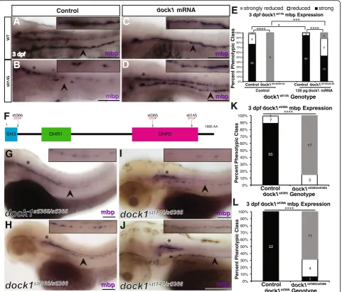
(WGS) of DNA pools from phenotypically mutant and phenotypically wild-type siblings. Analysis of WGS data [30] showed the causative mutation was located on chromosome 12 (Fig. 1e). Within the most highly linked region of Fig. 1stl145mutants exhibit decreasedmbpexpression in the PNS.a-d)Lateral views ofmbpexpression by WISH. Arrowheads indicate the PLLn. Asterisks indicate the central nervous system (CNS). Inset panels show a magnified view of the PLLn. Scale bars = 100μm.a)mbpat 3 dpf is strongly expressed in the PLLn of control larva (n= 93/96).b)stl145mutants at 3dpf exhibit reducedmbpexpression in the PLLn (n= 34).c)mbpexpression is strongly expressed in the PLLn of control larva at 5 dpf (n= 52/62).d)stl145mutants at 5 dpf expressmbp, but at reduced levels compared to control siblings (n= 27/30).e)Analysis of whole genome sequencing data showed that chromosome 12 exhibited the highest mutant to wild-type allele ratio.f)
Within the most highly linked region of chromosome 12,dock1was the only gene that contained an early stop codon.g)A schematic of the protein structure of Dock1 and the location of thestl145lesion. The SH3 and proline rich domains can bind adaptor proteins. The DHR-1 domain interacts with PtdIns(3,4,5)P3and the DHR-2 domain is the catalytic domain can that catalyzes the exchange of GDP for GTP in Rac1.h-i)Quantification of WISH formbp
chromosome 12, the most likely causative mutation was a C-to-T transition resulting in a premature stop codon within the Rac1 binding domain (DHR2 domain) encoding region
of dock1 (Fig. 1f, g). Genotyping revealed that dock1stl145/
stl145
homozygous mutations corresponded to decreased levels of mbp expression in the PNS at 3 dpf (p< 0.0001) and at 5 dpf (p < 0.0001) (Fig.1h-j). To definitively demon-strate that this premature stop codon was causative for the
stl145 phenotype, we performed a rescue experiment with
wild-type dock1 synthetic mRNA. Injection of 120 pg of full-length syntheticdock1mRNA into 1-cell stage embryos derived from an intercross of dock1stl145 heterozygotes suppressed thembp phenotype in stl145 mutants at 3 dpf (p= 0.0001) (Fig.2a-e). Additionally, we generated two new alleles ofdock1using TALENs. Thestl365allele generates a premature stop codon in the DHR2 domain, similar to the
stl145allele (Fig. 2f; Additional file2: Figure S2 A,B). The
stl366 allele causes a premature stop codon generated just
after the SH3 domain (Fig.2 f; Additional file2: Figure S2 C,D). Both alleles exhibit decreased mbp expression in the PNS (p< 0.0001) and fail to complement with thestl145 al-lele (dock1stl145/stl365= 4/4, dock1stl145/stl365= 6/6; Fig. 2 g-l). To confirm thatdock1is expressed in Schwann cells of zeb-rafish in addition to mammalian Schwann cells [20],
RT-PCR for dock1 was performed on cDNA from adult
PLLn, which is enriched in Schwann cell nuclei. This ana-lysis showed thatdock1is expressed in the PLLn (Additional file 2: Figure S2 E,F). Together, these results confirm that
the stl145 phenotype is the result of the premature stop
codon indock1. The phenotype of these new mutants sug-gests a previously unappreciated role of Dock1 in Schwann cell development.
Schwann cell myelination is significantly reduced in dock1stl145mutants at early stages
We next investigated which stages of Schwann cell deve-lopment are affected indock1stl145mutants. To interrogate if the decrease ofmbpexpression indock1stl145mutants is the result of decreased myelination, we employed TEM to analyze the ultrastructure of the PLLn at 3 dpf and 5 dpf. At 3 dpf, consistent with WISH for mbp, the percentage of myelinated axons in the PLLn ofdock1stl145mutants is significantly reduced compared to siblings (p< 0.0001), while the number of axons is not significantly altered (p= 0.0983) (Fig. 3a-d). At 5 dpf, dock1stl145/stl145 mu-tant axon number is similarly unaffected (p= 0.3031) while mutants did exhibit a significant decrease in the percentage of myelinated axons compared to controls (p= 0.0003), although this phenotype is more variable
compared to the mutant phenotype at 3 dpf (Fig. 3
e-h). dock1stl145/+ larvae do not exhibit decreased myelination compared to wild-type siblings at 3 dpf (p= 0.6549 percent myelinated axons;p= 0.7258 total axon number) or 5 dpf (p= 0.7297 percent myelinated axons;
p= 0.6924 total axon number); thus, wild-type and hetero-zygous siblings were combined as controls. The presence of myelinated axons at 5 dpf in dock1stl145/stl145 larvae, although fewer in number compared to controls, illustrates that Schwann cells do possess the capability to myelinate axons indock1mutants. Maternal zygotic (MZ) dock1stl145 mutants at 5 dpf also exhibit a reduction in the number of myelinated axons compared to MZ dock1stl145 heterozy-gotes (p= 0.0183) (Additional file3: Fig. S3 a-d). No signifi-cant difference in the percent myelinated axons was observed between zygotic dock1stl145 mutants and MZ
dock1stl145mutants (p= 0.8300). To test if a defect in
mye-lination is consistent between alleles, we performed TEM
ondock1stl366mutants and siblings. These mutants also
dis-play a significantly decreased percentage of myelinated axons at 3 dpf (p= 0.0072) with no significant difference in axon number (p= 0.3775) (Additional file 3: Figure S3 E-H). A slight reduction in the percent myelinated axons of the PLLn persists at 21 dpf in MZ dock1stl145 mutants
(p= 0.0155), while axon number (p= 0.5831) and
Schwann cell nuclei number (p= 0.1583) are not
significantly altered compared to MZ dock1stl145
het-erozygous controls (Additional file 4: Figure S4).
These results show that dock1 mutations lead to
re-ductions in the number of myelinated axons, and that these effects are more pronounced at early stages of development.
Neither Schwann cell migration nor number are affected indock1stl145mutants
To understand why myelination is decreased indock1stl145 mutants at 3 dpf, we examined earlier stages of Schwann cell development, beginning with SCPs. Importantly, glo-bal development of dock1stl145 mutants is normal and overt PLLn defects are not observed (Additional file 5: Figure S5 A-F). Acetylated tubulin staining shows that axons extend down the trunk of larvae and neuromast
number is not significantly altered (p= 0.7518),
(Additional file5: Figure S5 G-I).dock1stl145/+larvae and wild-type siblings do not exhibit a significant difference in neuromast number (p= 0.0727) and were thus combined as a control. Additionally, blood vessels (kdlr:mcherry positive) and somites (MF 20 positive) can develop in
zyg-oticdock1stl145mutants (Additional file5: Figure S5 J-M).
dock1stl145/stl145mutants also exhibit consistent and strong expression ofsox10 along the PLLn (p= 0.3522), demon-strating that SCP migration is not impaired and that Schwann cells populating the PLLn are thus poised to myelinate (Fig. 4b, c). Dock1 has been shown to regulate the actin cytoskeleton in other systems; therefore, we hypothesized that actin cytoskeletal dynamics might be altered during SCP migration in dock1stl145mutants. We
performed live-imaging of the PLLn in tg(foxd3:gfp);
dock1stl145wild-type, heterozygous, or mutant larvae that
also mosaically expressed sox10:Lifeact-RFP, which binds and fluorescently labels F-actin in cells expressing sox10 [42]. Live-imaging from ~ 30–33 hpf did not reveal any overt defects in migration or in actin cytoskeleton localization (Fig. 4d-k; Additional file 6: Movie S1, Additional file 7: Movie S2, Additional file 8: Movie S3
mbp mbp
mbp
3 dpf dock1stl366mbp Expression
3 dpf dock1stl365mbp Expression
Percent Phenotypic Class
dock1stl365Genotype
mbp
SH3 DHR1 DHR2
1 1866 AA
stl366
STOP stl365STOP stl145STOP
strongly reduced reduced strong
3 dpf mbp
Percent Phenotypic Class
dock1stl145Genotype
3 dpf dock1stl145mbp Expression
Control 120 pg dock1 mRNA
A
WT
stl145
Control dock1 mRNA
B
C
D
E
F
G
H
I
J
K
L
mbp mbp mbp 65 7 3 1 17 0% 10% 20% 30% 40% 50% 60% 70% 80% 90% 100%Control dock1stl365/stl365
22 1 4 11 0% 10% 20% 30% 40% 50% 60% 70% 80% 90% 100%
Control dock1stl366/stl366 29 50 13 8 3 9 1 6 3 0% 10% 20% 30% 40% 50% 60% 70% 80% 90% 100%
Controldock1stl145/stl145
Percent Phenotypic Class
dock1stl366Genotype
dock1stl145/stl145 Control
Fig. 2Mutations indock1cause decreasedmbpexpression in the PNS.a-d)Lateral views ofmbpexpression by WISH at 3 dpf. Arrowheads indicate PLLn. Asterisks indicates the CNS. Inset panels show a magnified view of PLLn. Scale bars = 100μm. a)Control larvae robustly express
mbpin the PLLn (n= 29/38).b)dock1stl145homozygous mutants exhibit strongly reducedmbpexpression in the PLLn (n= 6/6).c)Control larvae injected withdock1mRNA exhibit strong expression ofmbpin the PLLn (n= 50/53).d)dock1stl145homozygous mutants injected withdock1
mRNA robustly expressmbpin the PLLn (n= 13/25).e)Quantification of the percent phenotypic classes larvae were scored formbpexpression in the PLLn at 3 dpf. Control = pooled uninjected and phenol red injected larvae.f)A schematic of the Dock1 protein with the locations of the
stl366,stl365, andstl145lesions indicated.g-j)Lateral views ofmbpexpression by WISH at 3 dpf. Arrowheads indicate the PLLn. Asterisks indicate the CNS. Inset panels show a magnified view of PLLn. Scale bars = 100μm.g)dock1stl365homozygous mutants (n= 20) andh)dock1stl366
homozygous mutants exhibit reducedmbpexpression in the PLLn (n= 15/16).i)dock1stl145/stl365compound heterozygotes andj)dock1stl145/stl366
compound heterozygotes exhibit reducedmbpexpression in the PLLn.k, l)Quantification of WISH formbpfromdock1stl365(k)anddock1stl366(l)
and Additional file 9: Movie S4). In both control and
dock1stl145 mutant larvae, F-actin was consistently
local-ized to the back of migrating SCPs. To our knowledge, this is the first time live actin dynamics have been reported in migrating SCPs in vivo. High-resolution still images also show LifeAct distributed throughout the cell with the highest concentration at the back of SCPs (Additional file10: Figure S6).
Because migration is not affected indock1stl145mutants, we examined whether decreased myelination indock1stl145
mutant nerves was the result of fewer Schwann cells. To do this, we generated and analyzed 2 dpf double trans-genic tg(foxd3:gfp);tg(sox10:nls-eos);dock1stl145 larvae. The
sox10:nls-eos transgene enabled manual counting of
Schwann cell nuclei along the PLLn, while the foxd3:gfp transgene provided a co-label to ensure Schwann cell identity. Counting the number of double positive cells at 2 dpf showed thatdock1stl145/+and wild-type siblings do not exhibit a significant difference in cell number (p= 0.2218) and were thus combined as the control group. No
A B
C
E F
G H
D
A
C
B
D
E
F
G
D’
E’
F’
G’
H
I
J
K
H’
L
M
N
I’
J’
K’
significant difference in the number of Schwann cells between mutants and control siblings was observed (p= 0.1243), suggesting that a reduction in Schwann cell number is not a contributing factor to decreased myelination of the PNS in dock1stl145 mutants (Fig. 4 l-n). Overall, these experiments demonstrate that SCP migration and number are not overtly affected in
dock1stl145 mutants.
Defects in Schwann cell development are first observed during radial sorting and myelination initiation
Given that the dock1stl145 mutation does not alter SCP migration, we next asked if Schwann cell development was affected at the immature and pro-myelinating Schwann cell stages using TEM. At 60 hpf, Schwann cells in MZdock1stl145heterozygous siblings have begun to myelinate axons whereas MZ dock1stl145 mutants are extending processes into axon bundles and can be found in the promyelinating, but not myelinating state (Fig. 5 a-c′). This phenotype suggests that radial sorting by Schwann cells is delayed in in dock1stl145 mutants. We then hypothesized that radial sorting delays at 3 dpf in
dock1stl145 mutants might result in higher numbers of
Schwann cells associated with axons in a 1:1 ratio at 5 dpf as more Schwann cells have entered the pro-myelinating state. Indeed, compared to 3 dpf, a greater number of Schwann cells are associated in a 1:1 ratio with axons indock1stl145mutants (3 dpf:p= 0.6068; 5dpf: p= 0.0086) (Fig. 5 d-i). To further test if Schwann cells are developmentally delayed at the pro-myelinating state, we examined expression of krox20 (egr2), a tran-scription factor that initiates expression of myelin associ-ated genes. By WISH, krox20 expression is significantly decreased along the PLLn of dock1stl145/stl145 mutants compared to wild-type and heterozygous control siblings at 3 dpf (p< 0.0001), demonstrating that dock1stl145 mu-tant Schwann cells are developmentally delayed compared to their siblings (Fig. 5 j-l). This reduction in krox20 expression is not a result of an absence of Schwann cells because sox10 positive Schwann cells are present along
the PLLn by WISH at 3 dpf (p= 0.8141) (Fig.5 m-o). To-gether, these data show that dock1stl145 mutants exhibit delays in development that begin during radial sorting and extend throughout initial myelination of the PNS in zebrafish.
Discussion
A critical component of Schwann cell development is the remodeling of the cytoskeleton to promote shape changes to facilitate proper myelination of the PNS [3, 43]. Al-though some of the intracellular components involved in cytoskeletal rearrangements have been identified, such as Rac1, the full complement of proteins involved in this process has not been comprehensively defined. Through a forward genetic screen in zebrafish, we identified an early stop codon in the Rac1 binding domain ofdock1, an atypical GEF, that causes decreasedmbpexpression in the PNS at 3 dpf and 5 dpf. Rescue experiments and comple-mentation tests with two newly engineered alleles ofdock1 confirmed that mutations in dock1 result in decreased mbpexpression.
TEM analysis showed fewer myelinated axons in mu-tants at 3 dpf, 5 dpf, and 21 dpf, whereas axon number is not significantly affected at any stage assessed. However, we did note that several unmyelinated axons in some mu-tant nerves were abnormally large in diameter and had many mitochondria (data not shown). We demonstrated that reducedmbpexpression and the reduction of myelin-ated axons in mutants is not caused by absence or loss of Schwann cell number. While two other members of the Dock1-family of atypical GEFs, Dock7 and Dock8, affect SCP migration in mammals [20,21], this does not appear to be the case of Dock1 in zebrafish. This is not entirely unexpected, since loss of Rac1 in mouse Schwann cells did not affect Schwann cell migration [5, 6]. Although overt defects in migration were not detected using live-imaging, these experiments enabled visualization of F-actin localization, which showed that F-actin is localized at the back of migrating Schwann cells. This live-imaging data with Lifeact supports previously reported data from
(See figure on previous page.)
Fig. 4Schwann cell migration and number is not affected instl145mutants.a, b)Lateral view of WISH forsox10at 2 dpf. Arrowheads indicate the PLLn. Asterisks indicate the CNS.a)Strongly expressingsox10positive cells are located throughout the PLLn in control larvae (n= 21/23), similar tob)dock1stl145homozygous mutant larva (n= 18).c)Quantification of WISH forsox10at 2 dpf based on phenotypic classes and genotypes for thestl145lesion shows no significant difference in expression (p= 0.3522, Bars represent means ± SD; Chi-squared analysis).d, g’)
A C D F
B
H
I L O
J M
K N
C’ E G
3D culture of Schwann cells showing that migrating Schwann cells in vivo move in an amoeboid-like fashion [44], as contractions seem to occur at the back of the cell. In the future, it will be interesting to generatedock7 and
dock8 zebrafish genetic mutants and observe how
migra-tion and F-actin localizamigra-tion is affected in SCPs.
Although a significant reduction in the percent myeli-nated axons is observed at 5 dpf, Schwann cells in
dock1stl145 mutants do have the capability to myelinate
axons, suggesting thatdock1 is involved in the timing of myelination onset. It is also possible that other Dock1 family members compensate fordock1loss of function in our mutants. Further experiments are needed to deter-mine if Dock1 functions in a Schwann cell-autonomous or non-cell-autonomous manner. Consistent with data showing thatdock1 mutant Schwann cells are delayed in radial sorting and myelination, expression of krox20, a transcription factor essential for expression of myelin genes, is decreased in dock1stl145 mutant nerves. Impor-tantly, Schwann cell number is not affected indock1stl145 mutants and overall PLLn development is not affected in
dock1stl145 mutants compared to controls, as determined
by acetylated tubulin staining and counting neuromast number. Combined, these data demonstrate that Schwann cell radial sorting and myelination are delayed in dock1 mutants.
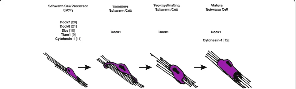
How might Dock1 regulate Schwann cell radial sorting and myelination? GEFs are proposed to aid in regulating the spatial and temporal activation of RhoGTPases, such as Rac1 [8]. During the course of development, Schwann cells undergo unique and critical cell shape changes during migration, radial sorting, and myelination. We hypothesize that to regulate the cytoskeletal rearrange-ments necessary for such functions, Schwann cells utilize a“tool-kit”of GEFs–both canonical and atypical – to activate RhoGTPases, which subsequently remodel the cytoskeleton. For example, Dock7 and Dock8, in addition to canonical GEFs, Dbl’s big sister (Dbs) [10] and Tiam1 [9], have already been shown to regulate SCP migration. Dock1 may be activated after Schwann cells have migrated to subsequently promote radial sorting and early myelination. Because few GEFs have been
(See figure on previous page.)
Fig. 5stl145mutants exhibit delays in radial sorting and decreased expression ofkrox20.a-g)TEM of cross-sections of the PLLn. Myelinated axons are pseudocolored in green and axons associated with promyelinating Schwann cells are pseudocolored in purple. Scale bars = 500 nm.a, b)Micrographs from the same PLLn within a MZdock1stl145heterozygote show Schwann cells myelinating axons at 60 hpf.C)An MZdock1stl145
homozygous larva does not have myelinated axons at 60 hpf, but Schwann cells are extending processes into axon bundles.C′)Magnification of inset fromCshows an axon surrounded by a pro-myelinating Schwann cell.d, e)Schwann cells can myelinate and sort axons at 3 dpf in wild-type (n = 6 animals, 10 nerves) and mutant larvae (n= 4 animals, 6 nerves).f, g)More sorted axons are present in mutants (n= 8 animals, 15 nerves) at 5 dpf compared to controls (n= 11 animals, 17 nerves). Quantification of the number of sorted axons ath)3 dpf andi)5 dpf shows a statistical difference at 5 dpf (unpairedtTest with Welch’s correction).j, k)Lateral view of WISH forkrox20at 3 dpf. Arrowheads indicate PLLn. Inset panels show a magnified view of the PLLn. Scale bar = 50μm.j)krox20is expressed along the PLLn of control larvae (n= 67) whereask)
dock1stl145homozygous mutants express little to nokrox20along the PLLn (n = 18/19).l)Quantification of WISH forkrox20at 3 dpf based on phenotypic classes and genotypes for thestl145lesion (p< 0.0001, Chi-squared analysis).m, n)Lateral view of WISH forsox10at 3 dpf. Arrowheads indicate the PLLn. Inset panels show a magnified view of the PLLn. Scale bar = 50μm.m)Control larvae exhibitsox10positive Schwann cells along the PLLn (n= 37) similar toN)dock1stl145homozygous mutants (n= 16).o)Quantification of WISH forsox10at 3 dpf based on phenotypic classes and genotypes for thestl145lesion (p= 0.8141, Chi-squared analysis). Bars represent means ± SD, **p< 0.001,
****p< 0.0001
Schwann Cell Precursor (SCP)
Immature Schwann Cell
Pro-myelinating Schwann Cell
Mature Schwann Cell
Dock1 Dock1 Dock1
Cytohesin-1 [12] Dock7 [20]
Dock8 [21]
Dbs [10]
Tiam1 [9] Cytohesin-1 [11]
shown to play a role in Schwann cell development in vivo, important next steps in elucidating the discrete sig-nals necessary for development will be to define the rep-ertoire of active GEFs, particularly after SCP migration (Fig.6). RhoGTPases, like Rac1, are ubiquitous and im-portant for initiating cytoskeletal rearrangements as well as other cell biological processes; therefore, differ-ent GEFs may be utilized to activate RhoGTPases cell-specifically. Previously, it has been shown that Rac1 activation regulates Schwann cell radial sorting and mye-lination [6] in addition to promoting the transition from Schwann cell migration to radial sorting [5]. Because Dock1 has been shown to specifically bind and
acti-vate Rac1 in various biological contexts, we
hypothesize that if Dock1 functions
cell-autono-mously, it may be one of many GEFs that activate Rac1 in Schwann cells. Multiple GEFs working to-gether in concert could increase the total levels of ac-tivated Rac1 during a critical period in Schwann cell
development to enable the process extensions
necessary for radial sorting and myelination. One explanation of our data is that Schwann cell radial
sorting and myelination is slower in dock1 mutants
because activated Rac1 levels have not reached a critical threshold to promote these processes. In the future, as better in vivo Rac1 sensors are developed, it will be interesting to test this hypothesis. Addition-ally, Dock1 may function redundantly with other GEFs. As radial sorting and myelination are critical for PNS health, many GEFs may converge on the same pathway such that if only one GEF is
dysfunc-tional, radial sorting and myelination can still
proceed, albeit at a slower rate. Alternatively, Dock1
may function in neurons and mutations in dock1 may
indirectly affect Schwann cell development.
The cell autonomy of Dock1 function and the up-stream signals that trigger Dock1 activation remain to be elucidated. Dock1 has a DHR1 domain that can bind phosphatidylinositols [45] located in the cell membrane, making Dock1 is an attractive candidate to serve as a link between cell surfaces receptor and the cytoskeleton. Additionally, Dock1 could be a representative of a class of drug targets for diseases affecting peripheral myelin, especially because GEFs may contribute to myelination disease states in hu-man patients [4, 46–48]. Although RhoGTPases are critical for cytoskeletal rearrangements, their ubiquity in many cell types limits their ability to serve as useful therapeutic drug targets. Alternatively, GEFs, particularly atypical GEFs like Dock1, could open a door to indirectly affect RhoGTPases in a more cell-specific manner and thus influence the cell shape changes that promote proper Schwann cell develop-ment and myelination.
Conclusions
In this study, we demonstrate that mutations in an atyp-ical GEF, dock1, result in defects in Schwann cell radial sorting and myelination. Schwann cells are slower to ex-tend processes into axon bundles and subsequently mye-linate fewer axons. Schwann cell number and migration are not affected in these mutants; however, Schwann cells in dock1 mutants fail to robustly express markers such as mbp and krox20in early development,
suggest-ing that dock1 aids in the temporal regulation of
Schwann cell radial sorting and development. Moreover, Dock1 may represent a link between extracellular signals and the intracellular cytoskeletal rearrangements neces-sary for radial sorting and myelination.
Additional files
Additional file 1:Figure S1. A-C)Lateral view of WISH formbp. Arrowheads indicate the PLLn. Asterisks indicate the CNS.A)
Representative image of a PLLn scored as“strong”expression, withmbp
strongly and continuously expressed along PLLnB)as“reduced” expression, with reduced but consistentmbpexpression along PLLn and
C)as“strongly reduced”with patchymbpexpression along the PLLn. (PDF 846 kb)
Additional file 2:Figure S2. A)Thestl365allele was generated by a TALEN that resulted in one base pair insertion causing an early stop.B)
Genotyping assay for thestl365lesion. The PCR amplified product is digested with EcoRV and run on a 3% agarose gel.C)Thestl366allele was generated by a TALEN that resulted in a 13 base-pair deletion caus-ing an early stop.D)Genotyping assay for thestl366lesion. The PCR amplified product is digested with HpyCH4III and run on a 3% agarose gel.E)RT-PCR fordock1on adult PLLn cDNA showsdock1is expressed in the PLLn.F)Control reaction performed with Milli-q water as a substrate. (PDF 362 kb)
Additional file 3:Figure S3. A-B)TEM of a cross-section of the PLLn at 5 dpf in MZ siblings. Myelinated axons are pseudocolored in green. Scale bars = 500 nm. A)Axons inMZdock1stl145heterozygotes (n= 4 animals, 6 nerves) contain many myelinated axons whereasB)MZdock1stl145
mu-tants have fewer myelinated axons (n= 5 animals, 8 nerves).C) Quantifi-cation of the percent myelinated axons.D)Quantification of the total number of axons (NS,p= 0.2926).E-F)TEM of a cross-section of the PLLn at 3 dpf indock1stl366siblings. Myelinated axons are pseudocolored in
green. Scale bars = 500 nm.E)Schwann cells in control siblings have myelinated more axons (n = 4 animals, 6 nerves) compared toF)
dock1stl366homozygous mutant nerves (n= 3 animals, 5 nerves).G) Quan-tification of the percent myelinated axons.H)Quantification of the total number of axons (NS,p= 0.3775). Bars represent means ± SD. *p< 0.05, **p< 0.01, unpairedtTest with Welch’s correction. (PDF 1677 kb) Additional file 4:Figure S4. A-B)TEM of a cross-section of the PLLn at 21 dpf in MZ siblings. Scale bars = 10μm. (A’-B′)Magnified images. Scale bars = 2μm.A-A’)Axons inMZdock1stl145heterozygotes (n = 4 animals, 5
nerves) contain many myelinated axons andB-B′)MZdock1stl145mutants have fewer myelinated axons (n= 2 animals, 3 nerves).C)Quantification of the percent myelinated axons.D)Quantification of the total number of axons (NS,p= 0.5831).E)Quantification of the total number of Schwann cell nuclei (NS,p= 0.1583). Bars represent means ± SD. *p < 0.05, unpairedtTest with Welch’s correction. (PDF 2275 kb) Additional file 5:Figure 5.Gross development is normal at 3 dpf comparingA)wild-type,B)heterozygous, andC)mutant larvae from adock1stl145
intercross. Scale bars = 500μm. D-F)Gross development is normal and swim bladders have inflated at 5 dpf comparingD)wild-type,E)heterozygous, andF)
mutant from adock1stl145intercross. Scale bars = 500μm.G)Acetylated tubulin
shows axons are present and well-fasiculated in both wild-type (n = 3) andH)
DASPEI labeling, did not vary between controls (n= 46) or mutants (n= 16) at 3 dpf (NS,p= 0.7518), indicating that global PLLn development is not affected. Bars represent means ± SD; unpairedtTest with Welch’s correction.J)Tg(kdlr:mcherry)
labeling blood vessels at 4 dpf in wild-type andK)dock1stl14/stl145mutants.L)MF
20 staining shows defined somite development in wild-type andM)dock1stl14/ stl145
mutant larvae at 1 dpf. Scale bars = 100μm. (PDF 5492 kb)
Additional file 6:Movie S1.Live-imaging of aTg(foxd3:gfp)wild-type larva (~ 30–33 hpf) injected withsox10:Lifeact-RFP.The larva was imaged every 3 min for 3 h. (AVI 8176 kb)
Additional file 7:Movie S2.Grayscale single channel movie of Lifeact as seen in Additional file6: Movie S1. (AVI 6489 kb)
Additional file 8:Movie S3.Live-imaging of aTg(foxd3:gfp) dock1stl145/ stl145
larva (~ 30–33 hpf) injected withsox10:Lifeact-RFP.The larva was im-aged every 3 min for 3 h. (AVI 5595 kb)
Additional file 9:Movie S4.Grayscale single channel movie of Lifeact as seen in Additional file8: Movie S3. (AVI 2810 kb)
Additional file 10:Figure S6. A)Zeiss Airyscan image ofTg(foxd3:gfp)
wild-type andB)Tg(foxd3:gfp) dock1stl145/+larva (~ 30 hpf) injected with
sox10:Lifeact-RFP.A’-B′)Lifeact-RFP localization within Schwann cell pre-cursors. Scale bars = 10μm. (PDF 557 kb)
Abbreviations
bp:Base pair; CNS: Central nervous system; dpf: Days post fertilization; GDP: Guanosine diphosphate; GEF: Guanine nucleotide exchange factor; GTP: Guanosine triphosphate; hpf: Hours post fertilization;mbp:Myelin basic protein; MF 20: Myosin heavy chain antibody; mm: Millimeter; mM: Millimolar; MZ: Maternal zygotic; ng: Nanogram; nl: Nanoliter; PBS: Phosphate buffered saline; PCR: Polymerase chain reaction; pg: Picogram; PLLn: Posterior lateral line nerve; PNS: Peripheral nervous system;
PtdIns(3,4,5)P3: Phosphatidylinositol (3,4,5)-trisphosphate; SCP: Schwann cell
precursor; SD: Standard deviation; TEM: Transmission electron microscopy; WISH: Whole mount in situ hybridization; WT: Wild-type;μg: Microgram; μm: Micrometer
Acknowledgements
We thank members of the Monk, Solnica-Krezel, and Kaufman laboratories for assistance with the screen and helpful discussions; the Washington University Center for Cellular Imaging; the Washington University Zebrafish Consortium; S. Kucenas (University of Virginia) for theTg(sox10(4.9):nls-eos)line; D. Lyons for the
sox10:lifeact-RFPplasmid; C. Shiau (University of North Carolina) for the
Tg(kdlr:mcherry)line; M. Mokallad (Washington University) for the acetylated alpha-tubulin antibody, and the Solnica-Krezel laboratory (Washington University) for the MF 20 antibody.
Author contributions
A.L.H., B.L.H, S.D.A., and K.R.M. performed the genetic screen and identified thestl145mutant. R.L.C. and K.R.M designed research, and R.L.C. performed research. R.L.C. and K.R.M analyzed data, and R.L.C. and K.R.M. wrote the paper. All authors edited and approved of the manuscript.
Funding
R.L.C. is supported by the National Science Foundation Graduate Research Fellowship (DGE-1745038). This work was also supported by the Philip and Sima Needleman Student Fellowship in Regenerative Medicine (A.L.H.) and by the National Institute of Neurological Disorders and Stroke (NINDS) Grants F31 NS096814 (A.L.H), F31 NS094004 (to B.L.H.), and F31 NS087801 (to S.D.A.), and by a National Institute of Child Health and Human Development Grant R01 HD80601 (to K.R.M.). K.R.M. is a Harry Weaver Neuroscience Scholar of the NMSS.
Availability of data and materials
Data, reagents, and zebrafish lines available on request from the authors.
Ethics approval and consent to participate
Zebrafish were reared in accordance with the Washington University IRB and animal protocols and were raised in the Washington University Zebrafish Consortium (http://zebrafish.wustl.edu/husbandry.htm).
Consent for publication
Not applicable.
Competing interests
The authors have no competing interests.
Publisher’s Note
Springer Nature remains neutral with regard to jurisdictional claims in published maps and institutional affiliations.
Author details
1Department of Developmental Biology, Washington University School of
Medicine, St. Louis, MO 63110, USA.2Vollum Institute, Oregon Health and
Science University, Portland, OR 97239, USA.3Institute of Neuroscience, University of Oregon, Eugene, OR 97403, USA.
Received: 15 May 2018 Accepted: 20 July 2018
References
1. Jessen KR, Mirsky R. The origin and development of glial cells in peripheral nerves. Nat Rev Neurosci. 2005;6:671–82.
2. Monk KR, Feltri ML, Taveggia C. New insights on Schwann cell development. Glia. 2015;63:1376–93.
3. Feltri ML, Poitelon Y, Previtali SC. How Schwann cells sort axons: new concepts. Neuroscientist. 2016;22:252–65.
4. Feltri ML, Suter U, Relvas JB. The function of RhoGTPases in axon ensheathment and myelination. Glia. 2008;56:1508–17.
5. Nodari A, Previtali SC, Dati G, Occhi S, Court FA, Colombelli C, et al. Alpha6beta4 integrin and dystroglycan cooperate to stabilize the myelin sheath. J Neurosci. 2008;28:6714–9.
6. Benninger Y, Thurnherr T, Pereira JA, Krause S, Wu X, Chrostek-Grashoff A, et al. Essential and distinct roles for cdc42 and rac1 in the regulation of Schwann cell biology during peripheral nervous system development. J Cell Biol. 2007;177:1051–61.
7. Guo L, Moon C, Niehaus K, Zheng Y, Ratner N. Rac1 controls Schwann cell myelination through cAMP and NF2/merlin. J Neurosci. 2012;32:17251–61. 8. Rossman KL, Der CJ, Sondek J. GEF means go: turning on RHO GTPases with
guanine nucleotide-exchange factors. Nat Rev Mol Cell Biol. 2005;6:167–80. 9. Yamauchi J, Miyamoto Y, Tanoue A, Shooter EM, Chan JR. Ras activation of a Rac1 exchange factor, Tiam1, mediates neurotrophin-3-induced Schwann cell migration. Proc Natl Acad Sci U S A. 2005;102:14889–94.
10. Yamauchi J, Chan JR, Miyamoto Y, Tsujimoto G, Shooter EM. The neurotrophin-3 receptor TrkC directly phosphorylates and activates the nucleotide exchange factor Dbs to enhance Schwann cell migration. Proc Natl Acad Sci U S A. 2005;102:5198–203.
11. Miyamoto Y, Torii T, Nakamura K, Takashima S, Sanbe A, Tanoue A, et al. Signaling through Arf6 guanine-nucleotide exchange factor cytohesin-1 regulates migration in Schwann cells. Cell Signal. 2013;25:1379–87. 12. Yamauchi J, Miyamoto Y, Torii T, Takashima S, Kondo K, Kawahara K, et al.
Phosphorylation of cytohesin-1 by Fyn is required for initiation of myelination and the extent of myelination during development. Sci Signal. 2012;5 ra69
13. Brugnera E, Haney L, Grimsley C, Lu M, Walk SF, Tosello-Trampont A-C, et al. Unconventional Rac-GEF activity is mediated through the Dock180-ELMO complex. Nat Cell Biol. 2002;4:574–82.
14. Côté J-F, Vuori K. Identification of an evolutionarily conserved superfamily of DOCK180-related proteins with guanine nucleotide exchange activity. J Cell Sci. 2002;115:4901–13.
15. Côté J-F, Vuori K. GEF what? Dock180 and related proteins help Rac to polarize cells in new ways. Trends Cell Biol. 2007;17:383–93.
16. Hasegawa H, Kiyokawa E, Tanaka S, Nagashima K, Gotoh N, Shibuya M, et al. DOCK180, a major CRK-binding protein, alters cell morphology upon translocation to the cell membrane. Mol Cell Biol. 1996;16:1770–6. 17. Reddien PW, Horvitz HR. CED-2/CrkII and CED-10/Rac control phagocytosis
and cell migration in Caenorhabditis elegans. Nat Cell Biol. 2000;2:131–6. 18. Ziegenfuss JS, Doherty J, Freeman MR. Distinct molecular pathways mediate
19. Laurin M, Fradet N, Blangy A, Hall A, Vuori K, Côté J-F. The atypical Rac activator Dock180 (Dock1) regulates myoblast fusion in vivo. Proc Natl Acad Sci U S A. 2008;105:15446–51.
20. Yamauchi J, Miyamoto Y, Chan JR, Tanoue A. ErbB2 directly activates the exchange factor Dock7 to promote Schwann cell migration. J Cell Biol. 2008;181:351–65.
21. Miyamoto Y, Torii T, Kawahara K, Tanoue A, Yamauchi J. Dock8 interacts with Nck1 in mediating Schwann cell precursor migration. Biochem Biophys Rep. 2016;6:113–23.
22. Monk KR, Talbot WS. Genetic dissection of myelinated axons in zebrafish. Curr Opin Neurobiol. 2009;19:486–90.
23. Moore CA, Parkin CA, Bidet Y, Ingham PW. A role for the myoblast city homologues Dock1 and Dock5 and the adaptor proteins Crk and Crk-like in zebrafish myoblast fusion. Development. 2007;134:3145–53.
24. Epting D, Wendik B, Bennewitz K, Dietz CT, Driever W, Kroll J. The Rac1 regulator ELMO1 controls vascular morphogenesis in zebrafish. Circ Res. 2010;107:45–55.
25. Schäker K, Bartsch S, Patry C, Stoll SJ, Hillebrands J-L, Wieland T, et al. The bipartite rac1 guanine nucleotide exchange factor engulfment and cell motility 1/dedicator of cytokinesis 180 (elmo1/dock180) protects endothelial cells from apoptosis in blood vessel development. J Biol Chem. 2015;290:6408–18.
26. McGraw HF, Snelson CD, Prendergast A, Suli A, Raible DW. Postembryonic neuronal addition in zebrafish dorsal root ganglia is regulated by notch signaling. Neural Dev. 2012;7:23.
27. Gilmour DT, Maischein H-M, Nüsslein-Volhard C. Migration and function of a glial subtype in the vertebrate peripheral nervous system. Neuron. 2002;34:577–88.
28. Wang Y, Kaiser MS, Larson JD, Nasevicius A, Clark KJ, Wadman SA, et al. Moesin1 and Ve-cadherin are required in endothelial cells during in vivo tubulogenesis. Development. 2010;137:3119–28.
29. Herbert AL, Fu M-M, Drerup CM, Gray RS, Harty BL, Ackerman SD, et al. Dynein/dynactin is necessary for anterograde transport of Mbp mRNA in oligodendrocytes and for myelination in vivo. Proc Natl Acad Sci U S A. 2017;114:E9153–62.
30. Sanchez NE, Harty BL, O’Reilly-Pol T, Ackerman SD, Herbert AL, Holmgren M, et al. Whole Genome Sequencing-Based Mapping and Candidate Identification of Mutations from Fixed Zebrafish Tissue. G3 (Bethesda). 2017; g3.300212.2017
31. Bedell VM, Wang Y, Campbell JM, Poshusta TL, Starker CG, Ii RGK, et al.In vivogenome editing using a high-efficiency TALEN system. Nature. 2012; 491:114–8.
32. Cunningham RL, Monk KR. Whole mount in situ hybridization and immunohistochemistry for zebrafish larvae. Methods Mol Biol. 2018; 1739:371–84.
33. Thisse C, Thisse B. High-resolution in situ hybridization to whole-mount zebrafish embryos. Nat Protoc. 2008;3:59–69.
34. Dutton KA, Pauliny A, Lopes SS, Elworthy S, Carney TJ, Rauch J, et al. Zebrafish colourless encodes sox10 and specifies non-ectomesenchymal neural crest fates. Development. 2001;128:4113–25.
35. Pogoda H-M, Sternheim N, Lyons DA, Diamond B, Hawkins TA, Woods IG, et al. A genetic screen identifies genes essential for development of myelinated axons in zebrafish. Dev Biol. 2006;298:118–31.
36. Lyons DA, Pogoda H-M, Voas MG, Woods IG, Diamond B, Nix R, et al. erbb3 and erbb2 are essential for schwann cell migration and myelination in zebrafish. Curr Biol. 2005;15:513–24.
37. Petersen SC, Luo R, Liebscher I, Giera S, Jeong S-J, Mogha A, et al. The adhesion GPCR GPR126 has distinct, domain-dependent functions in Schwann cell development mediated by interaction with laminin-211. Neuron. 2015;85:755–69.
38. Cunningham RL, Monk KR. Transmission Electron microscopy for zebrafish larvae and adult lateral line nerve. Methods Mol Biol. 2018;1739:385–400. 39. Czopka T, Lyons DA. Dissecting mechanisms of myelinated axon formation
using zebrafish. Methods Cell Biol. 2011;105:25–62.
40. Cunningham RL, Monk KR. Live imaging of Schwann cell development in zebrafish. Methods Mol Biol. 2018;1739:401–5.
41. Grimsley CM, Kinchen JM, Tosello-Trampont A-C, Brugnera E, Haney LB, Lu M, et al. Dock180 and ELMO1 proteins cooperate to promote evolutionarily conserved Rac-dependent cell migration. J Biol Chem. 2004;279:6087–97. 42. Riedl J, Crevenna AH, Kessenbrock K, Yu JH, Neukirchen D, Bista M, et al.
Lifeact: a versatile marker to visualize F-actin. Nat Methods. 2008;5:605–7.
43. Salzer JL. Schwann cell myelination. Cold Spring Harb Perspect Biol. 2015;7: a020529.
44. Cattin A-L, Burden JJ, Van Emmenis L, Mackenzie FE, Hoving JJA, Garcia Calavia N, et al. Macrophage-induced blood vessels guide Schwann cell-mediated regeneration of peripheral nerves. Cell. 2015;162:1127–39. 45. Côté J-F, Motoyama AB, Bush JA, Vuori K. A novel and evolutionarily conserved PtdIns(3,4,5)P3-binding domain is necessary for DOCK180 signalling. Nat Cell Biol. 2005;7:797–807.
46. Delague V, Jacquier A, Hamadouche T, Poitelon Y, Baudot C, Boccaccio I, et al. Mutations in FGD4 encoding the rho GDP/GTP exchange factor FRABIN cause autosomal recessive Charcot-Marie-tooth type 4H. Am J Hum Genet. 2007;81:1–16.
47. Stendel C, Roos A, Deconinck T, Pereira J, Castagner F, Niemann A, et al. Peripheral nerve demyelination caused by a mutant rho GTPase guanine nucleotide exchange factor, frabin/FGD4. Am J Hum Genet. 2007;81:158–64. 48. Verhoeven K, De Jonghe P, Van de Putte T, Nelis E, Zwijsen A, Verpoorten N,