Balwin N., et al. J Sci Res Pharm, 2018;7(11):138-144
World Inventia Publishers
J
ournal of
S
cientific
R
esearch in
P
harmacy
http://www.jsrponline.com/
Vol. 7, Issue 11, 2018 ISSN: 2277-9469
USA CODEN: JSRPCJResearch Article
GREEN SYNTHESIS OF SILVER NANOPARTICLES AND CHARACTERIZATION USING PLANT LEAF ESSENTIAL OIL
ROSEMARINUS OFFICINALIS AND THEIR ANTIFUNGAL ACTIVITY AGAINST HUMAN PATHOGENIC FUNGI
R.R. Thanighai arassu a, Balwin Nambikkairaj a *,D.R. Ramya a
a PG and Research Department of Zoology Eco-biology wing, Voorhees college, Vellore-632001, Tamil Nadu, INDIA.
Received on: 31-10-2018; Revised and Accepted on: 16-11-2018
ABSTRACT
P
resent study, we utilized the bio–reductive potential of the plant leaf essential oil Rosemarinus officinalis, for the synthesis of silver nanoparticles (AgNPs). The green synthesized AgNPs was achieved at 80°C and found to be highly stable in room temperature for some time. The AgNPs was found to be small dot round in shape with an average size of ~52nm in diameter. The AgNPs were characterized using ultraviolet-visible absorption spectroscopy fourier transform Infrared spectroscopy (FTIR), transmission electron microscopy (TEM),Scanning electron microscopy and Edx (SEM). The anti-fungal activities of the synthesized AgNPs were tested against a few human fungal pathogens Aspergillus niger, Aspergillus flavus, Candida albicans, Candida tropicalis and Candida kefyr showed high activity against these microorganisms. The results showed that the size and the shape can be easily controlled by employing the optimum concentrations ratio of AgNO3 solution and also varying temperature and incubation time. The synthesized AgNPs from Rosemarinus officinalis plant leaf oil can be used as a potent therapeutic agent and for anti-cancer activity.KEYWORDS: Aspergillus niger, Green synthesis, XRD, Silver nanoparticles, Tem.
INTRODUCTION
T
he discovery of the fact that nano silver (Ag) can fight against the microbes, manufacturers have incorporated Ag nano particles (Ag NPs) into more than 200 consumer products, including clothing and cosmetics. They are quickly gaining popularity for their multitude of uses. Despite tremendous benefits, the inevitable release of engineered NPs in the environment with the development of nanotechnology is a serious case of concern of environmental biologists world wide [Bhatt and tripathi, 2011]. As chemical synthesis is a popular approach to the production of metal Ag; however, it requires the use of toxic chemicals as reducing and/or capping substances [Song and kim, 2008; Gole and Murphy, 2004; Meltzer et al., 2001]. Therefore, there is a growing need to simultaneously develop eco-friendly biological processes for the synthesis of Ag that do not use toxic chemicals in the synthesis protocol.Green processes with the use of economic, efficient and ecofriendly catalysis are gaining much importance due to the benefits associated with their use. The major advantage of green synthesis of nanomaterials is their important role in protecting the environment [Saifuddin et al., 2009]. All the green and clean technologies are expected to minimize things that contribute to environmental problems: air and water emissions, greenhouse gases, non-renewable or toxic substances or materials. The utilization of products and materials from various plant species as well as several other biological materials for the synthesis of nano Ag particles are directly related to the mechanisms of nanotechnology and green chemistry. More and more research inputs are rendered for this and there has been an upsurge of interest in the
*Corresponding author:
Balwin Nambikkairaj
PG and Research Department of Zoology Eco-biology wing, Voorhees College, Vellore-632001, Tamil Nadu, INDIA.
* E-Mail: [email protected]
DOI:https://doi.org/10.5281/zenodo.1489509
biological synthesis of nano materials by using several plants, plant pure compounds and plant biochemical compounds in the past few years [Egorova and Revina, 2000; Mukherjee et al., 2002; Zhang et al., 2011].
Nanotechnology has been one of the rapidly growing interdisciplinary areas of science and technology that integrates material science and biology. This technology has broad applications in the fields including electronics, biomedical sciences, pharmaceutical industry, cosmetics preparation, water filtration, and catalytic systems. Developments in nanotechnology lead to the emergence of new materials which contribute to the environmental burden of engineered nanoparticles (NP). This has resulted in a growing environmental impact of direct exposures to NP, which thoroughly established (Brayner, 2008). It is, therefore, imperative to explore both the cost-effective and eco-friendly methods of synthesizing NP and to further evaluate their safety from the standpoint of eco-friendliness (Navarro et al., 2008a; Singh et al., 2009). Plant leaf extract mediated synthesis evaluation of NP is one such green technology that has the potential to address the public concern in the above perspectives. Application of green technology has reduced the use of hazardous reagents and solvents, improved the material and energy efficiency of chemical processes, and enhanced the design of non-toxic products.
This present study the silver nanoparticles were synthesised from plant leaf essential oil Rosemarinus officinalis and these nanoparticles was characterized. Antifungal activity test was done to know the biological activity of synthesized silver nanoparticles against the most pathogenic fungi.
MATERIALS AND METHODS
1. Plant leaf essential oils:
The plant leaf essential oils were purchased from Commercial center Aromax Trading Company, Chennai, Tamil Nadu (India). The silver nitrate (AgNO3) was purchased from HiMedia (Mumbai, India).
2. Fungal Strains:
precipitate was sampled for transmission electron microscopy (TEM) observation. TEM samples of the aqueous suspension of Ag nano particles were prepared by placing a drop of the suspension on carbon-coated copper grids and the films on the TEM grids were allowed to stand for 2 min, after which the extra solution was removed using a blotting paper and the grid was allowed to dry prior to measurement. TEM observations were performed on a HITACHI-JP/H7600 instrument (Japan) operated at an accelerating voltage of 100 kV. The size distribution of the resulting nanoparticles was estimated on the basis of TEM micrographs with the assistance of Sigma Scan Pro software (SPSS Inc., Version 4.01.003). Energy dispersive X-ray (EDX) analyses were performed on a JEOL JSM-6400 microscope (Japan) fitted with Oxford-6506 (England)EDX analyser.
6. FTIR Analysis:
Perkin-Elmer spectrometer FTIR Spectrum ONE in the range4000–400 cm−1 at a resolution of 4 cm−1.was used. The sample was mixed with KCl procured from Sigma. Thin sample disc was prepared by pressing with the disc preparing machine and placed in Fourier Transform Infra Red [FTIR] for the analysis of the nanoparticles.
7. XRD Analysis:
The silver nano particles was coated onto a glass substrate and the XRD measurements were carried out using a Philips X’Pert Pro X-Ray diffractometer, with the following working conditions: CuK_ Ni-filtered radiation;40 kV, 30 mA; divergence slit 0.47◦.
followed (Perez et al., 1990; Perez et al., 1999; Erdemogllu et al., 2003; Bagamboula et al., 2004). Each fungal isolate was suspended in Potato Dextrose broth Himedia Mumbai, Maharashtra (India) broth and diluted to approximately 105 colony forming unit (CFU) per mL. They were “flood inoculated onto the surface of Potato Dextrose agar and then dried. Five-millimeter diameter wells were cut from the agar using a sterile cork-borer, and 25 μl of the samples solutions were delivered into the wells. The plates were incubated for 48 h at room temperature. Antimicrobial activity was evaluated by measuring the zone of inhibition against the test microorganisms. Ethanol was used as solvent control. Amphotericin B was used as reference antifungal agent for molds and fluconazole was used for yeast like fungi. The tests were carried out in triplicate.
RESULTS
1. Invitro antifungal assay:
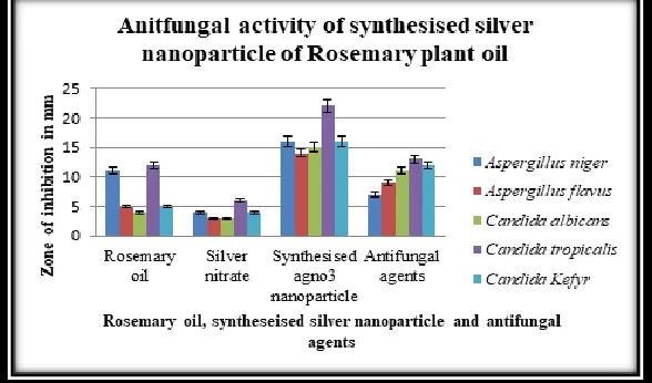
The plant essential oil Rosemarinus officinalis showed notable antifungal activity against Aspergillus niger, Aspergillus flavus, Candida
albicans, Candida tropicalis and Candida kefyr in (Table. 1). The essential
oil Rosemarinus officinalis was very highly active against Candida
tropicalis (12.36±0.48) and least against Candida albicans (4.35±0.35).
Silver nitrate solution was highly active against Candida tropicalis
(6.27±0.44) and least against Candida albicans (3.01±0.33). The silver nanoparticle Rosemarinus officinalis was also highly active against
Candida tropicalis (22.23±0.78) and least against Aspergillus flavus
(14.59±0.58). All fungi were found to be sensitive to all test essential oil
Rosemarinus officinalis and synthesized silver nanoparticle Rosemarinus
officinalis and mostly comparable to the standard reference antifungal
drug Amphotericin B and fluconazole to some extent.
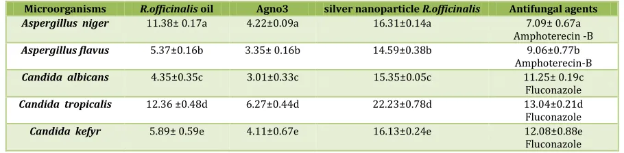
Table No. 1: Antifungal activity of synthesized silver nanoparticle Rosemarinus officinalis oil
Microorganisms R.officinalis oil Agno3 silver nanoparticle R.officinalis Antifungal agents
Aspergillus niger 11.38± 0.17a 4.22±0.09a 16.31±0.14a 7.09± 0.67a Amphoterecin -B
Aspergillus flavus 5.37±0.16b 3.35± 0.16b 14.59±0.38b 9.06±0.77b Amphoterecin-B
Candida albicans 4.35±0.35c 3.01±0.33c 15.35±0.05c 11.25± 0.19c Fluconazole
Candida tropicalis 12.36 ±0.48d 6.27±0.44d 22.23±0.78d 13.04±0.21d Fluconazole
Candida kefyr 5.89± 0.59e 4.11±0.67e 16.13±0.24e 12.08±0.88e Fluconazole
Fig. 1: Inhibition of growth of selected bacteria by synthesized silver nanoparticle Rosemarinus officinalis
2. Biosynthesis of silver nanoparticles:
The plant leaf essential oil Rosemarinus officinalis of 20 ml was added to 80 ml of 2 mM Agno3 solution in a conical flask, and the magnetic stirrer started vigorously stirred inside the conical flask which is on the hot plate the biosynthesis reaction started after continuous stirring with magnetic stirrer for twenty five minutes and the color reaction was observed in which clear Agno3 solution changed into brown color which indicates that formation of corresponding silver nanoparticles shown in (Fig. 2a and 2b). The UV–Vis spectra of silver nanoparticles synthesized by Rosemarinus officinalis leaf oil are showed narrow peak observed at 450 nm was seen in (Fig. 2c).
3. SEM and EDX analysis:
The scanning electron microscope showed the structure of synthesized silver nanoparticles was observed which is in condensed form (Fig 3a). The analysis of energy dispersive spectroscopy (EDS) of the silver nanoparticles the presence of elemental silver signal was confirmed (Fig 3b).
4. TEM analysis:
The transmission electron microscope image of silver nanoparticle was noted in (Fig 4). The picture suggests that the small dot round shaped silver nanoparticles is distributed widely with a diameter of 52 nm.
5. XRD analysis:
The X-ray diffraction pattern of silver nanoparticle was shown in (Fig 5).The XRD pattern thus clearly illustrates that the silver nano particles present green synthesis method are powdery in nature.
6. FTIR analysis:
FT IR spectroscopic studies were carried out to investigate to find possible bioreducing agents present in the plant oil . The spectra of plant oil were recorded before and after adding the silver nitrate solution (Fig. 6). The spectrum indicates major peak at 3472 cm-1.The FTIR spectrum also reveals the presence of alkenes C=C stretch vibration (1743 cm-1), ether C-O stretch (1087 cm-1).In addition to this, other peaks were obtained at 1645 cm-1, 1087 cm-1 and minor peaks at 2933cm-1, 1373 cm-1, 560 cm-1.
The major peak at 3472 cm-1 indicates the presence of OH group and Alkenyl C=C Stretch, Alcoholic C - O Stretch, which plays a major role in the synthesis of seed powder, mediated silver nanoparticles.
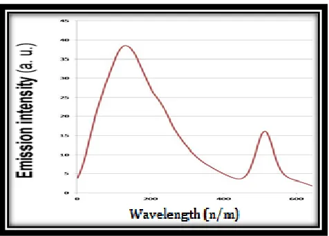
7. Fluroscence Spectroscopy analysis:
The fluroscent spectroscopy of silver nanoparticle were shown in (Fig 7).A broad emission band having prominent peak centered at 185∼ nm is observed for the plant oil as it is excited at 500 nm. In this study emission intensity gradually increases with the decreasing concentration of Agno3.This decreasing intensity suggest that due to the close proximity of emissive species with nanoparticles, quenching of emission take takes place through energy transfer process.
Fig. 2: A) Silver nitrate solution; B) Silver nanoparticleRosemarinus officinalis oil; C) UV–Vis spectrum analysis of silver nanoparticle
reduced by Rosemarinus officinalis plant leaf oil at 450 nm.
Fig. 3a: Scanning electron microscope image of silver nanoparticle synthesized by
plant leaf oil Rosemarinus officinalis
Fig. 3b: Sem- EDS spectrum showed the presence of silver signal
Fig. 4: Transmission electron microscope image of silver nanoparticle synthesized by
Fig. 5: XRD patterns of silver nanoparticles synthesized by plant leaf essential oil Rosemarinus officinalis
Fig. 6: FTIR spectrum of vacuum dried powder of silver nanoparticles synthesized by
plant leaf essential oil Rosemarinus officinalis
Fig. 7: Fluorescence emission spectra (excitation at 500 nm) of synthesized silver nanoparticles from
plant leaf essential oil Rosemarinus officinalis
DISCUSSION
T
he green syntheses of nanoparticles open a new possibility of conveniently synthesizing pure metallic or bimetallic nanoparticles using natural products. However, the mechanism of green route toThese optical properties are dominated by the collective oscillation of conduction electrons resulting from the interaction with electromagnetic radiation. Controlling the spontaneous precipitation of silver nanoparticles occurs in which medium, is called solgel medium. This controlling procedure is fully successful by different plant latex. When light absorbance capacity of solgel medium is increased, then size of nano particle is increased & when peak height for UV-Vis absorption (nm) is increased, then concentration of nano particles is increased. Nano particles have a large surface area compared with the total volume. The surface area to volume ration is interesting because chemical reactions typically occurs on surfaces, so nano particles that have a high surface to energy ration can be used in many interesting ways such as in catalysis.
Use of silver nanoparticle should emerge as one of the novel approaches in cancer therapy and when the molecular mechanism of targeting is better understood, the applications of silver nanoparticles are likely to expand further (Satyavani et al. 2011). The silver nanoparticles show efficient antimicrobial activity compared to other salts. Therefore, silver is ideally suited for effective control of germs, molds and fungus. Its benefit over the use of antibiotics can be used as a powerful strategy to combat the increasing spread of multidrug resistance resulting from broad use of antibiotics. Therefore, clinical efficiency of antibiotics has been compromised (Ghosh et al. 2012). The main biomolecules responsible for nanoparticle synthesis were polyphenols or flavonoids (Ghosh et al. 2012). In one of our recent study, the synthesis of silver nanoparticles using whole plant extracts of
Rosemarinus officinalis has been reported (Malabadi et al. 2012).
Antibacterial activity of silver nanoparticles was assessed by using disc diffusion method against Aspergillus niger, Aspergillus flavus, Candida albicans and Candida tropicalis. The results of this study also clearly indicated that silver nanoparticles synthesized from plant extracts of
Rosemarinus officinalis has many pharmaceutical applications for the
control of deadly pathogens (Malabadi et al. 2012). The significant and higher anti fungicidal activity of Aspergillus flavus are probably due to the presence of flavonoids in the plant (Malabadi et al. 2005; Malabadi et al. 2012). Shanker et al. (2003) demonstrated the rapid synthesis of
stable silver nanoparticles in high concentration using
proteins/enzymes extracted from Rosemarinus officinalis leaf. The reduction of metal ions and stabilization of the silver nanoparticles is believed to occur by an enzymatic process (Shankar etal. 2003).
Biological properties of nanoparticles are largely influenced by size, distribution and morphology (Satyavani et al. 2011; Malabadi et al. 2012a, 2012b; Farooqui et al. 2010). There are many applications of nanoparticles in medicine and they play an important role in drug delivery (Malabadi et al. 2012a, 2012b). Reduction of Ag (I) to Ag (o) can be achieved by chemical, electrochemical, and phytochemical reduction as well as thermal, ultrasound, microwave, gamma and electron irradiation (Hettiarachchi and Wickramarachchi, 2011). Silver nanoparticles are novel silver compounds composed of clusters of silver atoms developed using nanotechnology.
CONCLUSION
A
t first time, we reported that Rosemarinus officinalis plant leaf oil is found to be suitable for the synthesis AgNPs with 20 min andREFERENCES:
1. Bagamboula CF, Uyttendaele M, Debevere J. Inhibitory effect of thyme and basil essential oils, carvacrol, thymol, estragol, linalool and p- cymene towards Shigella sonnei and S. flexneri. Food Microbiol 2004;21:33–42
2. Bhatt I, Tripathi BN. Interaction of engineered nanoparticles with various components of the environment and possible strategies for their risk assessment. Chemosphere 2011 ;82:308-317.
3. Brayner R. The toxicological impact of nanoparticles. Nanotoday
2008;3:48–55
4. Chandran SP, M. Chaudhary, R. Pasricha A. Ahmad and M. Sastr. Synthesis of gold nanotriangles and silver nanoparticles using Aloe vera plant extract. Biotechnol Prog 2006y;22:577-583. 5. Dickson DPE. Nanostructured magnetism in living systems. J
Magn Magn Mater 1999;203:46-49.
6. Egorova EM, Revina AA. Synthesis of metallic nanoparticles in reverse micelles in the presence of quercetin. Colloid Surf A
2000;168:87-96.
7. Erdemogllu N, Ku¨ peli E, Yes ilada E. Anti-inflammatory and antinociceptive activity assessment of plants used as remedy in Turkish folk medicine. J Ethnopharmacol 2003;89:123-129. 8. Farooqui AMD, Chauhan PS, Moorthy SN, Shaik PK (2010)
Extraction of silver nanoparticles from the left extracts of Clerodendrum incerme. Digest J Nanomater Biostruct 2010;5: 43-49.
9. Ghosh S, Patil S, Ahire M, Kitture R, Kale S, Pardesi K, Cameotra SS, Bellare J, Dhavale DD, Jabgunde A, Chopade BA. Synthesis of silver nanoparticles using Dioscorea bulbifera tuber extract and evalution of its synergistic potential in combination with antimicrobial agents. Int J Nanomed 2012;7:483-496.
10. Gole A, Murphy CJ. Seed-mediated synthesis of gold nanorods: role of the size and nature of the seed. Chem Mater 2004;16: 3633-3640.
11. Groning R, J. Breitkreutz, V. Baroth and RS. Muller. Nanoparticles in plant extracts: factors which influence the formation of nanoparticles in black tea infusions. Pharmazie 2001 ;56:790-792.
12. Hettiarachchi MA, Wickramarachchi PASR. Synthesis of chitosan stabilized silver nanoparticles using gamma ray radiation and characterization. J Sci University of Kelaniya 2011;6:65-75. 13. Ingle A, M. Rai, A. Gade and M. Bawaska. Fusariumsolani: a novel
biological agent for the extracellular synthesis of silver nanoparticles. J Biobased Mater Bioenergy 2008r;2:243-7. 14. Malabadi RB. Antibacterial activity in the rhizome extract of
Costus speciosus (Koen.). J Phytol Res 2005;18(1):83-85. 15. Malabadi RB, Lokare- Naik S, Meti NT, Mulgund GS, Nataraja K,
Vijaya Kumar S. Synthesis of silver nanoparticles from in vitro derived plants and callus cultures of Clitoria ternatea; Evaluation of antimicrobial activity. Res in Biotech 2012a;3(5): 26-38.
using whole plant extracts of Clitoria ternatea. Res in Pharm
2012b;2(4):10-21.
17. Meltzer S, Resch R, Koel BE, Thompson ME, Madhukar A, Requicha A, Will P. Fabrication of nanostructures by hydroxylamine seeding of gold nanoparticle templates, Langmuir 2001;17:1713-1718.
18. Mukherjee P, Senapati S, Mandal D, Ahmad A, Khan M, Kumar R, Sastry M. Extracellular synthesis of gold nanoparticles by the fungus Fusarium oxysporum. ChemBioChem 2002;5:461-463. 19. Navarro E, Bann A, Behra A, Hartman NB, Filser J, Miao AJ, Quigg
A, Santischi PH, Sigg L. Environmental behavior and ecotoxicology of engineered nanoparticles. Ecotoxicol
2008a;17:372–386
20. Perez C, Pauli M, Bazerque P. An antibiotic assay by the agar-well diffusion method. Acta Biol Med Exp 1990;15:13-115. 21. Perez C, Agnese AM, Cabrera JL. The essential oil of Senecio
graveolens (Compositae): chemical composition and
antimicrobial activity tests. J Ethnopharmacol 1999;66:91-96. 22. Saifuddin N, Wong CW, Nur-Yasumira AA. Rapid biosynthesis of
silver nanoparticles using culture supernatant of bacteria with microwave irradiation. E-J Chem 2009;6:61-70.
23. Satyavani K, Gurudeeban S, Ramanathan T, Balasubramanian T. Biomedical potential of silver nanoparticles synthesized from calli cells of Citrullus colocynthis (L.) Schrad. J Nanotech 2011; 43(9):1-8.
24. Shankar SS, Ahmad A, Pasricha R, Sastry M. Bioreduction of chloroaurate ions by geranium leaves and its endophytic fungus yields gold nanoparticles of different shapes. J Mater Chem
2003a;13:1822-1826.
25. Shankar SS, A. Rai, A. Ahmad and MJ. Sastry. Rapid synthesis of Au, Ag and bimetallic Au shell nanoparticles using Neem. J Collid Interface Sci 2004;275:496-502.
26. Singh N, Manshian B, Gareth JS, Jenkins GJS, Griffiths SM, Williams PM, Maffeis TGG, Wright CJ, Doak SH. Nanogenotoxicology: the DNA damaging potential of engineered nanomaterials. Biomaterials 2009;30:3891-3914.
27. Song JY, Kim BS. Rapid biological synthesis of silver nanoparticles using plant leaf extracts. Bioproc Biosyst Eng
2008;32:79-84.
28. Zhang X, Yan Y, Tyagi RD, Surampalli RY. Synthesis of nanoparticles by microorganisms and their application in enhancing microbial reaction rates. Chemosphere 2011 ;82:489-494.
How to cite this article:
Balwin N., et al. GREEN SYNTHESIS OF SILVER NANOPARTICLES AND CHARACTERIZATION USING PLANT LEAF ESSENTIAL OIL
ROSEMARINUS OFFICINALIS AND THEIR ANTIFUNGAL ACTIVITY AGAINST HUMAN PATHOGENIC FUNGI. J Sci Res Pharm
2018;7(11):138-144.DOI:https://doi.org/10.5281/zenodo.1489509
Conflict of interest: The authors have declared that no conflict of interest exists.