Original Research Article
A prospective study of gestational trophoblastic disease profile
with special reference to mortality and pregnancy outcome
after successful management of the same
Abantika Bhattacharya
1, Tanmay Kanti Panja
2*, Amiya Bhattacharya
3,
Baijayanti Baur
1, Kishore P. Madhwani
4INTRODUCTION
Gestational trophoblastic disease (GTD) is a spectrum of cellular proliferations arising from the placental villous trophoblast encompassing four main clinicopathologic forms: hydatidiform mole (complete and partial), invasive mole, choriocarcinoma, and placental site
trophoblastic tumor (PSTT).1 The term “gestational trophoblastic neoplasia” (GTN) has been applied collectively to the latter 3 conditions, which can progress, invade, metastasize, and lead to death if left untreated.
Since the middle of the 20th century the management of trophoblastic disease took a quantum leap with the
ABSTRACT
Background: Gestational trophoblastic disease (GTD) is a group of rare tumors that involve abnormal growth of cells inside a woman's uterus. GTD does not develop from cells of the uterus like cervical cancer or endometrial (uterine lining) cancer do. Instead, these tumors start in the cells that would normally develop into the placenta during pregnancy. GTD is unique because the maternal lesions arise from the fetal tissue as a molar pregnancy. All forms of GTD can be treated. In most cases the treatment produces a complete cure. The study was conducted to assess the various presenting features of GTD and factors associated with it.
Methods: It was an observational hospital based prospective epidemiological study. Complete enumeration technique was followed and a total of 305 female patients were included in the sample. A pre-designed and pre-tested interview schedule was used to record different information and detailed history.
Results: Of the 305 patients studied, 67.2% were diagnosed with H. mole, 23% patients were diagnosed with gestational trophoblastic tumor, among them 4.9% had choriocarcinoma. Majority were primigravida and of blood group O type. Pregnancy outcome after successful management of GTD were 63.3% had full term pregnancy, 20% cases had repeat molar pregnancy, 10% had spontaneous abortion while 6.7% (2/30) had pre term delivery.
Conclusions: Gestational trophoblastic disease is seen most commonly in reproductive age group. If it is not diagnosed on time it can be fatal. This is a highly curable tumor even in the presence of distant metastasis.
Keywords: Gestational trophoblastic disease, Mortality, Pregnancy
1Department of Community Medicine, Midnapore Medical College, West Bengal, India 2Midnapore Medical College, West Bengal, India
3
Jalpaiguri District Hospital, West Bengal, India
4
Occupational Health Consultant, Mumbai, Maharashtra, India
Received: 19 July 2019
Revised: 04 September 2019
Accepted: 05 September 2019
*Correspondence:
Dr. Tanmay Kanti Panja,
E-mail: [email protected]
Copyright: © the author(s), publisher and licensee Medip Academy. This is an open-access article distributed under the terms of the Creative Commons Attribution Non-Commercial License, which permits unrestricted non-commercial use, distribution, and reproduction in any medium, provided the original work is properly cited.
introduction of various prognostic scoring, radioimmunoassay of β-HCG and introduction of chemotherapy. Today, inspite of usual dramatic presentation of a frightened pale young lady at the emergency with bleeding per vagina, passage of grape like vescicles or haemoptysis, modern gynaecological oncology has reached a stage where we can assure her and her anxious relatives that this condition is completely curable, and though her present pregnancy has been wasted, she can expect normal reproductive outcome in future. The mortality rate for invasive mole approached 15%, most often because of hemorrhage, sepsis, embolic phenomena, or complications from surgery.
Choriocarcinoma had a mortality rate of almost 100% when metastases were present and approximately 60% even when hysterectomy was done for apparent nonmetastatic disease. Gestational trophoblastic neoplasms are now some of the most curable of all solid tumors, with cure rates 90% even in the presence of widespread metastatic disease.2-4
This extensive study was undertaken to assess the various presenting features of GTD, factors associated with GTD like age of mother, obstetric history, parity and religion, blood group etc, to study the different clinical presentations of the same, to study the mortality profile of these patients and to assess their future pregnancy outcome.
METHODS
It was an observational hospital based prospective epidemiological study, conducted in the Eden Hospital of Medical College Kolkata during the time period May 2008- June 2009.
Complete enumeration technique was followed and a total of 305 female patients were included in the sample (n=Z2pq/e2, where, n=sample size, Z= standard normal variate=1.96 at 95% C.I., p=prevalence of GTD= 1 per 387 live births per year in the South east Asia=0.25%, q=1-p and e=relative error=20%).5 Calculated sample size was estimated to be 300. Ethical permission was obtained from Institutional Review Board.
Women with GTD, admitted in Eden Hospital, MCH either through emergency or outdoor with molar pregnancy diagnosed by ultrasound examination during routine antenatal checkup, molar pregnancy in the process of expulsion with severe bleeding per vagina and patients with invasive mole and choriocarcinoma with bleeding from vaginal metastasis and patients with haemoptysis from lung metastasis and elevated levels of β-HCG admitted to the hospital constituted our study population.
Out of total 305 patients studied prospectively, 205 patients were cases of hydatitiform mole, 15 cases were of choriocarcinoma and 55 cases were of invasive mole
and 30 cases of pregnancy after past successful management of GTD.
A pre-designed and pre-tested interview schedule was used to record information regarding the following variables: name, age, parity, religion, address, occupation of both husband and wife, socio-economic status and caste. Detailed history was obtained about duration of amenorrhoea, history of passage of grape like vesicles, vaginal bleeding-time of onset, duration of bleeding episode, amount, colour, whether continuous or recurrent and any associated pain abdomen. Patients were also asked about excessive nausea and vomiting, weight loss, palpitation, any history of convulsion, respiratory difficulty, perceptions of foetal movement. Other features of systemic metastasis were elicited such as haemoptysis, any focal neurological deficit or vaginal growth. Detailed personal history as regards dietary history, history of allergy, addictions and family history about presence of molar pregnancy among sisters, mother and aunts as well as her blood group were also noted. Husband’s history regarding age, occupation, addictions, history of previous marriage with conceptions resulting in H. Mole and blood groups were included in the schedule.
The second part of the schedule consisted of General Survey and Gynaecological examination, where a thorough general examination of the patients were noted including general condition, built, nutrition, shock, degree of anaemia, presence of oedema, jaundice, engorged neck veins, respiratory distress, tremors pulse, blood pressure and temperature. Abdominal examination like shape, distension, fullness of flanks, any suprapubic bulging were noted and abdomen palpated for palpable uterus, fundal height correlating with period of amenorrhoea, consistency of uterus (whether doughy or firm), palpation of foetal parts, shifting dullness and presence of any lump was checked. Abdomen was also auscultated with a stethoscope for presence of foetal heart sounds. Finally vaginal examination was carried out by both inspection using a speculum for vaginal bleeding, its character, passage of grape like vesicles, presence of any metastatic deposits, condition of cervix and nodules. Per vaginal examination was done to note the size of uterus, any adnexal mass, tenderness and condition of cervix. A thorough systemic examination was also done by palpating the liver, auscultating the lungs, palpating the spleen and central nervous system for any evidence of metastasis.
Third part of the schedule recorded information on investigations performed from bed head tickets, OPD tickets and laboratory reports like haemoglobin concentration, total and differential count of WBC,
Platelet count, ESR, ABO blood grouping,
10 weeks was used as cut off for diagnosis of invasive mole. In those patients in whom the β-HCG titre had not reached normal levels by 10 weeks, diagnosis of persistent trophoblastic disease was established and given chemotherapy.
After successful completion of chemotherapy with resolution of signs and symptoms and β-HCG level below 10 mIU/ml, patients were discharged and asked to attend Gyne OPD for follow up. The main indications of surgery were: suction and evacuation of HM, hysterectomy in cases of persistent GTT with family complete with indications for surgery.
Follow up was done at regular intervals and at each visit the following symptoms and signs were enquired irregular vaginal bleeding, pain abdomen, respiratory difficulty. Per vaginal examination was done to detect uterine enlargement, size of theca lutein cysts, vaginal metastasis etc.
Β-HCG levels were studied to ascertain steadily falling levels and maintenance levels below 10 mIU/ml. Chest X ray and USG pelvis were done in selected cases. Follow up was continued for a period of at least one year. Patients who attended this institution for pregnancy after successful management of GTD was routinely followed up in the Ante natal clinic and most of these patients had a successful outcome of their pregnancy.
Inclusion criteria
Those who agreed to participate in the study.
Exclusion criteria
Those who not willing to participate in the study and severely ill.
Data were entered into MS-Excel sheet and checked for accuracy. Tests were computed using SPSS version 20.0.
RESULTS
The present study was conducted at Eden Hospital Medical College, Kolkata among 305 women with GTD, admitted in Eden Hospital, MCH either through emergency or outdoor with molar pregnancy diagnosed by ultrasound examination during routine antenatal check-up, molar pregnancy in the process of expulsion with severe bleeding per vagina and patients with invasive mole and choriocarcinoma with bleeding from vaginal metastasis and patients with haemoptysis from lung metastasis and elevated levels of β-HCG. Of the 305 patients studied, 205 (305/205, 67.2%) were diagnosed with H. mole, 100 patients (100/305, 23%) patients were diagnosed with gestational trophoblastic tumor, among them 15 patients (15/305, 4.9%) had choriocarcinoma (Table 1).
Majority of the study population belonged to 19-29 year age group and were Hindu. Majority were primigravida and of blood group O type (Table 2).
Table 1: Distribution of cases according to type of disease (n=305).
Type of case No. of cases %
Hydatidiform mole (n=205)
Complete mole 92 30.2
Partial mole 113 37.0
Gestational trophoblastic tumour (GTT) (n=100)
Non-metastatic 55 18.0
Choriocarcinoma 15 4.9
Pregnancy after successful
management of H. mole 30 9.8
Total 305 100
Table 2: Distribution of cases according to various characteristic variables (n=305).
Patient variables
H.mole and pregnancy after successful
management of H.mole (n=235) (%)
Invasive mole (non metastatic GTT) (n=55) (%)
Chorio carcinoma
(n=15) (%) Total (%)
Age (in years)
<18 28 (9.2) 9 (2.9) 2 (0.6) 39 (12.7)
19-29 112 (36.7) 18 (5.9) 8 (2.7) 128 (45.3)
30-39 42 (13.8) 13 (4.3) 3 (1.1) 58 (19.2)
>40 53 (17.4) 15 (4.9) 2 (0.5) 70 (22.8)
Parity
Primigravida 115 (37.7) 17 (5.6) 4 (1.3) 136 (44.6)
Second gravid 53 (17.3) 12 (3.7) - 65 (21)
Third gravid 32 (10.5) 14 (4.6) 5 (1.6) 51 (16.7)
Fourth gravid 25 (8.2) 11 (3.6) 5 (3.3) 46 (15.1)
Fifth or more gravid 10 (3.3) 1 (0.3) 1 (0.3) 12 (3.9)
Religion
Patient variables
H.mole and pregnancy after successful
management of H.mole (n=235) (%)
Invasive mole (non metastatic GTT) (n=55) (%)
Chorio carcinoma
(n=15) (%) Total (%)
Muslim 87 (28.5) 22 (7.2) 6 (2) 115 (37.7)
Christian 3 (1) 1 (0.3) - 4 (1.3)
Blood group
A 55 (18) 16 (5.2) 8 (2.7) 79 (25.9)
B 76 (24.9) 9 (2.9) 5 (1.7) 90 (29.5)
AB 11 (3.6) 4 (1.3) 1 (0.3) 16 (5.2)
O 93 (30.5) 26 (8.5) 1 (0.3) 120 (39.3)
Pregnancy events leading to chorio carcinoma (n=15)
Post molar - - 11 (73.3) -
Post abortal - - 3 (20) -
Post full term - - 1 (6.7) -
Degree of anemia at the time of presentation (Hb gm%)
<6 85 (27.8) 12 (3.9) 5 (1.7) 102 (33.4)
6-8 88 (28.8) 21 (6.8) 7 (2.4) 116 (38)
9-11 46 (15.1) 19 (6.2) 2 (0.6) 67 (21.9)
>11 16 (5.2) 13 (4.3) 1 (0.3) 30 (9.8)
Period of Amenorrhoea in completed weeks (cases except chorio-carcinoma) at the time of presentation (n=290)
H. mole and pregnancy after successful management of H. mole (n=235) (%)
Invasive mole (non- metastatic GTT) (n=55) (%)
Choriocarcinoma (n=15) (%)
Total (%) (n=290)
<8 24 (8.3) 16 (5.5) - 40 (13.8)
8-12 96 (33.1) 32 (11.0) - 128 (44.1)
13-16 68 (23.4) 5 (1.7) - 73 (25.2)
17-20 43 (14.8) 1 (0.3) - 44 (15.2)
>20 4 (1.4) 1 (0.3) - 5 (1.7)
Height of uterus to period of amenorrhoea (POA) in cases of H.mole (n=235) Total (%)
>POA 176 (74.9) - - 176 (74.9)
Corresponding to POA 51 (21.7) - - 51 (21.7)
<POA 8 (3.4) - - 8 (3.4)
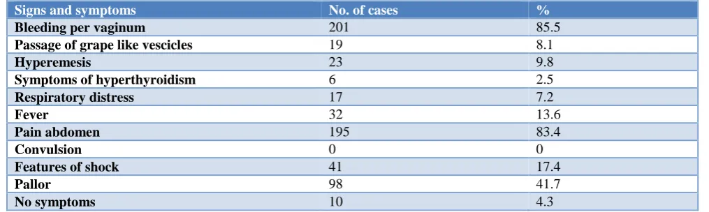
Table 3: Presenting signs and symptoms in case of H.mole (n=235).*
Signs and symptoms No. of cases %
Bleeding per vaginum 201 85.5
Passage of grape like vescicles 19 8.1
Hyperemesis 23 9.8
Symptoms of hyperthyroidism 6 2.5
Respiratory distress 17 7.2
Fever 32 13.6
Pain abdomen 195 83.4
Convulsion 0 0
Features of shock 41 17.4
Pallor 98 41.7
No symptoms 10 4.3
*: Multiple response type.
Of different pregnancy events leading to
choriocarcinoma, post molar was most common type (11/15, 73.33%) followed by post abortal (3/15, 20%) and full term molar pregnancy was least common (1/15, 6.7%) (Table 2).
Relation of H. mole to the period of amenorrhoea during diagnosis (except cases of chorio carcinoma) revealed that 44.1% (128/290) of patients reported amenorrhoea between 8 to 12 weeks duration while as high as 15.2% (44/290) reported amenorrhoea between 17-20 weeks and 1.7% reported more than 20 weeks (Table 2).
Relation of height of uterus to period of amenorrhoea at the time of presentation of H. mole showed that in only 21.7% cases (51/235) height of uterus corresponded to the period of amenorrhoea while 75% (176/235) cases had uterine height more than period of amenorrhoea and only 3.4% (8/235) cases had a height less than period of amenorrhoea (Table 2).
Comparison of presenting signs and symptoms in cases of H. mole revealed that as high as 85.5% (201/235) of cases complained of bleeding per vagina, 8.1% cases had passage of grape like vesicles, 9.8% complained of
hyperemesis, 2.5% cases had features of
hyperthyroidism, 7.2% had respiratory distress, 13.6% cases complained of fever, 83.4% cases had pain abdomen, 17.4% cases had features of shock, 41.7% had pallor while 4.3% cases had no symptoms (Table 3).
Table 4: Maternal mortality in the present study (n=275).
Type of case No. of
cases
No. of deaths %
H.mole 205 12 5.8
Invasive mole
(non-metastatic GTT) 55 2 3.6
Chorio carcinoma 15 12 80
Total 275 26 9.5
No deaths were reported in pregnant patients who conceived after successful management of GTD.
Maternal mortality in the present study was found to be 9.5% (26/275). No deaths were reported in pregnant patients who conceived after successful management of GTD (Table 4).
Table 5: Pregnancy outcome after management of GTD (n=30).
Pregnancy outcome Number %
Full term pregnancy 19 63.3
Pre-term pregnancy 2 6.7
Spontaneous abortion 3 10
Repeat molar pregnancy 6 20
Congenital malformations - -
Total 30 100
Pregnancy outcome after successful management of GTD were as follows: 63.3% (19/30) had full term pregnancy, 20% (6/30) cases had repeat molar pregnancy, 10%
(3/30) had spontaneous abortion while 6.7% (2/30) had pre term delivery (Table 5).
DISCUSSION
GTD forms a spectrum of proliferative abnormalities of trophoblast ranging from benign H. mole to highly malignany choriocarcinoma.
The prevalence of GTD was highest in the age group of 19-29 years probably because maximum number of pregnancies occurs in this age group. On the contrary, Bagshawe et al reported 10 fold increased risk over the age of 40 years.3 Highest number of cases were primigravida. Another study found 32.8% cases occurred in primi, 18.7% cases among second gravid4 while Orr opined that parity did not have any influence on molar pregnancy.5 Chakraborty et al found 44% of patients of H. mole among primi and 42% among second gravid.6
60% of cases were reported among Hindu and 37.7% among Muslims. Similar findings were observed by Precdicayil where he reported prevalence of molar pregnancy in 80% Hindus and 20% Muslims.7
Patients having O blood group were in highest proportions were AB blood group were in least proportions.
With respect to degree of anaemia 33.4% patients had haemoglobin level <6 gm% of them, 27.8% had H. mole and 1.7% had choriocarcinoma, while only 9.8% had haemoglobin level >11 gram%. Berkowits reported that anaemia with haemoglobin concentration of less than 10 gram% was found in 50% cases of GTD.8
On the contrary, Soto-Wright found anaemia in only 5% of H. mole patients.9
The high degree of anaemia in this study could be due to lower socio-economic status of the population, delayed diagnosis preceded by significant bleeding P/V and that about 40% of Indian women are anaemic.10
At the time of presentation, 40% patients had amenorrhoea 12 to 20 weeks. Ignorance, lack of awareness with poor socio-economic condition, absence of diagnostic facilities could account for their delayed presentation.
Bleeding P/V was the most common symptom in this study found in 85.5% of cases. Passage of grape like vesicles was found in 8.1% cases, 9.8% complained of
hyperemesis, 2.5% cases had features of
hyperthyroidism, 7.2% had respiratory distress, 13.6% cases complained of fever, 83.4% cases had pain abdomen, 17.4% cases had features of shock, 41.7% had pallor while 4.3% cases had no symptoms. According to Rose et al uterine bleeding was the most common sign and varied from spotting to profuse haemorrhage.12 Bleeding P/V was found in 97% of patients by Berkowitz.8
Twiggs observed respiratory complications in 27% of women with molar pregnancy greater than 16 weeks size.13
Maternal mortality in the present study was found to be 9.5%. Of them 5 patients of H. mole succumbed due to pulmonary embolism, 6 died to bleeding P/V and hypovolaemic shock and the one died due to shock following emergency S&E.
30 patients conceived after successful management of GTD and among them 19 (63.3%) had successful full term pregnancy outcome.
CONCLUSION
Spectrum of gestational trophoblastic diseases varies from benign to malignant disease. The disease can be treated successfully even in the presence of distant metastasis if diagnosed and treated on time. If it is not diagnosed on time, even hydatiform mole can be fatal. Hence it is important to diagnose and treat the condition early, as the curative rate of Gestational trophoblastic disease is very high.
Funding: No funding sources Conflict of interest: None declared
Ethical approval: The study was approved by the Institutional Ethics Committee
REFERENCES
1. John R, Gestational trophoblastic disease I: epidemiology, pathology, clinical presentation and diagnosis of gestational trophoblastic disease, and management of hydatidiform mole. Am J Obstetr Gynecol. 2010;203(2):531-9.
2. Hancock BW, Seckl MJ, Berkowitz RS, Cole LA, eds. Gestational trophoblastic disease, 3rd ed.
London, UK: International Society for the Study of Trophoblastic Diseases; 2009.
3. Soper JT. Gestational trophoblastic disease. Obstet Gynecol. 2006;108:176-87.
4. Berkowitz RS, Goldstein DP. Current management of gestational trophoblastic disease. Gynecol Oncol. 2009;112:654-62.
5. Orr JW, Austin JM, Hatch KD, Shingleton HM, Younger JB, Boots LR. Acute pulmonary edema associ ated with molar pregnancies: a high-risk factor for devel- opment of persistent trophoblastic disease. Am J Obstet Gynecol. 1980;136:412-5. 6. Chakraborty R, Srinivasan MR, Raskin S.
Estimation of the incidenceof a rare genetic disease through a two-tier mutation survey. Am J Hum Genet. 1993;52:1129-38.
7. Peedicayil A, Regi A, Jasper PM. High Risk Gestational Trophoblastic Disease. The J Obstetr Gynaecol India. 1999: 98-101.
8. Berkowits RS, Goldstein DP. The management of molar pregnancy and gestational trophoblastic tumors. In: Knapp RC, Berkowitz RS, editors. Gynecologic Oncology. 2nd ed. New York: McGraw-Hill; 1993: 328-38.
9. Soto-Wright V, Bernstein M, Goldstein DP, Berkowitz RS. The changing clinical presentation of complete molar pregnancy. Obstet Gynecol. 1995;86:775-9.
10. Rakesh PS, Gopichandran V, Jamkhandi D, Manjunath K, George K, Prasad J. Determinants of postpartum anemia among women from a rural population in southern India. Int J Womens Health. 2014;6:395-400.
11. Curry SL, Schlaerth JB, Kohorn EI, Boyce JB, Gore H, Twiggs LB, et al. Hormonal contraception and tropho- blastic sequelae after hydatidiform mole. A Gynecologic Oncology Group study. Am J Obstet Gynecol. 1989;160:805-9.
12. Rose PG, O'toole RV, Rofagha SK, Qualman S, Boutselis JG. Malignant peripheral primitive neuroectodermal tumor of the uterus. J Surgical Oncol. 1987;35(3):165-9.
13. Twiggs LB, Morrow CP, Schlaerth JB. Acute pulmonary complications of molar pregnancy. Am J Obstet Gynecol. 1979;135:189-94.
Cite this article as: Bhattacharya A, Panja TK, Bhattacharya A, Baur B, Madhwani KP.A