Journal of Nature and Science, Vol.1, No.7, e139, 2015
Immunology
Role of T Follicular Helper cells in Multiple Sclerosis
Nathalie Schmitt
Baylor Institute for Immunology Research, Dallas, TX 75204, USA
Multiple Sclerosis (MS) is a chronic, inflammatory and neurodegenerative disease which results from the destruction of myelin and associated collateral tissue damage within the central nervous system (CNS). MS is a highly diverse disease with different clinical profiles. During the past decade, several new treatment options have been introduced, but no treatment completely stops the disease progression. Therefore deeper understanding of the disease mechanism is necessary to develop novel therapeutic strategies. While yet to be proven, there is evidence suggesting the involvement of T follicular helper (Tfh) cells, a CD4 T cell subset specialized for the provision of help to B cells, in the pathogenesis of MS. In this review, I will discuss the potential pathogenic roles of Tfh cells in the course of MS. Journal of Nature and Science, 1(7):e139, 2015
T follicular helper cells | CD4 T cell | Multiple Sclerosis
Introduction
MS affects approximately 2.5 million persons worldwide [1]. Although usually non-fatal, MS often progresses over the time and substantially affects the quality of life of the patients. The precise etiology of MS is unknown but both genetic and environmental factors confer susceptibility to MS [2, 3]. While some investigators have proposed that MS is a primarily neurodegenerative disease accompanied with secondary inflammation, MS is widely recognized as an autoimmune disorder [4]. The contribution of the immune system to the pathology of MS is supported by the findings in genome-wide association studies (GWAS) and subsequent targeted genome studies. In particular, these studies strongly suggest the involvement of CD4 T cells in the pathogenesis. The strongest association of genetic susceptibility to MS is with MHC class II alleles [5] and the majority of the risk loci are mapped to gene important for immune function particularly genes involved in CD4 T cell differentiation[3].
Diverse clinical forms of MS
MS is a highly diverse disease with different clinical profiles. About 85% of MS patients first develop a form called relapsing remitting disease (RRMS), characterized by multiple episode of neurological disability with complete or almost complete recovery during the relapses. The majority of these patients eventually develop a clinically distinct form called secondary progressive MS (SPMS). The transition from RRMS to SPMS is characterized by a gradual delay in the recovery of neurological functions following the attacks, a shortening of the period of remission, and the apparition of new neurological symptoms even between relapses. The patients will finally present with a regular steady increase of neurological dysfunction and associated physical disabilities. About 15% of the patients present with steadily worsening clinical disabilities from the time of the initial diagnostic without (primary progressive MS) or accompanied with occasional exacerbations (progressive relapsing MS). Whether the same type of CD4 T cells play a pathogenic role in these subgroups, or whether distinct types of CD4 T cells are primarily associated with a particular subgroup is currently unknown.
Insights from mouse EAE models
A number of insights regarding the disease mechanism of MS have been obtained from the studies with experimental autoimmune encephalomyelitis (EAE) mouse models. EAE is induced by immunizing mice with myelin antigens. While T cell-deficient mice
are resistant to the induction of EAE, restoration of the CD4 T cell population before immunization, or adoptive transfer of inflammatory CD4 T cells specific for myelin antigen is sufficient to cause EAE [6, 7]. In addition, transgenic mice carrying T cell receptor recognizing an MHC-class II restricted epitope of myelin antigen spontaneously develop EAE [8]. These observations indicate that EAE is mediated by inflammatory myelin antigen-specific CD4 T cells.
Earlier studies in the 80’s with EAE models concluded that IFN-γ-producing Th1 cells were the major CD4 T cell subset causing EAE [9]. Transfer of myelin antigen-specific Th1 cells was capable of inducing EAE, and blocking the effect of IL-12 with polyclonal neutralizing antibodies was able to abrogate EAE induction [10]. However, later studies demonstrated that the effect of the polyclonal anti-IL-12 antibody was not mediated solely by blocking the function of IL-12 per se, which is composed of IL-12p40 and IL-12p35 subunits. While IL-12p40-/- mice were
highly resistant to the induction of EAE, IL-12p35-/- mice were
found to be susceptible [11]. These observations lead to the discovery that IL-23, a cytokine composed of IL-12p40 and IL-23p19 subunits, is critical cytokine for EAE induction [12]. IL-23 efficiently expands Th17 cells and confers Th17 cells with encephalitogenicity that produce various proinflammatory cytokines such as IL-17A, IL-17F, IL-22, and GM-CSF [13]. The pathogenicity of Th17 cells is also attributed to their expression of the chemokine receptor CCR6. CCL20, the ligand of CCR6, is constitutively expressed by epithelial cells of the choroid plexus, and promotes the entry of CCR6+ CD4 T cells into the CNS [14]. Mice
lacking CCR6 are highly resistant to the induction of EAE [15], indicating the importance of the CCL20-CCR6 axis in the pathogenesis.
While both Th1 and Th17 cells contribute to the development of EAE, a fraction of Th17 cells was found to co-express IL-17 and IFN-γ. Importantly, these IFN-γ+ Th17 cells are more pathogenic
than Th cells producing IFN-γ or IL-17 alone in the EAE model [16]. Furthermore, recent studies show that GM-CSF-/- mice are resistant
to EAE induction, and GM-CSF derived from encephalitogenic CD4 T cells is more critical than IFN-γ or IL-17 for the induction of EAE [17-21]. GM-CSF production by Th17 cells can be enhanced by IL-23, and suppressed by TGF-β [20].
In summary, both Th1 and Th17 cells contribute to the pathogenesis of EAE, and their encephalitogenicity is largely affected by the microenvironment in lymphoid organs and/or inflammatory sites.
Observations in human MS
Many studies have been performed aiming at determining the nature of pathogenic CD4 T cells in MS patients. One approach is to determine the differences between MS patients and control subjects in the cytokine concentrations and the global cytokine expression patterns of CD4 T cells in blood and cerebrospinal fluid (CSF). A recent study with a cohort of RRMS patients showed that the concentration of IL-17A and IL-17F was higher in the serum and
_________
Conflict of interest: No conflicts declared. *
Corresponding Author. Nathalie Schmitt, PhD. Baylor Institute for Immunology Research, 3434 Live Oak, Dallas, TX 75204, USA. Phone: +1-214-818-6542; Fax: +1-214-820-4813
E-mail: nathalis@baylorhealth.edu
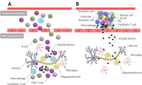
Figure 1. Two different types of CNS inflammation in multiple sclerosis. (A) Many CNS lesions are found in the white matter of the CNS. Loss of the
integrity of the blood brain barrier permits the entry of various immune cells including CD4 T cells, monocytes-macrophages, B cells and cytotoxic T cells into the CNS parenchyma. In the inflammatory sites, CD4 T cells participate in the damages of the oligodendrocytes and neuron fibers through the release of pro-inflammatory cytokines including IL-17, GM-CSF and IFN-γ. (B) Some CNS lesions are also found in cortex. These lesions are often found in the proximity of lymphocyte aggregates containing T and B cells and/or ectopic lymphoid follicles (ELF) in the subarachnoid space. The formation of ELF is found approximately 40% of SPMS patients. Tfh cells, in particular the Th17-type subset, might play a critical role for the formation and maintenance of the ELF. Soluble mediators secreted by immune cells in the ELF diffuse to the gray matter in the CNS. Presumably these soluble mediators induce neuronal damage either by a direct cytotoxic action on neuron or oligodendrocytes or indirectly by activating the microglia.
CSF of the patients, and the concentration of IFN-γ was also higher in the serum [22]. This supports the involvement of Th17 and Th1 cells in the pathogenesis of RRMS. RRMS patients also displayed higher frequencies of blood CD4 T cells expressing IL-17A and GM-CSF upon stimulation with PMA and ionomycin [22, 23]. Consistently, another report demonstrated that the frequencies of CD4 T cells expressing IL-17 were elevated in blood and CSF of RRMS patients during relapses [24]. In contrast, the frequency of IFN-γ+ CD4 T cells were similar between healthy subjects and
RRMS patients, and between stable and relapse phases in RRMS patients [24].
Another approach is to determine the frequency and the type of myelin antigen specific CD4 T cells in blood and CSF of MS patients. Earlier studies in the 90’s analyzed the frequency of myelin antigen-specific CD4 T cells by measuring in vitro the cell proliferation (by [H3]-thymidine uptake) in response to the
stimulation with myelin antigens. These studies concluded that the frequency of myelin specific CD4 T cells in blood was similar between healthy subjects and MS patients [25]. Yet, more recent analyses by using HLA class II tetramers demonstrated that myelin oligodendrocyte glycoprotein (MOG)-specific CD4 T cells in blood were present at a slightly higher frequency in RRMS patients than healthy subjects [26]. This suggests the frequency of myelin antigen-specific CD4 T cells might be increased in MS patients.
Nonetheless, the quality of myelin antigen-specific CD4 T cells seems more dramatically different between MS patients and control subjects. Studies in the late 90’s suggested that myelin antigen-specific CD4 T cells in MS patients are largely composed of memory cells, while those in control subjects are largely composed
cells was not extensively performed due to their rarity in blood [26], a recent study systematically analyzed their functions in RRMS patients and healthy subjects by screening a large number of T cell libraries generated from blood samples. Among blood CCR6+
memory CD4 T cells, myelin antigen-reactive T cells from RRMS patients showed enhanced production of IFN-γ, IL-17, and GM-CSF compared to control subjects, while those from control subjects produced more IL-10 [29]. Furthermore, myelin antigen-specific CD4 T cells in RRMS patients displayed inflammatory Th17 gene signatures [29]. These studies further support the involvement of Th1 and Th17 cells in the pathogenesis of MS. Several mechanism can be considered by which Th17 cells cause neuronal damages in MS. For example, there is a study demonstrating that human Th17 cells promote the blood-brain barrier disruption by producing IL-17 and IL-22, and also directly display cytotoxic activity against neurons [30]. Thus, consistent with findings in mouse EAE models, Th1 and Th17 cells are likely involved in the disease course of human MS.
Tfh cells in the CSF of MS patients
Journal of Nature and Science, Vol.1, No.7, e139, 2015
many other human autoimmune diseases including systemic lupus erythematosus, rheumatoid arthritis, and Sjogren’s syndrome, by promoting the development of autoantibodies [32].
While MS is largely considered as a T cell-mediated disease, there is a line of evidence demonstrating the involvement of B cells and their antibody production in the disease course. First, treatment of RRMS patients with Rituximab, an anti-CD20 monoclonal antibody, induces a decrease of clinical and imaging activity [33]. Second, many studies show that patients with MS display antibodies recognizing myelin antigens in serum as well as in CSF [33]. Third, transfer of human anti-MOG antibodies enhanced demyelination of the CNS in a mouse EAE model [34].
While where and how Tfh cells interact with B cells in MS remains unknown, there is evidence that Tfh cells can migrate to the CNS in MS patients. The concentration of the chemokine CXCL13, the ligand of CXCR5, is elevated in the CSF of RRMS patients and is correlated to the number of CD4 T cells in the CSF. Furthermore, treatment of RRMS patients with Rituximab was accompanied with a reduction of the infiltration of B and T cells in the CNS together with a decrease of CXCL13 concentration in the CSF [35]. The correlation between the levels of CXCL13 and T cells in the CSF of MS patients suggests that CXCR5+ Tfh cells migrate and retain in
the CSF in response to elevated expression of CXCL13 during the course of MS.
Importantly, studies with brain tissues from MS patients showed the production of CXCL13 in actively demyelinating lesions but not in chronic inactive lesions [36, 37]. This suggests that CXCR5+ Tfh
cells can be also recruited into actively demyelinating lesions. The presence of IL-21-expressing CD4 T cells in both acute and chronic active lesions in MS patients further suggests the activation of Tfh cells in situ [38]. IL-21 secreted by Tfh cells in situ might promote the proliferation of B cells and their differentiation into cells producing autoantibodies. Alternatively, IL-21 might also enhance the cytotoxic activity of myelin antigen-specific cytotoxic T cells, and indirectly promote the damage of neuronal tissues
Role of Tfh cells in the formation of ectopic lymphoid structure
The most important role of Tfh cells in the pathology of MS might be associated with the generation of ectopic lymphoid structure (ELF) in the CNS (Figure 1). ELFs contain B cells, Tfh cells, and a follicular dendritic cell (FDC) network, and support a germinal center-like response. The formation of ELF is observed in approximately 40% of SPMS patients [39, 40]. Such ELF is usually found in meninges adjacent to large subpial lesions. In mice, the formation of ELF is preceded by an ectopic expression of lymphoid chemokines including CXCL13. The first lymphoid infiltrates are mainly composed of T cells, and are followed by a progressive influx of B cells and the development of organized lymphoid structures [41]. Indeed, the FDC network within the ELF of MS patents express CXCL13, and recruits Tfh cells [42]. Activated Tfh cells secrete CXCL13 and might further support the expansion and maintenance of ELFs.
The presence of meningeal ELF in SPMS patient is associated with a younger age of disease onset, shorter time to disease progression, and younger age at death [40]. Furthermore, meningeal ELF is associated with a more pronounced demyelination, microglia activation, and neuronal loss in cerebral cortex [39, 40]. These observations strongly suggest that meningeal ELF contributes to the disease progress. Given that grey matter demyelinated lesions are located adjacent to or some distance from ELFs [40], it is plausible that soluble factors produced at ELFs, such as cytokines and antibodies against myelin antigens, diffuse toward the cortex and cause demyelination. These soluble factors might directly induce neuronal demyelination or indirectly via activation of other cells such as microglia.
Specialized Tfh subset promoting ELF formation?
Recent studies in mice and humans show that Tfh lineage cells in lymphoid organs are composed of subsets that differ in their localization, phenotype and functions [32]. Then which Tfh subset plays a major role in the pathogenesis of MS? Almost nothing is known. Herein I would like to discuss a candidate Tfh subset based on current findings.
Studies on human blood CXCR5+ memory CD4 T cells, called
blood memory Tfh cells [43], have identified multiple functionally distinct subsets [44-46]. These subsets can be defined by the differential expression of these markers: the chemokine receptors CXCR3 and CCR6; and the co-stimulatory molecules ICOS and PD-1 [43]. In particular, two chemokine receptors, CXCR3 and CCR6, define the three major functionally distinct subsets: Th1, Th2, and Th17 type cells (Th1: CXCR3+CCR6-; Th2: CXCR3-CCR6-;
and Th17: CXCR3-CCR6+) [43]. While Th2 and Th17-like Tfh
cells induce naïve B cells to produce Immunoglobulins and to undergo isotype switching through secretion of IL-21, Th1-like Tfh cells do not help naïve B cells [44]. Therefore, Th2 and Th17-like blood Tfh cells represent the most efficient helpers for naïve B cells. Although remained to be demonstrated, these distinct blood memory Tfh subsets likely reflect the heterogeneous subsets within Tfh-lineage cells in secondary lymphoid organs and also potentially in ELFs.
This strategy to define the functionally distinct blood Tfh subsets has been applied to characterize the type of Tfh response associated with autoimmune diseases [44, 47-49] and induced by vaccinations [50]. An initial report demonstrated that patients with juvenile dermatomyositis show the skewing of blood Tfh subsets towards Th2 and Th17-like cells (thus increased efficient B cell helpers)[44]. Similarly, patients with other autoimmune diseases such as adult SLE [48] and Sjogren’s syndrome [47] were shown to display an increase of Th17-like blood Tfh cells.
Interestingly, progressive MS patients were also shown to display the same alteration in the composition of blood memory Tfh subsets: low Th1-like cells and high Th17-like cells [49]. Thus, Tfh cells, in particular Th17-like subset, might be involved in the disease course of progressive MS. Indeed, there is evidence in mouse EAE models suggesting the involvement of Th17 cells in the formation of ELF in the CNS. Transfer of Th17 but not Th1 or Th2 cells induced the formation of ELF in the subarachnoid space of EAE mice [51]. Furthermore, Th17 cells were transformed in the ELF into the phenotype of Tfh cells and expressed multiple Tfh markers such as CXCR5, ICOS, and Bcl6 [51]. This suggests that Th17 cells can join the formation of ELF in the CSF, and acquire the phenotype of Tfh cells in situ. Alternatively, it is plausible that Th17 cells that promote the formation of ELF in the CNS of MS patients might be Th17-type Tfh cells.
Conclusions
The nature of the pathogenic CD4 T cells in human MS is much less characterized compared to those in mouse EAE models. More effort on the analyses of primary CD4 T cells obtained from MS patients is necessary to gain direct insights into MS pathogenesis. With this regard, these studies can be dramatically benefitted by recent advance in technologies which permit analysis of the phenotype, genomics, and epigenomics at a single cell level. Such effort might eventually provide evidence linking between myelin antigen-specific Tfh cells and progressive MS and/or the development of grey matter lesions.
Acknowledgment
1. Frohman, E.M., Racke, M.K., and Raine, C.S. (2006). Multiple sclerosis--the plaque and its pathogenesis. N Engl J Med 354, 942-955.
2. Ebers, G.C. (2008). Environmental factors and multiple sclerosis. Lancet Neurol 7, 268-277.
3. International Multiple Sclerosis Genetics, C., Wellcome Trust Case Control, C., Sawcer, S., Hellenthal, G., Pirinen, M., Spencer, C.C., et al.
(2011). Genetic risk and a primary role for cell-mediated immune mechanisms in multiple sclerosis. Nature 476, 214-219.
4. Ransohoff, R.M., Hafler, D.A., and Lucchinetti, C.F. (2015). Multiple sclerosis-a quiet revolution. Nat Rev Neurol 11, 246.
5. Sawcer, S., and Hellenthal, G. (2011). The major histocompatibility complex and multiple sclerosis: a smoking gun? Brain 134, 638-640.
6. Stromnes, I.M., and Goverman, J.M. (2006). Passive induction of experimental allergic encephalomyelitis. Nat Protoc 1, 1952-1960.
7. Stromnes, I.M., and Goverman, J.M. (2006). Active induction of experimental allergic encephalomyelitis. Nat Protoc 1, 1810-1819.
8. Lafaille, J.J., Nagashima, K., Katsuki, M., and Tonegawa, S. (1994). High incidence of spontaneous autoimmune encephalomyelitis in immunodeficient anti-myelin basic protein T cell receptor transgenic mice. Cell 78, 399-408.
9. Sriram, S., Solomon, D., Rouse, R.V., and Steinman, L. (1982). Identification of T cell subsets and B lymphocytes in mouse brain experimental allergic encephalitis lesions. J Immunol 129, 1649-1651.
10. Leonard, J.P., Waldburger, K.E., and Goldman, S.J. (1995). Prevention of experimental autoimmune encephalomyelitis by antibodies against interleukin 12. J Exp Med 181, 381-386.
11. Becher, B., Durell, B.G., and Noelle, R.J. (2002). Experimental autoimmune encephalitis and inflammation in the absence of interleukin-12. J Clin Invest 110, 493-497.
12. Cua, D.J., Sherlock, J., Chen, Y., Murphy, C.A., Joyce, B., Seymour, B., et al. (2003). Interleukin-23 rather than interleukin-12 is the critical cytokine for autoimmune inflammation of the brain. Nature 421, 744-748.
13. Langrish, C.L., Chen, Y., Blumenschein, W.M., Mattson, J., Basham, B., Sedgwick, J.D., et al. (2005). IL-23 drives a pathogenic T cell population that induces autoimmune inflammation. J Exp Med 201, 233-240.
14. Sallusto, F., Impellizzieri, D., Basso, C., Laroni, A., Uccelli, A., Lanzavecchia, A., et al. (2012). T-cell trafficking in the central nervous
system. Immunol Rev 248, 216-227.
15. Reboldi, A., Coisne, C., Baumjohann, D., Benvenuto, F., Bottinelli, D., Lira, S., et al. (2009). C-C chemokine receptor 6-regulated entry of TH-17 cells into the CNS through the choroid plexus is required for the initiation of EAE. Nat Immunol 10, 514-523.
16. Kebir, H., Ifergan, I., Alvarez, J.I., Bernard, M., Poirier, J., Arbour, N., et al. (2009). Preferential recruitment of interferon-gamma-expressing TH17 cells in multiple sclerosis. Ann Neurol 66, 390-402.
17. Haak, S., Croxford, A.L., Kreymborg, K., Heppner, F.L., Pouly, S., Becher, B., et al. (2009). IL-17A and IL-17F do not contribute vitally to
autoimmune neuro-inflammation in mice. J Clin Invest 119, 61-69.
18. Ferber, I.A., Brocke, S., Taylor-Edwards, C., Ridgway, W., Dinisco, C., Steinman, L., et al. (1996). Mice with a disrupted IFN-gamma gene are susceptible to the induction of experimental autoimmune encephalomyelitis (EAE). J Immunol 156, 5-7.
19. Codarri, L., Gyulveszi, G., Tosevski, V., Hesske, L., Fontana, A., Magnenat, L., et al. (2011). RORgammat drives production of the cytokine GM-CSF in helper T cells, which is essential for the effector phase of autoimmune neuroinflammation. Nat Immunol 12, 560-567.
20. El-Behi, M., Ciric, B., Dai, H., Yan, Y., Cullimore, M., Safavi, F., et al.
(2011). The encephalitogenicity of T(H)17 cells is dependent on IL-1- and IL-23-induced production of the cytokine GM-CSF. Nat Immunol 12, 568-575.
21. McQualter, J.L., Darwiche, R., Ewing, C., Onuki, M., Kay, T.W., Hamilton, J.A., et al. (2001). Granulocyte macrophage
colony-stimulating factor: a new putative therapeutic target in multiple sclerosis. J Exp Med 194, 873-882.
22. Tao, Y., Zhang, X., Chopra, M., Kim, M.J., Buch, K.R., Kong, D., et al.
(2014). The role of endogenous IFN-beta in the regulation of Th17 responses in patients with relapsing-remitting multiple sclerosis. J Immunol 192, 5610-5617.
23. Rasouli, J., Ciric, B., Imitola, J., Gonnella, P., Hwang, D., Mahajan, K., et al. (2015). Expression of GM-CSF in T Cells Is Increased in Multiple Sclerosis and Suppressed by IFN-beta Therapy. J Immunol
24. Brucklacher-Waldert, V., Stuerner, K., Kolster, M., Wolthausen, J., and Tolosa, E. (2009). Phenotypical and functional characterization of T helper 17 cells in multiple sclerosis. Brain 132, 3329-3341.
25. Zhang, J., Markovic-Plese, S., Lacet, B., Raus, J., Weiner, H.L., and
26. Raddassi, K., Kent, S.C., Yang, J., Bourcier, K., Bradshaw, E.M., Seyfert-Margolis, V., et al. (2011). Increased frequencies of myelin oligodendrocyte glycoprotein/MHC class II-binding CD4 cells in patients with multiple sclerosis. J Immunol 187, 1039-1046.
27. Scholz, C., Patton, K.T., Anderson, D.E., Freeman, G.J., and Hafler, D.A. (1998). Expansion of autoreactive T cells in multiple sclerosis is independent of exogenous B7 costimulation. J Immunol 160, 1532-1538.
28. Lovett-Racke, A.E., Trotter, J.L., Lauber, J., Perrin, P.J., June, C.H., and Racke, M.K. (1998). Decreased dependence of myelin basic protein-reactive T cells on CD28-mediated costimulation in multiple sclerosis patients. A marker of activated/memory T cells. J Clin Invest 101, 725-730.
29. Cao, Y., Goods, B.A., Raddassi, K., Nepom, G.T., Kwok, W.W., Love, J.C., et al. (2015). Functional inflammatory profiles distinguish myelin-reactive T cells from patients with multiple sclerosis. Sci Transl Med 7, 287ra274.
30. Kebir, H., Kreymborg, K., Ifergan, I., Dodelet-Devillers, A., Cayrol, R., Bernard, M., et al. (2007). Human TH17 lymphocytes promote
blood-brain barrier disruption and central nervous system inflammation. Nat Med 13, 1173-1175.
31. Crotty, S. (2014). T follicular helper cell differentiation, function, and roles in disease. Immunity 41, 529-542.
32. Ueno, H., Banchereau, J., and Vinuesa, C.G. (2015). Pathophysiology of T follicular helper cells in humans and mice. Nat Immunol 16, 142-152.
33. von Budingen, H.C., Bar-Or, A., and Zamvil, S.S. (2011). B cells in multiple sclerosis: connecting the dots. Curr Opin Immunol 23, 713-720.
34. Zhou, D., Srivastava, R., Nessler, S., Grummel, V., Sommer, N., Bruck, W., et al. (2006). Identification of a pathogenic antibody response to native myelin oligodendrocyte glycoprotein in multiple sclerosis. Proc Natl Acad Sci U S A 103, 19057-19062.
35. Piccio, L., Naismith, R.T., Trinkaus, K., Klein, R.S., Parks, B.J., Lyons, J.A., et al. (2010). Changes in B- and T-lymphocyte and chemokine
levels with rituximab treatment in multiple sclerosis. Arch Neurol 67, 707-714.
36. Corcione, A., Casazza, S., Ferretti, E., Giunti, D., Zappia, E., Pistorio, A., et al. (2004). Recapitulation of B cell differentiation in the central
nervous system of patients with multiple sclerosis. Proc Natl Acad Sci U S A 101, 11064-11069.
37. Krumbholz, M., Theil, D., Cepok, S., Hemmer, B., Kivisakk, P., Ransohoff, R.M., et al. (2006). Chemokines in multiple sclerosis:
CXCL12 and CXCL13 up-regulation is differentially linked to CNS immune cell recruitment. Brain 129, 200-211.
38. Tzartos, J.S., Craner, M.J., Friese, M.A., Jakobsen, K.B., Newcombe, J., Esiri, M.M., et al. (2011). IL-21 and IL-21 receptor expression in lymphocytes and neurons in multiple sclerosis brain. Am J Pathol 178, 794-802.
39. Magliozzi, R., Howell, O., Vora, A., Serafini, B., Nicholas, R., Puopolo, M., et al. (2007). Meningeal B-cell follicles in secondary progressive multiple sclerosis associate with early onset of disease and severe cortical pathology. Brain 130, 1089-1104.
40. Howell, O.W., Reeves, C.A., Nicholas, R., Carassiti, D., Radotra, B., Gentleman, S.M., et al. (2011). Meningeal inflammation is widespread and linked to cortical pathology in multiple sclerosis. Brain 134, 2755-2771.
41. Bombardieri, M., Barone, F., Lucchesi, D., Nayar, S., van den Berg, W.B., Proctor, G., et al. (2012). Inducible tertiary lymphoid structures,
autoimmunity, and exocrine dysfunction in a novel model of salivary gland inflammation in C57BL/6 mice. J Immunol 189, 3767-3776.
42. Serafini, B., Rosicarelli, B., Magliozzi, R., Stigliano, E., and Aloisi, F. (2004). Detection of ectopic B-cell follicles with germinal centers in the meninges of patients with secondary progressive multiple sclerosis. Brain Pathol 14, 164-174.
43. Schmitt, N., Bentebibel, S.E., and Ueno, H. (2014). Phenotype and functions of memory Tfh cells in human blood. Trends Immunol 35, 436-442.
44. Morita, R., Schmitt, N., Bentebibel, S.E., Ranganathan, R., Bourdery, L., Zurawski, G., et al. (2011). Human blood CXCR5(+)CD4(+) T cells are counterparts of T follicular cells and contain specific subsets that differentially support antibody secretion. Immunity 34, 108-121.
45. He, J., Tsai, L.M., Leong, Y.A., Hu, X., Ma, C.S., Chevalier, N., et al.
(2013). Circulating Precursor CCR7(lo)PD-1(hi) CXCR5(+) CD4(+) T Cells Indicate Tfh Cell Activity and Promote Antibody Responses upon Antigen Reexposure. Immunity 39, 770-781.
Journal of Nature and Science, Vol.1, No.7, e139, 2015
47. Li, X.Y., Wu, Z.B., Ding, J., Zheng, Z.H., Li, X.Y., Chen, L.N., et al.
(2012). Role of the frequency of blood CD4(+) CXCR5(+) CCR6(+) T cells in autoimmunity in patients with Sjogren's syndrome. Biochem Biophys Res Commun 422, 238-244.
48. Le Coz, C., Joublin, A., Pasquali, J.L., Korganow, A.S., Dumortier, H., and Monneaux, F. (2013). Circulating TFH Subset Distribution Is Strongly Affected in Lupus Patients with an Active Disease. PLoS One 8, e75319.
49. Romme Christensen, J., Bornsen, L., Ratzer, R., Piehl, F., Khademi, M., Olsson, T., et al. (2013). Systemic inflammation in progressive multiple
sclerosis involves follicular T-helper, Th17- and activated B-cells and correlates with progression. PLoS One 8, e57820.
50. Bentebibel, S.E., Lopez, S., Obermoser, G., Schmitt, N., Mueller, C., Harrod, C., et al. (2013). Induction of ICOS+CXCR3+CXCR5+ TH cells
correlates with antibody responses to influenza vaccination. Sci Transl Med 5, 176ra132.
51. Peters, A., Pitcher, L.A., Sullivan, J.M., Mitsdoerffer, M., Acton, S.E., Franz, B., et al. (2011). Th17 cells induce ectopic lymphoid follicles in
central nervous system tissue inflammation. Immunity 35, 986-996.