Original Article
129 |P a g e
Int J Med Res Prof.2016; 2(2); 129-36. P-ISSN:2454-6356; E-ISSN:2454-6364
Analysis of the Dynamic Pattern of Foreign Bodies in
Otorhinolaryngology in a Tertiary Care Hospital
Jawaid Ahmad Zargar
1, Sachin Gupta2*, Gopika Kalsotra
3,
Ghulam Mohammad Mir
4, Parmod Kalsotra
51
Asstt Surgeon, Department of Health, J&K,
2Senior resident,
Department of ENT & HNS, ASCOMS, Sidhra, Jammu,
3
Lecturer,
4PG Student,
5Professor, Department of ENT & HNS, SMGS, GMC Jammu, INDIA.
Article History
Received: 30 Jan 2016 Revised: 04 Feb 2016 Accepted: 12 Feb 2016
*Correspondence to:
Dr. Sachin Gupta Senior resident, Department of ENT & HNS,
ASCOMS, Sidhra, Jammu
ABSTRACT
Aims: To study the epidemiological profile of foreign bodies in ENT with respect to different parameters.
Subjects and Methods: A retrospective, hospital based study was conducted on the patients attending the ENT Department of S.M.H.S Hospital, Srinagar extending over a period of one year w.e.f. 01.11.2006 to 31.10.2007 and included both OPD and admitted patients of all age groups and both sexes with the history of foreign body lodgment/ingestion or inhalation.
Results: The incidence of foreign bodies in ENT OPD in Srinagar was 1.2%, more in males (63%) as common to females (36%). Most common age group was 0-8 and least common is 16-24. Most common site of foreign body lodgment was in ear followed by nose and oesophagus which together constitute the bulk (76%) of the total incidence. The inanimate foreign bodies far exceeded to the animate ones, beans and grains being the most common. 90.45% of the patients presented within one day of foreign body lodgment. Most of them (96%) had no similar past history. Complications during foreign body removal occurred in 5.68% of the patients.
Conclusion: Foreign body impaction was more commonly encountered in children especially in males probably due to the fact that male children are more curious and due to their exploring nature and tendency to put things in the body cavities. Proper care and a high degree of suspicion can prove decisive and help to avert many catastrophies due to accidental or deliberate insertion of foreign bodies.
KEYWORDS:
Epidemiology, Foreign body, Ingestion, Inhalation.INTRODUCTION
The tendency of a child to accidently or intentionally lodge objects in the numerous orifices and cavities of the head and neck and the critical physiologic importance of the upper airway, makes foreign bodies a subject of major importance when discussing the otolaryngologic emergencies.
Clinical features due to foreign body (FB) lodgement in ENT area not only depend upon the anatomic site involved but also on the age of the patient and the degree of obstruction and inflammatory response produced by the foreign body, symptoms ranging from asymptomatic to acute life threatening condition. This is one of the leading causes of unintentional death in the home, especially among children under the age of five years. Another possibility is the presence of multiple foreign bodies in same or different orifices, especially in mentally abnormal and psychotic patients.
Classification of Foreign Bodies in ENT1 1. Animate 2. Inanimate Animate:
Maggots, screw worms & their larvae, black, carpetbeetles, ascaris, cockroaches, mosquitos, housefly are common offenders.
Inanimate:
a. Vegetable foreign bodies are commonly peas, beans, dried pulses and nuts.
b. Mineral matter may be pieces of pencil, paper, sponge, pieces of metal, plastic toys, washers, nuts, nails, screws, buttons, studs, pebbles, beads and cotton wool etc.
c. Arising from surgery pieces of polyp, bone, cartilage, swabs, instruments and packs may be left behind.
Different Sites for Lodgement of Foreign Bodies May be Classified as Under:
Foreign bodies in Aerodigestive tract.
Foreign bodies in Ear.
Foreign bodies in Soft tissue Neck and related structures.
The foreign body may lie in any part of the nasal fossa,
but those which are seen by an Otolaryngologist are commonly beyond the constriction at the nasal vestibule. Animate FB such as maggots and worms cause symptoms of bilateral nasal obstruction, headaches and serosanguinous fluid discharge. FB that may get neglected presents with symptoms of unilateral purulent nasal discharge and nasal obstruction. (Fig1A, 1B).
Fig1A: A child with long standing foreign body nose. Fig 1B: Rhinolith retrieved from the same patient
Fig 2A: Foreign Body being removed from Crico-pharynx in a patient
Fig 2B: Chicken meat bolus retrieved as foreign body in the same patient
Foreign bodies may rarely get impacted in the nasopharnyx due to unskillful removal attempts. The foreign body may remain there without causing any symptoms or the patient may complain of something behind the nose or the patients may complain of bilateral nasal obstruction.
Foreign bodies especially fish bones get commonly lodged in the posterior one-third of the tongue, tonsillar pillars, vallecullae and the posterior pharyngeal wall. This causes difficulty in swallowing, pain in throat or even respiratory symptoms. If the foreign body is neglected it may lead to abscess formation.
Foreign bodies in the larynx and trachea constitute a medical emergency and require immediate medical attention. The initial symptoms of a foreign body in this region are typically dramatic. The aspiration instantly produces a spasm of coughing but unless there is marked respiratory embarrassment the mucosa rapidly adapts to the abnormal situation. Following are the most common symptoms that may indicate the presence of foreign body in respiratory tract:
Choking or gaging when the object is first inhaled.
Wheezing
Foreign bodies in subglottis can masquerade as laryngotracheobronchitis or croup.
Although the initial symptoms listed above may subside, the foreign body may still be obstructing the airway. Occlusion of the larynx may cause acute obstruction, which is a dire emergency
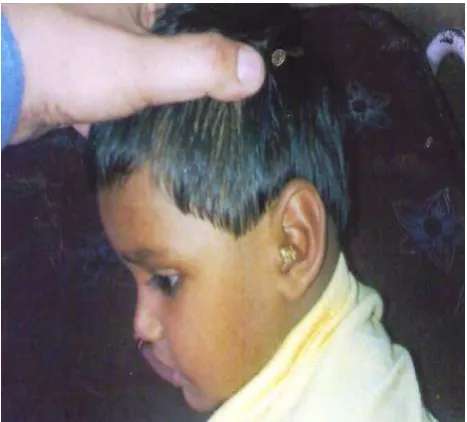
The patient with foreign body lodged in the oesophagus can be asymptomatic while others may present with symptoms of dysphagia, drooling, pain, vomiting, cough, stridor, increased salivation or a persistent foreign body sensation The removal of oesophageal foreign body is hazardous because the wall is thin and may become further weakened by inflammation (Figure 2A and 2B). Foreign bodies in ear can be either rings stuck in lobule or anything inserted in the external auditory canal (Figure 3). Inserted foreign bodies are usually found in the external auditory canal mostly just medial to isthmus or may pass through the tympanic membrane to lie in the middle ear cavity or even lodge in the inner ear after penetrating the external and middle ear. Some objects placed in the ear may not cause symptoms, while other objects, such as food and insects, may cause pain in the ear, redness, or drainage. Hearing may be affected if the object is blocking the ear canal.
Fig 3: A child with foreign body (cotton) ear
Foreign Bodies have also been reported to be lodged in the soft tissues of the neck2, adjacent to great vessels of the neck, and other related structures.
MATERIALS AND METHODS
The study was conducted on the patients attending the ENT Department of S.M.H.S Hospital, Srinagar extending over a period of one year w.e.f. 1.11.2006 to 31.10.2007 and included both OPD and admitted patients of all age groups and both sexes with the history of foreign body lodgment/ingestion or inhalation. A detailed history of foreign body with respect to age, sex,
residence, type/nature of foreign body (animate/ inanimate), site/side of lodgment, duration, mode of entry, symptoms was recorded. It was also noted whether the patient presented to the ENT Department of his own or was referred from any other health care centre. General physical examination, relevant systemic examination and a detailed local examination was carried out which included:
Otoscopic examination and/ or examination under microscope for aural foreign bodies.
Anterior rhinoscopy, posterior rhinoscopy, nasal endoscopy for nasal foreign bodies.
Oropharyngeal examination including indirect laryngoscopy where ever possible.
Radiological investigations such as X- Rays, barium studies and CT scan were carried out in patients of this series as per the need. Those patients requiring foreign body removal under general anaesthesia were suitably investigated and foreign bodies were removed skillfully under GA.
RESULTS
A total of 35220 patients attended the department in the said period and out of them 440 cases met the selection criterion and were enrolled for the study.
The incidence of foreign body lodgement at various sites in this hospital attending population of our part of the country was found to be 1.2% or 124 per 10000 new patients.
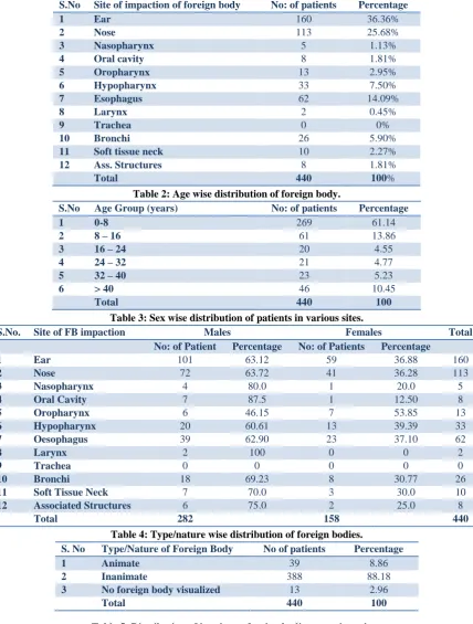
Site wise distribution of patients with foreign bodies is as shown in table 1. Foreign bodies in the ear are most common (36.6%). Foreign bodies of ear, nose and oesophagus together constituting more than (76%) of the total foreign bodies.
Age wise distribution (table 2) shows that the foreign bodies were most common in the age group of 0-8 years (61.14%). The next common age group was 8-16 years (13.86%), followed by > 40 years age group (10.45%). The least common age group was 16 – 24 years (4.55%). Ear foreign bodies are most common in the age group of 0 – 8 years (58.75%), followed by (15%) in the 8 – 16 age group. 86.73% of nasal foreign bodies occurred in the age group of 0 – 8 years and 10.62% of nasal foreign bodies in the study were found in the age group of 8 – 16 year.
Foreign bodies in the oral cavity 3 (37.5%) were found in the 24 – 32 age group and 2 each (25% each) in the age groups of 0 – 8 years and 32 – 40 years followed by 1(12.5%) in the age group of 8 – 16 years. Similarly, foreign bodies at other sites were mostly found in the age group of 0 – 8 followed by older age groups.
Table 1: Different sites of foreign body impaction/suspected foreign body impaction.
S.No Site of impaction of foreign body No: of patients Percentage
1 Ear 160 36.36%
2 Nose 113 25.68%
3 Nasopharynx 5 1.13%
4 Oral cavity 8 1.81%
5 Oropharynx 13 2.95%
6 Hypopharynx 33 7.50%
7 Esophagus 62 14.09%
8 Larynx 2 0.45%
9 Trachea 0 0%
10 Bronchi 26 5.90%
11 Soft tissue neck 10 2.27%
12 Ass. Structures 8 1.81%
Total 440 100%
Table 2: Age wise distribution of foreign body.
S.No Age Group (years) No: of patients Percentage
1 0-8 269 61.14
2 8 – 16 61 13.86
3 16 – 24 20 4.55
4 24 – 32 21 4.77
5 32 – 40 23 5.23
6 > 40 46 10.45
Total 440 100
Table 3: Sex wise distribution of patients in various sites.
S.No. Site of FB impaction Males Females Total
No: of Patient Percentage No: of Patients Percentage
1 Ear 101 63.12 59 36.88 160
2 Nose 72 63.72 41 36.28 113
3 Nasopharynx 4 80.0 1 20.0 5
4 Oral Cavity 7 87.5 1 12.50 8
5 Oropharynx 6 46.15 7 53.85 13
6 Hypopharynx 20 60.61 13 39.39 33
7 Oesophagus 39 62.90 23 37.10 62
8 Larynx 2 100 0 0 2
9 Trachea 0 0 0 0 0
10 Bronchi 18 69.23 8 30.77 26
11 Soft Tissue Neck 7 70.0 3 30.0 10
12 Associated Structures 6 75.0 2 25.0 8
Total 282 158 440
Table 4: Type/nature wise distribution of foreign bodies.
Table 5: Distribution of inanimate foreign bodies at various sites.
Site Beans Grains Plastic Stone Paper Vegetable Coins Denture Fish
bone
Others
Ear 35 25 7 8 11 - - - - 34
Nose 29 4 11 5 2 4 - - - 44
Nasopharynx 2 - 1 - - - 2
Oral cavity - - 3 - - - 3 2
Oropharynx - - - 7 6
Hypopharynx - - 2 - - - 26 - - 5
Oesophagus - - 3 - - - 29 11 3 14
Larynx - 4 - - - 4
Bronchi 3 6 2 - - - 12
STN - - - 3 - - - 5
S. No Type/Nature of Foreign Body No of patients Percentage
1 Animate 39 8.86
2 Inanimate 388 88.18
3 No foreign body visualized 13 2.96
Table 6: Nature and no. of Animate foreign bodies.
S.No Site Cockroaches Mosquito Ant Bug Housefly Total
1 Ear 15 7 7 4 4 37
2 Nose 1 0 0 0 1 2
3 Nasopharynx 0 0 0 0 0 0
4 Oral Cavity 0 0 0 0 0 0
5 Oropharynx 0 0 0 0 0 0
6 Hypo pharynx 0 0 0 0 0 0
7 Oesophagus 0 0 0 0 0 0
8 Larynx 0 0 0 0 0 0
9 Bronchi 0 0 0 0 0 0
10 STN 0 0 0 0 0 0
11 Ass. Structures 0 0 0 0 0 0
Total 16 7 7 4 5 39
Table 7: Mode of entry of foreign body impaction/suspected foreign body impaction.
S. No Site Accidental %
Self-inflicted
% Iatrogenic % Total
1 Ear 149 93.12 7 4.38 04 2.50 160
2 Nose 104 92.03 8 7.07 1 0.88 113
3 Nasopharynx 5 100.0 0 0 0 0 5
4 Oral cavity 6 75.0 1 12.5 1 12.5 8
5 Oropharynx 13 100.0 0 0 0 0 13
6 Hypopharynx 33 100.0 0 0 0 0 33
7 Oesophagus 62 100.0 0 0 0 0 62
8 Larynx 2 100.0 0 0 0 0 2
9 Trachea 0 0 0 0 0 0 0
10 Bronchi 26 100.0 0 0 0 0 26
11 Soft tissue neck 10 100.0 0 0 0 0 10
12 Associated Structures. 8 100.0 0 0 0 0 8
Total 418 16 6 440
Table 8: Duration wise distribution of foreign body impaction.
S.No Duration of F.B lodgement No: of patients Percentage
1 Wihin 1 day 398 90.45
2 1 day – 1 week 37 8.41
3 1 week – 1 month 3 0.68
4 > 1 month 2 0.45
Total 440 100
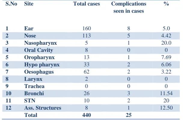
Table 9: Distribution of patients with FB at various sites according to complications observed during removal.
S.No Site Total cases Complications
seen in cases
%
1 Ear 160 8 5.0
2 Nose 113 5 4.42
3 Nasopharynx 5 1 20.0
4 Oral Cavity 8 0 0
5 Oropharynx 13 1 7.69
6 Hypo pharynx 33 2 6.06
7 Oesophagus 62 2 3.22
8 Larynx 2 0 0
9 Trachea 0 0 0
10 Bronchi 26 3 11.54
11 STN 10 2 20
12 Ass. Structures 8 1 12.50
Animate foreign bodies constitute 8.86% of total foreign bodies, while as inanimate foreign bodies constitute 88.18% (table 4) . In only 2.95% cases of patients with complaint of foreign body lodgment, no foreign body could be visualized in this study. Animate foreign bodies were found in ear and nose only, while as inanimate foreign bodies were found in all site except trachea in the study
Beans were the commonest inanimate foreign bodies found in this study. Coins were the second commonest types. (Table 5) Out of a total of 39 animate foreign bodies 37 were found in ear alone and cockroaches were most common in this study (Table 6).
Mode of entry of foreign bodies
95% of foreign body lodgement was accidental in nature, while as in 3.64% it was self-inflicted and in only 1.36% cases it was iatrogenic. Accidental nature of foreign body lodgement was for more common at all sites in this study (table 7).
As evident from the table 8, most of the patients, about 90.45% (398 out of 440) presented within 1 day of the foreign body lodgement, whereas about 8.4% presented within 1 week, (0.68%) patients presented within one month and only 0.45% presented within 1 year.
Only 4.32% of patients with foreign bodies had past history of foreign body lodgement. Past history of foreign body lodgement was mostly found in nose (8.85%) in this study. It was 6.06% in hypopharynx, 3.22% in oesophagus, 3.12% in ear, while there was no past history in other sites in this study.
Complications occurred in only 5.68% of patients during foreign body removal. As is evident from the table 9, complication was found in ear foreign bodies (5%), nose (4.42%), nasopharynx (20%), oropharynx (7.69%), hypopharynx (6.06%), oesophagus (3.22%), bronchi (11.54%), STN (20%) and associated structures (12.50%).
DISCUSSION
Foreign body impaction is very common in clinical practice and foreign body impaction in the ENT region may sometimes produce fatal outcome. The present study was undertaken in recognition of the problem and was designed to investigate the clinico-epidemiological features of foreign body impaction in northern part of the country, managed in a tertiary care (referral) cum teaching hospital over a period of one year. In the present study 440 patients of foreign body impaction at various sites in head and neck who presented to the ENT Department in one year period during which a total of 35220 patients were examined.
Site wise distribution
This included 160 cases (36.36%) of ear foreign bodies, 113 cases (25.68%) of nasal foreign bodies, 5 cases (1.13%) of nasopharyngeal foreign bodies, 8 cases (1.81%) of oral cavity foreign bodies, 13 cases (2.95%)
of oropharyngeal foreign bodies, 33 cases (7.50%) of hypopharyngeal foreign bodies, 62 cases (14.09%) of oesophageal foreign bodies, 2 cases (0.45%) of laryngeal foreign bodies, 26 cases (5.90%) of bronchial foreign bodies, 10 cases (2.27%) of foreign bodies in the soft tissues of the neck and 8 cases (1.81%) of foreign bodies in associated structures of head and neck.
The high incidence of ear foreign bodies was also reported by Ngo et al.3 who in their case study recorded 117 cases (33.14%) of ear foreign body where as 125 cases (35.41%) of nasal foreign bodies were found out of 353 cases of foreign body lodgement in children. Endican et al.4 in their study of I037 ENT foreign bodies in children observed 68.6% cases of ear foreign bodies, 26% in nose, 2.5% of pharynx, 2.0% in esophagus, laryngotracheobronchial tree (LTB) in 2.0% cases and Kumar et al.5 reported the nasal foreign bodies as the most common occurance 176 (45%) out of 399 total foreign bodies, followed by ear foreign bodies 102 (25%), Oesophagus 83 (21%) and bronchus 18 (4%). The lower incidence of bronchial foreign body in this part of the world may be because of the reason that many cases go unnoticed because of unawareness on the part of the parents and the symptoms are mistaken for pneumonia or other respiratory illness.
Age Distribution
The most common age group involved in the present study was 0 – 8 years age group with 269 (61.14%) of total 440 cases occurring in this age group. This was followed by 8 – 16 years age group with 61 (13.86%) of foreign bodies. According to Banerjee (6) in his study on iatrogenic foreign bodies the most common age group affected was 0 – 10 years (52.5%) whereas second most common age group affected was 11 – 20 years (38.9%). The similar observation was made by Ngo et al 3 who observed 43.3% cases in the age group of 4 – 8 years and followed by those less than 4 years old.
Sex Wise Distribution of foreign bodies
The present study showed a definite male preponderance with 282 (64.09%) males and 158 (35.91) females out of the total of 440 cases. This could be attributed to the fact that male children are more active and curious searching for a mischief. Banerjee6 reported slight female preponderance of 52% against 48% in males in case of 250 iatrogenic foreign bodies studied by them while Reilly et al.7 reported 53% occurrence in male children and 47 % in female children out of 5528 children with foreign bodies studied by them.
Nature wise distribution
In the present study a total of 388 inanimate foreign bodies were recovered at various site and out of it highest number was constituted by beans 73 (18.81%), followed by 43 (11.08%) coins. Usually bean grains, as it happens to other common edible seeds fall on the kitchen floor during cooking and are found by children and in some cases mothers would gave their children beans to play with. Furthermore, children have an immature protective cough reflex compared to adults and due to narrower airway, morbidity and mortality are higher in children. Coins were found in children who have a tendency of putting coins in the mouth which may be inadvertently swallowed. Reilly et al.7 have reported coins as the commonest foreign body in their study.
Meat bolus impaction and denture swallowing were also common in our study. In elderly edentulous patients, inadequate mastication leads to impaction of a bolus of food. An ill-fitting or broken denture may be accidently swallowed during a meal or an epileptic seizure. An artificial denture may obliterate tactile sensation in the roof of the mouth so that the patients fail to detect fish or meat bone in the mouth thus leading to its impaction. Care must be thus taken while eating meat and fish bones in such cases.
Thompson et al.10 in their study of external auditory canal foreign bodies reported 72% of foreign bodies as beads and beans. Saha et al.11 reported in his study on 1476 ear foreign bodies 303 (20.52%) as vegetable foreign bodies, 1048 (71%) as non-hygroscopic nonmetallic foreign body, 52 (3.5%) metallic foreign bodies and 73 (4.94%) as animate foreign bodies. Figueiredo et al.12 observed sponge fragment 26.86%, plastic 18.09%, beans 14.76%, paper fragment 5.47% of nasal foreign bodies. Milhome et al.13 observed grains 25% as the total nasal foreign bodies.
Nayak et al.14 have also observed coins as the commonest foreign bodies in children and meat and fish bones in adults in their study of crico-pharyngeal and oesophageal foreign bodies. Macpherson et al.15 in their study of oesophageal foreign bodies reported coins in 69%. Liz et al.16 observed food boluses, coins, fish bone, denture prosthesis and chicken bones as oesophageal foreign bodies in their study of upper gastrointestinal foreign bodies.
Daniilidis et al.17 in their study of foreign bodies in airways found pumpkin seeds 25% of all cases. Lone et al.18 reported in their study of tracheobronchial foreign bodies beans (24%), Peas (18%) and corns (10%). Kalyanappagol et al.19 in their study on tracheobronchial foreign body aspirations in children that groundnut was the commonest foreign body.
Mode of Entry Wise Distribution of foreign body Out of 440 foreign bodies in our study 418 (95%) were accidently lodged or impacted, 16 (3.64%) were self-inflicted and only 6 (1.36%) were iatrogenic in nature.
Out of 160 ear foreign bodies 149 (93.12%) were accidental, 7 (4.38%) self-inflicted and 4 (2.5%) iatrogenic. Out of 113 nasal foreign bodies 104 (92.03%) were accidental, 8 (7.07%) self inflicted and 1 (0.88%) iatrogenic. Out of 8oral cavity foreign bodies 6 (75%) were accidental, 1 (12.5%) each self inflicted and iatrogenic. The foreign bodies at rest of sites were all accidental in nature. Abrenica et al.20 in their study of 648 oesophageal and tracheobronchial foreign bodies have reported that accidental swallowing and aspiration of foreign body while playing was the cause.
Duration Wise Distribution of foreign bodies
In the present study of 440 foreign bodies 398 (90.45%) patients reported within 1 day of foreign body impaction, 37 (8.41%) reported between 1 day to 1 week, 3 (0.68%) reported between 1 week to 1 month while 2 patients (0.45%) reported after > 1 month of foreign body impaction. In case of bronchial foreign bodies 26.92% of patients presented within 1 day to 1 week and in 1 case (3.85%) between 1 week to 1 month. This may be explained by the fact that it takes 7- 14 days after the aspiration to develop serious complications such as consolidation or collapse which might prompt parents about the seriousness of the problem.
Banerjee et al.6 reported in their study of 250 iatrogenic foreign bodies of children, 210 (84%) presented within 1 week and only 40 (16%) presented later than that while Ngo et al 3 reported in their study an average delay in presentation of 3 days. In the present study 80-100% of foreign bodies presented within 1 day of foreign body impaction except bronchus.
with bronchial FB that duration of symptoms varied from less than 6 hours to 3 months.
History of associated illness
The incidence of associated illness in case of FB lodgement at various sites varied from 2 – 7%. Macpherson et al.15 have reported in their study of oesophageal FB that 17 % of patients had preexisting oesophageal disease. Liz et al.16 in their study 1088 upper gastrointestinal FB have reported the associated gastrointestinal diseases as oesophageal carcinoma (33%), stricture (23.9%) diverticulum (15.9%), post gastrectomy (11.4%), hiatus hernia (10.2%) and achlasia (5.7%).
CONCLUSION
In the present study it was clear that children were more affected than adults which are in agreement with the literature. It can be concluded that young children are curious and will insert FB into their ear and noses, usually with object found at home and thus result in its accidental impaction. It has been observed that the incidence of FB dies down as the baby grows. Presumably the attainment of mental maturity abates them from not doing such mischief ever again or in lesser frequencies. Parents should be educated to abstain from giving nuts, seeds, coins or small toys to young children and keep proper supervision of small children while playing. Little concern and simple measures can prevent seemingly innocuous objects turning into life threatening FB and causing avoidable catastrophies.
REFERENCES
1. Anandkumar C, Ashokkumar C, Lathi A, Kishve S.
Complications associated with foreign bodies in
tracheobronchial tree in rural population. Int J Healthcare and Biomedical Research 2014; 2(2): 137-40
2. Sardana DS, Sardana P. Unusual foreign body in the neck. Ind J Otolaryngol Head Neck Surg 1999; (special ed.); 65-66 3. Ngo A, Ng KC, Sim TP. Otorhinolaryngeal foreign bodies in children presenting to the emergency department. Singapore Med J 2005; 46(4): 172 - 8.
4. Endican S, Garap JP, Dubey SP. Ear, Nose and throat foreign bodies in Malanesian children: An analysis of 1037 cases. Int J Pediatr Otol Rhinol Laryngol2006;70(9): 1539 - 45. 5. Kumar S, Gulati A. Pediatric emergencies in otolaryngology in a metropolitan city. Ind Pediatr 1999; 36: 1256 - 8.
6. Banerjee S Concept of foreign body-its past & present Ind J Otolaryngol Head Neck Surg 1999; (special edition); 23-9 7. Reilly B K, Stool D, Chen X, Rider G, Stool S E, Reilly J S. Foreign body injury in children in the twentieth century: A modern comparison to the Jackson collection. Int J Pediatr Otol Rhinol Laryngol 2003; 67(1): S171 - S174.
8. Shafi M, Yousufani A H, Hussain S I. Foreign Bodies in External Auditory Canals: Experience of 653 Cases Over 8 Years. J Liaquat Uni Med Health Sci 2010; 09(02):70-4 9. Al-Juboori AN. Aural Foreign Bodies: Descriptive Study of 224 Patients .International Journal of Otolaryngology 2013, 4. 10. Thompson SK, Wein RO, Dutcher PO. External auditory canal foreign body removal: management practices and outcomes. Laryngoscope2003; 113: 1912-5
11. Saha S, Chandra S, Mondal PK, Das S. Emergency Otorhinolaryngological cases in Medical College Kolkota A statistical analysis.Ind J Otolaryngol Head Neck Surg 2005; 57(3); 219 - 25
12. Figueirido RR, Azevedo AA, Kos AO, Tomita S. Nasal foreign bodies: Description of types and complications in 420 cases. Rev Bras Otol Rhinol Laryngol 2006; 72(1): 18 - 23. 13. Milhome SC, Nardi C, Kobari K, Sena P, Teixera. Nasal foreign body: An experience performed at a Medical College in Marilia. Int Archives Otol Rhinol Laryngol 2006; 10(4): 4 - 6. 14. Nayak SR, Kirtana MV, Shah AK & Karnik PP. Foreign bodies in the Cricopharyngeal region & Oesophagus. (A review of of 226 cases) J Postgrad Med 1984; 30 (4); 214-8 15. Macpherson RI, Hill JG, Othersen HE, Tagge EP, Smith CD. Esophageal foreign bodies in children: Diagnosis, treatment and complications. AJR 1996; 166: 919 - 20 16. Li ZS, Sun ZX, Zou DW, Xu GM, Wu RP, Liao Z. Endoscopic management of foreign bodies in the upper gastrointestinal tract: Experience with 1088 cases in China. Gastrointest Endosc 2006; 64(4): 485 - 92.
17. Daniilidis J, Symeonidis B, Triaridis K, Kouloulas A Foreign body in the airways. A review of 90 cases. Arch Otolaryngol 1977;103: 570-3
18. Lone SA, Lateef M. Foreign body in Tracheobronchial tree. JK Science 2004; 6(2): 77 - 80.
19. Kalyanappagol VKT, Kulkarni NH, Bidri LH. Management of Tracheobronchial foreign body aspirations in paediatric age group. A 10 year retrospective analysis Ind J anaesth 2007; 51(1): 20- 3.
20. Abrenica RB, Chua AH. Esophageal and Tracheobronchial foreign bodies. A 10 year retrospective study. Philippine J Otolaryngol Head Neck Surg 2004; 19 (1-2): 33-40.
21. Merchant SN, Kirtane MV, Shah KB, Karnik PP. Foreign bodies in the Bronchi (A 10 year review of 132 cases) J Postgrad Med 1984; 30(4): 219-23
22. Crysdale WS , Sendi KS, Yoo J. Oesophageal Foreign bodies in children 15- year review of 484 cases. Ann Otol Rhinol Laryngol 1991; 100; 320-4
23. Mishra A, Shukla GK, Bhatia N. Aural foreign bodies. Ind J Pediatr 2000; 67(4): 267-9.
24. Yadav SPS, Singh J, Aggarwal N, Goel A. Airway foreign bodies in children: Experience of 132 cases. Singapore Med J 2007; 48(9): 850
Source of Support: Nil.
Conflict of Interest: None Declared.
Copyright: © the author(s) and publisher. IJMRP is an official publication of Ibn Sina Academy of Medieval Medicine & Sciences, registered in 2001 under Indian Trusts Act, 1882.
This is an open access article distributed under the terms of the Creative Commons Attribution Non-commercial License, which permits unrestricted non-commercial use, distribution, and reproduction in any medium, provided the original work is properly cited.