www.impactjournals.com/oncotarget/ Oncotarget, 2016, Vol. 7, (No. 49), pp: 81012-81025
Preclinical evaluation of the safety and pathogenicity of a live
attenuated recombinant influenza A/H7N9 seed strain and
corresponding MF59-adjuvanted split vaccine
Huilin Ou1,*, Wei Yao2,*, Nanping Wu1, Frederick X.C. Wang3, Tianhao Weng1, Chengcong Han2, Xiangyun Lu1, Dongshan Yu1, Haibo Wu1, Linfang Cheng1, Honglin Chen4, Hangping Yao1, Lanjuan Li1
1State Key Laboratory for Diagnosis and Treatment of Infectious Diseases, Collaborative Innovation Center for Diagnosis and Treatment of Infectious Diseases, The First Affiliated Hospital, School of Medicine, Zhejiang University, Hangzhou, China 2Department of Pre-clinical Research and Development, Zhejiang Tianyuan Bio-Pharmaceutical Co., Ltd., Hangzhou, China 3Department of Bioengineering, Erik Jonsson School of Engineering and Computer Science, The University of Texas at Dallas,
Texas, USA
4State Key Laboratory for Emerging Infectious Diseases, Carol Yu Centre for Infection, The University of Hong Kong, Hong
Kong, China
*These authors have contributed equally to this work
Correspondence to: Lanjuan Li, email: ljli@zju.edu.cn
Hangping Yao, email: yaohangping@zju.edu.cn
Keywords: H7N9, safety and toxicity, H7N9 vaccine, H7N9 seed strain
Received:July 07, 2016 Accepted:October 12, 2016 Published: October 19, 2016
ABSTRACT
Developing a safe and effective H7N9 influenza vaccine was initiated in early spring 2013, following human infections with a novel avian influenza A (H7N9) virus. In this study, a candidate H7N9 vaccine seed strain is produced using reverse genetics, with HA and NA derived from a human H7N9 virus and the remaining genes from the PR8 backbone virus which grows well in eggs. We verified that the virulence and transmissibility of the recombinant H7N9 vaccine seed strain were decreased as compared to wild-type H7N9 virus, to levels comparable with PR8. Using the seed virus, we produced a monovalent split influenza A (H7N9) MF59-adjuvanted vaccine that was immunogenic in mice. Our H7N9 vaccine is selected for clinical investigation and potential human use. To assess the safety of our H7N9 vaccine, we performed acute toxicity, repeated dose toxicity and active systemic anaphylaxis tests. Our results showed that, under the conditions used in this study, the NOEAL (no obvious adverse effect level) was 30 μg/0.5 mL.
INTRODUCTION
The outbreak of a previously unrecognized novel
reassortant avian influenza A (H7N9) virus in China
in March 2013 provoke people's attention [1], the
outbreak rapidly spread to other Chinese provinces and municipalities [2]. World Health Organization has reported a total of 571 cases, 212 deaths (37.1%) by February 23,
2015 [3].
Antiviral drugs are critical tools for treating infection with the novel H7N9 virus; however, this approach is
fraught with budgetary restrictions and concerns regarding
misuse and the development of drug resistance. A more
feasible requires the production of safe and effective
vaccines [4]. The ideal influenza vaccine seed virus
must be low pathogenic for safe use in a manufacturing setting. The application of plasmid-based reverse genetics systems did aid the development and production of
vaccines greatly [5, 6], it offers the benefits including the removal of pathogenic traits at the plasmid stage [7].
We applied reverse genetics technologies to construct
candidate viruses that harbor the hemagglutinin (HA) and neuraminidase (NA) glycoproteins of A/Zhejiang/DTID-ZJU01/2013(H7N9), with the remaining genes derived from A/Puerto Rico/8/34(H1N1; PR8). There are several H7N9 seed strains, most of them were produced using
the HA and NA sequences from the A/Shanghai/2/2013 and A/Anhui/1/2013 strains [8]. Importantly, The A/ Zhejiang/1/2013 strain applied in our vaccine was used for the first time. Accordingly, our research was conducted to evaluate recombinant clones in BALB/c mice and
ferrets to determine whether the recombinants displayed desirable characteristics including lower virulence and
transmissibility. The development of vaccines against
H7-containing subtypes have been hampered on account of the
poor immunogenicity [9–10]. The oil-in-water adjuvant MF59, which we have paired with our split virus vaccine, has been reported to enhance efficacy, permit dose sparing,
lower the antigen dose and improve the antibody response
that required to induce protection [11, 12]. In contrast, adding classic alum to subunit influenza or split vaccines
has only been reported to improve it to a small extent [13, 14].
The safety profiles of vaccines continue to undergo
considerable scrutiny, with a variety of issues raised following the withdrawal of a tetravalent rhesus-human
reassortant rotavirus vaccine, RRV-TV, in 1999 [15].
Moreover, vaccines are typically given to a large number of both healthy individuals and vulnerable populations,
including infants and young children [16]. Vaccines
fall within the scope of medicinal products, and as such must undergo strict preclinical safety evaluation prior
to licensing. This MF59-adjuvanted split H7N9 vaccine
prepared from recombinant virus has been proven
immunogenic and has shown prophylactic efficacy against A/Zhejiang H7N9 virus in preclinical studies [17]. Here,
preclinical safety testing including acute toxicity, repeated dose toxicity and active systemic anaphylaxis tests were performed on our vaccine to assess the safety of the vaccine for potential clinical application.
RESULTS
Virulence and transmissibility of vaccine seed virus
The pathogenicity of different viruses was compared using the quantitative estimation minimal lethal dose
(MLD50). The laboratory strain PR8, caused morbidity and mortality in infected mice, with a MLD50 of 10 -3.5/0.05 mL. The wt H7N9 virus was highly pathogenic in
mice (MLD50, 10-0.7/0.05 mL), whereas the pathogenicity
of recombinant H7N9 vaccine seed was attenuated (MLD50, 10-1.6/0.05 mL), compared with wt H7N9 virus.
No ferret deaths occurred in any of the six groups. In both the wt H7N9 and PR8 virus infection groups, we
observed reduced locomotor activity and typical viral upper respiratory symptoms such as sneezing and runny
nose beginning around 3 dpi and progressing until 8 dpi. Respiratory symptoms were observed in one ferret in the H7N9 vaccine seed virus infection group at 7 dpi, but disappeared within 24 h. One and two ferrets in the
wt H7N9 virus transmission group were found to have clinical symptoms at day 6 and day 7, respectively, after
housing with infected ferrets, with all clinical symptoms
disappearing by day 10. No obvious clinical symptoms
were observed in the other two transmission groups. These
observations support that the wt H7N9 virus can transmit
by aerosol, causing mild symptoms in ferrets, while the
recombinant H7N9 vaccine seed virus does not.
We weighed the three infection groups before
infection and again at 2, 4, 6, 8, 10, 12 and 14 dpi, and
the three transmission groups before caging together with
the infection groups and again on days 2, 4, 6, 8, 10, 12
and 14 after caging together. Total body weight throughout
the course of infection (14 days) for each group is shown in Figure 1A. A trend towards a reduction in weight at
1 dpi was observed in all three infection groups and
continued until day 8 dpi before reaching a plateau. There was no significant decrease in body weights in the H7N9 vaccine seed virus transmission group or PR8 transmission group. Ferrets in the wt H7N9 virus transmission group
started losing weight at day 2 after caging together, with
weights remaining unchanged at days 4, 6 and 8, and then increasing. No significant differences in body weight (p > 0.05) were observed across the three infection groups or
the three transmission groups.
Body temperature was assessed twice daily and
found to fluctuate between the morning and the evening (Figure 1B and 1C). The morning temperatures of the
three infection groups increased at 2, 6, and 10 dpi. Both morning and afternoon temperatures peaked at 2 dpi. The morning temperatures of the three transmission groups
fluctuated upwards at day 1 and persisted until day 6 after caging together. Body temperatures of the H7N9 vaccine seed virus and PR8 transmission groups fluctuated within the physiological temperature range with no significant differences observed. The temperatures of the wt H7N9
virus transmission group increased at day 2 and day 6 after
caging together with mean temperatures of 39.3 ± 0.1°C and 39.2 ± 0.4°C, respectively.
Virus titers from the H7N9 vaccine seed infection group were significantly lower than the wt H7N9 virus infection group (p < 0.05; Figure 1E), especially at 1 and 5 dpi. Titers were also lower than in the PR8 infection group (p < 0.05), especially at 3 dpi. The peak of viral
shedding from ferrets in the transmission groups was
between 5 and 8 dpi, which was consistent with the
timeframe of virus shedding from ferrets in the infection groups. The virus titers from nasal turbinate swabs of
the wt H7N9 transmission group were even higher than from the wt H7N9 infection group at certain times during observation period; the peak virus titer from the wt H7N9 virus transmission group was 4.92 log10 TCID50/mL, while that of the wt H7N9 virus infection group was 4.30 log10 TCID50/mL. The virus titer of the H7N9 virus seed transmission group was significantly lower than the other two transmission groups (both p < 0.05).
The serum HI titers of all the groups were negative at 3 dpi and 14 dpi (Figure 1F). The serum HI titers varied across the three infection groups with the PR8 infection group showing the highest titers (20480), followed by the H7N9 seed infection group (453), and finally the wt H7N9 infection group (113). Fourteen days after caging together, the HI titers of 2 ferrets in the wt H7N9 transmission group were 80 and 160 respectively, which suggests
these two ferrets may have been infected through aerosol transmission.
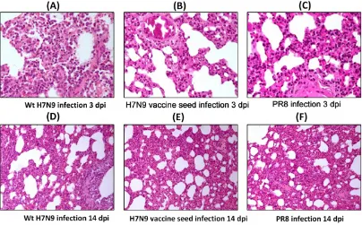
The extent and characteristics of the lesions were
variable among the three infection groups (Figure 2). Pulmonary vasodilation occurred in all three infection
groups at 3 dpi and more serious damage such as focal widened alveolar septum, epithelial cell degeneration and
shedding and inflammatory cell infiltration were detected in wt H7N9 virus infected ferrets. Lesions were mitigated in all these groups at 14 dpi. The wt H7N9 infected ferrets
showed the most severe lesions at both 3 dpi and 14 dpi.
No inflammatory reactions or other histopathological
changes were observed in any of the following tissues: spleen, intestines, liver, heart, kidney, olfactory bulb and brain.
Toxicity of the H7N9 split vaccine
There were no unscheduled deaths during the
course of the study. No vaccine-related effects on clinical signs, body weight (Figure 3), food consumption,
ophthalmologic examinations, coagulation, urinalysis, body temperature or local tolerance were observed.
[image:3.612.71.545.402.645.2]Acute toxicity studies showed that NEUT and WBC in both males and females from group 3 (H7N9 split virus with MF59 adjuvant) and group 4 (MF59 adjuvant control) increased, with values being comparable to the NS (normal saline) control group (group 1; Table 1A). The albumin/globulin (A/G) ratio in both males and females
Figure 1: Body weight, body temperature, viral load, viral titers and serum HI titer of ferrets from the virulence and transmissibility test.A. Weight of ferrets up to 14 dpi. B. Morning temperature of the six groups over the course of infection. C.
in group 3 and group 4 decreased (Table 1B). There were no H7N9 avian influenza vaccine-related organ weight changes, H7N9 avian influenza vaccine with adjuvant, or adjuvant-related organ weight changes observed in the spleen. Compared with the NS control group, the mean absolute and relative (to brain and body weight) weight of the spleen of females in the group treated with adjuvant increased slightly (22% to 24%; Table 1C), without gross lesions. All spleen weights for other animals were within the range of the NS control groups with the exception one
male animal in group 4 which had a higher spleen weight.
Similar changes did not occur in other males; therefore, it
was considered unrelated to the study treatments.
NEUT and EOS in both males and females
undergoing repeated dose toxicity testing in group 3
(H7N9 split virus with MF59 adjuvant) and group 4 (MF59 adjuvant control) increased after dosing on day 17 and day 32 (Table 2A). A/G ratio in both males and
females in group 3 and group 4 decreased after dosing
[image:4.612.105.509.235.486.2]on day 17 and day 32, when compared to the NS control (Table 2B). The ratio change was due to a significant increase in globulin, and decrease in albumin. In the dosing phase, mean absolute and relative (to body and brain) spleen weights were increased in males and females treated with the influenza vaccine with adjuvant and increased to statistically significant levels (p < 0.05)
Figure 2: Lung histopathology of ferrets inoculated intranasally inoculated with 106 TCID50 of wt H7N9 virus, H7N9
vaccine seed or PR8 at 3 dpi and 14 dpi. Multiple 4-μm-thick sections were stained with H&E. Original magnification A-C. ×200, D-F. ×100.
[image:4.612.69.548.563.677.2]Table 1: Hematology: NEUT and WBC; Serum Chemistry: ALB (Albumin), GLB (Globulin) and A/G (%) and Spleen weight (absolute weight, spleen/body ratio and spleen/brain ratio) in both males and females rats from acute toxicity test in group 2 (H7N9 split virion), group 3 (H7N9 split virion with MF59 adjuvant) and group 4 (MF59 adjuvant control) with values being comparable to the NS control group (Group 1)
Table 1A
Dose (mg/kg) Study Day
30 μg/0.5 mL
H7N9 Male:10/5, Female:10/5
15 μg/0.25 mL H7N9
+ MF59 0.25 mL Male:10/5, Female:10/5
0.25 mL PBS + MF59 0.25 mL Male:10/5, Female:10/5 Number of animals
NEUT (103/μL) (%) Day 2 ↓0.5 ↑12.65 ↑402.84*** ↑706.08*** ↑366.95*** ↑765.21***
Day 14 ↓36.75 ↑3.32 ↑4.3 ↑42.71 ↑0.72 ↑14.57
WBC (103/μL) (%) Day 2 ↓14.68 ↑7.84 ↑26.25 ↑61.33** ↑17.69 ↑77.25**
[image:5.612.60.562.110.512.2]Day 14 ↓40.43** ↑7.67 ↓19.38 ↑0.87 ↓26.79* ↑18.02
Table 1B
Dose (mg/kg) Study Day
30 μg/0.5 mL
H7N9 Male:10/5, Female:10/5
15 μg/0.25 mL H7N9
+ MF59 0.25 mL Male:10/5, Female:10/5
0.25 mL PBS + MF59 0.25 mL Male:10/5, Female:10/5 Number of animals
Day 2
ALB (g/L) ↓2 − ↓1.9 ↓0.8 ↓2 ↓2.1 GLB (g/L) ↓1.6 ↓0.2 ↑1.7 ↑2.1 ↑1.8 ↑2.3
A/G (%) ↑0.62 ↑0.88 ↓11.11*** ↓9.34** ↓12.03*** ↓12.9***
Day 14
ALB (g/L) ↓1.1 ↓1.3 ↓.1.8 ↓3.5 ↓0.6 ↓2.2 GLB (g/L) ↓0.8 ↑1.6 ↓0.9 ↑0.6 ↓0.6 ↑1.8 A/G (%) ↑0.24 ↓2.71 ↓1.72 ↓4.02 ↑0.8 ↓3.77
Table 1C
Dose (mg/kg) Index
30 μg/0.5 mL
H7N9 Male:10/5, Female:10/5
15 μg/0.25 mL H7N9
+ MF59 0.25mL Male:10/5, Female:10/5
0.25 mL PBS + MF59 0.25 mL Male:10/5, Female:10/5 Number of animals
Spleen weight
Spleen (g) ↓0.0231 ↑0.0227 ↓0.0415 ↑0.0245 ↓0.0688 ↑0.1106 Spleen/Body (10-3) ↓0.0033 ↑0.0476 ↓0.0805 ↑0.0506 ↓0.2374 ↑0.4543
Spleen/Brain ↓0.0008 ↑0.002 ↓0.0093 − ↓0.0119 ↑0.0537 “−” indicates that all changes were small in magnitude or were comparable to the control groups. ***, ** and* indicate p < 0.001, 0.01, and 0.05, respectively when compared with the NS control. (A) NEUT and WBC changes after dosing on day 2 (B) Serum ALB, GLB and A/G (%) changes of after dosing on day 2 (C) Spleen weight (absolute weight, spleen/body ratio and spleen/brain ratio) changes after dosing on Day 14.
in females treated with adjuvant alone (Table 2C). The
weight change of the spleen was associated with minimal
extramedullary hematopoiesis in some females. Following the recovery period, the hematology (NEUT and EOS), serum chemistry (A/G ratio) and spleen weight changes had resolved, with values comparable to the NS control
group.
At the dosing phase of repeated dose toxicity testing, treatment with the influenza vaccine with adjuvant, and/or treatment with adjuvant alone, caused mixed cell infiltration in the interstitium of the intramuscular injection sites (Figure
4A). The severity of this change was slightly higher in animals treated with the influenza vaccine with adjuvant than in those treated with adjuvant alone. We also observed
lymphoid hyperplasia in the follicles and plasmacytosis in
the medulla of the lymph nodes (popliteal and inguinal; Figure 4B and Figure 4D). These lymph nodes were the draining nodes for the injection and these changes were an expected effect of the treatment. Mixed cell infiltration in the
perinodular connective tissue of the popliteal lymph nodes,
Table 2: Hematology: NEUT and EOS; Serum Chemistry: ALB (Albumin), GLB (Globulin) and A/G (%) and spleen weight (absolute weight, spleen/body ratio and spleen/brain ratio) in both male and female rats from repeated dose toxicity testing in group 2 (H7N9 split virion), group 3 (H7N9 split virion with MF59 adjuvant) and group 4 (MF59 adjuvant control) with values being comparable to the NS control group (group 1)
Table 2A Dose (mg/kg)
Study Day 30 μg/0.5 mLH7N9 Male:10/5, Female:10/5
15 μg/0.25 mL H7N9
+ MF59 0.25 mL Male:10/5, Female:10/5
0.25 mL PBS + MF59 0.25 mL Male:10/5, Female:10/5 Number of animals
NEUT (103/μL) (%) Day 17 −a − ↑50.14*** ↑143.29*** ↑54.96*** ↑75.58***
Day 32 − − ↑44.83 ↑66.75* ↑7.92 ↓0.48 Day 57 − − ↑2.83 ↓32.56 ↑29.09 ↓28.37 EOS (103/μL) (%) Day 17 − − ↑149.73*** ↑227.27*** ↑98.46*** ↑145.46***
[image:6.612.58.557.114.663.2]Day 32 − − ↑366.67*** ↑613.04*** ↑69.44** ↑215.22*** Day 57 − − ↑40 ↑83.33 ↑20 ↑8.33
Table 2B Dose (mg/kg)
Study Day 30 μg/0.5 mLH7N9 Male:10/5, Female:10/5
15 μg/0.25 mL H7N9
+ MF59 0.25 mL Male:10/5, Female:10/5
0.25 mL PBS + MF59 0.25 mL Male:10/5, Female:10/5 Number of animals
Day 17 ALB (g/L) ↑0.1 ↓2.1 ↓3.2*** ↓5*** ↓2.4*** ↓4.5*** GLB (g/L) ↓0.8 ↓1 ↑3*** ↑2.4*** ↑2.6*** ↑1.3
A/G (%) − − ↓17.05*** ↓18.36*** ↓14.06*** ↓14.07*** Day 32 ALB (g/L) ↓0.4 ↓0.8 ↓2.2*** ↓4** ↓1.8*** ↓3.1*
GLB (g/L) ↓0.6 ↑0.3 ↑3.7*** ↑4.8*** ↑1.6* ↑1 A/G (%) − − ↓18.60*** ↓24.56*** ↓10.72*** ↓11.53*** Day 57 ALB (g/L) ↑0.3 ↓1.7 ↑0.3 ↓1.3 ↓0.4 ↑2.3
GLB (g/L) ↓0.7 ↓0.4 ↑2.5 ↑3.3 ↓1.2 ↑1.1 A/G (%) − − ↓8.06 ↓11.57* ↑3.70 ↑2.57
Table 2C Dose (mg/kg)
Index 30 μg/0.5 mLH7N9 Male:10/5, Female:10/5
15 μg/0.25 mL H7N9
+ MF59 0.25 mL Male:10/5, Female:10/5
0.25 mL PBS + MF59 0.25 mL Male:10/5, Female:10/5 Number of animals
Day 32 Spleen (g) ↑0.0638 ↑0.0201 ↑0.1103 ↑0.0725 ↑0.0399 ↑0.0849* Spleen/Body (10-3) ↑0.1304 ↑0.0617 ↑0.2348 ↑0.2504 ↑0.0343 ↑0.3262*
Spleen/Brain ↑0.0203 ↑0.0203 ↑0.0421 ↑0.0421* ↑0.0194 ↑0.0399* Day 57 Spleen (g) ↓0.0019 ↑0.1035 ↓0.0755 ↓0.0079 ↓0.0421 ↓0.0102
Spleen/Body (10-3) ↓0.0138 ↑0.0175 ↓0.0978 ↑0.0646 ↓0.0082 ↓0.0195
(Figure 4E) was observed. These findings were considered an extension of the local injection reaction because of the close proximity to the intramuscular injection site(s). Extramedullary hematopoiesis in the spleen (Figure 4F)
was also observed, which correlated with increased organ weights. This change was considered to be an adaptive
response to the inflammatory reaction incited by the injection of adjuvant or influenza vaccine with adjuvant, which correlated with the NEUT and EOS increase in hematology analysis and A/G decrease in serum chemistry. At the end of recovery phase (Figure 5), only mononuclear
cell infiltration and fibroplasias at the injection sites, and
lymphoid hyperplasia and plasmacytosis in the popliteal lymph nodes were noted, indicating most of the changes had resolved following the recovery phase, which correlated with the recovery observed in the hematology and serum chemistry results.
No abnormalities were noted during the induction phase of the active systemic anaphylaxis test. On the
days of challenge dosing, no abnormalities were recorded for groups 1-4 immediately following the challenge
[image:7.612.122.492.217.653.2]injection, up to 3 h after injection. For animals in group
Figure 4: At day 32 (dosing phase) of the repeated dose toxicity test, H&E staining was performed of various organs from rats treated with H7N9 vaccine with MF59 adjuvant or NS control.A. Intramuscular injection sites (original magnification, ×40), B. popliteal lymph nodes (original magnification, ×200), C. biceps femori (original magnification, NS control, ×40, H7N9 + MF59, ×200), D. inguinal lymph nodes (original magnification, ×40), E. sciatic nerve (original magnification, ×100), F. spleen
5 (albumin, positive control), strong signs of labored respiration, unsteady gait and/or purpura, as well as weak
signs of restlessness, piloerection, and nose scratching, were observed in six animals within 5 min of intravenous challenge.
Hematology and clinical pathology changes were
restricted to those animals receiving the vaccine with
adjuvant or adjuvant alone. These included significant increases in WBC and NEUT in the acute toxicity test, increases in EOS and NEUT in repeated dose toxicity testing and decreases in the A/G ratio. These changes were considered an immune response to the adjuvant. There were no macroscopic findings in the observation period.
There were no organ weight changes or microscopic
findings that were attributed to administration of the influenza vaccine alone. During repeated dose toxicity
testing, increased mean absolute and relative spleen weights were observed in males and females treated
with influenza vaccine with adjuvant and statistically significant increases were observed in females treated with adjuvant alone. This change correlated with minimal
extramedullary hematopoiesis in some females, and was
considered to be an adaptive response to the inflammatory reaction incited by the injection of influenza vaccine with adjuvant or adjuvant alone. Consequently, under the conditions in this study, the NOEAL (no obvious adverse effect level) was determined to be 30 μg/0.5 mL.
DISCUSSION
Previous studies have reported the safety of vaccines prepared using alternative seed strains [18]. However,
most of the studies are clinical reports and evaluate only
the clinical adverse events. Systematic reports including
potential physiological and pathological changes are rare.
Pathogenicity experiments in mice and ferrets confirmed
that we successfully developed a safe vaccine seed strain which demonstrated lower virulence and transmissibility
as compared with wt H7N9 virus. Furthermore, preclinical safety and toxicity tests of the MF59-adjuvanted split influenza vaccine provided no indication of a potential risk of administering the vaccine up to 30 μg/0.5 mL dose in
the experimental animals.
We applied reverse genetics to construct candidate
vaccine which contained the HA and NA genes of A/ Zhejiang/DTID-ZJU01/2013 virus and six genes that encoded internal proteins from the PR8 virus. According
to a previous analysis of antigenic epitopes of avian
influenza A (H7N9) viruses [19, 20], the antigenic variation of HA fragments between A/Zhejiang H7N9 virus and other H7N9 viruses is minor. The H7N9/PR8
virus has many desirable characteristics, including that it can be parenterally administered in an inactive form. Moreover, the lack of pathogenicity or infectivity in chickens should provide for the production of an
[image:8.612.161.452.395.678.2]based vaccine. We utilized a ferret (Mustela putorius furo)
model to investigate virulence and transmissibility of this
H7N9 vaccine seed strain because they are susceptible to
natural infection and develop respiratory disease and lung
pathology similar to humans [21]. Pathogenicity studies showed that the H7N9/PR8 reassortant was not highly
virulent, and transmissibility for mice or ferrets, which
was in accordance with ideal influenza vaccine seed strain
requirements.
Unfortunately, an adjuvant is demanded because
of the comparatively week immunogenicity of split
vaccines [9, 22]. MF59 was applied because according
to the report that it can obviously reduce the antigen
requirements of the split influenza vaccine and can achieve the protective effect with a single dose [10, 23]. Ideal adjuvant properties include a strong enhancement of the immune response, as well as a favorable safety profile. Similar to the classic immunoadjuvant Al(OH)3, the safety
and potency of MF59 has been described in many human
clinical trials [24]. The safety of vaccines must be strictly assessed, and every vaccine batch must be examined
by the National Control Laboratories according to the guidelines published by each country. However, rare cases
of the development of autoimmune diseases following
influenza vaccinations with MF59 adjuvant [25] and
alum-associated macrophagic myofasciitis syndrome [26] have been reported. The preclinical study reported here supports
the good safety of the MF59-adjuvanted H7N9 split vaccine by suggesting a lack of toxicological findings. Toxicity tests of the split H7N9 vaccine in rats and guinea pigs indicated a large safety margin. A dose of a high concentration of the MF59-adjuvanted H7N9 split vaccine
by the intramuscular route in rats showed no effects at
levels far above a typical clinical dose volume of 0.5 mL
for these types of materials. Therefore, on a weight-to-weight basis, a safety margin of approximately 240-fold
(rat = 250 g) was achieved for a 60 kg person. Only after
repeated intramuscular administration of the vaccine with
MF59 or MF59 alone in rats did we observe changes in
hematology and clinical pathology, and these changes
were considered an immune response to the adjuvant or adaptive response to the inflammatory reaction. Overall, the vaccine candidate showed no toxicological findings of significance at levels greatly in excess of those proposed for clinical use. However, the causes of clinical adverse
events are often unclear, and careful attention should be paid especially during the use of concomitant medications.
In conclusion, the comprehensive preclinical
evaluation program conducted here documented the safety
of the recombinant H7N9 vaccine seed strain and safety of the intramuscular MF59-adjuvanted split influenza vaccine in animal models. We believe this MF59-adjuvanted H7N9 split vaccine is able to protect against H7N9 virus without major HA antigenic drift. Further human testing
will be needed to determine whether it could represent a candidate vaccine.
MATERIALS AND METHODS
Ethics statement
Research has been carried out in accordance with the ethical standards, and according to the Declaration of Helsinki and national and international guidelines.
The investigation has been approved by the authors' institutional review board.
Animals, adjuvant and cell lines
Six-week-old BALB/c mice and 4- to 6-month-old castrated male pet ferrets (Mustela putorius furo) were purchased from the Institute of Laboratory Animal Science, Chinese Academy of Medical Sciences. Six-week-old rats were supplied by BioLASCO Taiwan Co., Ltd. Four- to six-week-old guinea pigs were provided by Charles River (Beijing, China). We housed animals
in the departmental animal facility under the following
requirements: temperature 22 ± 3°C, humidity 65 ± 5%, and a 12 h light/dark cycle. The animals were fed a
standard diet and water ad libitum, and were acclimatized
for one week prior to the experiments. MF59 adjuvant was kindly provided by Tianyuan Bio-Pharmaceutical, which is affiliated with Novartis Vaccine Inc. The non-permissive renal canine epithelial cell line MDCK was obtained from ATCC (Rockville, MD, USA). All the tests performed conformed to Good Laboratory Practice (GLP).
Virus preparation
When using live influenza virus, we performed it in a biosafety level (BSL) 3 laboratory according to standard BSL3 guidelines. The reassortant virus was constructed
using plasmid-based reverse genetics through transfection
of eight individual plasmids into Vero cells [5, 6]. The plasmids harbored the HA and NA genes from the A/ Zhejiang/DTID-ZJU01/2013(H7N9) virus, which was isolated from a patient in Zhejiang Province of China in 2013, and six internal genes from PR8. These viruses were propagated in specific-pathogen-free (SPF), embryonated chicken eggs and harvested, clarified, ultrafiltrated, zonal centrifuged, purified, cleaved, sterilization filtered and identified. All production processes were completed in a sterile workshop at Tianyuan Bio-Pharmaceutical in accordance the requirements of China Food and Drug Administration (CFDA) standard.
Vaccine seed virus MLD50 determination
was determined in groups of six mice. The MLD50 was calculated using the Reed and Muench method [27].
Virulence and transmissibility of the vaccine seed virus
The wt H7N9 virus, H7N9 vaccine seed virus and PR8 virus control were tested for virulence and transmissibility in total of 24 (350 g to 900 g) ferrets.
The ferrets were divided into six groups for infection
testing (wt H7N9 virus infection, H7N9 vaccine seed virus infection and PR8 virus infection), or transmission testing (wt H7N9 virus transmission, H7N9 vaccine seed virus transmission and PR8 virus transmission). Ferrets were housed individually. Four ferrets per infection group
were inoculated with 106 TCID50 of virus. On day 1
post-infection (dpi), a naïve ferret was introduced into each
cage with the inoculated ferrets to measure direct contact
transmission. All animals were observed for clinical
signs of respiratory disease twice daily. Body weights were recorded and turbinate swabs collected every other
day. Ferrets in the three infection groups were sacrificed (two at each time point) at 3 dpi and 14 dpi and lungs were removed, fixed in 10% buffered formalin, and embedded in paraffin. Blood samples were collected just prior to sacrifice, and serum samples were tested for hemagglutination inhibition (HI) titers. Viral load and viral titers in the lung (each lobe), turbinate, olfactory bulb, brain (anterior and posterior), spleen and duodenum were determined by quantitative real-time PCR and end-point titration in MDCK cells respectively. Lung (each lobe),
spleen, intestines, liver, heart, kidney, olfactory bulb, brain
(anterior and posterior), and other organs were reserved for pathological examination. In each transmission group,
the period of infection, viral load and viral titers were
determined in the same manner as described above. HI
titers were examined at 14 days post-conversion.
HI test
All sera were processed using receptor-destroying enzyme to prevent non-specific inhibition, as described previously [11]. Prior to testing, 4 hemagglutinating units
of virus was mixed 1:1 with a two-fold serial dilution
series of serum and incubated at 37°C for 1 h. Following the incubation, 50 μL of 1% chicken erythrocytes in phosphate buffered saline (PBS) were added, mixed, incubated for 1 h at 4°C, and agglutination patterns read within 10 min. All tests were performed in “V” bottom microtiter plates and carried out in duplicate. The HI-titer was defined as the reciprocal of the last dilution of serum
that completely inhibits hemagglutination.
RT-PCR
The nasal turbinates were gently rubbed with a wet cotton swab tip for 5 seconds to collect the swab samples.
Before extraction, 0.5 mL Hank’s Balanced Salt Solution (HBSS) was added into the secretions in NS to adjust to 1 mL volume. Samples were extracted from 200 μL as previously described [28]. Quantitative RT-PCR using Influenza A H7N9 virus nucleic acid fluorescent PCR rapid detection kits (DaAn Gene) was performed to amplify the HA and NA genes of the H7N9 virus, with 18S rRNA serving as an internal control.
Virus titrations
We determined virus titrations as described by
end-point titration in MDCK cells [29]. MDCK cells
were inoculated with tenfold serial dilutions of culture
supernatants. Cells were washed twice with PBS one hour after inoculation and cultured in 200 μL of infection medium (MEM containing 100 IU/mL penicillin, 100 μg/ mL streptomycin, 4% bovine serum albumin [fraction V, Gibco-BRL], and 4 μg/mL trypsin [Gibco-BRL]). Three
days after inoculation, the supernatants of cell cultures were tested for agglutinating activity using turkey erythrocytes as an indicator of infection of the cells.
Infectious virus titers were calculated from five replicates each of the homogenized tissue samples [27].
Histopathology of vital organs
All vital organs including the lungs (each lobe),
spleen, intestines, liver, heart, kidney, olfactory bulb, and
brain (anterior and posterior) from the three infection groups were harvested and immersed in 4% formaldehyde at least for 24 h. After fixation of the tissues and processing in paraffin wax, 4 μm sections were prepared and stained with H&E, examined and photographed.
Safety and toxicity evaluation of the split virus vaccine and MF59 adjuvant acute toxicity in rats
According to the WHO guidelines on vaccine safety evaluation (WHO technical reports, No. 927, 2005), the recommended dosing volume for each injection is no more than 30 μg/0.5 mL. Therefore, in this study, the highest single dose for the H7N9 avian influenza vaccine was 30 μg/0.5 mL. Forty Sprague Dawley (SD) rats were randomly assigned to four groups of 5/sex/group. Rats from groups 1-4 were intramuscularly injected with split H7N9 vaccine (30 μg/0.5 mL), split H7N9 vaccine (15 μg/0.25 mL) and MF59 0.25 mL, MF59 (PBS 0.25 mL and MF59 0.25 mL), or NS 0.5 mL, respectively. The dose was administered by intramuscular injection, with a half dose infected in each hind limb. Animals were observed for 14
days then fasted overnight prior to being necropsied.
Repeated dose toxicity in rats
from groups 1-4 were intramuscularly injected with split H7N9 vaccine (30 μg/0.5 mL), split H7N9 vaccine (15 μg/0.25 mL) and MF59 0.25 mL, M59 (PBS 0.25 mL and MF59 0.25 mL), or NS 0.5 mL, respectively, three times on days 1, 15, and 29, as described above. Ten rats of each sex per group and 5/sex/group rats underwent exsanguination from the abdominal aorta after isoflurane
anesthesia and blood sample collection and were
necropsied on day 32 (dosing phase), and day 57 (recovery phase), respectively.
Active systemic anaphylaxis test of split vaccine in guinea pigs
A total of 30 guinea pigs of both sexes were randomly assigned to five groups of 5/sex/group. For induction, groups 1-5 were intramuscularly injected with of 0.5 mL NS, 0.25 mL MF59 plus 0.25 mL PBS, 30 μg/0.5 mL H7N9 vaccine, 30 μg/0.25 mL H7N9 vaccine plus 0.25 mL MF59 and 5 mg/0.5 mL albumin,
respectively, once every other day for a total of three
injections (on days 1, 3, and 5). The challenge was performed on days 19 and 26. For the challenge, all animals were intravenously injected with doses twice
those of the induction phase.
Clinical signs
Cage-side observation was conducted daily for the duration of the study period. Viability (morbidity and mortality) checks were performed twice daily. Detailed
observations were conducted once pre-test, once prior to dosing on day 1, once at approximately 3 h post-dosing on day 1, and weekly thereafter during the observation period.
Body weight
Each animal was weighed once pre-test, once on day 1 prior to dosing, once between 24 h and 48 h post-dosing,
and once weekly throughout the remaining observation period.
Food consumption
Daily food consumption was measured once for all
animals at day -2 during the pre-dosing period, and once
weekly throughout the study from day -1 (except during fasting periods). Food was withdrawn overnight prior to
scheduled blood collections for hematological, coagulation and serum biochemical determinations, urine collections, and scheduled necropsies.
Ophthalmological examination
Eyes were examined via slit lamp and indirect
ophthalmoscope, once pre-dose, once halfway through the
experimental period and once on the last day. A mydriatic (containing 1.0% tropicamide) was used to dilate the
pupils for ophthalmology examination.
Local tolerance (injection site)
Injection sites had previously been clipped of hair and the injection site identified by indelible ink. The injection site was maintained free of excessive hair during the observation period. The injection site was scored using the Draize scoring system. The injection site was scored prior to each injection and at approximately 24 h and 48 h after injection.
Body temperature
Body temperatures were measured once prior to day
1 and pre-dose on day 1, once at approximately (± 30 min)
3 h, 6 h, and 24 h post-dose and once prior to scheduled necropsy.
Hematological analysis
Blood samples for interim hematology, and clinical chemistry evaluation were obtained from the
jugular vein of conscious animals. Blood samples for
hematology, coagulation, and clinical chemistry were obtained from the abdominal aorta of animals at necropsy
as a terminal procedure after isoflurane anesthesia. Five hundred microliters of blood were transfer into a CBC collection tube (K2EDTA, BD, USA) and analyzed using a hematology analyzer (Advia 2120, Seimens, USA). Additionally, 2 mL blood were transferred into a vacutainer (sodium citrate 3.2%, BD) and centrifuged at 3000 rpm for
10 min. The plasma was isolated and used to determine
aggregation time using a coagulometer (BE Compact-X, Behnk Elektronik, Germany). The following parameters were anlayzed: HCT (hematocrit), WBC (white blood cell count), HGB (hemoglobin concentration), RBC (red blood cell count), MCV (mean corpuscular volume), MCHC (mean corpuscular hemoglobin concentration), MCH (mean corpuscular hemoglobin), MPV (mean platelet volume), RDW (RBC distribution width), PLT (platelet count), NEUT (neutrophils), EOS (eosinophils), BASO (basophils), MONO (monocytes), LYMP (lymphocytes), retic (reticulocyte count), blood smear for cytology, PT (prothrombin time), FIB (fibrinogen), APTT (activated partial thromboplastin time).
Blood chemistry
A/G (albumin/globulin ratio), TG (triglycerides), TCHO (total cholesterol), TBILNA (total bilirubin), ALP (alkaline phosphatase), Na (sodium), GGT (gamma glutamyl transferase), K (potassium), GLU (glucose), Cl (chloride), UREA (urea), CK (creatine kinase).
Urinalysis
During the final week of study, urinalysis was conducted to determine BIL (bilirubin), turbidity (color and clarity), KET (ketones), SG (specific gravity), BLO (occult blood), pH, PRO (protein), GLU (glucose) and URO (urobilinogen) using Multistix 10 SG reagent strips (Bayer, U.S.A.), and a urine analyzer (AX-4030, Aikelai, Japan). Microscopy studies were also performed for
examination of sediment.
Necropsy findings
Scheduled terminated study animals underwent exsanguination from the abdominal aorta after isoflurane anesthesia. Gross observation was performed of the external surface, the cranial cavity, all orifices, and all
thoracic and abdominal organs.
Organ weight
Following necropsy, the absolute and relative weight ratio (organ-to-body and organ-to-brain) of following
organs was measured: adrenal glands, brain, heart, liver, kidneys, epididymis, lungs with main stem bronchi, ovaries, pituitary gland, prostate gland, thymus, spleen,
testes, thyroid glands with parathyroid gland(s) and uterus (including cervix).
Histopathology
The following tissues were obtained: liver, kidney,
adrenal gland, bone, aorta and bone marrow (sternum), bone (femur, including stifle joint), fallopian tubes, harderian gland, heart, injection sites, lungs with main
stem bronchi, brain, spleen, epididymis, uterus, vagina,
trachea, nerves (sciatic), esophagus, mammary glands (inguinal, female only), pituitary gland, prostate gland, salivary glands, lymph nodes (inguinal, mandibular, mesenteric and popliteal), thymus, stomach, urinary bladder, small/large intestine, eyes with optic nerve(s), ovaries, seminal vesicles, skin (inguinal), skeletal muscle (biceps femoris), spinal cord (cervical, thoracic, lumbar),
testes, thymus, gross lesions, submandibular gland and
pancreas. Tissues were trimmed and fixed in 10% neutral buffered formalin (NBF) with the following exceptions: eyes with optic nerves were fixed in 2.5% glutaraldehyde solution, and testes as well as epididymis were fixed in modified Davidson’s solution for 24 to 72 h before being transferred to 10% NBF. Tissues were removed from fixative, processed, embedded in paraffin, sectioned,
stained with hematoxylin and eosin (H&E), and examined
microscopically.
Statistical analysis
Males and females were analyzed separately. The viral load, virus titer, body weight, organ weight, biochemistry data and hematological data were analyzed
for homogeneity of variance using Levene’s test. One-way ANOVA was conducted for data shown to have homogeneity of variance. If homogeneity of variance and significance between test groups was confirmed, the Scheffe’s test was performed. If heterogeneity of variance and significance between test groups was determined, Dunnett’s test was conducted. All analysis were performed using the SPSS program.
ACKNOWLEDGMENTS
This work was supported by the State Project for Essential Drug Research and Development [grant number 2015ZX09101044] and the Science & Technology Key Program of Zhejiang China [grant number 2014C03039].
CONFLICTS OF INTEREST
Two authors Wei Yao and Chengcong Han have declared they have conflict interests relevant to the submitted work. They are employees of Zhejiang Tianyuan Bio-Pharmaceutical Co., Ltd. (which was affiliated with Novartis Vaccine Inc.) and participated in the manufacture of H7N9 vaccine and MF59 adjuvant, however, they had
nothing to do with the investigation of the safety and toxicity of this vaccine. The other authors declare no competing interests.
Authors’ contributions
LJ Li, HP Yao, NP Wu and HL Chen developed the concept and designed the studies. HP Yao, HL Ou, XY Lu, DS Yu, HB Wu and LF Cheng developed and established the methodologies and assays. W Yao and C Han prepared the H7N9 vaccine. HP Yao, HL Ou, Frederick XC Wang and TH Weng conducted data acquisition, analysis and interpretation. HP Yao, HL Ou wrote, reviewed, and revised the manuscript. LJ Li and HP Yao supervised and administered the studies. All authors read and approved the final manuscript.
REFERENCES
2. Li Q, Zhou L, Zhou M, Chen Z, Li F, Wu H, Xiang N, Chen E, Tang F, Wang D, Meng L, Hong Z, Tu W, et al. Epidemiology of human infections with avian influenza A(H7N9) virus in China. N Engl J Med. 2014; 370: 520-32.
3. World Health Organization., WHO risk assessment of human infection with avian influenza A (H7N9) virus. WHO Risk Assessment (2015) Available at: 2015. http:// www.who.int/entity/influenza/human_animal_interface/ influenza_h7n9/Risk_Assessment/en/index.html. (Accessed: 1th February 2016).
4. Fries LF, Smith GE, Glenn GM. A recombinant viruslike particle influenza A (H7N9) vaccine. N Engl J Med. 2013; 369: 2564-6.
5. Hoffmann E, Neumann G, Kawaoka Y, Hobom G, Webster RG. A DNA transfection system for generation of influenza A virus from eight plasmids. Proc Natl Acad Sci U S A. 2000; 97: 6108-13.
6. Neumann G, Watanabe T, Ito H, Watanabe S, Goto H, Gao P, Hughes M, Perez DR, Donis R, Hoffmann E, Hobom G, Kawaoka Y. Generation of influenza A viruses entirely from cloned cDNAs. Proc Natl Acad Sci U S A. 1999; 96: 9345-50.
7. Nicolson C, Major D, Wood JM, Robertson JS. Generation of influenza vaccine viruses on Vero cells by reverse genetics: an H5N1 candidate vaccine strain produced under a quality system. Vaccine. 2005; 23: 2943-52.
8. World Health Organization., Summary of status of development and availability of avian influenza A (H7N9) candidate vaccine viruses. WHO Risk Report (2015) Available at: www.who.int/influenza/vaccines/virus/ candidates_reagents/a_h7n9/en/. (Accessed: 1th August 2016).
9. Atmar RL, Keitel WA, Patel SM, Katz JM, She D, El Sahly H, Pompey J, Cate TR, Couch RB. Safety and immunogenicity of nonadjuvanted and MF59-adjuvanted influenza A/H9N2 vaccine preparations. Clin Infect Dis. 2006; 43: 1135-42.
10. Nicholson KG, Colegate AE, Podda A, Stephenson I, Wood J, Ypma E, Zambon MC. Safety and antigenicity of non-adjuvanted and MF59-adjuvanted influenza A/Duck/ Singapore/97 (H5N3) vaccine: a randomised trial of two potential vaccines against H5N1 influenza. Lancet. 2001; 357: 1937-43.
11. Arguedas A, Soley C, Abdelnour A, Sales V, Lindert K, Della Cioppa G, Clemens R, Costa Rican HNVSG. Assessment of the safety, tolerability and kinetics of the immune response to A/H1N1v vaccine formulations with and without adjuvant in healthy pediatric subjects from 3 through 17 years of age. Hum Vaccin. 2011; 7: 58-66.
12. Clark TW, Pareek M, Hoschler K, Dillon H, Nicholson KG, Groth N, Stephenson I. Trial of 2009 influenza A (H1N1) monovalent MF59-adjuvanted vaccine. N Engl J Med. 2009; 361: 2424-35.
13. Bresson JL, Perronne C, Launay O, Gerdil C, Saville M, Wood J, Hoschler K, Zambon MC. Safety and immunogenicity of an inactivated split-virion influenza A/ Vietnam/1194/2004 (H5N1) vaccine: phase I randomised trial. Lancet. 2006; 367: 1657-64.
14. Bernstein DI, Edwards KM, Dekker CL, Belshe R, Talbot HK, Graham IL, Noah DL, He F, Hill H. Effects of adjuvants on the safety and immunogenicity of an avian influenza H5N1 vaccine in adults. J Infect Dis. 2008; 197: 667-75.
15. Murphy TV, Gargiullo PM, Massoudi MS, Nelson DB, Jumaan AO, Okoro CA, Zanardi LR, Setia S, Fair E, LeBaron CW. Intussusception among infants given an oral rotavirus vaccine. New England Journal of Medicine. 2001; 344: 564-72.
16. Ellenberg SS, Foulkes MA, Midthun K, Goldenthal KL. Evaluating the safety of new vaccines: summary of a workshop. Am J Public Health. 2005; 95: 800-7.
17. Ou H, Yao H, Yao W, Wu N, Wu X, Han C, Cheng L, Chen K, Chen H, Li L. Analysis of the immunogenicity and bioactivities of a split influenza A/H7N9 vaccine mixed with MF59 adjuvant in BALB/c mice. Vaccine. 2016; 34: 2362-70.
18. Sobhanie M, Matsuoka Y, Jegaskanda S, Fitzgerald T, Mallory R, Chen ZY, Luke C, Treanor J, Subbarao K. Evaluation of the Safety and Immunogenicity of a Candidate Pandemic Live Attenuated Influenza Vaccine (pLAIV) Against Influenza A(H7N9). Journal of Infectious Diseases. 2016; 213: 922-9.
19. Liu M, Song TR, Hua S, Wu AP, Jiang TJ. Computational analysis of antigenic epitopes of avian influenza A (H7N9) viruses. Science China-Life Sciences. 2015; 58: 687-93.
20. De Groot AS, Moise L, Liu R, Gutierrez AH, Terry F, Koita OA, Ross TM, Martin W. Cross-conservation of T-cell epitopes: now even more relevant to (H7N9) influenza vaccine design. Hum Vaccin Immunother. 2014; 10: 256-62.
21. Maher JA, DeStefano J. The ferret: an animal model to study influenza virus. Lab Anim (NY). 2004; 33: 50-3.
22. Stephenson I, Nicholson KG, Colegate A, Podda A, Wood J, Ypma E, Zambon M. Boosting immunity to influenza H5N1 with MF59-adjuvanted H5N3 A/Duck/Singapore/97 vaccine in a primed human population. Vaccine. 2003; 21: 1687-93.
23. Baras B, Stittelaar KJ, Simon JH, Thoolen RJ, Mossman SP, Pistoor FH, van Amerongen G, Wettendorff MA, Hanon E, Osterhaus AD. Cross-protection against lethal H5N1 challenge in ferrets with an adjuvanted pandemic influenza vaccine. PLoS One. 2008; 3: e1401.
24. Pellegrini M, Nicolay U, Lindert K, Groth N, Della Cioppa G. MF59-adjuvanted versus non-adjuvanted influenza
vaccines: integrated analysis from a large safety database.
25. Cheng MP, Kozoriz MG, Ahmadi AA, Kelsall J, Paquette K, Onrot JM. Post-vaccination myositis and myocarditis in a previously healthy male. Allergy Asthma Clin Immunol. 2016; 12: 6.
26. Gherardi R, Coquet M, Cherin P, Authier F, Laforêt P, Belec L, Figarella-Branger D, Mussini J, Pellissier J, Fardeau M. Macrophagic myofasciitis: an emerging entity. The Lancet. 1998; 352: 347-52.
27. Reed LJ, Muench H. A simple method of estimating fifty per cent endpoints. American journal of epidemiology. 1938; 27: 493-7.
28. Kuypers J, Wright N, Morrow R. Evaluation of quantitative and type-specific real-time RT-PCR assays for detection of
respiratory syncytial virus in respiratory specimens from
children. J Clin Virol. 2004; 31: 123-9.