Copyright © 2002, American Society for Microbiology. All Rights Reserved.
Clinical and Laboratory Features of
Mycobacterium mageritense
Richard J. Wallace Jr.,
1* Barbara A. Brown-Elliott,
1Leslie Hall,
2Glenn Roberts,
2Rebecca W. Wilson,
1,3Linda B. Mann,
1Christopher J. Crist,
1Sher H. Chiu,
4Robbie Dunlap,
4Maria J. Garcia,
5J. Todd Bagwell,
6and Kenneth C. Jost Jr.
4Departments of Microbiology1and Pathology,3University of Texas Health Center, Tyler, and Mycobacteriology
Mycology Branch, Texas Department of Health,4and Austin Infectious Disease Consultants,6Austin,
Texas; Mayo Clinic, Rochester, Minnesota2; and Departmento de Medicina Preventiva,
Facultad de Medicina, Universidad Autonoma de Madrid, Madrid, Spain5
Received 28 November 2001/Returned for modification 22 January 2002/Accepted 8 May 2002
Six clinical isolates of the nonpigmented, rapidly growing speciesMycobacterium mageritensewere recovered from sputum, bronchial wash, blood, sinus drainage, and two surgical wound infections from separate patients in Texas, New York, Louisiana, and Florida. The isolates matched the ATCC type strain by PCR restriction enzyme analysis of the 65-kDahspgene sequence of Telenti, high-performance liquid chromatography, bio-chemical reactions, and partial 16S rRNA gene sequencing. These are the first isolates of this species to be described in the United States and the first isolates to be associated with clinical disease. Susceptibility testing of all known isolates of the species revealed all isolates to be susceptible or intermediate to amikacin, cefoxitin, imipenem, and the fluoroquinolones and sulfonamides but resistant to clarithromycin. Because of their phenotypic and clinical similarity to isolates of theMycobacterium fortuitumthird biovariant complex (sorbitol positive), isolates ofM. mageritense are likely to go undetected unless selected carbohydrate utilization or molecular identification methods are used.
In 1997 Domenech et al. (4) described a new nonpigmented species of rapidly growing mycobacterium that they named
Mycobacterium mageritense. The name was derived from Mag-erit, the old Arabic name of Madrid, where four of the five isolates had been recovered. The isolates were from sputum and came from two hospitals in Spain. None were known to be clinically significant. The isolates were nonpigmented and had
similarities to bothM. smegmatisand theM. fortuitumcomplex
(1, 4, 10, 18).
Utilizing PCR restriction enzyme analysis (PRA) of the
Telenti fragment (11, 14) of the 65-kDahspgene, we identified
six clinical isolates whose PRA patterns matched the pattern of
M. mageritense. We compared these strains to the initial strains from Spain by using standard growth and biochemical tests,
partial 16S rRNA gene sequencing, PRA of the 65-kDa hsp
gene, high-performance liquid chromatography (HPLC), and determination of antimicrobial susceptibilities.
MATERIALS AND METHODS
Organisms.Eleven isolates ofM. mageritense, including the five original Span-ish isolates described in 1997 (ATCC 700351T, strains 1635, 1470, 1636, and
1336) (4), three isolates from Texas (strains 1852, 1582, and 2056), one from Florida (strain 1301), one from New York (strain 2048), and one from Louisiana (strain 1846), were studied.M. fortuitumATCC 6841TandM. smegmatisATCC
19420Twere used as control strains for HPLC and PRA, whileStaphylococcus
aureusATCC 29213 andM. peregrinumATCC 700686 were used as controls for susceptibility testing.
Growth and biochemical characterization. All 11 isolates identified asM. mageritensewere examined for colony morphology, pigmentation, and growth
within 7 days on Trypticase soy agar and Middlebrook 7H10 agar at 30, 35, and 45°C, as well as for arylsulfatase activity at 3 days (graded from 0 to 4⫹), nitrate reductase activity (graded from 0 to 5⫹), iron uptake, and urease activity (graded from 0 to 4⫹) (5). Carbohydrate utilization testing includedD-mannitol, i-myo -inositol, citrate,D-galactose,L-rhamnose,D-trehalose,D-glucitol (sorbitol), D-xylose, andL-arabinose (10, 15). Acetamide (Becton Dickinson Biosciences, Sparks, Md.) utilization was also determined in addition to susceptibility to kanamycin, cephalothin, and polymyxin B by using a disk diffusion technique on Mueller-Hinton agar (17, 20).
Susceptibility testing.Susceptibility to 10 antimicrobial agents was determined by broth microdilution in cation-supplemented Mueller-Hinton broth (2, 3, 12, 13, 17, 23). The antimicrobials tested were amikacin, cefoxitin, ciprofloxacin, clarithromycin, doxycycline, imipenem, minocycline, sulfamethoxazole, tobramy-cin, and linezolid. Because not all isolates were tested simultaneously, some isolates were not tested against minocycline or linezolid. Breakpoints were those suggested by the National Committee for Clinical Laboratory Standards for rapidly growing mycobacteria (7), except with linezolid and minocycline. Break-points for minocycline (not addressed by the National Committee for Clinical Laboratory Standards) were made the same as those for doxycycline, which is addressed in the document (7), and linezolid breakpoints were those recently proposed for rapidly growing mycobacteria (19). Quality control was performed withS. aureusATCC 29213 andM. peregrinumATCC 700686 (6).
HPLC.Mycolic acids were prepared as described previously (1). They were then dissolved in 200l of chloroform containing 200g of 4 bromomethyl-6,7-dimethoxycoumarin and 200g of 18-crown-6 ether and transferred to a 2-ml autosampler vial into which 100l of a 2% potassium bicarbonate solution had been evaporated previously. The vial was heated at 60°C for 15 min and allowed to evaporate. The mycolic esters were dissolved in 500l of chloroform con-taining internal standards and analyzed by fluorescence detection HPLC (FL-HPLC) as described previously (1).
PRA.All isolates ofM. mageritensewere subjected to PCR amplification of a 439-bp segment of the 65-kDahspgene, as originally described by Telenti et al. (14). Ground cell supernatants were used as DNA templates (11, 22) by using the appropriate positive and negative controls (14).
Seven restriction endonucleases (BstEII,HaeIII,AciI,HhaI,MspI,HinfI, and
BsaHI) were used. Restriction fragments were electrophoresed on 3% metaphor agarose (4-bp resolution; FMC Bioproducts, Rockland, Maine) containing ethidium bromide in a Mini-SubCell electrophoresis system (Bio-Rad, Hercules, Calif.) at 95 V for 1.5 to 2.0 h. Fragment sizes (in base pairs) were estimated on a computerized Bio Image system (Millipore, Bedford, Mass.) (1).
* Corresponding author. Mailing address: The University of Texas Health Center, Department of Microbiology, 11937 US Hwy 271, Tyler, TX 75708. Phone: (903) 877-7680. Fax: (903) 877-7652. E-mail: Richard.Wallace@uthct.edu.
2930
on May 15, 2020 by guest
http://jcm.asm.org/
16S rRNA partial gene sequencing.16S rRNA gene sequencing was per-formed by using standard techniques (8). Sequencing analysis of the first 500 bp in the 16S rRNA gene that includes the hypervariable A region was performed with an ABI Prism 3100 genetic analyzer (Applied Biosystems, Foster City, Calif.) and a MicroSeq 16S 500 16S rDNA sequencing module. Data analysis was carried out with MicroSeq version 1.4 (Applied Biosystems).
RESULTS
Organisms.Eleven isolates ofM. mageritensewere studied. Five isolates were isolated in Spain from 1987 to 1989 and described in 1997 (4). One of the five isolates was submitted to the American Type Culture Collection and is designated
ATCC 700351T. The six United States isolates were submitted
for susceptibility testing and identification from 1999 to 2002 to the Mycobacteria/Nocardia Laboratory of the University of Texas Health Center at Tyler. The first isolate was recovered from sputum culture, the second was recovered from a bron-choscopy sample, the third was obtained from a thigh wound culture from a woman with localized cellulitis following a li-posuction procedure, the fourth was from the blood of an immune-suppressed patient with a central catheter and clinical sepsis, the fifth was from a patient with severe sinusitis, and the sixth was from a patient with a wound infection and, probably, osteomyelitis following an open fracture which had undergone open reduction and internal fixation (Table 1). The history of the second patient is provided below.
Case summary. The patient was a 37-year-old previously healthy white female referred by a local plastic surgeon in July 2000. She had a liposuction procedure in her medial thigh area in January 2000. At an instrument insertion site she had a “sore that never healed.” She continued to have drainage off and on from this area despite therapy with oral cephalosporins. A small nodule remained in the thigh. The thigh lesion began to drain more profusely. On 17 July 2000, a culture of this drain-age grew an acid-fast bacillus on routine media that was
sub-sequently identified asM. mageritense. She was referred to one
of the authors (J.T.B.) on 21 July, and a soft tissue infection involving a rapidly growing mycobacterium was suspected. Her physical exam was normal except for a small, 2-cm circular nodule on the left medial thigh with a scant amount of serous drainage. She was started on doxycycline and ciprofloxacin. She was seen again on 29 August 2000. The small nodule was still present but much improved, and no drainage was present.
At her request, the antibiotic therapy was discontinued. She was seen again in October 2000, after she had noticed about a week of drainage from the same thigh nodule. She was re-started on antibiotics. An elliptical excision of the nodule was performed on 8 November 2000. A culture showed no growth. She was continued on ciprofloxacin alone at that time. She was seen again 17 January 2001 and had persistent drainage, al-though the nodule was much smaller. This area was excised again with excellent results. She was placed on ciprofloxacin following that last visit and was doing well with no evidence of recurrence 3 months later.
Growth and biochemical characterization.All 11 isolates of
M. mageritenseproduced nonpigmented smooth or rough col-onies within 7 days on Trypticase soy agar and Middlebrook 7H10 agar at 30 and 35°C (Table 2). Growth at 45°C was variable. Isolates were strongly positive for 3-day arylsulfatase production, nitrate reductase, iron uptake, and urease. All five of the Spanish isolates and the six United States isolates
uti-lizedD-mannitol, i-myo-inositol,L-rhamnose,D-glucitol
(sorbi-tol),D-trehalose, and acetamide. Approximately 50% of
iso-lates from both groups utilizedD-galactose, and most or all of
the isolates were unable to utilizeL-arabinose, citrate, orD
-xylose.
Susceptibility testing.All 11 isolates ofM. mageritensewere susceptible to ciprofloxacin and sulfamethoxazole. All isolates were susceptible or intermediate in susceptibility to amikacin, linezolid, imipenem, and cefoxitin and were intermediate or resistant to tobramycin. All 11 isolates were resistant to
clar-ithromycin, with MICs for 8 of 11 being⬎32g/ml. By disk
diffusion testing, only 50% of the Spanish isolates other than the type strain were susceptible to polymyxin B while all of the United States isolates were susceptible. For kanamycin, all of the Spanish isolates were susceptible while all of the United States isolates were resistant. None of the isolates were inhib-ited by cephalothin. Susceptibility results for all drugs are shown in Tables 2 and 3.
When grown simultaneously to stationary phase with the lipids extracted after the same incubation time, the ATCC type
strain ofM. mageritense(ATCC 700351T), the remaining four
Spanish strains, and five of the six United States isolates had matching FL-HPLC patterns that were distinct from those
produced by M. smegmatis and M. fortuitum. The M.
mag-TABLE 1. Clinical features of the six United States isolates ofM. mageritense
Isolate Age/sexa Source specimen Location Disease Other disease Treatment; outcome
1852 37/F Surgical wound (thigh) Texas Wound infection following
liposuction None Doxycycline,ciprofloxacin; surgery, cured
1582 Unknown/F Sputum Texas Unknown Unknown Unknown
1301 32/F Blood Florida Catheter-related sepsis Immune suppression Amikacin, linezolid
2048 51/F Sinus New York Severe sinusitis Unknown Amikacin (topical),
ciprofloxacin, clarithromycin 2056 25/M Surgical wound, knee
effusion Texas Wound infection, possibleosteomyelitis Recent open femurfracture with open reduction
Amikacin, imipenem; surgery, improved 1846 42/M Bronchial wash Louisiana Clinical significance unknown HIV Macrolide
aAge given in years. F, female; M, male.
on May 15, 2020 by guest
http://jcm.asm.org/
[image:2.587.44.541.83.223.2]eritense isolates had a relatively flat first set of peaks and a single major peak in the second set of peaks (Fig. 1). As previously described (1), a minor difference in peak retention
time permitted a distinction betweenM. mageritenseand M.
fortuitum; all but one of theM. mageritensestrains produced a
peak at approximately 5.64 min, whereas the correspondingM.
fortuitum peak eluted 0.04 min earlier. One isolate (2048)
exhibited a different pattern, with two major peaks and some-what different retention times.
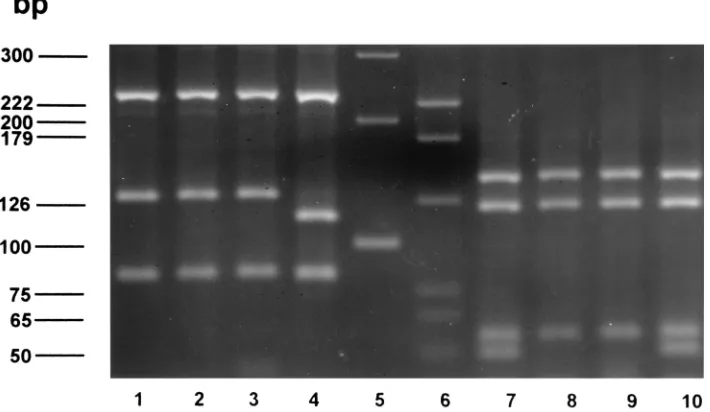
PRA.By PRA, the 11 isolates ofM. mageritensegave
iden-tical restriction patterns with the seven restriction enzymes, except that three of the six clinical isolates from the United
States had an extra cutting site forBsaHI (this enzyme
[image:3.587.46.544.81.407.2]com-monly exhibits intraspecies variation with mycobacteria and
TABLE 2. Laboratory features of the ATCC type strain, Spanish isolates, and United States clinical strains ofM. mageritense
Feature
Presence (⫹), absence (⫺), or type of feature for indicated strain or isolate(s)
ATCC 700351T Spanish isolates (%) United States Isolates
1582 1852 1301 2048 2056 1846
Pigment (early/late) ⫺/⫺ ⫺/⫺(0) ⫺/⫺ ⫺/⫺ ⫺/⫺ ⫺/⫺ ⫺/⫺ ⫺/⫺
Colony morphology Smooth Smooth Smooth Smooth Smooth Rough Rough Rough
Growth (2 wk)
⬍7 days ⫹ ⫹(100) ⫹ ⫹ ⫹ ⫹ ⫹ ⫹
30°C ⫹ ⫹(100) ⫹ ⫹ ⫹ ⫹ ⫹ ⫹
35°C ⫹ ⫹(100) ⫹ ⫹ ⫹ ⫹ ⫹ ⫹
42°C ⫹ NDa ⫹ ⫹ ⫹ ⫹ ND ND
45°C ⫹/⫺b ⫹/⫺(50) ⫺ ⫺ ⫹/⫺ ⫹ ⫹ ⫹/⫺
Growth on MacConkey agar
without crystal violet ⫹ ⫹(100) ND ⫹ ND ND ND ND
Arylsulfatase (3 days) ⫹(4⫹) ⫹(100) ⫹(3⫹) ⫹(3⫹) ⫹(4⫹) ⫹(4⫹) ⫹(4⫹) ⫹(4⫹)
Catalase (68°C) ⫺ ⫺(0) ⫺ ⫺ ⫺ ⫺ ⫺ ⫺
Nitrate reduction ⫹(5⫹) ⫹(100) ⫹(5⫹) ⫹(5⫹) ⫹(5⫹) ⫹(5⫹) ⫺ ⫹(5⫹)
Iron uptake ⫹ ⫹(100) ⫹ ⫹ ⫹ ⫹ ⫹ ⫹
Urease ⫹(4⫹) ⫹(100) ⫹(4⫹) ⫹(4⫹) ⫹(4⫹) ⫹(4⫹) ⫹(4⫹) ⫹(4⫹)
Carbohydrate utilization
L-Arabinose ⫹/⫺ ⫺(0) ⫺ ⫺ ⫺ ⫺ ⫺ ⫺
Citrate ⫺ ⫺(0) ⫹/⫺ ⫺ ⫹/⫺ ⫹ ⫹/⫺ ⫹/⫺
D-Galactose ⫺ ⫹/⫺(50) ⫹ ⫹ ⫹ ⫺ ⫹ ⫺
i-myo-Inositol ⫹ ⫹(100) ⫹ ⫹ ⫹ ⫹ ⫹ ⫹
D-Mannitol ⫹ ⫹(100) ⫹ ⫹ ⫹ ⫹ ⫹ ⫹
L-Rhamnose ⫹ ⫹(100) ⫹ ⫹ ⫹ ⫹ ⫹ ⫹
D-Glucitol (sorbitol) ⫹ ⫹(100) ⫹ ⫹ ⫹ ⫹ ⫹ ⫹
D-Trehalose ⫹ ⫹(100) ⫹ ⫹ ⫹ ⫹ ⫹ ⫹
D-Xylose ⫺ ⫺(0) ND ⫺ ⫺ ⫹/⫺ ⫺ ⫺
Acetamide ⫹ ⫹(100) ⫹ ⫹ ⫹ ⫹ ⫹ ⫹
Inhibition
Polymyxin B ⫺ ⫹/⫺(50) ⫹ ⫹ ⫹ ⫹ ⫹ ⫹
Cephalothin ⫺ ⫺(0) ⫺ ⫺ ⫺ ⫺ ⫺ ⫺
Kanamycin ⫹ ⫹(100) ⫺ ⫺ ⫺ ⫺ ⫺ ⫺
aND, not determined. b⫹/⫺, weak or variable.
TABLE 3. Susceptibility results of isolates ofM. mageritense
Drug Resistancebreakpoint (g/ml)a
MIC (g/ml) against:
Spanish isolates United States isolates
ATCC 700351T 1635 1470 1636 1336 1852 1582 1301 2048 2056 1846
Amikacin ⱖ64 2 2 2 2 8 32 32 32 32 32 32
Cefoxitin ⱖ128 16 8 16 16 16 32 32 16 16 32 16
Ciprofloxacin ⱖ4 ⱕ0.125 0.25 0.25 0.25 0.5 0.25 0.25 ⱕ0.25 0.5 0.5 0.25
Clarithromycin ⱖ8 ⬎64 16 ⬎64 64 16 ⬎64 ⬎64 8 ⬎32 ⬎32 ⬎64
Doxycycline ⱕ16 4 0.5 1 2 2 4 8 2 ⬎64 16 32
Imipenem ⱖ16 2 ⱕ1 4 2 4 4 8 ⱕ1 2 2 NDb
Linezolid ⱖ32 1 ND ND ND ND 4 8 ⱕ2 16 8 8
Minocycline ⱖ16 4 0.5 1 2 2 ND ND ND ND ND ND
Sulfamethoxazole ⱖ64 2 2 4 2 2 32 8 32 8 ⱕ4 8
Tobramycin ⱖ16 8 8 8 8 8 ⬎16 16 ⬎16 ⬎16 ⬎16 ⬎16
aSee reference 7. bND, not determined.
on May 15, 2020 by guest
http://jcm.asm.org/
[image:3.587.41.547.569.712.2]other aerobic actinomycetes) (R. W. Wilson and R. J. Wallace, Jr., unpublished data). Their pattern was unique among those of all species in our current database (Fig. 2).
16S rRNA sequencing. The ATCC type strain of M. mag-eritense(ATCC 700351T) and four of the clinical isolates (1852, 1582, 2048, and 1846) had identical 500-bp sequences that included the hypervariable region A. Two isolates (1301 and
2056) differed from the other five isolates by a single G3A
nucleotide change at position 446 (99.8% identify with the ATCC type strain).
DISCUSSION
These are the first cases of clinical disease due toM.
mag-eritenseto be published and the first six isolates of the species
to be identified in the United States, and this is the first report of drug susceptibilities for this species. We have also further detailed laboratory characteristics of this new species.
The clinical disease produced byM. mageritenseis similar to
disease produced by theM. fortuitumthird biovariant complex
(17, 21) and includes skin and soft tissue infections as well as health care-associated infections. From a therapeutic
stand-point, United States isolates ofM. mageritense are generally
more difficult to treat, as amikacin MICs for all six isolates to
date have been high (32 g/ml), 50% of the isolates were
resistant to doxycycline, and all were resistant to clarithromy-cin. Drugs that offer therapeutic potential include imipenem, cefoxitin, sulfonamides, linezolid, and the newer quinolones, although quinolone monotherapy should be avoided when pos-sible because of the risk of developing mutational resistance (16); in addition, there is no published experience with the use
of linezolid for theM. fortuitumgroup. Surgical debridement
can be useful (as in case 2) but is not required for most diseases given the drugs available for therapy.
The susceptibility patterns of the 11 isolates of M.
mag-eritensewere very similar. However, there were differences in aminoglycoside susceptibilities between the Spanish isolates and the United States isolates. The six United States isolates were all intermediate to amikacin, with amikacin MICs of 32
g/ml, and resistant to kanamycin, while the five Spanish
iso-lates were susceptible to both aminoglycosides. These two dif-ferent aminoglycoside susceptibility patterns have previously
been shown to be present in isolates of theM. fortuitumthird
biovariant complex (17). The isolates were also uniformly re-sistant to clarithromycin, a finding previously noted for all
isolates of the sorbitol-positive M. fortuitum third biovariant
complex (of which most isolates have been tentatively
desig-natedM. houstonense[17]). High-level resistance to
clarithro-mycin (MIC ofⱖ32g/ml) is seen only in these two taxonomic
[image:4.587.49.277.76.239.2]groups among all members of theM. fortuitum group. With
FIG. 1. FL-HPLC patterns of three strains ofM. mageritenseand two rapidly growing mycobacterial reference strains.
FIG. 2. PRA restriction fragment length polymorphism pattern from isolates ofM. mageritenseand anM. fortuitumcontrol. Lanes 1 to 4:BstEII digests fromM. mageritenseATCC 700351T, clinical isolates 1582 and 1852, and control isolateM. fortuitumATCC 6841T, respectively. Lanes 5
and 6: 100-bp and pGEm markers, respectively. Lanes 7 to 10:HaeIII digests with the same isolates as those used for theBstEII digests.
on May 15, 2020 by guest
http://jcm.asm.org/
[image:4.587.117.472.489.695.2]susceptibility patterns, the two groups are distinguishable by cefoxitin susceptibility, as the cefoxitin MICs for all of the
sorbitol-positive third biovariant isolates areⱖ64g/ml (17)
while those for all of the isolates ofM. mageritenseare ⱕ32
g/ml (Table 3).
The first study ofM. mageritensesuggested that it was
phe-notypically most closely related to M. smegmatis, based on
growth at 45°C and a variable 3-day arylsulfatase reaction (4). The present study, however, showed that many isolates of the species grow poorly at 45°C and all had a positive 3-day aryl-sulfatase reaction when tested by the method of Kent and Kubica (5). These features, combined with the absence of pigmentation and growth on MacConkey agar without crystal violet (10), result in responses identical to those of other
spe-cies currently grouped within theM. fortuitum complex.
Ac-cording to results of the additional recommended tests for evaluating rapidly growing mycobacteria, namely those of ni-trate reductase, iron uptake, and carbohydrate utilization of
D-mannitol, i-myo-inositol, and citrate (10), the isolates ofM.
mageritensehave reactions identical to those of theM.
fortui-tumthird biovariant complex. They are also sorbitol positive,
as are approximately 40% of the members of this complex (17).
Initial phenotypic studies suggested that the M. fortuitum
third biovariant complex (defined as isolates that matched the
growth and biochemical characteristics of M. fortuitum but
were alsoD-mannitol and i-myo-inositol positive) (10, 17) was
heterogeneous and likely comprised multiple taxa. They were
subdivided into two groups according toD-glucitol (sorbitol)
utilization (17), but studies of the electrophoretic patterns of
their -lactamase showed multiple patterns among isolates
which were cefoxitin susceptible in both sorbitol groups, sug-gesting that multiple taxa might be present (24). The data on the previous susceptibilities to cefoxitin of isolates in the
sor-bitol-positive group showed the MICs to be low (ⱕ32g/ml)
for 4 of 33 isolates (12%) according to the same broth suscep-tibility method (17) as that used in the present study. In
ret-rospect, these four isolates may well have been isolates ofM.
mageritense, and this suggests that 10 to 15% of sorbitol-posi-tive isolates will belong to this taxon. In contrast, with isolates ofM. fortuitum, M. chelonae, and M. abscessus, each species
had a single unique -lactamase pattern. The recently
de-scribedM. septicum (9) and nowM. mageritense support the
concept of this group being a complex, as both of these new species meet prior phenotypic definitions of the third biovari-ant complex. Additionally, ongoing studies suggest that at least
five more species are present in this complex, including M.
houstonenseand M. bonickei (M. F. Schinsky, R. E. Morey, M. P. Douglas, A. G. Steigerwalt, R. W. Wilson, M. M. Floyd, M. I. Daneshvar, B. A. Brown-Elliott, R. J. Wallace, Jr., M. M. McNeil, D. J. Brenner, and J. M. Brown, submitted for publi-cation).
Given that the isolates ofM. mageritensehave the growth,
biochemical, and drug susceptibility patterns ofM. fortuitum
third biovariant complex sorbitol-positive isolates (except for cefoxitin MICs) (17), it is easy to see how isolates of the former could be missed. One carbohydrate not routinely tested that
would separate the two taxa isL-rhamnose, as the M.
mag-eritenseisolates were all positive and⬍5% ofM. fortuitumthird biovariant complex isolates have previously been reported to be positive (R. J. Wallace, Jr., unpublished data). One method
for separation of the species and/or taxa of theM. fortuitum
group is to screen them with mannitol. Most isolates within this
group will be M. fortuitum, which produces a negative test.
These can be identified without any additional testing. All
other members of theM. fortuitumgroup are mannitol
posi-tive. For isolates that are mannitol positive, the additional sugars of inositol, sorbitol, and rhamnose will readily separate these four remaining species and/or taxa (Table 4). The com-bination of intermediate amikacin MICs, resistance to
kana-mycin, and low cefoxitin MICs (ⱕ32 g/ml) is also highly
suggestive of this new taxon. The most accurate methods for identification are molecular, with PRA and partial 16S rRNA gene sequencing readily separating the two taxa. The use of molecular studies, greater attention to susceptibility patterns,
or carbohydrate utilization withL-rhamnose should allow for
increased recognition of this species as a human pathogen.
ACKNOWLEDGMENTS
We gratefully acknowledge Debbie Moyeno at Memorial Laborato-ries in Jacksonville, Fla., for providing clinical laboratory assistance. We also thank Joanne Woodring for preparation of the manuscript.
REFERENCES
1.Brown, B. A., B. Springer, V. A. Steingrube, R. W. Wilson, G. E. Pfyffer, M. J. Garcia, M. C. Menendez, B. Rodriguez-Salgado, K. C. Jost, S. H. Chiu, G. O. Onyi, E. C. Bo¨ttger, and R. J. Wallace, Jr.1999. Description of Mycobacte-rium wolinskyiandMycobacterium goodii, two new rapidly growing species related toMycobacterium smegmatisand associated with human wound in-fections: a cooperative study from the International Working Group on Mycobacterial Taxonomy. Int. J. Syst. Bacteriol.49:1493–1511.
2.Brown, B. A., J. M. Swenson, and R. J. Wallace, Jr.1992. Broth microdilu-tion MIC test for rapidly growing mycobacteria, p. 5.11.1.InH. D. Isenberg (ed.), Clinical microbiology procedures handbook. American Society for Microbiology, Washington, D.C.
3.Brown, B. A., R. J. Wallace, Jr., G. O. Onyi, V. De Rosas, and R. J. Wallace III.1992. Activities of four macrolides, including clarithromycin, against
Mycobacterium fortuitum,Mycobacterium chelonae, andM. chelonae-like or-ganisms. Antimicrob. Agents Chemother.36:180–184.
4.Domenech, P., M. S. Jimenez, M. C. Menendez, T. J. Bull, S. Samper, A. Manrique, and M. J. Garcia.1997.Mycobacterium mageritensesp. nov. Int. J. Syst. Bacteriol.47:535–540.
5.Kent, P. T., and G. P. Kubica.1985. Public health mycobacteriology: a guide for the level III laboratory. U.S. Department of Health and Human Services, Centers for Disease Control, Atlanta, Ga.
6.National Committee for Clinical Laboratory Standards.1999. Performance standards for antimicrobial susceptibility testing; ninth informational sup-plement. NCCLS document M100-S9. National Committee for Clinical Lab-oratory Standards, Wayne, Pa.
7.National Committee for Clinical Laboratory Standards.2000. Susceptibility testing of mycobacteria, nocardia and other aerobic actinomycetes. Tentative standard, 2nd ed. NCCLS document M24-T2. National Committee for Clin-ical Laboratory Standards, Wayne, Pa.
[image:5.587.301.542.93.191.2]8.Patel, J. B., D. G. B. Leonard, X. Pan, J. M. Musser, R. E. Berman, and I. Nachamkin.2000. Sequence-based identification ofMycobacteriumspecies using the MicroSeq 500 16S rDNA bacterial identification system. J. Clin. Microbiol.38:246–251.
TABLE 4. Carbohydrate utilization for recognition of species and taxa presently within theM. fortuitumgroup
Species or taxon
Presence (⫹) or absence (⫺) of utilization of indicated carbohydrate
Mannitol Inositol Sorbitol Rhamnose
M. fortuitum ⫺ ⫺ ⫺ ⫺
M. peregrinum ⫹ ⫺ ⫺ ⫺
M. fortuitumthird biovariant
Sorbitol positive ⫹ ⫹ ⫹ ⫺
Sorbitol negative ⫹ ⫹ ⫺ ⫺
M. mageritense ⫹ ⫹ ⫹ ⫹
on May 15, 2020 by guest
http://jcm.asm.org/
9.Schinsky, M. F., M. M. McNeil, A. M. Whitney, A. G. Steigerwalt, B. A. Lasker, M. M. Floyd, G. G. Hogg, D. J. Brenner, and J. M. Brown.2000.
Mycobacterium septicumsp. nov., a new rapidly growing species associated with catheter-related bacteraemia. Int. J. Syst. Evol. Micro.50:575–581. 10.Silcox, V. A., R. C. Good, and M. M. Floyd.1981. Identification of clinically
significantMycobacterium fortuitumcomplex isolates. J. Clin. Microbiol.14: 686–691.
11.Steingrube, V. A., J. L. Gibson, B. A. Brown, Y. Zhang, R. W. Wilson, M. Rajagopalan, and R. J. Wallace, Jr.1995. PCR amplification and restriction endonuclease analysis of a 65-kilodalton heat shock protein gene sequence for taxonomic separation of rapidly growing mycobacteria. J. Clin. Microbiol. 33:149–153.
12.Swenson, J. M., C. Thornsberry, and V. A. Silcox.1982. Rapidly growing mycobacteria: testing of susceptibility to 34 antimicrobial agents by broth microdilution. Antimicrob. Agents Chemother.22:186–192.
13.Swenson, J. M., R. J. Wallace, Jr., V. A. Silcox, and C. Thornsberry.1985. Antimicrobial susceptibility of five subgroups ofMycobacterium fortuitum
andMycobacterium chelonae. Antimicrob. Agents Chemother.28:807–811. 14.Telenti, A., F. Marchesi, M. Balz, F. Bally, E. C. Bo¨ttger, and T. Bodmer.
1993. Rapid identification of mycobacteria to the species level by polymerase chain reaction and restriction enzyme analysis. J. Clin. Microbiol.31:175– 178.
15.Tsukamura, M.1984. Identification of mycobacteria, p. 54–58. Mycobacte-riosis Research Laboratory of the National Chubu Hospital. Obu, Aichi, Japan.
16.Wallace, R. J., Jr., G. Bedsole, G. Sumter, C. V. Sanders, L. C. Steele, B. A. Brown, J. Smith, and D. R. Graham.1990. Activities of ciprofloxacin and ofloxacin against rapidly growing mycobacteria with demonstration of ac-quired resistance following single-drug therapy. Antimicrob. Agents Che-mother.34:65–70.
17.Wallace, R. J., Jr., B. A. Brown, V. A. Silcox, M. Tsukamura, D. R. Nash, L. C. Steele, V. A. Steingrube, J. Smith, G. Sumter, Y. Zhang, and Z. Blacklock.1991. Clinical disease, drug susceptibility, and biochemical pat-terns of the unnamed third biovariant complex ofMycobacterium fortuitum. J. Infect. Dis.163:598–603.
18.Wallace, R. J., Jr., B. A. Brown, V. A. Steingrube, and R. Wilson.1997. Request for clarification of the difference betweenM. mageritensesp. nov. andM. smegmatis. Int. J. Syst. Bacteriol.47:1277–1278.
19.Wallace, R. J., Jr., B. A. Brown-Elliott, S. C. Ward, C. J. Crist, L. B. Mann, and R. W. Wilson.2001. Activities of linezolid against rapidly growing my-cobacteria. Antimicrob. Agents Chemother.45:764–767.
20.Wallace, R. J., Jr., J. M. Swenson, V. A. Silcox, and R. C. Good.1982. Disk diffusion testing with polymyxin and amikacin for differentiation of Myco-bacterium fortuitumandMycobacterium chelonei. J. Clin. Microbiol.16:1003– 1006.
21.Wallace, R. J., Jr., J. M. Swenson, V. A. Silcox, R. C. Good, J. A. Tschen, and M. S. Stone.1983. Spectrum of disease due to rapidly growing mycobacteria. Rev. Infect. Dis.5:657–679.
22.Wilson, R. W., V. A. Steingrube, B. A. Brown, and R. J. Wallace, Jr.1998. Clinical application of PCR-restriction enzyme pattern analysis for rapid identification of aerobic actinomycete isolates. J. Clin. Microbiol.36:148– 152.
23.Woods, G. L., J. S. Bergmann, F. G. Witebsky, G. A. Fahle, A. Wanger, B. Boulet, M. Plaunt, B. A. Brown, and R. J. Wallace, Jr.1999. Multisite reproducibility of results obtained by the broth microdilution method for susceptibility testing ofMycobacterium abscessus, Mycobacterium chelonae,
andMycobacterium fortuitum. J. Clin. Microbiol.37:1676–1682.
24.Zhang, Y., R. J. Wallace, Jr., V. A. Steingrube, B. A. Brown, D. R. Nash, V. A. Silcox, and M. Tsukamura.1992. Isoelectric focusing patterns of -lactama-ses in the rapidly growing mycobacteria. Tuber. Lung Dis.73:337–344.