Functional diversification of duplicate genes through subcellular
adaptation of encoded proteins
Ana C Marques, Nicolas Vinckenbosch, David Brawand and
Henrik Kaessmann
Address: Center for Integrative Genomics, University of Lausanne, CH-1015 Lausanne, Switzerland.
Correspondence: Ana C Marques. Email: ana.marques@unil.ch. Henrik Kaessmann. Email: henrik.kaessmann@unil.ch
© 2008 Marques et al.; licensee BioMed Central Ltd.
This is an open access article distributed under the terms of the Creative Commons Attribution License (http://creativecommons.org/licenses/by/2.0), which permits unrestricted use, distribution, and reproduction in any medium, provided the original work is properly cited.
Subcellular localization of duplicate genes
<p>Analysis of the subcellular localization patterns of duplicate genes revealed that protein subcellular adaptation represents a common mechanism for the functional diversification of duplicate genes.</p>
Abstract
Background: Gene duplication is the primary source of new genes with novel or altered functions. It is known that duplicates may obtain these new functional roles by evolving divergent expression patterns and/or protein functions after the duplication event. Here, using yeast (Saccharomyces cerevisiae) as a model organism, we investigate a previously little considered mode for the functional diversification of duplicate genes: subcellular adaptation of encoded proteins.
Results: We show that for 24-37% of duplicate gene pairs derived from the S. cerevisiae whole-genome duplication event, the two members of the pair encode proteins that localize to distinct subcellular compartments. The propensity of yeast duplicate genes to evolve new localization patterns depends to a large extent on the biological function of their progenitor genes. Proteins involved in processes with a wider subcellular distribution (for example, catabolism) frequently evolved new protein localization patterns after duplication, whereas duplicate proteins limited to a smaller number of organelles (for example, highly expressed biosynthesis/housekeeping proteins with a slow rate of evolution) rarely relocate within the cell. Paralogous proteins evolved divergent localization patterns by partitioning of ancestral localizations ('sublocalization'), but probably more frequently by relocalization to new compartments ('neolocalization'). We show that such subcellular reprogramming may occur through selectively driven substitutions in protein targeting sequences. Notably, our data also reveal that relocated proteins functionally adapted to their new subcellular environments and evolved new functional roles through changes of their physico-chemical properties, expression levels, and interaction partners.
Conclusion: We conclude that protein subcellular adaptation represents a common mechanism for the functional diversification of duplicate genes.
Background
Gene duplication is an important evolutionary mechanism, providing genomes with the genetic raw material for the emergence of genes with new or altered functions [1]. Several
evolutionary fates of the two duplicate gene copies are possi-ble and have been described. For instance, one of the two cop-ies may be redundant and accumulate deleterious mutations that eventually render it a non-functional pseudogene [1].
Published: 12 March 2008
Genome Biology 2008, 9:R54 (doi:10.1186/gb-2008-9-3-r54)
Received: 25 October 2007 Revised: 29 January 2008 Accepted: 12 March 2008 The electronic version of this article is the complete one and can be
natural selection if an increase in gene dosage of the ancestral gene is beneficial [2], or if a change of the stoichiometry of proteins in complexes (for example, after whole genome duplication (WGD) events) would be deleterious [3,4]. Finally, if both gene copies are preserved after the duplication event, they may functionally diverge in two major ways.
In the classic scenario termed neofunctionalization [1], one of the duplicates evolves a new function (usually defined as a new biochemical function of the encoded protein), while the other retains the ancestral function of the progenitor gene. An alternative model - termed subfunctionalization - posits that the ancestral functions are partitioned between the two dupli-cates such that their joint levels and patterns of activity are equivalent to the single ancestral gene [5-7]. 'Gene function' in this model is defined as either a function of the encoded proteins [6,8] or the expression pattern of the gene [5,9]. In addition, a combination of these two scenarios ('subneofunc-tionalization') was recently proposed [10].
The subcellular localization of a protein is key to its function in the cell [11]. In view of this and prompted by the observa-tion that a number of individual reports describe gene fami-lies that encode proteins that differ with respect to their subcellular localization (see, for example, [12,13]; for more individual examples, see also [14]), we set out to systemati-cally investigate an - as yet - little considered alternative mechanism for the functional diversification of duplicate genes, namely, the subcellular relocalization and adaptation of their encoded proteins [14] (which may or not be followed or accompanied by changes of gene expression patterns and/ or functional/biochemical properties of the proteins).
To this end, we used the yeast Saccharomyces cerevisiae as a model, for three reasons. First, the subcellular localization of a large proportion (approximately 75%) of its proteins was recently established [15]. Second, in addition to other dupli-cates, the WGD event in this species, which occurred approx-imately 100 million years ago [16], resulted in a large set of well-defined duplicate gene pairs with the same age (that is, they have the same divergence time). Finally, a wide range of genome- and proteome-wide functional data sets are availa-ble for this organism. Thus, the S. cerevisiae
genome/pro-patterns of protein subcellular adaptation after gene duplication.
Results and discussion
Subcellular divergence is common among yeast whole-genome duplicates
Using protein localization data (22 compartments; obtained by green fluorescent protein (GFP)-fusion analysis) covering 75% of the S. cerevisiae proteome [15], we established the subcellular localization of proteins encoded by 900 yeast genes, forming 450 pairs of WGD-derived duplicate genes [17] (see Materials and methods for details). Among these, we collected 238 pairs for which both paralogs are unambigu-ously assigned to at least one subcellular compartment (Table 1; see Materials and methods). For 88 of these protein pairs (approximately 37%), we found that the two duplicates are located in at least one different subcellular compartment (Table 1 and Additional data file 1).
The localization data we used was previously shown to be in 80% agreement with data (small and large-scale) from the
Saccharomyces Genome Database [15,18], suggesting that the subcellular assignments are generally reliable. However, to assess to what extent experimental artifacts may poten-tially have influenced the analysis of subcellular divergence between duplicates, we performed a second analysis using earlier S. cerevisiae localization data generated by epitope-tagging [19]. The two localization analyses present considera-ble differences in their experimental setup and the number of cellular compartments covered. Thus, the error sources and potentially misassigned subcellular localizations are expected to be different between the two datasets (for details see [15]).
We found no significant difference in the proportion of paral-ogous protein pairs showing distinct subcellular localizations between the GFP (88 of 238 pairs, approximately 37%) and epitope data (53 of 124 pairs, approximately 43%; two-tailed
P = 0.31, Fisher's exact test; Table 1). We also considered 75 paralogous protein pairs for which localization was assigned in both the GFP and the epitope fusion analyses. Among these, 18 (24%) showed a distinct subcellular localization in both experimental sets (Table 1). Thus, we estimate that
Table 1
Subcellular localization data for S. cerevisiae proteins in this study
Number of S. cerevisiae proteins* Number of WGD duplicate pairs† Number of WGD pairs with distinct
localizations for the two members GFP tagging‡ 3,919 (62.9%) 238 (52.8%) 88 (37.0%)
Epitope tagging§ 2,745 (44.0%) 124 (27.5%) 53 (42.7%)
GFP/epitope tagging overlap¶ 2,716 (43.5%) 75 (16.7%) 18 (24%)
approximately 24-37% of the S. cerevisiae WGD pairs show protein localization differences, consistent with a recent esti-mate (approxiesti-mately 19%) based on Gene Ontology (GO) annotation [20]. This suggests that a significant proportion of yeast duplicates have diverged in terms of their subcellular localization.
All following analyses are based on the GFP-fusion localiza-tion data [15], since they represent the most extensive and reliable localization survey of the budding yeast proteome available. WGD-derived duplicates with distinct cellular localization will be referred to as D-pairs, and those with the same subcellular distribution as S-pairs.
Subcellular localization change and protein function
As some biological processes (for example, phosphorylation) are widespread in the cell, whereas others, such as transcrip-tion, are restricted to certain organelles (nucleus, mitochon-dria), one may expect that ancestral functions may impose different constraints with respect to the subcellular diversifi-cation potential of duplicates.
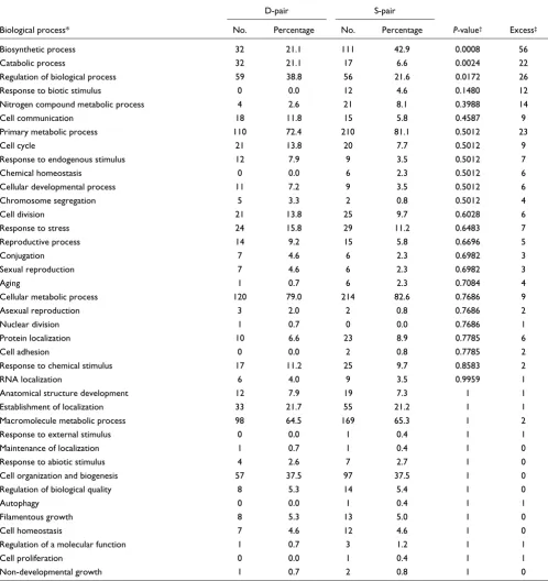
To assess whether a gene's biological function indeed influ-ences the subcellular localization fate of proteins after dupli-cation, we tested for general functional differences between genes in S- and D-pairs using GO annotation. In this analysis, we assume that the current GO distribution of duplicates overall reflects that of their ancestors. Two GO categories stand out (Table 2). While S-pairs show a significant excess of genes involved in biosynthetic processes, D-pairs are signifi-cantly enriched with genes involved in catabolism (P ≤ 0.01 after false discovery rate correction [21]). We note that, gen-erally, S. cerevisiae proteins (excluding the WGD duplicates) involved in catabolism are located in, on average, 1.47 com-partments, while those that contribute to biosynthesis local-ize in 1.35 compartments, a significantly different distribution (two-tailed P < 0.01, Mann-Whitney U test). This suggests that the a priori wider subcellular distribution of proteins involved in catabolic pathways facilitates functional divergence through subcelullar relocalization after gene duplication when compared to biosynthetic proteins, which show more restricted localization patterns.
Next, we analyzed the extent of amino acid divergence in D-and S-pair duplicates. To this end, we used a related yeast species, Kluyveromyces waltii [17], which diverged from S. cerevisiae before the WGD event, as an outgroup, and esti-mated the non-synonymous substitution rate (that is, the number of non-synonymous substitutions per non-synony-mous site, dN) on the lineages leading to each one of the two
S. cerevisiae duplicates using a maximum-likelihood approach [22] (see Materials and Methods for details). This analysis revealed a difference in the dN distribution between genes in S- and D-pairs (Figure 1; Additional data file 1; two-tailed P < 10-5, Mann-Whitney U test); S-pair genes generally
show lower non-synonymous substitution rates than those in D-pairs.
Consistent with previous observations [17], cases of extreme decelerated evolution (one of the duplicates has a dN = 0) among S-pairs include protein coding genes that are known to be highly constrained, such as ribosomal genes (28 pairs), histones (2 pairs) and elongation factors (2 pairs). Selection for increased gene dosage and/or decreased dosage imbal-ance may explain the intensity of purifying selection observed for these 'housekeeping' duplicates [1,3,23]. The fact that these duplicates did not change their subcellular localization is likely due to the specificity of their biological function, which is restricted to certain compartments and may gener-ally preclude subcellular shifts, as suggested by our data above.
We also found that S-pair genes show higher expression levels than D-pair genes (median = 1.3 copies per cell versus 0.8 copies per cell; two-tailed P < 10-5, Mann-Whitney U test),
consistent with the idea that many S-pairs represent duplica-tions of housekeeping genes. This difference is also reflected at the protein level; D-pair genes (median = 5,436.9 pmol) express significantly more protein than S-pair genes (mean = 35,788.3 pmol, two-tailed P < 0.01, Mann-Whitney U test).
Thus, generally, biological function appears to be a strong determinant for the propensity of duplicates to relocate in the cell. While duplicate proteins encoded by slowly evolving housekeeping genes with high expression levels (for example, genes involved in biosynthetic process, such as ribosomal genes) tend to preserve ancestral localization patterns (and functions) after duplication, duplicates from other categories, such as those involved in catabolic processes, are much more likely to evolve divergent localization patterns.
Functional divergence of duplicates through neo- or sublocalization
If divergent subcellular localization between duplicates was a consequence of sublocalization alone, the joint number of dif-ferent compartments per protein pair (that is, combining both duplicates) would be expected to be the same as that of
the common ancestral protein. Conversely, the number of compartments per pair should be higher than that of the progenitor if neolocalization contributed to subcellular diversification.
Summary of GO analysis for D- and S-pair duplicates
D-pair S-pair
Biological process* No. Percentage No. Percentage P-value† Excess‡
Biosynthetic process 32 21.1 111 42.9 0.0008 56 Catabolic process 32 21.1 17 6.6 0.0024 22 Regulation of biological process 59 38.8 56 21.6 0.0172 26 Response to biotic stimulus 0 0.0 12 4.6 0.1480 12 Nitrogen compound metabolic process 4 2.6 21 8.1 0.3988 14 Cell communication 18 11.8 15 5.8 0.4587 9 Primary metabolic process 110 72.4 210 81.1 0.5012 23
Cell cycle 21 13.8 20 7.7 0.5012 9
Response to endogenous stimulus 12 7.9 9 3.5 0.5012 7
Chemical homeostasis 0 0.0 6 2.3 0.5012 6
Cellular developmental process 11 7.2 9 3.5 0.5012 6 Chromosome segregation 5 3.3 2 0.8 0.5012 4
Cell division 21 13.8 25 9.7 0.6028 6
Response to stress 24 15.8 29 11.2 0.6483 7 Reproductive process 14 9.2 15 5.8 0.6696 5
Conjugation 7 4.6 6 2.3 0.6982 3
Sexual reproduction 7 4.6 6 2.3 0.6982 3
Aging 1 0.7 6 2.3 0.7084 4
Cellular metabolic process 120 79.0 214 82.6 0.7686 9
Asexual reproduction 3 2.0 2 0.8 0.7686 2
Nuclear division 1 0.7 0 0.0 0.7686 1
Protein localization 10 6.6 23 8.9 0.7785 6
Cell adhesion 0 0.0 2 0.8 0.7785 2
Response to chemical stimulus 17 11.2 25 9.7 0.8583 2
RNA localization 6 4.0 9 3.5 0.9959 1
Anatomical structure development 12 7.9 19 7.3 1 1 Establishment of localization 33 21.7 55 21.2 1 1 Macromolecule metabolic process 98 64.5 169 65.3 1 2 Response to external stimulus 0 0.0 1 0.4 1 1 Maintenance of localization 1 0.7 1 0.4 1 0 Response to abiotic stimulus 4 2.6 7 2.7 1 0 Cell organization and biogenesis 57 37.5 97 37.5 1 0 Regulation of biological quality 8 5.3 14 5.4 1 0
Autophagy 0 0.0 1 0.4 1 1
Filamentous growth 8 5.3 13 5.0 1 0
Cell homeostasis 7 4.6 12 4.6 1 0
Regulation of a molecular function 1 0.7 3 1.2 1 1
Cell proliferation 0 0.0 1 0.4 1 1
Non-developmental growth 1 0.7 2 0.8 1 0
*We selected GO level 3, since this constitutes a good compromise between the number of genes annotated and the depth of the information contained in each class [46]. †After false discovery rate correction. ‡Represents the difference between the observed number of genes in the
[image:4.612.55.553.122.650.2]To assess the contribution of neo- and sublocalization to the functional diversification of duplicates and given the lack of subcellular localization data for ancestral proteins, we used the average number of subcellular compartments of yeast sin-gleton gene products (that is, genes that show no evidence of paralogs in the S. cerevisiae genome; see Materials and meth-ods for details) as a proxy for the subcellular representation of WGD duplicate progenitors (akin to a previous analysis of yeast duplicates [10]).
We observed that the joint number of distinct compartments per D-pair (mean = 2.31 ± 0.63, median = 2) is significantly higher than that observed for singleton proteins (mean = 1.30 ± 0.49, median = 1, two-tailed P < 10-5, Mann-Whitney U
test). In contrast, there is no difference between the distribu-tions of the number of subcellular compartments for S-pairs
(mean = 1.27 ± 0.42, median = 1) and singletons (two-tailed P
= 0.2, Mann-Whitney U test), suggesting that the increase in the number of compartments observed for D-pairs is due to neolocalization events among D-pair proteins.
A potential caveat of this analysis is that the types of proteins
represented in D-pairs might generally and a priori be
present in a larger number of compartments, as also indi-cated by the analysis of the number of compartments for cat-abolic/biosynthetic proteins discussed above. To control for this, we compared the number of distinct compartments per D-pairs and singletons for proteins within the same GO classes. To ensure adequate sample sizes, we focused on the 8 GO categories that contain more than 30 proteins for both D-pairs and singletons (Table 3). This analysis shows that for all eight comparisons, the joint number of compartments per D-pair is significantly higher than that observed for singletons (Table 3; two-tailed P < 10-4, Mann-Whitney U test). This
sug-gests that the elevated number of compartments for D-pairs is indeed a result of neolocalization and not due to a wide cel-lular representation of ancestral progenitor proteins, prior to duplication. Based on the observed excess (mean, approxi-mately 0.98) of D-pairs relative to singletons from the same functional categories, we estimate that, on average, approxi-mately one compartment is gained by neolocalization per duplication event between duplicates showing subcellular divergence. In addition to the elevated number of compart-ments per D-pairs, we find that the average number of com-partments per D-pair protein (approximately 1.53) is significantly higher than that of singletons in 7 of 8 compari-sons (two-tailed P < 0.05, Mann-Whitney U test). This result further underscores that neolocalization probably predomi-nated over sublocalization during yeast duplicate evolution.
To further assess and illustrate the types and extent of subcel-lular relocalizations in the evolution of yeast gene families, we Distribution of non-synonymous substitution rates (dN) for duplicate
[image:5.612.55.297.85.264.2]genes in S- and D-pairs (estimated for the time since the whole-genome duplication event - see text for details)
Figure 1
Distribution of non-synonymous substitution rates (dN) for duplicate genes in S- and D-pairs (estimated for the time since the whole-genome duplication event - see text for details).
0 0.1 0.2 0.3 0.4 0.5 0.6 0.7 0.8 0.9 1 >1 0
10 20 30 40
D-pair
S-pair
dN
% o
f g
e
n
e
s
Illustration of the different evolutionary fates of (functional) duplicate genes Figure 2
Illustration of the different evolutionary fates of (functional) duplicate genes. Each gene/protein is represented in different colors: red, ancestral, 'A'; green, duplicate copy A1; and blue, duplicate copy A2. Different shapes of proteins (circle, square, and triangle) indicate different functions. Three different subcellular localizations (nucleus, cytoplasm, and cytoplasmic membrane) are indicated in a schematic cell. We note that only the major possible scenarios are illustrated here.
gene A
gene A1
gene A2
duplication neolocalization
sub
localization
neofunctionalization
[image:5.612.56.555.538.674.2]used the K. waltii ortholog(s) as outgroups and reconstructed the phylogeny for 45 WGD yeast families (15 D- and 30 S-pair-containing gene families) with at least 1 additional mem-ber and mapped the subcellular localizations of these onto the phylogenies (Figure 3 and Additional data file 2). In 16 fami-lies, the subcellular localization has remained completely pre-served among the members (Additional data file 2). For the remaining 29 families, we analyzed changes in protein loca-tion, assuming that the scenario requiring the smallest
number of subcellular changes, given the observed data (par-simony principle), reflects the true pattern of events.
For 16 of the 29 families, we could infer the most likely sce-nario of subcellular diversification. Eight families show instances of neolocalization (Additional data file 2). For example, members of the ubiquitin-conjugating enzyme fam-ily, involved in protein degradation [24], are generally located in the cytoplasm and the nucleus (Figure 3a). However,
Comparison between the number of different compartments per D-pairs, proteins in D-pairs, and singleton proteins from the same GO categories
Singleton D-protein D-pair Biological process* Total no. Average no. of
compartments
Total no. Average no. of compartments
P-value† Total no. Average no. of
compartments
P-value‡
Regulation of biological process 64 1.30 59 1.56 0.020 32 2.31 1.92E-10 Macromolecule metabolic process 247 1.28 98 1.54 0.001 52 2.31 1.57E-19 Cell organization and biogenesis 160 1.28 57 1.60 0.021 37 2.38 2.27E-14 Primary metabolic process 325 1.30 110 1.52 0.005 57 2.28 1.93E-20 Catabolic process 53 1.47 32 1.41 0.370 16 2.19 1.19E-04 Establishment of localization 81 1.17 33 1.55 0.013 19 2.37 1.82E-09 Biosynthetic process 162 1.30 32 1.53 0.044 18 2.11 1.52E-07 Cellular metabolic process 372 1.32 120 1.52 0.006 62 2.27 1.42E-21 Regulation of biological process 64 1.30 59 1.56 0.020 32 2.31 1.92E-10 The data are derived from 8 categories with at least 30 proteins per group - see text for details. *GO level 3. †Mann-Whitney U test comparing
D-proteins and singletons. ‡Mann-Whitney U test comparing D-pairs and singletons.
[image:6.612.58.554.128.293.2]Subcellular localizations of the (a) UBC and (b) AIR family members and subcellular localization changes inferred based on the phylogeny Figure 3
Subcellular localizations of the (a) UBC and (b) AIR family members and subcellular localization changes inferred based on the phylogeny. The common name and yeast protein identifier (in brackets) of the protein are indicated. The schematic representation of a yeast cell depicts three possible localizations: nucleus (small circle), endoplasmatic reticulum (eclipse around nucleus), and cytoplasm (remainder of the cell). The co-localization of the protein with one of the yeast subcellular compartments is indicated by grey shading.
(b)
AIR protein family
(a)
UBC protein family
UBC11
UBC9
UBC13
UBC1
UBC5
UBC4
QR18
CDC34 RAD6
neolocalization sublocalization
GIS2
AIR1 AIR2
[image:6.612.57.553.446.666.2]UBC7p (also known as QR18) neolocalized to the endoplas-mic reticulum (ER; Figure 3a), where it became essential for the degradation of misfolded proteins [25].
Sublocalization events occurred in four protein families. For instance, GIS2 (cytoplasmic) and AIR2 (nuclear) of the AIR protein family partitioned their ancestral compartments (the cytoplasm and nucleus - still seen for their AIR1 paralog; Fig-ure 3b). Consistent with their specific localizations, GIS2 spe-cialized in a function in the RAS/cAMP signaling pathway [26], whereas AIR2 became specifically involved in the processing and export of mRNAs from the nucleus [27].
In the remaining four families, both neo- and sublocalization events appear to have occurred (Additional data file 2). In addition to the neolocalization event described above, the UBC family also reveals an instance of sublocalization based on GFP data; UBC13 lost the ancestral nuclear localization (Figure 3a). Thus, the UBC family shows both neo- and sub-localization of family members.
Subcellular shifts and signal peptide evolution
The information required for sorting of proteins to different cellular compartments is encoded in their sequence, some-times in distinct targeting motifs [11]. Consequently, differ-ences in the subcellular localization of paralogous proteins should be due to protein sequence changes and should, in principle, be detectable. However, the identification of pro-tein targeting sequence determinants has proven to be a difficult task [11]. To elucidate the molecular basis of subcel-lular relocalization of duplicates, we focused our analysis on the best characterized targeting sequences; amino-terminal signal peptides (SPs) that target proteins to the mitochondria or the ER. These types of SPs are typically 13-36 amino acids long and are usually cleaved from the mature peptide [28].
We estimated the amino acid divergence between WGD pro-teins pairs with ER and/or mitochondrial localization (36 pairs in total, among which 21 are S-pairs and 15 are D-pairs; Table 4). We then determined the aminoacid divergence in the first either 13 or 36 amino acids (putative signal peptide region) and in the mature peptide (protein sequence without signal peptide), and then compared it between the 21 S- and 15 D-pairs (Table 4).
The average amino acid divergence in the SP is higher for pro-tein pairs for which the ER/mitochondrial localization is not preserved (the median divergence based on the 13 amino acid SP is 0.92, and based on the 36 amino acid SP is 0.86) than for those pairs that maintained the same subcellular localiza-tion (13 amino acid SP, 0.69; 36 amino acid SP, 0.67), a sig-nificantly different distribution (two-tailed P < 0.05, Mann-Whitney U test). In contrast, we observed no significant dif-ference for the accumulation of amino acid substitutions in the mature peptide (for neither of the mature peptide sizes tested) between the two sets of proteins (two-tailed P > 0.6,
Mann-Whitney U test; Table 4), which excludes the possibil-ity that proteins that changed their subcellular localization generally show a faster rate of protein evolution and, there-fore, show an elevated SP divergence. Thus, at least for proteins targeted to the mitochondria and to the ER, differ-ences in subcellular localization between duplicates are asso-ciated with accelerated signal peptide sequence evolution. Conceivably, this acceleration may have been driven by posi-tive Darwinian selection. A recent study demonstrating selec-tively driven optimization of a mitochondrial targeting signal of a protein from a young primate gene (L Rosso and col-leagues, unpublished) suggests that this is a plausible scenario.
The NTG1/2 base excision repair and TRR1/2 thioredoxin reductase WGD gene pairs provide striking examples of how subcellular reprogramming through changes in targeting sequences may occur (Figure 4). Through a comparison with
the NTG orthologous protein from K. waltii, we determined
that NTG1 gained an amino-terminal signal after the WGD event (mainly through a number of amino acid substitutions) that targets it to mitochondria, while NTG2 maintained the ancestral nuclear localization (Figure 4a). Conversely, TRR1 lost the ancestral capacity to localize to mitochondria, due to a deletion of its amino-terminal mitochondrial targeting sequence (Figure 4b). Thus, while keeping their ancestral enzymatic functions [29,30], both NTG1 and TRR1 obtained new functional roles through neolocalization changes, caused by gain and loss of (mitochondrial) targeting sequences, respectively.
Functional adaptation to new subcellular environments
Functional adaptation of duplicate proteins to new subcellu-lar compartments may occur in several ways. Given that organelles generally display distinct physico-chemical prop-erties that are reflected in the propprop-erties of their proteome and transcriptome [31,32], relocalized duplicates may show physico-chemical adaptations that allow them to optimally function in their new (in the case of neolocalization) or more restricted (sublocalization) cellular environments.
We first tested whether duplicate proteins reveal evidence for adaptation to the pH of the compartments to which they localize. To this end, we analyzed the pI (isoelectric point) of duplicates, since the pI distribution of proteins is specific to compartments and likely associated with the compartments' pH [32]. We observed a significantly different distribution of fold differences in pI between D- and S-pair duplicates (two-tailed P < 10-3, Mann-Whitney U test; see Additional data file
remains possible that the elevated pI divergence of D-pair proteins may simply reflect the generally higher amino acid divergence observed for D-pair relative to S-pair duplicates (see above).
To distinguish between these two possibilities, we tested whether the observed substitutions between proteins are biased in terms of the pI of the accumulated amino acids. This analysis revealed that 24 of the 88 D-pairs display a signifi-cantly skewed accumulation of substitutions regarding the pI
of their amino acids (P < 0.05, Pearson's chi-square test; Bonferroni-corrected for multiple (238) tests). In other words, for 24 pairs, the two paralogs have accumulated a sig-nificantly larger number of amino acids with a higher or smaller pI, respectively, than expected by chance for such a pairwise comparison (50%). This is a significantly higher pro-portion of pairs (one-tailed P < 0.05, Fisher's exact test) com-pared to that of S-pairs (26/150 pairs), for which the difference in pI cannot be explained by subcellular localiza-tion differences. These analyses suggest that D-pair proteins
Amino acid divergence between WGD protein pairs
D-pairs S-pairs P-value*
Amino-teminus signal peptide
13 amino acids 0.92 0.69 0.019†
36 amino acids 0.86 0.67 0.017†
Mature peptide
13 amino acids 0.57 0.50 0.596
36 amino acids 0.57 0.48 0.785
*Two-tailed Mann-Whitney U test. †Significant at the 5% level.
[image:8.612.49.560.117.231.2]Subcellular relocalization and signal peptide evolution Figure 4
Subcellular relocalization and signal peptide evolution. Signal peptides (36 amino-terminal residues) and experimentally determined subcellular localizations of the (a) NTG1/NTG2 and (b) TRR1/TRR2 duplicate pairs (derived from the S. cerevisiae WGD event) are shown. K. waltii orthologous sequences are used as outgroups. Predotar [39,40] was used to predict subcellular localizations based on the protein sequences. The (predicted) subcellular localization of the K. waltii proteins was considered to represent the ancestral state. Identical residues in all peptide sequences are represented with (*) under the corresponding position in the protein alignment.
(b) TRR signal peptide evolution (a) NTG signal peptide evolution
TRR2 - Thioredoxin reductase TRR1 - Thioredoxin reductase
cytoplasm nuclear
NTG2 - DNA base excision repair NTG1 - DNA base excision repair
mitochondrial mitochondrial
subcellular localization prediction
NTG1 NTG2 outgroup
mitochondrial 0.69
-0.05
ER -0.01 0.01
elsewhere 0.31 0.99 0.94
subcellular localization prediction
TRR1 TRR2 outgroup
mitochondrial -0.64 0.87
ER 0.01
[image:8.612.58.557.385.651.2]
show adaptation to the pH/pI properties of new or altered cellular environments through the fixation of certain amino acids by natural selection.
The expression level of a gene was reported to also be related - at least in part - to the subcellular localization of its product [31]. This may be due to the different volumes of the various compartments (for example, larger compartments would require more protein molecules according to this hypothesis [31]). We computed the fold difference in mRNA transcript abundance [33] for our set of yeast duplicates. The average expression difference between genes in D-pairs (mean, 0.88) is higher than that between S-pair genes (0.71), a significantly different distribution (two-tailed P < 0.05, Mann-Whitney U
test). The elevated expression divergence of D-pair duplicates may indicate that they generally adapted to the expression level requirements of their compartments, for example, through changes in their regulatory sequences.
Subcellular adaptation, protein-protein interactions, and the evolution of new functions
The subcellular localization of a protein determines its ability to interact with other proteins in its local environment. Therefore, subcellular diversification of duplicates should often entail changes in their interactions with other proteins. In the case of sublocalization, the descendant duplicate (assuming that it required protein partners for functioning) is bound to lose interaction partners that were specific to the lost compartment(s). Conversely, proteins that occupy new subcellular niches may obtain new interaction partners.
Using a database containing extensive S. cerevisiae protein interaction data [34], we observed that the two members in D-pairs share a significantly smaller fraction of interactors (median = 6.4%) than duplicates in S-pairs (median = 13.7%, two-tailed P < 0.05, Mann-Whitney U test; Figure 5). This
result is likely not due to a difference in the number of differ-ent interactors determined for the two sets of protein pairs (median = 9 and 8 interactors per S- and D-pairs, respec-tively; two-tailed P = 0.48, Mann-Whitney U test). Thus, as predicted, subcellular divergence of duplicates appears to lead to a pronounced divergence in terms of their interaction with other proteins.
Subcellular relocalization may allow for the possibility that duplicate proteins evolve new functions (in the case of neolo-calization) or functionally specialize (in the case of sublocali-zation, where both duplicates localize to distinct compartments) by evolving interactions with proteins that are located in their own compartment(s) but not in that of their duplicate copies. To test this, we assessed how often an interactor is located in the same compartment as the D-pair duplicate with which it interacts. We then compared this value to the extent of co-localization of these interactors with the other protein of the pair (with which no interaction was found).
For 1,270 interactions that are not shared between D-pair proteins (involving 955 interactors and 82/88 D-pair pro-teins), 684 show co-localization of the interactor and the duplicate with which it interacts. This represents a signifi-cantly larger overlap than that observed between these inter-actors and the non-interacting paralogs of the pairs (582/ 1,270, two-tailed P < 10-4, Fisher's exact test). We note,
how-ever, that - as expected (given the shared history of the two duplicates of a D-pair) - this subcellular overlap is greater than that observed between random protein pairs (1,354/ 2,628, two-tailed P < 10-4, Fisher's exact test). These results
support the notion that subcellular diversification allowed duplicates to obtain new functions and/or functionally spe-cialize by evolving interactions with proteins that are specific to their compartment(s). Given that neolocalization seems frequent (see above), duplicates appear to often have obtained novel functional roles by evolving interactions with compartment-specific proteins - unattainable to their single copy progenitors.
Conclusion
In this study, we have begun to assess the role of subcellular relocalization and adaptation for the emergence of new or altered gene functions after duplication, using yeast as a model organism. Our work suggests that subcellular gence has played a significant role for the functional diver-gence of duplicate genes. It has affected roughly one-third of yeast WGD duplicates, in particular those involved in biolog-ical processes with a wider subcellular distribution (for exam-ple, catabolism).
Although subcellular redistribution of duplicate proteins involved repartitioning/loss of ancestral compartments,
relo-calization of proteins to previously unoccupied
[image:9.612.55.298.85.264.2]Distribution of the proportion of shared interactors for genes in S- and D-pairs
Figure 5
Distribution of the proportion of shared interactors for genes in S- and D-pairs.
0 10 20 30 40 50 60 70 80 90 100 0
10 20 30 40 50
D-pair
S-pair
% shared interactors per protein
%
o
led to an overall gain of compartments among duplicates. Thus, duplicate genes appear to frequently have obtained new functional roles through the process of subcellular relocaliza-tion. The finding that relocalized proteins have obtained new interaction partners and lost ancestral ones underscores this notion. Interestingly, we found that relocalized proteins show adaptations to the physico-chemical properties of their altered cellular environments through the selective fixation of amino acid substitutions.
A number of individual reports have revealed differences in subcellular localization of paralogous proteins in humans and other mammals (for example, [12]; see also references in [14]). Our study here motivates and warrants systematic sur-veys that address the role of subcellular adaptation in the functional diversification of mammalian (duplicate) genes. These should also aim to explore recent duplications (most duplications in the yeast genome - including those studied here - are old), in order to better understand the timing and selective pressures associated with this process. In fact, two individual recent cases from apes have shed initial light on the early stages of subcellular adaptation (L Rosso and col-leagues, unpublished). These demonstrate that subcellular adaptation may indeed occur through both neolocalization (L Rosso and colleagues, unpublished) and sublocalization (L Rosso and colleagues, unpublished), and that subcellular adaptation may be accompanied or followed by adaptive changes of the biochemical function of the protein (L Rosso and colleagues, unpublished). Moreover, they show that sub-cellular shifts may be adaptive, driven by positive selection, and may occur through a few selected changes in specific (signal) sequences (consistent with our analysis of duplicated target sequences presented here), thus allowing for rapid retargeting of duplicate proteins during evolution.
We conclude that in addition to changes in their expression and biochemical function, selectively driven subcellular adaptation has played an important role for the functional diversification of duplicate genes and the emergence of new gene functions in both uni- and multicellular organisms. Thus, generally, investigating the subcellular phenotype of duplicate genes may provide valuable clues to their function and fate.
Materials and methods
S. cerevisiae WGD genes and other paralogs
We retrieved the gene IDs of 900 S. cerevisiae WGD paralogs (organized in 450 gene pairs) as well as the IDs, orthologs, and nucleotide/protein sequence of K. waltii orthologs from the supplemental data of [17]. Nucleotide and amino acid sequences for all S. cerevisiae WGD gene pairs and non-WGD paralogs (as defined by Ensembl gene family annotations) were retrieved from the Ensembl database, release 45 [35,36].
Subcellular localization data were retrieved from [37]. Only proteins unambiguously assigned to at least one of the 22 analyzed subcellular compartments were used. Another glo-bal protein localization data set [19,38] was used for compar-ison. Subcellular localizations (Figure 4) were predicted using Predotar [39,40].
Non-synonymous substitution rates
We used MUSCLE 3.6 [41] to construct codon-based nucle-otide alignments of S. cerevisiae WGD gene pairs and their corresponding K. waltii orthologous genes. To estimate the
rate of non-synonymous changes, dN, along the different
branches of the S. cerevisiae/K. waltii gene trees, we used the CODEML free-ratio model as implemented in the PAML 3.15 package [22].
Phylogenetic reconstructions
We used a maximum likelihood approach, PROML, as imple-mented in the PHYLIP 3.67 software package, to reconstruct the phylogeny of the protein families (protein sequence align-ment infiles were generated using MUSCLE 3.6).
S. cerevisiae singletons
To identify proteins without paralogs (singletons), an all-against-all BLASTP similarity search (E-value = 0.1) was con-ducted. Proteins without hits against other proteins in this search were considered to be singletons.
Gene ontology analysis
GO analyses were conducted using FatiGO [42,43].
Gene expression analyses
S. cerevisiae gene expression levels - measured as the number of mRNA copies per cell - were retrieved from [33]. The aver-age absolute protein abundance in pmol was retrieved from the literature (supplemental data of [44]). The fold difference of gene expression per gene pair was calculated as the abso-lute difference between the numbers of mRNA copies or pro-tein concentration per gene normalized by the average mRNA copy number or protein concentration per gene pair.
Isoelectric point and hydrophathy data
Hydrophathy and pI data were collected from the literature (supplemental data of [44]).
Protein-protein interactions
Protein interactors for all proteins in D- and S-pairs were col-lected from the BioGRID repository [34,45].
Abbreviations
Authors' contributions
ACM, NV and HK conceived and designed the experiments. ACM, NV and DB performed analysis. ACM and HK wrote the paper. All authors read and approved the final manuscript.
Additional data files
The following additional data are available with the online version of this paper. Additional data file 1 is a table listing all whole genome duplicate pairs and relevant information for each member. Additional data file 2 contains the subcellular localizations of members from 45 S. cerevisiae protein fami-lies (each containing one WGD protein pair) and most parsi-monious subcellular localization changes inferred based on the phylogeny.
Additional data file 1
WGD proteins (in pairs), their subcellular localizations and other properties
WGD proteins (in pairs), their subcellular localizations and other properties.
Click here for file Additional data file 2
Subcellular localizations of members from 45 S. cerevisiae protein families (each containing one WGD protein pair) and most parsi-monious subcellular localization changes inferred based on the phylogeny
The WGD pair is indicated in header for each tree. The compart-ments inferred to have been gained by neolocalization are under-lined in red; those inferred to have resulted from sublocalization are underlined in green. Trees where subcellular relocalization events could not be inferred are labeled accordingly ('not clear'). Click here for file
Acknowledgements
This research was supported by funds available to HK from the European Union (STREP: PKB140404) and the Swiss National Science Foundation.
References
1. Ohno S: Evolution by Gene Duplication Berlin: Springer Verlag; 1970. 2. Li WH: Molecular Evolution Sunderland MA: Sinauer Associates; 1997. 3. Aury JM, Jaillon O, Duret L, Noel B, Jubin C, Porcel BM, Segurens B, Daubin V, Anthouard V, Aiach N, Arnaiz O, Billaut A, Beisson J, Blanc I, Bouhouche K, Câmara F, Duharcourt S, Guigo R, Gogendeau D, Katinka M, Keller AM, Kissmehl R, Klotz C, Koll F, Mouël A, Lepère G, Malinsky S, Nowacki M, Nowak JK, Plattner H, et al.: Global trends of whole-genome duplications revealed by the ciliate Paramecium tetraurelia. Nature 2006, 444:171-178.
4. Papp B, Pal C, Hurst LD: Dosage sensitivity and the evolution of gene families in yeast. Nature 2003, 424:194-197.
5. Force A, Lynch M, Pickett FB, Amores A, Yan YL, Postlethwait J: Preservation of duplicate genes by complementary, degen-erative mutations. Genetics 1999, 151:1531-1545.
6. Hughes AL: The evolution of functionally novel proteins after gene duplication. Proc Biol Sci 1994, 256:119-124.
7. Stoltzfus A: On the possibility of constructive neutral evolution. J Mol Evol 1999, 49:169-181.
8. Hughes AL, Friedman R: Expression patterns of duplicate genes in the developing root in Arabidopsis thaliana. J Mol Evol 2005, 60:247-256.
9. Lynch M, Force A: The probability of duplicate gene preserva-tion by subfuncpreserva-tionalizapreserva-tion. Genetics 2000, 154:459-473. 10. He X, Zhang J: Rapid subfunctionalization accompanied by
prolonged and substantial neofunctionalization in duplicate gene evolution. Genetics 2005, 169:1157-1164.
11. Emanuelsson O, von Heijne G: Prediction of organellar targeting signals. Biochim Biophys Acta 2001, 1541:114-119.
12. Reymond A, Meroni G, Fantozzi A, Merla G, Cairo S, Luzi L, Riganelli D, Zanaria E, Messali S, Cainarca S, Guffanti A, Minucci S, Pelicci PG, Ballabio A: The tripartite motif family identifies cell compartments. EMBO J 2001, 20:2140-2151.
13. Schmidt TR, Doan JW, Goodman M, Grossman LI: Retention of a duplicate gene through changes in subcellular targeting: an electron transport protein homologue localizes to the golgi. J Mol Evol 2003, 57:222-228.
14. Byun-McKay SA, Geeta R: Protein subcellular relocalization: a new perspective on the origin of novel genes. Trends Ecol Evol 2007, 22:338-344.
15. Huh WK, Falvo JV, Gerke LC, Carroll AS, Howson RW, Weissman JS, O'Shea EK: Global analysis of protein localization in bud-ding yeast. Nature 2003, 425:686-691.
16. Wolfe KH, Shields DC: Molecular evidence for an ancient dupli-cation of the entire yeast genome. Nature 1997, 387:708-713. 17. Kellis M, Birren BW, Lander ES: Proof and evolutionary analysis
of ancient genome duplication in the yeast Saccharomyces cerevisiae. Nature 2004, 428:617-624.
18. Saccharomyces Genome Database [http://www.yeastge nome.org/]
19. Kumar A, Agarwal S, Heyman JA, Matson S, Heidtman M, Piccirillo S, Umansky L, Drawid A, Jansen R, Liu Y, Cheung KH, Miller P, Gerstein M, Roeder S, Snyder M: Subcellular localization of the yeast proteome. Genes Dev 2002, 16:707-719.
20. Wapinski I, Pfeffer A, Friedman N, Regev A: Natural history and evolutionary principles of gene duplication in fungi. Nature 2007, 449:54-61.
21. Benjamini Y, Hochberg Y: Controlling the false discovery rate: a practical and powerful approach to multiple testing. Statist Soc B 1995, 55:289-300.
22. Yang Z: PAML: a program package for phylogenetic analysis by maximum likelihood. Comput Appl Biosci 1997, 13:555-556. 23. Zhang L, Li WH: Mammalian housekeeping genes evolve more
slowly than tissue-specific genes. Mol Biol Evol 2004, 21:236-239. 24. Jentsch S, Seufert W, Sommer T, Reins HA: Ubiquitin-conjugating enzymes: novel regulators of eukaryotic cells. Trends Biochem Sci 1990, 15:195-198.
25. Friedlander R, Jarosch E, Urban J, Volkwein C, Sommer T: A regula-tory link between ER-associated protein degradation and the unfolded-protein response. Nat Cell Biol 2000, 2:379-384. 26. Balciunas D, Ronne H: Yeast genes GIS1-4: multicopy
suppressors of the Gal- phenotype of snf1 mig1 srb8/10/11 cells. Mol Gen Genet 1999, 262:589-599.
27. Inoue K, Mizuno T, Wada K, Hagiwara M: Novel RING finger pro-teins, Air1p and Air2p, interact with Hmt1p and inhibit the arginine methylation of Npl3p. J Biol Chem 2000, 275:32793-32799.
28. Hegde RS, Bernstein HD: The surprising complexity of signal sequences. Trends Biochem Sci 2006, 31:563-571.
29. You HJ, Swanson RL, Harrington C, Corbett AH, Jinks-Robertson S, Senturker S, Wallace SS, Boiteux S, Dizdaroglu M, Doetsch PW: Sac-charomyces cerevisiae Ntg1p and Ntg2p: broad specificity N-glycosylases for the repair of oxidative DNA damage in the nucleus and mitochondria. Biochemistry 1999, 38:11298-11306. 30. Pedrajas JR, Kosmidou E, Miranda-Vizuete A, Gustafsson JA, Wright
AP, Spyrou G: Identification and functional characterization of a novel mitochondrial thioredoxin system in Saccharomyces cerevisiae. J Biol Chem 1999, 274:6366-6373.
31. Drawid A, Jansen R, Gerstein M: Genome-wide analysis relating expression level with protein subcellular localization. Trends Genet 2000, 16:426-430.
32. Ho E, Hayen A, Wilkins MR: Characterization of organellar pro-teomes: a guide to subcellular proteomic fractionation and analysis. Proteomics 2006, 6:5746-5757.
33. Holstege FC, Jennings EG, Wyrick JJ, Lee TI, Hengartner CJ, Green MR, Golub TR, Lander ES, Young RA: Dissecting the regulatory circuitry of a eukaryotic genome. Cell 1998, 95:717-728. 34. Stark C, Breitkreutz BJ, Reguly T, Boucher L, Breitkreutz A, Tyers M:
BioGRID: a general repository for interaction datasets. Nucleic Acids Res 2006, 34 (Database issue):D535-D539. 35. Hubbard TJ, Aken BL, Beal K, Ballester B, Caccamo M, Chen Y, Clarke
L, Coates G, Cunningham F, Cutts T, Down T, Dyer SC, Fitzgerald S, Fernandez-Banet J, Graf S, Haider S, Hammond M, Herrero J, Holland R, Howe K, Johnson N, Kahari A, Keefe D, Kokocinski F, Kulesha E, Lawson D, Longden I, Melsopp C, Megy K, Meidl P, et al.: Ensembl 2007. Nucleic Acids Res 2007, 35 (Database issue):D610-D617. 36. Ensembl Database [http://www.ensembl.org]
37. Yeast GFP Fusion Localization Database [http://yeast gfp.ucsf.edu/]
38. Subcellular Localization Epitope [http://ygac.med.yale.edu] 39. Small I, Peeters N, Legeai F, Lurin C: Predotar: A tool for rapidly
screening proteomes for N-terminal targeting sequences. Proteomics 2004, 4:1581-1590.
40. Predotar [http://urgi.versailles.inra.fr/predotar/predotar.html] 41. Edgar RC: MUSCLE: multiple sequence alignment with high
accuracy and high throughput. Nucleic Acids Res 2004, 32:1792-1797.
42. Al-Shahrour F, Minguez P, Vaquerizas JM, Conde L, Dopazo J: BABE-LOMICS: a suite of web tools for functional annotation and analysis of groups of genes in high-throughput experiments. Nucleic Acids Res 2005, 33 (Web server issue):W460-W464. 43. FatiGO [http://fatigo.bioinfo.cnio.es]