International Journal of Emerging Technology and Advanced Engineering
Website: www.ijetae.com (ISSN 2250-2459,ISO 9001:2008 Certified Journal, Volume 3, Issue 10, October 2013)
704
An Unsupervised method for brain MRI Segmentation
Zhang Gang
1, Zhou Dan
2, Huang Ying
3, Huang Xiaobo
4, Zhang Yong
5, Luo Weishi
6, Zhang Yang
7,
Qian Dongxiang
8, LiJun
9, Hong Jiaming
101.2.3.4School of Automation, Guangdong University of Technology, Guangzhou, 510006, China
5,6,7Department of Neurosurgery, Guangdong NO.2 Provincial People’s Hospital, Guangzhou, 510317, China 8,9Department of Neurosurgery, the Third Affiliated Hospital of Guangzhou Medical University
Guangzhou, 510150, China
10School of Medical Information Engineering, Guangzhou University of Chinese Medicine
Guangzhou, 510006, China
Abstract—Computer aid diagnosis for brain MRI image is widely used in hospitals. An important step in it is to recognize different regions within an MRI image according to medical experience. In this paper, we propose an unsupervised learning algorithm for automatic segmentation of MRI images. Different from previous methods, our method achieves the idea of visual segmentation, which simulates the thinking procedure of doctors. And prior knowledge can be incorporated in our model. Evaluation results on a synthetic brain MRI dataset and a real dataset show the effectiveness of the proposed method.
Keywords—brain MRI segmentation, normalized cut, unsupervised learning, prior knowledge, template matching
I. INTRODUCTION
Magnetic resonance imaging (MRI) is a widely used technique for anatomical assessment of human brain structures, which is an important method for brain disease diagnosis [1]. MRI provides a comprehensive insight of what happen in patient’s brain, including common structures of brains, e.g. white matter (WM), grey matter (GM), cerebrospinal fluid (CSF) and voxels, and lesion regions located in single common structures or overlapped areas of them [2].
Since brain diseases are common for human life, and brain MRI is a usual inspection step for diagnosis, increasingly large amount of brain MRI images are required to analyse and recognize. Hence an automatic analysis system for brain MRI is of considerable meaning. Currently in the related literatures, there are at least two levels for such system. The first level is a system that can automatically recognize common structures of a brain, as well as mark some uncommon regions [3]. The second level is a system based on the first one, while incorporating some diagnosis functions [4]. Due to the complexity of brain MRI images, it is not feasible to directly diagnose with original MRI images. Systems belong to the first level may provide valuable information for diagnosis. Hence, it has attracted much attention recently.
The study of brain MRI image segmentation can be roughly divided into two categories based on different observation and assumption of MRIs. The idea of the first category is based on pixel expansion [5]. This kind of methods requires supervise information as initial inputs. Seeds for each common region should be picked out manually before running the method. And an expansion procedure is adopted to enlarge these regions through capturing near similar pixel points. The second category goes another direction. It assumes that different regions are separated by borders, either clear or rough. A large body of kind of methods relied on image alignment against some predefined templates [6].
Based on our observation, the second category obeys much our essential idea. In a cognition view, when a doctor inspects a brain MRI image, visual disjoint regions would be generated at first through recognition of borders [7]. In this procedure knowledge or medical experience is not considered. However, to the best of our knowledge, the image characteristic of visual disjoint regions has not been fully studied and exploited in current methods either in supervised or semi-supervised learning framework.
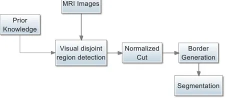
In this paper we directly model the experience implied in the procedure when a doctor inspects a brain MRI image. Our method regards this procedure as a fully unsupervised one. We propose an extended segmentation method based on a famous image cutting algorithm, namely Normalized Cut, which regards each image as a graph with pixels as nodes and similarities between pixels as weights of edge. To precisely segment a given MRI image, prior knowledge is also incorporated into our framework by leveraging the similarity function defined between pixels. Figure 1 sketches the main framework of this paper.
International Journal of Emerging Technology and Advanced Engineering
Website: www.ijetae.com (ISSN 2250-2459,ISO 9001:2008 Certified Journal, Volume 3, Issue 10, October 2013)
[image:2.612.56.282.143.249.2]705
Figure 1. The main steps of this paper.
This paper is organized as following. In Section II we briefly review some considerable work related to this paper. In Section II we present the main method. In Section IV some evaluation results are reported with some comparison to previous successful methods. And finally we conclude the paper in Section V.
II. RELATED WORK
We review some considerable work related to this paper. The work falls into two categories. One is on the topic of MRI image analysis through machine intelligent methods. And the other is on the topic of unsupervised graph cutting algorithm and its application, which founds the base of this work. Reddy et al. [8] proposed an iterative conditional model (ICM) algorithm to detect abnormality of brain MRI image segmentation. They proposed an iterative conditional model algorithm for segmentation of MRI image and Markov random field (MRF) model for abnormality detection. Their method is a supervised learning method which relied on the selection of starting points (seeds) and the number of regions must be set before running the algorithm. The abnormality detection through MRF is based on the segmentation generated by ICM. Since MRF is a probabilistic method, it seeks a consistent and balanced situation with some unknown distribution. Their method is unstable when the MRI images are of low quality. Soesanti, et al. [9] proposed a method for brain MRI images segmentation based on fuzzy logic and spatial information. They introduced the theory of fuzzy logic to describe the region overlapping and rough border in a MRI image. They proposed a modified version of fuzzy C-means to cluster pixels in a brain MRI image so as to generate segments. To make the generated segments contiguous, they incorporate spatial information in the clustering algorithm. They defined pixel adjacency through a matrix. The method was totally unsupervised. However, their method did not reflect the idea of visual segmentation. This is because C-means based method maximizes the inner-cluster similarity and minimizes the inter-cluster similarity.
But for MRI images, information from means of pixels does not often reflect the ground truth status of the corresponding local part.
On another hand, the study of unsupervised graph cutting algorithm in machine learning makes it possible for the proposed method. To fully support the visual disjoint regions generation, a simple but direct way is to regard an image as a graph. Shi and Malik [10] proposed an algorithm, namely Normalized Cut (NCut), for image segmentation only based on pixel difference. Their method can extract global impression of a given image, instead of focusing on local features and their consistencies. Their method regards an image as a graph and models the segmentation problem as an optimization one with a well-defined target function. However, their method requires high computational cost. And prior knowledge on the final segmentation or adjacency between regions cannot be applied to the model. Later, Maji et al. [11] proposed an updated version of NCut for image segmentation. Their method has the ability to use some kinds of priors, which can be used for constrained image segmentation. Their method can yield segmentation results correlated to given priors.
III. THE METHOD
Our work is originated from a series of NCut based image segmentation algorithms.
A. Problem definition
Suppose we have a set of brain MRI images denoted as ,and an image is represented as a by matrix. In this work, we only consider 256-grey images. Each image is associated with a segmentation matrix, indicating a manual segmentation of it, and with an integer vector indicating the type information of each region. The definition of type information vector is shown in Figure 2.
Figure 2. Definition of type information
International Journal of Emerging Technology and Advanced Engineering
Website: www.ijetae.com (ISSN 2250-2459,ISO 9001:2008 Certified Journal, Volume 3, Issue 10, October 2013)
706
B. Graph representation
In this subsection, we present how to represent a brain MRI image di as a weighted undirected graph, denoted as a
node set Vi and an edge set Ei. The edge is form between a
pair of nodes. We only connect adjacent pixels with edges. Each edge has a weight defined as the similarity between two end points associated with e. The similarity is defined through a heat kernel over node space as Eq. (1) shows:
(1)
Where is the square Euclidean distance
between. If is smaller than a pre-set value , is set to 0. A small would push the graph to be sparse, which reduces the computational cost of the algorithm. Note that the generated graph may not be a connected graph, which naturally forms a general segment of the original graph.
Another graph construction strategy is kNN, meaning that for a node it only connects edges with nodes in the set of its nearest neighbours. The kNN strategy can guarantee the generated graph to be a full connected graph. In this work, we use the first threshold-based graph construction strategy.
C. Unsupervised Graph Cutting
In this section we present our main method for image segmentation with graph representation. First it is necessary to define a cut of a graph as two node sets and where and . In graph theory we can also define the dissimilarity of a cut as:
(2)
Eq. (2) means that the sum of weights of edges between two parts of a cut describes the quality of the cut. Among all cuts of a graph, there is at least one cut have smallest dissimilarity defined in Eq. (2), which is the optimal cut.
However, the minimum dissimilarity favours small node subsets. To generate relatively balanced cut, a modified criteria is defined by Shi et al. in [10]:
(3)
Where evaluates the total
connection weights between segment A and all nodes in the graph.
The smaller value indicates the better cutting according to the weights defined in the graph. Eq. (3) encourages large segments to be generated, since small segments may lead to small value of g, resulting in large value of Ncut. With almost the same idea, association between segments A and B can be defined as Eq. (4):
(4)
A connection can be established between Eq. (3) and Eq. (4) as following:
(5)
Let D be a diagonal matrix with and be a symmetric matrix where is the weight of edge connecting vertex i and j. Following [10] and [11]’s analysis, the cutting problem can be summarized as:
(6)
Where r is a Boolean vector with size |V|, whose value is 1 if the corresponding vertex belongs to segment A and 0
otherwise. Let and ,
. Eq. (6) can be solved through expressing it as a Rayleigh quotient [12].
Note that each time the algorithm performs only bipartition. To further generate more regions of a given brain MRI image, the algorithm must be run several times. Thought it prefers large regions, the bipartition guarantee the generated regions to be of different sizes.
Another problem is how to choose sub region to perform further cutting. We define a measurement, namely purity to indicate whether a region should be cut. The purity is defined as Eq. (7):
(7)
International Journal of Emerging Technology and Advanced Engineering
Website: www.ijetae.com (ISSN 2250-2459,ISO 9001:2008 Certified Journal, Volume 3, Issue 10, October 2013)
707
D. Prior knowledge
It is known that brain MRI image has some normal structures. And it is also accepted that if such information can be modelled and fed to a proper model, we can achieve good performance of the model. To do this, we need to formularize the prior knowledge suitable for our model. A template based region constraint is introduced as prior knowledge. The main idea is the definition of region adjacency by pixel inequality. To shed light on this, we add a system of linear constraints on Eq. (6). Two types of constraint are designed. The first type is pixel value constraint. It means that pixels should be put into the same segment if their values are fallen into a pre-set range. This type of constraint can also be expressed in another way, i.e. some pixels should not be put into the same segment. The second is region adjacency constraint. It works together with manual label to the generated regions. We will describe more details in the next section.
IV. EVALUATION
[image:4.612.336.552.157.228.2]We evaluate the proposed algorithm on a synthetic data set and a real clinical data set. The synthetic data set comes from an online brain MRI image simulated project [12]. It provides a web program that generates brain MRI image with different noise level and severity. The location and size of lesion regions are randomly generated but within a medical acceptable range. We generate 150 images with different setting parameters, among which there are 100 lesion images as well as 50 normal images. Figure 3 shows some images generated with this simulated program.
Figure 3. Sample brain images
Another evaluation data set is a real clinical data set of brain diseases from IBSR project [13]. Details of the synthetic data set are listed in Table I.
TABLEI
PROPERTIES OF SYNTHETIC DATA SET
Lesions Noise Clear Rotate
S1 2 Low 3 N
S2 1 Low 3 Y
S3 3 Med 2 N
S4 2 High 2 Y
S5 2 High 1 N
In Table I, 4 parameters control the quality of the generated data set. The column ―Lesions‖ means the number of lesion regions. ―Noise‖ means the level of mixture Gaussian noise to be added to the generated image. ―Clear‖ stands for clarity of the generated image. ―Rotate‖ means the Z-axis rotation of the image.
Table II shows the detailed properties of the real data set from IBSR project.
TABLEII
PROPERTIES OF THE REAL DATA SET
Description Size
R1 Normal Subject 112
R2 Tumor 1 + 2 141
R3 20 Normals 98
R4 T1-Weighted-Brain-Scan 102
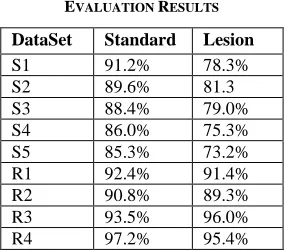
We launch two experiments to evaluate the proposed method. The first is to evaluate the segmentation accuracy of the proposed algorithm. The evaluation measurement is the ratio between the generated regions and the ground truth regions. Since the proposed method is an unsupervised learning method, the whole data set can be used as test data set. In this evaluation, we set the number of regions to be 7 based on medical experience. Table II and III show the accuracy of different settings of the algorithm in both synthetic and real data sets. We report the accuracy of both standard regions and lesion regions generation.
TABLEII EVALUATION RESULTS
DataSet Standard Lesion
S1 91.2% 78.3%
S2 89.6% 81.3
S3 88.4% 79.0%
S4 86.0% 75.3%
S5 85.3% 73.2%
R1 92.4% 91.4%
R2 90.8% 89.3%
R3 93.5% 96.0%
[image:4.612.339.542.349.411.2] [image:4.612.50.289.492.576.2] [image:4.612.373.516.569.694.2]International Journal of Emerging Technology and Advanced Engineering
Website: www.ijetae.com (ISSN 2250-2459,ISO 9001:2008 Certified Journal, Volume 3, Issue 10, October 2013)
708
The second is to evaluate the effect of prior knowledge. To do this, we manual define three pixel constraints for each image on 4 standard regions, i.e. inequality of pixel value range. Table III shows the evaluation result.
TABLEIII
EVALUATION RESULTS WITH PRIOR KNOWLEDGE
DataSet S+p1 L+p1 S+p2 L+p2
S1 93.1% 88.3% 96.2% 86.4% S2 91.3% 89.0% 95.6% 89.7% S3 88.6% 76.9% 90.5% 92.4% S4 89.4% 79.8% 88.0% 80.3% S5 85.3% 73.2% 83.9% 85.1% R1 96.5% 98.5% 97.4% 98.2% R2 92.0% 91.1% 95.3% 94.4% R3 90.4% 92.3% 90.1% 93.5% R4 97.5% 96.7% 96.9% 97.3%
In Table III, ―S‖ and ―L‖ stand for standard and lesion regions respectively. ―p1‖ and ―p2‖ stand for the first and second type of prior knowledge. We can see that the proper introduction of prior knowledge significantly improve the performance of the model.
V. CONCLUSIONS
We have proposed an unsupervised learning framework for brain MRI image segmentation. The proposed method regards each MRI image as a graph in either r-ball or kNN meaning. And then perform an extended version of Normalized Cut to obtain optimal cuttings. Our method also supports prior knowledge to improve the accuracy of segmentation. Two kinds of prior knowledge were defined in our framework. Evaluation on a synthetic and a real data set show the effectiveness of the proposed method. Further work includes different prior knowledge definition and the design of more powerful model for capturing the essential principles of brain MRI images.
Acknowledgment
The authors would like to thank Xiao-bo Huang for his professional advice for this paper. This work is supported by the Science and Technology Planning Project of Haizhu District, Guangzhou (2011-YL-05), the 2012 College Student Career and Innovation Training Plan Project (1184512043), the 2011 Higher Education Research Fund of GDUT (2013Y04).
REFERENCES
[1] Tranos Zuva, Oludayo O, Olugbara, Sunday O. Ojo and Seleman M Ngwira, ―Image Segmentation, Available Techniques, Developments and Open Issues,‖ Canadian Journal on Image Processing and Computer Vision Vol. 2, No. 3, MARCH 2011.
[2] Chuan-Yu Chang, Yue-Fong Lei, Chin-Hsiao Tseng, and Shyang-Rong Shih ―Thyroid Segmentation and Volume Estimation in Ultrasound Images‖ IEEE transactions on biomedical engineering, vol. 57, no. 6, JUNE 2010.
[3] Livier C, Maxime S, Pierre-Y, indiau H, Delingette S, Warfield K, Gregoire M, Nicholas A (2005). ‖Realistic Simulation of the 3-D Growth of Brain Tumors in MR Images Coupling Diffusion With Biomechanical Deformation‖, IEEE on Medical Imaging p. 24-33. [4] Chattopadhyay G, Chattopadhyay S (2009). Autoregressive forecast
of monthly total ozone concentration‖ A neurocomputing approach, Computers and Geosciences 35: 1925–1932.
[5] Bandyopadhyay G, Chattopadhyay S (2007). Single hidden layer Artificial Neural Network models versus multiple linear regression model in forecasting the time series of total ozone‖, Intern. J. Environ. Sci. Technol. 4(1), pp. 141–149.
[6] N. Senthilkumaran and R. Rajesh, ―Brain Image Segmentation using Granular Rough Sets‖, International Journal of Arts and Sciences, U.S.A., Vol.3, No.1, 2009, pp. 69-78.
[7] Bouchet A, Pastore J and Ballarin V, ‖Segmentation of Medical Images using Fuzzy Mathematical Morphology‖, JCS and T, Vol.7, No.3, October 2007, pp.256-262.
[8] Ramaswamy Reddy, E.V.Prasad, L.S.S Reddy. Abnormality Detection of Brain MR Image Segmentation using Iterative Conditional Mode Algorithm. International Journal of Applied Information Systems, Vol. 5, No.2, January 2013, pp. 56-65. [9] Indah Soesanti, Adhi Susanto, Thomas Sri Widodo, Maesadji
jokronagoro. MRI Brain Images Segmentation Based on Optimized Fuzzy Logic and Spatial Information. International Journal of Video & Image Processing and Network Security IJVIPNS-IJENS Vol. 11 No: 04, pp. 6-11.
[10] Shi, J. & Malik, J. Normalized Cuts and Image Segmentation IEEE Trans. Pattern Anal. Mach. Intell., IEEE Computer Society, 2000, 22, pp. 888-905
[11] Subhransu Maji, Nisheeth K. Vishnoi, and Jitendra Malik. Biased Normalized Cuts. In Proceedings of CVPR 2011, pp. 2057-2064. [12] BrainWeb: http://brainweb.bic.mni.mcgill.ca/brainweb/
International Journal of Emerging Technology and Advanced Engineering
Website: www.ijetae.com (ISSN 2250-2459,ISO 9001:2008 Certified Journal, Volume 3, Issue 10, October 2013)
709
Authors
Gang Zhang is PhD candidate in the School of Information Science and Technology at SUN YAT-SEN University, China. He received his MSc Degree in Computer Software and Theory from SUN YAT-SEN University, China, in 2005. His current research interests include data mining, machine learning, and its applications to bioinformatics and Traditional Chinese Medicine. Now he is a lecturer in School of Automation, Guangdong University of Technology.
Zhang Yong, M.D. Director of Neurosurgery department of Guangdong No.2 provincial hospital. The top expert of China in the field of cranial nerve diseases.
Huang Ying is MD of Faculty of Automation, Guangdong University of Technology. Her research direction includes intelligent information processing and computer vision.
Luo Weishi, Attending doctor of neurosurgery. Mater of neurosurgery. specializing in diagnosis and treatment of cranial nerve diseases.
Zhang Yang, Neurosurgery resident, specializing in intraoperative neural electrophysiological monitoring.
QianDongXiang is PhD,MD of the Third
Affiliated Hospital of Guangzhou medical University. As the director of Neurosurgery department, he does great job in clinical work, medical education and medical research concurrently. His research direction is injury and repair of central nervous system. According to dedication in much academic area, he gains lots of honor in Neuroscience academia. And, He was commended to assume the responsibility for various social duty, such as the associate director of Neurosurgery branch in Guangzhou institute of medicine, the national commission of Society for Neuroscience of China, the commission of Neurosurgeon branch in Guangdong physicians society, and so on.
LiJun is a PhD candidate in the School of Guangzhou medical University. He received his Bachelor of Science Degree in clinical medicine from Guangzhou medical University, China, in 2007. His research direction is injury and repair of central nervous system. Now he is a lecturer of the Third Affiliated Hospital of Guangzhou medical University.