Copyright © 1998, American Society for Microbiology
Insertion Element IS3-Based PCR Method for Subtyping
Escherichia coli O157:H7
CURT J. THOMPSON,1CLAIRE DALY,1TIMOTHY J. BARRETT,2JANE P. GETCHELL,1 MARY J. R. GILCHRIST,1ANDMIKE J. LOEFFELHOLZ1*
State Hygienic Laboratory, University of Iowa, Iowa City, Iowa 52242,1and Foodborne
and Diarrheal Diseases Branch, Division of Bacterial and Mycotic Diseases, National Center for Infectious Diseases, Centers for Disease
Control and Prevention, Atlanta, Georgia 303332
Received 6 November 1997/Returned for modification 19 December 1997/Accepted 3 February 1998
An Escherichia coli O157:H7 subtyping method based on PCR amplification of variable DNA sequences between the repetitive element IS3 was developed. Template DNA was prepared by boiling cells in Chelex. Two separate IS3 PCR amplifications were performed for each isolate: one with a single primer (primer IS3A) and one with two primers (primers IS3A and IS3B). The IS3 PCR subtyping method was applied to 35 epidemi-ologically related and unrelated E. coli O157:H7 isolates that had been previously characterized by pulsed-field gel electrophoresis (PFGE). PFGE identified 25 different subtypes (difference of one or more bands). PCR with single primer IS3A and primer pair IS3A-IS3B identified 6 and 14 different subtypes, respectively. By com-bining the results of the two PCR amplifications, 15 different IS3 PCR subtypes were identified. While not as sensitive as PFGE, IS3 PCR subtyping grouped all outbreak-related isolates. IS3 PCR banding patterns were reproducible between amplifications and between subcultures. IS3 PCR could serve as a simple, rapid screen-ing method for the identification of unrelated E. coli O157:H7 isolates.
Escherichia coli O157:H7 has recently emerged as an
im-portant cause of diarrhea and hemolytic-uremic syndrome. While the most common source of E. coli O157:H7 infections is ground beef (7), other sources have been reported, including whole cuts of beef (11), lettuce (1), apple juice and cider (4), lake water (9), and secondary person-to-person transmission (3). Because of the clonal nature of E. coli O157:H7 isolates (18), highly sensitive molecular biology-based subtyping meth-ods are required to differentiate unrelated strains. Pulsed-field gel electrophoresis (PFGE) (2) and analysis of restriction frag-ment length polymorphisms with radiolabeled probes (13) are sensitive methods for distinguishing outbreak-related and non-outbreak-related E. coli O157:H7 strains. In contrast to PFGE and restriction fragment length polymorphism procedures, the performance of which requires several days, PCR-based sub-typing procedures are simple and can provide results from a pure culture in a single day. A number of PCR-based subtyping methods are based on the amplification of variable genomic DNA sequences located between repetitive sequences. Some repeat motifs that have been targeted for use in PCR-based subtyping methods include the repetitive extragenic palidrome (REP) and the enterobacterial repetitive intergenic consensus (ERIC) sequences (17) present in a variety of bacteria, the polymorphic GC-rich repetitive sequence and the insertion sequence IS6110 present in Mycobacterium tuberculosis (6, 12), and IS1245 and IS1311 present in Mycobacterium avium (10). In spite of their ease of use and potential versatility, PCR-based subtyping procedures have disadvantages, including the lack of interlaboratory standardization and insufficient sensi-tivity for the differentiation of unrelated strains of some mi-croorganisms. One of our laboratories (State Hygienic Labo-ratory) was unable to distinguish epidemiologically unrelated
strains of E. coli O157:H7 and Salmonella infantis when tar-geting the ERIC sequence (unpublished results).
Natural populations of E. coli harbor several different inser-tion sequence (IS) classes, including IS3. IS3 was found to be significantly associated with E. coli strains from animals (14). Most E. coli strains possess up to seven copies of IS3, located on the chromosome and plasmids (8, 14). ISs, because of their mobile nature, have been shown to be useful targets for PCR-based subtyping methods. This study describes the develop-ment and evaluation of a rapid IS3-based PCR method for the subtyping of E. coli O157:H7. Previously described primers targeted to the ends of IS3 (5) were modified to amplify DNA fragments between copies of IS3, and PCR amplification con-ditions were optimized to generate reproducible DNA banding patterns useful for discriminating between E. coli O157:H7 isolates.
MATERIALS AND METHODS
Bacterial strains.Thirty-five E. coli O157:H7 isolates of known PFGE subtype
were obtained from the collection of one of us (T. J. Barrett) at the Centers for Disease Control and Prevention (CDC). The sources of the isolates and the PFGE results are summarized in Table 1. PFGE was performed as described previously (2). Agarose-embedded DNA was digested with XbaI followed by separation with a CHEF DR-II or CHEF Mapper system (Bio-Rad Laborato-ries, Hercules, Calif.) and a linearly ramped pulse time of 5 to 50 s.
Bacterial growth and DNA preparation for IS3 PCR. Strains were grown
overnight at 37°C on Trypticase soy agar plates with 5% sheep blood. One to three colonies were picked and suspended in 100ml of 10% (wt/vol) Chelex 100 (Bio-Rad) in a 1.5-ml microcentrifuge tube. Following heating at 100°C for 15 min, the tube was centrifuged at maximum speed for 2 min and the supernatant was carefully removed to avoid the Chelex-cell pellet. The DNA concentration in the supernatant was determined by measuring the A260and, if necessary, was adjusted to 100 ng/ml.
IS3 PCR subtyping.Two 25-ml PCR mixtures were prepared for each isolate:
one containing primer IS3A and one containing primers IS3A and IS3B. The sequences and locations of the primers in IS3 (16) are depicted in Fig. 1. For PCR in 0.2-ml thin-walled tubes, 200 ng (2ml) of DNA was added to 23ml of a PCR master mixture containing 10 mM Tris (pH 8.3), 50 mM KCl (13PCR Buffer II; Perkin-Elmer, Norwalk, Conn.), a 400mM concentration of each of dATP, dCTP, dGTP, and dTTP, 3 mM MgCl2, 1 U of AmpliTaq Gold DNA polymerase (Perkin-Elmer), and either primer IS3A at 6mM or primers IS3A
* Corresponding author. Mailing address: State Hygienic Labora-tory, University of Iowa, 102 Oakdale Campus, Iowa City, IA 52242. Phone: (319) 335-4500. Fax: (319) 335-4555. E-mail: michael-loeffel [email protected].
1180
on May 15, 2020 by guest
http://jcm.asm.org/
and IS3B each at 3mM. A Perkin-Elmer TC9600 thermal cycler was used for amplification. The amplification program consisted of an initial denaturation at 94°C for 5 min; 50 cycles of 94°C for 1 min, 35°C for 1 min, and 72°C for 2 min; and a final 7-min extension at 72°C. The amplification products were analyzed by electrophoresis in 1.5% agarose gels and were detected by ethidium bromide staining.
Interpretation of IS3 PCR patterns.The isolates were considered different if
there were one or more differences in band positions.
RESULTS
Optimization of IS3 PCR.The primers and deoxynucleoside triphosphates were titrated to determine the concentrations that yielded (i) the most PCR product and (ii) variable banding
patterns among different isolates (data not shown). In reac-tions with primer pair IS3A-IS3B, AmpliTaq Gold DNA poly-merase yielded more PCR product than amplification with standard AmpliTaq, with minor changes in the DNA banding patterns being found (data not shown). The banding pattern differences did not change the final subtyping results for the four isolates tested. Negative control reactions containing wa-ter in place of DNA occasionally contained one to four spuri-ous DNA bands which usually did not comigrate with E. coli bands. The banding patterns for the negative controls were variable. Comparison of annealing temperatures of 35 and 58°C showed that except for some differences, primarily with
[image:2.612.51.549.81.453.2]FIG. 1. Location of the PCR primers IS3A and IS3B in the IS3 sequence.
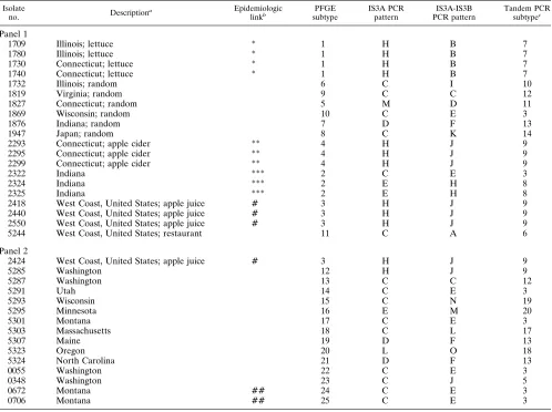
TABLE 1. Summary of E. coli O157:H7 isolates and subtyping results
Isolate
no. Descriptiona Epidemiologiclinkb subtypePFGE IS3A PCRpattern PCR patternIS3A-IS3B Tandem PCRsubtypec
Panel 1
1709 Illinois; lettuce * 1 H B 7
1780 Illinois; lettuce * 1 H B 7
1730 Connecticut; lettuce * 1 H B 7
1740 Connecticut; lettuce * 1 H B 7
1732 Illinois; random 6 C I 10
1819 Virginia; random 9 C C 12
1827 Connecticut; random 5 M D 11
1869 Wisconsin; random 10 C E 3
1876 Indiana; random 7 D F 13
1947 Japan; random 8 C K 14
2293 Connecticut; apple cider ** 4 H J 9
2295 Connecticut; apple cider ** 4 H J 9
2299 Connecticut; apple cider ** 4 H J 9
2322 Indiana *** 2 C E 3
2324 Indiana *** 2 E H 8
2325 Indiana *** 2 E H 8
2418 West Coast, United States; apple juice # 3 H J 9 2440 West Coast, United States; apple juice # 3 H J 9 2550 West Coast, United States; apple juice # 3 H J 9 5244 West Coast, United States; restaurant 11 C A 6 Panel 2
2424 West Coast, United States; apple juice # 3 H J 9
5285 Washington 12 H J 9
5287 Washington 13 C C 12
5291 Utah 14 C E 3
5293 Wisconsin 15 C N 19
5295 Minnesota 16 E M 20
5301 Montana 17 C E 3
5303 Massachusetts 18 C L 17
5307 Maine 19 D F 13
5323 Oregon 20 L O 18
5324 North Carolina 21 D F 13
0055 Washington 22 C E 3
0348 Washington 23 C J 5
0672 Montana ## 24 C E 3
0706 Montana ## 25 C E 3
aIsolates listed are all patient isolates. Foods listed were implicated as the vehicles but not the sources of the isolates. bIsolates with a common symbol are epidemiologically related.
cCombined results of separate amplifications with IS3A and IS3A-IS3B.
on May 15, 2020 by guest
http://jcm.asm.org/
bands larger than 600 bp, the banding patterns of the PCR products were quite similar in tests at both temperatures (data not shown). This suggests that at 35°C primer annealing is largely sequence specific and not random. The melting tem-peratures for primers IS3A and IS3B, are 63.2 and 65.1°C, respectively. Overall, the intensities of the DNA bands were substantially greater when the 35°C annealing temperature was used. The final subtyping results for the four isolates tested were the same at both annealing temperatures. The higher annealing temperature virtually eliminated DNA bands occa-sionally observed for the negative controls.
Reproducibility of IS3 PCR.DNA extracted from two iso-lates was subjected to three separate amplifications on differ-ent days with the same thermal cycler. Amplification was per-formed with primer pair IS3A-IS3B and with primer IS3A alone. The banding patterns of the PCR products were iden-tical among the three amplifications for both isolates tested with both primer configurations (data not shown). To exam-ine the between-subculture reproducibility of the IS3 PCR method, DNA from three consecutive subcultures of two iso-lates was extracted after overnight growth. DNA extracts from all subcultures were stored at220°C and were then amplified at the same time by using both primer configurations. The banding patterns of the PCR products were identical among all subcultures.
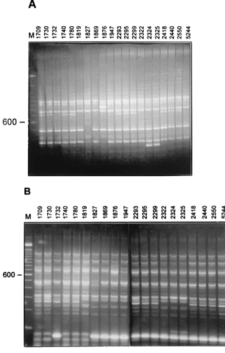
Analysis of E. coli O157:H7 isolates by IS3 PCR.To deter-mine the ability of the IS3 PCR to distinguish between isolates from unrelated outbreaks and to group epidemiologically re-lated isolates, we examined a panel (panel 1) consisting of 14 patient isolates from five different outbreaks and six random isolates (Table 1). PFGE identified 11 different strains (differ-ence of one or more bands). The PFGE results were consistent with the epidemiologic data. PCR with primer IS3 identified five strains (Table 1; Fig. 2A). Isolates from several outbreaks were classified as subtype H, and four of the six random iso-lates as well as the isolate from a restaurant were classified as subtype C. PCR with primer pair IS3A-IS3B was more discrim-inatory, identifying 10 strains (Table 1; Fig. 2B). There were two discrepancies between the PCR and the PFGE results. First, isolates 2322, 2324, and 2325, which were epidemiolog-ically related, were all classified as subtype 4 by PFGE. How-ever, with both the IS3A and the IS3A-IS3B primer configu-rations, isolate 2322 produced PCR banding patterns distinct from those generated by isolates 2324 and 2325. Repeat PFGE at the State Hygienic Laboratory showed several apparent band differences between the pattern produced by isolate 2322 and the pattern produced by isolates 2324 and 2325 (data not shown). Second, IS3 PCR was unable to differentiate the West Coast apple juice strain (PFGE subtype 3) from the Connect-icut apple cider strain (PFGE subtype 4).
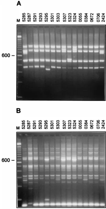
To further test the discriminatory power of the IS3 PCR, a second panel (panel 2) consisting of 15 patient isolates, only 2 of which were epidemiologically related, was examined. PFGE identified 15 different strains (Table 1). Two isolates from the same outbreak, 0672 and 0706, had different PFGE patterns. PCR with primer IS3A identified five strains (Fig. 3A). Nine of the isolates were IS3A subtype C. PCR with primer pair IS3A-IS3B identified eight strains (Fig. 3B). By combining the PCR results from both primer configurations (tandem PCR), nine strains were identified. Isolates 0672 and 0706 were identical by IS3 PCR.
DISCUSSION
Bacterial subtyping results should be able to distinguish epidemiologically unrelated strains and show that
[image:3.612.309.540.71.434.2]common-source strains are the same. Additionally, subtyping results must be reproducible. A sensitive but irreproducible method has little utility. However, a reproducible method that is slight-ly less sensitive may be useful as an adjunct to a more sensitive method if it allows the fast and easy screening of large numbers of isolates. Such a method may serve to rapidly group isolates into clusters, eliminating the need to analyze unrelated isolates by more sensitive, labor-intensive methods. It may also allow investigators to rapidly and easily determine whether an in-creased rate of E. coli O157:H7 isolations is due to an outbreak or a temporal clustering of sporadic cases. On the basis of our observations, the IS3 PCR subtyping method described here could serve as a rapid means for identifying unrelated E. coli O157:H7 strains, with further testing of isolates with the same IS3 PCR pattern being done by methods such as PFGE. IS3 PCR may not be sufficiently sensitive to be used as a stand-alone subtyping method for E. coli O157:H7. The 35 patient isolates analyzed in this study represented 24 different known sources and were grouped into 25 strains by PFGE, 14 strains by IS3A-IS3B PCR, and 15 strains by tandem IS3 PCR (com-bined results of separate amplifications with primer IS3A and with IS3A-IS3B primer pair). Isolates that differed by one or more bands by PFGE were considered to be unrelated strains. While Tenover et al. (15) have suggested that seven or more band differences by PFGE are required to classify bacterial FIG. 2. Panel 1 isolates from CDC. PCR patterns were generated with primer IS3A (A) and primer pair IS3A-IS3B (B). Lane M, 100-bp DNA ladder. The numbers on the left are in base pairs.
on May 15, 2020 by guest
http://jcm.asm.org/
isolates as unrelated, Barrett et al. (2) claim that this criterion may be too restrictive for the highly clonal E. coli O157:H7 organism and that isolates with PFGE patterns that differ by a single band are often not related. In this study, isolates 0672 and 0706, which were epidemiologically linked, were consid-ered to be unrelated by PFGE (three band differences). They had identical PCR patterns. However, it should be noted that five additional, unrelated isolates also had the same PCR pat-tern.
The PCR method described here uses primers that were previously used to amplify a large portion of the IS3 element (14). We reversed the orientation of the primers so that DNA sequences located between the IS3 copies would be amplified. While some E. coli isolates have been shown to lack IS3 (14), all E. coli O157:H7 strains tested to date (approximately 100) generated at least six bands by the IS3 PCR method. As has been described for other PCR-based subtyping methods that target specific repeated elements (12), it is possible that the IS3 primers anneal to sequences other than IS3. We have not sequenced the IS3 PCR products to determine this. The ob-servation that the PCR banding patterns obtained with the IS3A-IS3B primer pair were quite similar at annealing tem-peratures of both 35 and 58°C indicates that primer annealing is largely sequence specific rather than random. Most impor-tantly, by using the conditions described here, the IS3 PCR
method is reproducible between amplifications. Further inter-laboratory reproducibility studies are warranted.
PCR optimization experiments showed high product yield over relatively wide ranges of primer and deoxynucleoside triphosphate concentrations (data not shown). We used con-centrations (as described in the Materials and Methods sec-tion) that were well within these ranges to avoid potential vari-ability in the intensities of the DNA bands. Under otherwise identical amplification conditions, AmpliTaq Gold DNA poly-merase was found to generate substantially more PCR product than standard AmpliTaq and several additional DNA bands. The final strain designations were the same with either DNA polymerase. AmpliTaq Gold is a modified form of DNA poly-merase that requires heat for activation. According to the manufacturer, PCR mixtures containing AmpliTaq Gold usu-ally generate fewer spurious DNA bands and more intense specific bands because at room temperature there is no exten-sion of primers that bind to nonspecific sequences prior to the initial denaturation step of thermal cycling. However, this does not appear to explain the greater product yield that we ob-served with AmpliTaq Gold, since we were unable to detect the disappearance or reduction in the intensity of any bands, in-cluding low-molecular-weight bands that usually represent primer artifacts. Rather, it appears that the overall efficiency of AmpliTaq Gold is greater than that of standard AmpliTaq DNA polymerase in this assay.
In summary, IS3 PCR subtyped E. coli O157:H7 isolates with moderate sensitivity. To our knowledge, this is the first report of the application of a PCR-based subtyping method that targets a specific DNA sequence for the identification of
E. coli O157:H7 strains. IS3 PCR is a simple, rapid, and
repro-ducible method that could be used to screen E. coli O157:H7 solates. Isolates with identical IS3 PCR banding patterns should be further analyzed by PFGE.
REFERENCES
1. Ackers, M., B. Mahon, E. Leahy, T. Damrow, L. Hutwagner, T. Barrett, W.
Bibb, P. Hayes, P. Griffin, and L. Slutsker.1996. An outbreak of Escherichia
coli O157:H7 infections associated with leaf lettuce consumption, western
Montana, abstr. K43, p. 257. In Program and abstracts of the 36th Inter-science Conference on Antimicrobial Agents and Chemotherapy. American Society for Microbiology, Washington, D.C.
2. Barrett, T. J., H. Lior, J. H. Green, R. Khakhria, J. G. Wells, B. P. Bell, K. D.
Greene, J. Lewis, and P. M. Griffin.1994. Laboratory investigation of a
multistate food-borne outbreak of Escherichia coli O157:H7 by using pulsed-field gel electrophoresis and phage typing. J. Clin. Microbiol. 32:3013–3017. 3. Belongia, E. A., M. T. Osterholm, J. T. Soler, D. A. Ammend, J. E. Braun,
and K. L. MacDonald. 1993. Transmission of Escherichia coli O157:H7
infection in Minnesota child day-care facilities. JAMA 269:883–888. 4. Besser, R. E., S. M. Lett, J. T. Weber, M. P. Doyle, T. J. Barrett, J. G. Wells,
and P. M. Griffin.1993. An outbreak of diarrhea and hemolytic uremic
syndrome from E. coli O157:H7 in fresh-pressed apple cider. JAMA 269: 2217–2220.
5. Bisercic, M., and H. Ochman. 1993. Natural populations of Escherichia coli and Salmonella typhimurium harbor the same classes of insertion sequences. Genetics 133:449–454.
6. Friedman, C. R., M. Y. Stoeckle, W. D. Johnson, Jr., and L. W. Riley. 1995. Double-repetitive-element PCR method for subtyping Mycobacterium
tuber-culosis clinical isolates. J. Clin. Microbiol. 33:1383–1384.
7. Griffin, P. M., and R. V. Tauxe. 1991. The epidemiology of infections caused by Escherichia coli O157:H7, other enterohemorrhagic E. coli, and the asso-ciated hemolytic uremic syndrome. Epidemiol. Rev. 13:60–98.
8. Hu, S., K. Ptashne, S. N. Cohen, and N. Davidson. 1975.absequence of F is IS3. J. Bacteriol. 123:687–692.
9. Keene, W. E., J. M. McAnulty, F. C. Hoesly, L. P. Williams, K. Hedberg,
G. L. Oxman, T. J. Barrett, M. A. Pfaller, and D. W. Fleming. 1994. A
swimming-associated outbreak of hemorrhagic colitis caused by Escherichia
coli O157:H7 and Shigella sonnei. N. Engl. J. Med. 331:579–584.
10. Picardeau, M., and V. Vincent. 1996. Typing of Mycobacterium avium isolates by PCR. J. Clin. Microbiol. 34:389–392.
11. Rodrigue, D. C., E. E. Mast, K. D. Greene, J. P. Davis, M. A. Hutchinson,
J. G. Wells, T. J. Barrett, and P. M. Griffin.1995. A university outbreak of
Escherichia coli O157:H7 infections associated with roast beef and an
[image:4.612.81.255.69.413.2]un-usually benign clinical course. J. Infect. Dis. 172:1122–1125. FIG. 3. Panel 2 isolates from CDC. PCR patterns were generated with
primer IS3A (A) and with primer pair IS3A-IS3B (B). Lane M, 100-bp DNA ladder. The numbers on the left are in base pairs.
on May 15, 2020 by guest
http://jcm.asm.org/
12. Ross, B. C., and B. Dwyer. 1993. Rapid, simple method for typing isolates of
Mycobacterium tuberculosis by using the polymerase chain reaction. J. Clin.
Microbiol. 31:329–334.
13. Samadpour, M. 1995. Molecular epidemiology of Escherichia coli O157:H7 by restriction fragment length polymorphism using shiga-like toxin genes. J. Clin. Microbiol. 33:2150–2154.
14. Sawyer, S. A., D. E. Dykhuizen, R. F. Dubose, L. Green, T.
Mutangadura-Mhlanga, D. F. Wolczyk, and D. L. Hartl.1987. Distribution and abundance
of insertion sequences among natural isolates of Escherichia coli. Genetics
115:51–63.
15. Tenover, F. C., R. D. Arbeit, R. V. Goering, P. A. Mickelsen, B. E. Murray,
D. H. Persing, and B. Swaminathan.1995. Interpreting chromosomal DNA
restriction patterns produced by pulsed-field gel electrophoresis: criteria for bacterial strain typing. J. Clin. Microbiol. 33:2233–2239.
16. Timmerman, K. P., and C. P. D. Tu. 1985. Complete sequence of IS3. Nucleic Acids Res. 13:2127–2139.
17. Versalovic, J., T. Koeuth, and J. R. Lupski. 1991. Distribution of repetitive DNA sequences in eubacteria and application to fingerprinting of bacterial genomes. Nucleic Acids Res. 19:6823–6831.
18. Whittam, T. S., I. K. Wachsmuth, and R. A. Wilson. 1988. Genetic evidence of clonal descent of Escherichia coli O157:H7 associated with hemorrhagic colitis and hemolytic uremic syndrome. J. Infect. Dis. 157:1124–1133.