Cloning and identification of human Sca as a
novel inhibitor of osteoclast formation and bone
resorption.
S J Choi, … , G D Roodman, S V Reddy
J Clin Invest.
1998;
102(7)
:1360-1368.
https://doi.org/10.1172/JCI2667
.
Increased osteoclast activity is responsible for the enhanced bone destruction in
postmenopausal osteoporosis, Paget's disease, bone metastasis, and hypercalcemia of
malignancy. However, the number of known inhibitory factors that block osteoclast formation
and bone resorption are limited. Therefore, we used an expression-cloning approach to
identify novel factors produced by osteoclasts that inhibit osteoclast activity. A candidate
clone was identified and isolated from a human osteoclast-like multinucleated cell (MNC)
cDNA library, named osteoclast inhibitory peptide-1 (OIP-1), and the cDNA sequence was
determined. This sequence matched that of the recently identified human stem cell antigen,
was structurally similar to the mouse Ly-6 gene family, and the sequence predicted it was a
glycosyl phosphatidyl inositol (GPI)-anchored protein that had a cleavable COOH-terminal
peptide. Western blot analysis of conditioned media from 293 cells transfected with the
OIP-1 cDNA clone confirmed that OIP-OIP-1 was released into the media as a membrane-bound
GPI-linked protein. Interestingly, both recombinant OIP-1 expressed in Escherichia coli
(which does not have GPI linker) and OIP-1 expressed by mammalian cells significantly
reduced osteoclast-like MNC formation induced by 1,25-dihydroxyvitamin D3 or
PTH-related protein in mouse and human bone marrow cultures, and inhibited 45Ca release from
prelabeled bone in fetal rat organ cultures. In contrast, recombinant OIP-1 did not inhibit the
growth of a variety of other cell types. These data indicate that OIP-1 […]
Research Article
Find the latest version:
J. Clin. Invest.
© The American Society for Clinical Investigation, Inc. 0021-9738/98/10/1360/09 $2.00
Volume 102, Number 7, October 1998, 1360–1368 http://www.jci.org
Cloning and Identification of Human Sca as a Novel Inhibitor of Osteoclast
Formation and Bone Resorption
Sun Jin Choi,* Rowena D. Devlin,* Cheikh Menaa,* Hoyeon Chung,* G. David Roodman,*‡ and Sakamuri V. Reddy*
*Department of Medicine/Hematology, University of Texas Health Science Center, San Antonio, Texas 78284; and ‡the Audie Murphy
Veterans Administration Hospital, San Antonio, Texas 78284
Abstract
Increased osteoclast activity is responsible for the enhanced bone destruction in postmenopausal osteoporosis, Paget’s disease, bone metastasis, and hypercalcemia of malignancy. However, the number of known inhibitory factors that block osteoclast formation and bone resorption are limited. There-fore, we used an expression-cloning approach to identify novel factors produced by osteoclasts that inhibit osteoclast activity. A candidate clone was identified and isolated from a human osteoclast-like multinucleated cell (MNC) cDNA library, named osteoclast inhibitory peptide-1 (OIP-1), and the cDNA sequence was determined. This sequence matched that of the recently identified human stem cell antigen, was structurally similar to the mouse Ly-6 gene family, and the sequence predicted it was a glycosyl phosphatidyl inositol (GPI)-anchored protein that had a cleavable COOH-termi-nal peptide. Western blot aCOOH-termi-nalysis of conditioned media from 293 cells transfected with the OIP-1 cDNA clone confirmed that OIP-1 was released into the media as a membrane-bound GPI-linked protein. Interestingly, both recombinant OIP-1 expressed in Escherichia coli (which does not have GPI linker) and OIP-1 expressed by mammalian cells signif-icantly reduced osteoclast-like MNC formation induced by 1,25-dihydroxyvitamin D3 or PTH-related protein in mouse
and human bone marrow cultures, and inhibited 45Ca
re-lease from prelabeled bone in fetal rat organ cultures. In contrast, recombinant OIP-1 did not inhibit the growth of a variety of other cell types. These data indicate that OIP-1 is a novel, specific inhibitor of osteoclast formation and bone resorption. (J. Clin. Invest. 1998. 102:1360–1368.) Key words: osteoclast • bone marrow cultures • hSca • expression cloning • inhibitors
Introduction
Osteoclasts are the primary cells responsible for bone resorp-tion. Recent evidence has suggested that the osteoclast is a secretory cell that produces factors that can stimulate or
in-hibit its own formation/activity (1). To identify secretory fac-tors produced by osteoclasts that regulate osteoclast formation and/or activity, we used an expression-cloning approach with a cDNA library derived from highly purified human osteoclast-like cells formed in vitro (2). We have recently identified a new osteoclast stimulatory factor (OSF)1 by this approach, which can stimulate formation of human and mouse osteoclast-like cells in vitro and bone resorption in bone organ cultures (3).
Importantly, in addition to stimulatory factors of osteoclast activity, osteoclasts also produce factors that inhibit osteoclast activity. Oursler et al. (4) reported that TGF-b is produced by osteoclasts and is then activated in the bone microenviron-ment. Pfeilschifter and Mundy (5) had previously demon-strated that latent TGF-b was activated by osteoclasts during bone adsorption, and Chenu et al. (6) have shown that TGF-b inhibits all stages of osteoclast formation.
Using this approach, we report the identification and ini-tial characterization of a novel glycosyl phosphatidyl inosi-tol (GPI)-linked osteoclast inhibitory factor (OIP-1), which blocks osteoclast formation in both long-term human and mu-rine bone marrow cultures and 45Ca release from fetal rat long bones stimulated by 1,25-dihydroxyvitamin D3 (1,25(OH)2D3) or PTH-related protein (PTHrP).
GPI-linked proteins are membrane-bound proteins that can be shed from the cell surface in membrane-bound vesicles or cleaved and released by the action of phospholipase C (PLC), PLD, or other proteolytic enzymes. These proteins have been reported to act as cell activators, cell communica-tors, and signaling molecules in hematopoietic cells (7). Our data suggest that OIP-1 is a GPI-linked protein that is released from the cell surface as a membrane-bound protein and nega-tively regulates osteoclast formation/activity.
Methods
Material. DNA transfection kits were obtained from Stratagene (La Jolla, CA), and cDNA synthesis kits were purchased from Pharmacia Biotech Inc. (Piscataway, NJ). PCR kits were obtained from Perkin-Elmer Cetus Corporation (Foster City, CA). All restriction enzymes used were from New England Biolabs Inc. (Beverly, MA) and GIBCO BRL (Gaithersburg, MD), and chemicals were from Sigma Chemical Co. (St. Louis, MO). The 23c6 monoclonal antibody was generously provided by Dr. Michael Horton (The Rayne Institute, London, UK).
Screening the mammalian expression library. A human osteoclast-like cell cDNA expression library was prepared as described previ-Address correspondence to Dr. Sakamuri V. Reddy,
Medicine/He-matology, University of Texas Health Science Center, 7703 Floyd Curl Drive, San Antonio, TX 78284. Phone: 567-4832; FAX: 210-567-1956.
Received for publication 29 December 1997 and accepted in re-vised form 29 July 1998.
ously (2), and pools containing 2,000 independent clones each were screened. Briefly, column-purified plasmid DNA (10 mg; Qiagen, Inc., Chatsworth, CA) of each pool was transiently transfected into 293 cells (4 3 105) grown in individual 35-mm wells, using the calcium
phosphate method with a kit from Stratagene according to the manu-facturer’s protocol. 12 h after the start of the DNA transfection pro-cess, the cells were washed with 3 ml of serum-free DME and fed with 1.5 ml of the same medium. Conditioned medium from each pool was collected after 48 h and tested at different concentrations (0.1–10%, vol/vol) for its capacity to inhibit tartrate-resistant acid phosphatase positive (TRAP(1)) osteoclast-like multinucleated cell (MNC) for-mation in mouse bone marrow cultures stimulated with 1029 M
1,25(OH)2D3. Pools that significantly and reproducibly inhibited
TRAP(1) MNC formation in murine marrow cultures were then tested for their capacity to inhibit osteoclast-like MNC formation in human bone marrow cultures by counting the number of 23c6 anti-body-reactive MNCs formed, as described below. Pools that con-tained inhibitory activity were progressively subfractionated until they contained only one cDNA clone. This cDNA clone was then se-quenced by standard techniques, and the sequence was compared with the DNA database present in GenBank.
Murine bone marrow cultures. Bone marrow was collected asep-tically from BALB/c mice by flushing the tibiae with 1 ml of a-MEM using a tuberculin syringe fitted with a 27.5 gauge needle. The cells were washed twice and resuspended in a-MEM–10% FCS. The mar-row cell suspension was incubated in sterile 10-cm plastic tissue cul-ture dishes for 2 h. The nonadherent cells were collected and culcul-tured in quadruplicate for 6 d in 48-well plates at a density of 1.0 3 106 cells/
well in a-MEM–10% FCS supplemented with 1029 M 1,25(OH) 2D3 as
described by Takahashi et al. (8). The cultures were fixed with 4.5 mM citric acid, 2.25 mM sodium citrate, 3 mM sodium chloride, 3% formaldehyde, and acetone, and were washed twice in distilled water. The cultures were then stained for TRAP activity using an acid phos-phatase staining kit (Sigma Chemical Co.). The TRAP(1) MNCs containing three or more nuclei were counted.
Human bone marrow MNC cultures. Long-term human marrow cultures were performed as described previously (9). In brief, human bone marrow nonadherent mononuclear cells from normal volun-teers were cultured at 106 cells/ml in a-MEM–20% horse serum with
or without 1029 M 1,25(OH)
2D3 and with varying concentrations of
OIP-1–conditioned media. Half of the culture medium was changed weekly. After 3 wk, the cultures were harvested and stained with the 23c6 mAb, which identifies the osteoclast vitronectin receptor.
Effects of OIP-1–conditioned media on bone resorption in fetal rat organ cultures.Timed pregnant rats were injected with 250 mCi of
45Ca at day 18 of gestation, and 1 d later the rats were sacrificed by
cervical dislocation and the embryos removed. The explanted radii and ulnae were cultured on circles of membrane filter, mixed ester, 0.45 mm (Markson LabSales Inc., Hillsboro, OR) on stainless steel grids in 0.5 ml of BGJ medium (Sigma Chemical Co.) supplemented with 1 mg/ml BSA (Sigma Chemical Co.) and penicillin-streptomycin (50 U/ml and 50 mg/ml), in a controlled atmosphere of 5% CO2 in air
at 378C, as described by Raisz and Niemann (10). Bones were incu-bated for 24 h in control medium to allow for the removal of the ex-changeable 45Ca and then transferred to fresh medium. Bone
resorp-tion was stimulated with 1029 M 1,25(OH)
2D3 or 20 ng/ml PTHrP in
the presence or absence of varying concentrations of OIP-1–condi-tioned media. Control and experimental media were then changed af-ter 72 h, and the bone explants were incubated for a total of 5 d. Bone-resorbing activity was measured as percent of total 45Ca
re-leased from the bone into the medium over 5 d of incubation. The or-gan culture assay reflects bone resorption due to osteoclast forma-tion, as well as that by preexisting mature osteoclasts.
Northern blot analysis of the tissue distribution of OIP-1.The OIP-1 cDNA was digested with restriction endonuclease BamHI, separated by agarose gel electrophoresis, and recovered from the gel using a DEAE membrane (S&S Co., Dassel, Germany) according to the manufacturer’s protocol. a-[32P]dCTP radiolabeled OIP-1 cDNA probe
with specific activity of greater than 2 3 109 cpm/mg DNA was
pre-pared using a random priming kit (GIBCO BRL), and Northern blot analysis was done with a human Multiple Tissue Northern (MTN) blot (CLONTECH Laboratories Inc., Palo Alto, CA). The MTN fil-ter was prehybridized for 3 h and then hybridized for 20 h with 107
cpm/ml aqueous hybridization fluid (63 SSPE, 1% SDS, 23 Den-hardt’s solution, 100 mg/ml salmon sperm DNA). The filters were washed with 0.23 SSC and 0.1% SDS, exposed in photographic cas-settes, and visualized by autoradiography.
In situ hybridization. Human bone marrow cells were cultured in the presence of 1,25(OH)2D3 in LabTek chamber slides (Fisher
Scien-tific, Houston, TX) as described. The cells were fixed in 4% para-formaldehyde and permeabilized by treatment with 0.2 M HCl for 5 min at room temperature. Nonspecific background hybridization was reduced by treatment with 0.25% (vol/vol) acetic anhydride in 0.1 M triethanolamine-HCl, pH 8.0, for 5 min. The slides were washed in RNAse-free water and prehybridized for 1 h at 428C in a solution con-taining 50% formamide, 53 Denhardt’s solution, 53 SSC, 50 mM sodium phosphate, pH 6.5, 250 mg/ml sheared salmon sperm DNA, 250 mg/ml yeast tRNA, and 0.1% SDS. Hybridization was carried out in 50 ml of the same buffer containing 35S-labeled antisense or sense
oligo-mers to OIP-1 mRNA (106 cpm/ml) at 428C for 24 h. The slides were
washed at 378C in 23 SSC to remove unbound probe. The slides were then washed once for 30 min in 50% formamide/23 SSC/2-ME at 428C, twice for 10 min in 23 SSC–0.1% SDS at room temperature, and once for 10 min in 0.13 SSC–0.1% SDS at room temperature. The slides were air dried, and autoradiography was performed using nuclear tracking emulsion (NTB2; Eastman Kodak Co., Rochester, NY) and developing reagents as previously described (11, 12). The slides were counterstained with 0.5% (vol/vol) methyl green. Cells hybridized with a sense oligomer served as controls for these experiments.
Expression of recombinant OIP-1 protein in Escherichia coli. From the pcDNA1 mammalian expression clone, the coding region for the OIP-1 cDNA was amplified by PCR with two primers, GCC-ATATGAAGATCTTCTTGCCAG and GAGGATCCGGTCAGG-GGCCAAACCGC, and subcloned into the PET/14b vector between the NdeI and BamHI sites. This vector adds an NH2-terminal His tag
fusion at the NdeI site, which can be cleaved by thrombin. After transformation, the plasmid construct (14/b OIP-1) was verified by restriction mapping and sequence analysis, and retransformed into E. coli (BL-21). 50 ml of LB medium was inoculated with a single colony of bacteria E. coli (BL21) 14/b OIP-1 and incubated for 4 h as a seed culture. 2 liters of culture media was then inoculated with 1% (vol/ vol) of the seed culture and incubated for 2 h more at 378C. Recombi-nant OIP-1 expression was induced with 1 mM IPTG for 4 h, and the cells were sedimented by centrifugation. The cell pellet was washed with 50 ml of PBS and resuspended in 20 ml of PBS in the presence of 1 mM PMSF (Sigma Chemical Co.). After sonication on ice, the dis-rupted cells were sedimented at 12,000 g for 20 min at 48C. The pellet was resuspended in 20 ml of 6 M guanidine hydrochloride solution with 20 mM sodium phosphate buffer, pH 7.5, 0.5 M NaCl, and 10 mM imidazole. After centrifugation (at 12,000 g) for 20 min, the su-pernatant was removed carefully and loaded onto a His-Bind Resin (Novagen Co., Cambridge, MA) according to the manufacturer’s pro-tocol. Recombinant OIP-1 fusion protein was eluted with an imida-zole gradient (50–75 mM), and the eluate was dialyzed against milli Q water overnight. The precipitate was collected by centrifugation and washed with milli Q water three times and dissolved in 10 mM HCl with 20% glycerol. This purified recombinant OIP-1, which encom-passes the entire open reading frame, was used to generate polyclonal OIP-1 antisera as described below after the NH2-terminal sequence
of the recombinant protein was determined to confirm its identity and homogeneity.
ml of the same immunogen emulsified in PBS buffer. Three repeat challenges were performed every 14 d. The specific reactivity of the OIP-1 antiserum was evaluated by its capacity to detect recombinant OIP-1 in a Western blot.
SDS-PAGE and Western immunoblotting. Conditioned media from 293 cells transiently transfected with the OIP-1 cDNA clone and cell lysates from these cells were collected. The conditioned media were further subjected to ultracentrifugation at 40,000 rpm using a Beck-man SW50 rotor (Palo Alto, CA) for 4 h at 48C. The pellets and su-pernatant fractions obtained were collected separately. The samples were boiled in SDS sample buffer and loaded onto a 15% PAGE slab gel, which was stained with Coomassie blue. Electrophoretic transfer of protein from the polyacrylamide gel to nitrocellulose (S&S) was performed using a Semi-Dry-Blotting Unit (Fisher Hamilton Inc., Madison, WI) at 20 V for 45 min. After transfer, the nitrocellulose membrane was blocked with 5% skim milk and then blotted with the OIP-1 polyclonal antibody at 1:3,000 dilution. The nitrocellulose mem-brane was then washed with TBST buffer and reacted with horserad-ish peroxidase–labeled sheep anti–rabbit IgG and visualized with the ECL system (Amersham Life Science Int., Buckinghamshire, UK) on Kodak X-AR5 film (Eastman Kodak Co.) according to the manufac-turer’s protocol.
Statistical analysis. Results are reported as the mean6SEM for five replicate samples and were compared by Student’s t test. Results were considered significantly different for P, 0.05.
Results
Screening of the cDNA expression library.The cDNA expres-sion library was initially screened by testing the effects of con-ditioned media from transfected 293 cells for their capacity to inhibit osteoclast-like MNC formation in mouse bone marrow cultures. Conditioned media (1–10%) were added to mouse bone marrow cultures containing 1029 M 1,25(OH)
2D3. The TRAP(1) MNCs were counted and compared with the con-trol cultures treated with conditioned media from 293 cells transfected with the empty pcDNA1 vector. Five pools that re-producibly inhibited MNC formation in murine marrow cul-tures treated with 1029 M 1,25(OH)
2D3 were identified from the original 200 pools. The inhibitory pools were then screened by PCR for the presence of TGF-b or g-IFN, factors known to inhibit osteoclast formation. g-IFN cDNA was detected in one of the pools (data not shown), which was not screened further. Each of the remaining 4 inhibitory pools was divided into 12 subpools, containing 100–200 clones per subpool. The sub-pools were individually transfected into 293 cells, and the con-ditioned media were tested for their effects on TRAP(1) MNC formation stimulated with 1,25(OH)2D3 in mouse bone marrow cultures. One of the subpools, 105-3, inhibited MNC formation in three independent experiments and was further fractionated into individual clones in 96-well plates. Condi-tioned medium from the individual clones was then tested for its capacity to inhibit MNC formation as described above.
Effect of OIP-1–conditioned media on TRAP(1) MNC for-mation. The first inhibitory clone was designated OIP-1. In the mouse bone marrow cultures treated with 1,25(OH)2D3, conditioned media from 293 cells transfected with the OIP-1 cDNA clone (OIP-1 conditioned medium) decreased TRAP(1) MNC formation by z 60% at a concentration of 10% (vol/vol) and inhibited TRAP(1) MNC formation in a dose-dependent fashion at concentrations from 1–10% (vol/vol) (Fig. 1 A). In human bone marrow cultures, OIP-1–conditioned media de-creased osteoclast-like MNC formation (23c6(1) MNC)
in-duced by 1029 M 1,25(OH)
2D3z 60%, compared with the con-trol cultures at the concentration of 5% (vol/vol) (Fig. 1 B). OIP-1–conditioned media also inhibited MNC formation z 50% in murine and human marrow cultures treated with PTHrP (20 ng/ml) (data not shown).
Effects of OIP-1–conditioned media on bone resorption in fetal rat bone organ cultures.45Ca-labeled fetal rat long bones were stimulated with 1029 M 1,25(OH)
[image:4.612.315.556.58.465.2]2D3 and treated with
Figure 1. Effects of conditioned media from 293 cells transfected with the OIP-1 cDNA on mouse and human bone marrow osteoclast-like MNC formation. Varying concentrations (0–20% vol/vol) of con-ditioned media (CM) from 293 cells transfected with the OIP-1 cDNA, the OIP-1 cDNA in reverse orientation (OIP-1R), or the empty pcDNA1 vector were added to normal mouse bone marrow cultures (A) or human marrow cultures (B) stimulated with 1029 M
1,25(OH)2D3, as described in Methods. Results are presented as the
varying concentrations of OIP-1–conditioned media. OIP-1– conditioned media significantly inhibited 45Ca release com-pared with conditioned media from 293 cells transfected with the empty pcDNA1 vector. OIP-1–conditioned media also in-hibited PTHrP-stimulated bone resorption (20 ng/ml) in a dose-dependent manner and inhibited 45Ca release z 50% at a concentration of 32% (vol/vol), compared with no inhibition by conditioned media from 293 cells transfected with the empty pcDNA1 vector only (Fig. 2).
Identification of OIP-1 clone as the cDNA for the human stem cell antigen (hSca) and human RIG-E. The OIP-1 cDNA was 1122 bp nucleotides in length. It encoded a 393 bp open reading frame, polyadenylation signal, and a poly(A) tail (Fig. 3). Comparison of the OIP-1 cDNA sequence with the other sequences in the GenBank database revealed that it was iden-tical to the recently reported hSca (13, 14), also designated as retinoic acid–induced gene expression (RIG-E) (15). This gene (OIP-1/hSca/RIG-E) shares some homology with CD59 (16) and with the human epidermal growth factor receptor (14). OIP-1 also shares structural homology with the mouse Ly-6 gene family, which are cysteine-rich small peptides ex-pressed in hematopoietic cells (14, 17). Analysis of the de-duced amino acid sequence of the OIP-1 cDNA predicted that it was a GPI-anchored cell surface protein that contained a 20 amino acid NH2-terminal cleavable signal peptide, a 79 amino acid extracellular peptide, and a 32 amino acid COOH-termi-nal GPI-linker peptide.
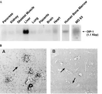
Tissue and cellular distribution of OIP-1 expression. North-ern blot analysis of RNAs from multiple human tissues showed the highest expression of OIP-1 in liver and lower levels in heart, lung, pancreas, kidney, skeletal muscle, placenta, and brain (Fig. 4 A). We also detected significant levels of OIP-1
mRNA expression in human bone marrow and MG-63 osteo-blast-like cells on separate Northern hybridization analyses (Fig. 4 A). Analysis of human bone marrow cultures by in situ hybridization showed that both MNCs and mononuclear cells expressed OIP-1 mRNA (Fig. 4 B).
Expression of recombinant OIP-1 protein. Recombinant OIP-1 protein was expressed in E. coli BL 21 using the PET/ 14b system after induction with 1 mM IPTG. The pellet that contained the inclusion bodies was solubilized with 6 M Gu-HCl and purified with a His-Bind Resin column as described above. Recombinant OIP-1 was eluted with a 50–70 mM imi-dazole buffer gradient, precipitated by dialysis against water, and solubilized with 10 mM HCl. The recombinant OIP-1 was not soluble in physiologic buffers unless 6 M GnHCl was added (data not shown). About 200 mg of rOIP-1 was purified from 1 liter of E. coli culture with more than 95% purity.
Effect of recombinant E. coli–derived OIP-1 on TRAP(1) MNC formation and bone resorption in fetal rat bone organ cultures. We also tested the effects of recombinant E. coli– derived OIP-1 on osteoclast-like MNC formation in mouse and human bone marrow cultures to determine if an OIP-1 protein, which still contained the putative COOH-terminal GPI-linker peptide and lacked the GPI-linker and was not
gly-Figure 2. The effects of conditioned media from 293 cells transfected with OIP-1 cDNA on bone resorption in fetal rat long bone cultures.
45Ca-labeled fetal rat long bones stimulated with PTHrP (20 ng/ml) or
[image:5.612.315.555.62.378.2]media (no PTHrP) were treated with varying concentrations (8–32% vol/vol) of conditioned media (CM) from 293 cells transfected with the OIP-1 cDNA or the empty vector (pcDNA1). Results represent the mean of four determinations for a typical experiment. Similar re-sults were seen in three experiments. *P , 0.05 compared with con-trol value.
Figure 3. DNA sequence and deduced amino acid sequence of OIP-1. The OIP-1 gene is 1.1 kb and encodes 131 amino acids. The 20 NH2-terminal amino acids encode a cleavable signal peptide and the
32 COOH-terminal amino acids encode a potential GPI-linker pep-tide. 99 Asn is the potential site for the GPI-linker and also serves as a possible NH2-glycosylation site. The COOH-terminal peptide
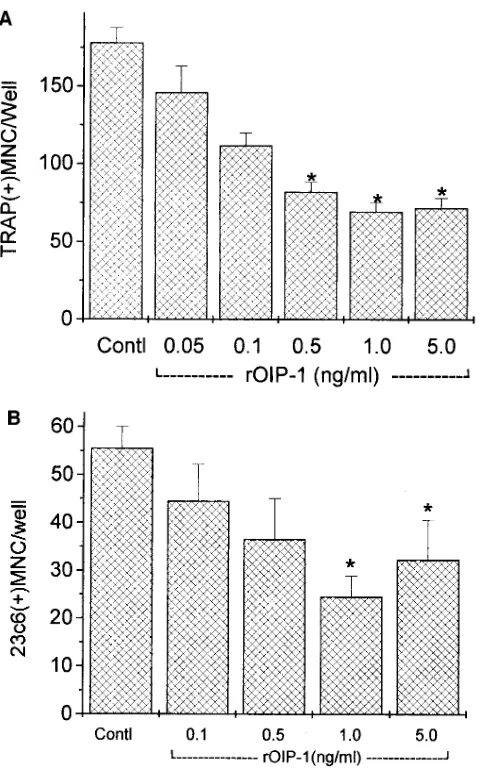
[image:5.612.58.297.444.636.2]cosylated, was biologically active. Surprisingly, in the mouse bone marrow cultures stimulated with PTHrP, recombinant OIP-1 decreased TRAP(1) MNC formation z 60% at a con-centration of 1 ng/ml. Recombinant OIP-1 also inhibited TRAP(1) MNC formation in the presence of 1,25(OH)2D3 in a dose-dependent fashion at concentrations of 0.05–5 ng/ml (Fig. 5 A). In human bone marrow cultures, recombinant OIP-1 also decreased 23c6(1) MNC formation induced by 1029 M 1,25(OH)2D3z 60% at the concentration of 1 ng/ml (Fig. 5 B) and inhibited MNC formation in murine and human marrow cultures treated with PTHrP z 60% (data not shown). Recom-binant OIP-1 was added to mouse bone marrow cultures at dif-ferent time points, and the osteoclast formation was measured. As shown in Fig. 6, addition of OIP-1 for only the first 2 d of the cultures significantly inhibited osteoclast formation. Addi-tion of OIP-1 after the first 2 d of the cultures did not inhibit osteoclast formation. Recombinant OIP-1 also inhibited 45Ca release from 45Ca-labeled fetal rat long bones stimulated with 1029 M 1,25(OH)
2D3z 50% at 10 ng/ml compared with the ve-hicle control assay, and inhibited 45Ca release stimulated by PTHrP (20 ng/ml) in a dose-dependent manner (Fig. 7).
Analysis of the intracellular and extracellular forms of OIP-1. We tested conditioned media from 293 cells and CHO cells that had been transiently transfected with the OIP-1 cDNA, as well as conditioned media from human bone marrow cultures that had formed osteoclast-like cells, for the presence of OIP-1 by Western blot analysis using antisera raised in rabbits against E. coli–derived recombinant OIP-1. As shown in Fig. 8 A, the OIP-1 in these conditioned media was slightly larger (16 kD) than rOIP-1. This difference in the electrophoretic mobility of the mammalian OIP-1 most likely results from addition of the
GPI anchor, which is added as a post-translational modifica-tion to OIP-1 in eukaryotic but not prokaryotic cells. In human bone marrow culture–conditioned media, the higher molecular weight band that was detected could be due to cell-specific post-translational modifications such as glycosylation. Since GPI-linked proteins have a cleavable COOH-terminal peptide and can be shed from the cell surface in membrane-bound ves-icles or the GPI anchor cleaved by PLC, we analyzed the intra-cellular and extraintra-cellular forms of OIP-1. Cell lysates and ul-tracentrifugation fractions of conditioned media from 293 cells transiently transfected with the OIP-1 cDNA were subjected to immunoblot analysis. The cell lysates contained the pre-dicted 14 kD intracellular form of OIP-1 under reducing and nonreducing conditions, and was similar in size to the recombi-nant OIP-1 expressed in E. coli. No OIP-1 was detected in the ultracentrifugation supernatants. A band migrating with an ap-parent molecular weight of 16 kD was detected under reducing conditions with DTT and b-mercaptoethanol treatment in the ultracentrifugation pellet. PI-PLC treatment of the OIP-1 present in the pellet decreased the apparent molecular weight of OIP-1 to 14 kD, and PNGase F (New England Biolabs) treatment further decreased the apparent molecular weight of OIP-1 to 11 kD (Fig. 8 B).
Effect of recombinant OIP-1 on other cell types. Cells of different lineages other than osteoclasts were treated with rOIP-1 to assess the inhibitory potential of OIP-1 for other cell types. Cell lines were treated with rOIP-1 (5 ng/ml) for 62 h, and the number of viable cells was determined. The growth of all of these cell lines was not inhibited by treatment with re-combinant OIP-1 compared with the control vehicle or media treatment (Table I).
Figure 4. (A) Tissue distribution of OIP-1. A MTN blot was hybridized with a 32
P-labeled OIP-1–specific probe generated with random label kit and processed as de-scribed in Methods. The highest levels of OIP-1 mRNA expression were present in liver. OIP-1 mRNA was also detected in the human MG-63 osteoblast cell line and human bone marrow. (B) Cellular localiza-tion of OIP-1 mRNA in human marrow cultures. Human marrow cultures were treated with 1,25(OH)2D3 for 3 wk and
[image:6.612.58.405.58.379.2]Table I. Effects of OIP-1 on the Growth of Different Cell Types
Cell line Cell type Control 10 mM HCl OIP-1 (5 ng/ml)
U937 Human monocytic 6.1 3 105 5.9 3 105 6.4 3 105
KMT-2 Human hematopoietic 1.9 3 105 1.4 3 105 1.8 3 105
ANB-IL-6 Human myeloma 1.4 3 105 1.2 3 105 1.6 3 105
ARH-77 Human myeloma 2.5 3 105 3.4 3 105 3.1 3 105
FDCP-1 Mouse hematopoietic 9.4 3 105 7.8 3 105 8.2 3 105
PSV-10 Human marrow stroma 2.1 3 105 2.5 3 105 2.8 3 105
ST-2 Mouse marrow stroma 3.1 3 105 2.6 3 105 3.2 3 105
SAKA Human marrow stroma 3.6 3 104 5.2 3 104 4.3 3 104
HELA Human cervical epithelium 2.9 3 105 1.5 3 105 3.4 3 105
Cells were plated at 1 3 105/ml at the initiation of culture in the presence or absence of 5 ng/ml rOIP-1 or an equivalent amount of vehicle or culture
media. Results represent mean of triplicate cultures for a typical experiment. Results do not differ significantly for each treatment group for a partic-ular cell line. A similar pattern of results was seen in two independent experiments.
Discussion
[image:7.612.58.298.257.641.2]We have used an expression cloning approach to identify a novel inhibitor of human and murine osteoclast formation and bone resorption, OIP-1. OIP-1 inhibited osteoclast-like cell formation in human and murine marrow cultures in a dose-dependent fashion and inhibited osteoclastic bone resorption in fetal rat long bone cultures. Furthermore, OIP-1 only inhib-ited osteoclast formation when added at the proliferative stage (days 1–3) of murine marrow cultures (18), suggesting that OIP-1 blocks osteoclast precursor proliferation. The inhibitory effects of OIP-1 appear to be selective to cells in the osteoclast lineage, and not due to nonselective cytotoxicity, since the growth of cells from other lineages was not inhibited by OIP-1 at concentrations that markedly inhibit osteoclast-like cell for-mation. The relative potency of OIP-1 in terms of its capacity
Figure 5. The effects of recombinant OIP-1 expressed in E. coli on mouse and human bone marrow osteoclast-like MNC formation. Varying concentrations of rOIP-1 (0.05–5 ng/ml) were added to nor-mal mouse (A) or human (B) bone marrow cultures stimulated with 1029 M 1,25(OH)
2D3. Control vehicle contained 2 ml of 10 mM HCl.
Results are reported as the number of TRAP(1) MNCs/well (A) and 23c6(1) MNCs/well (B), and represent the mean6SEM for four de-terminations. A similar pattern of results was found in four indepen-dent experiments. *P , 0.05 compared with the control value.
Figure 6. Time course studies of the effects of rOIP-1 on osteoclast formation in murine marrow culture. OIP-1–conditioned media was added to cultures treated with 1029 M 1,25(OH)
2D3 for the days
[image:7.612.317.554.464.648.2]to inhibit osteoclast formation and bone resorption in fetal rat bone organ cultures is similar to that reported for calcitonin cultures and TGF-b, although TGF-b was a more potent inhib-itor of human osteoclast-like cell formation (6, 19).
OIP-1 mRNA was expressed by a wide variety of tissues and was also expressed by osteoclasts and MG-63 cells. MG-63 cells are very similar to primary human osteoblasts (20), sug-gesting that human osteoblasts may also express OIP-1. These data further suggest that OIP-1 may act as both an autocrine and paracrine regulator of osteoclast activity.
Sequence analysis of OIP-1 predicted that it was a GPI-linked peptide with cleavable COOH-terminal and NH2 -termi-nal sig-termi-nal peptides and N-glycosylation sites. GPI-linked pro-teins have the GPI anchors attached at the cell surface, and the COOH-terminal peptide cleaved at the time of the attachment of the GPI anchor (21). Immunoblot analysis of the released form of OIP-1 supported these structural features. PLC treat-ment of OIP-1 suggested that there was a 2 kD GPI linker present on the secreted form of OIP-1. PNGase treatment of OIP-1 resulted in a further decrease in the apparent molecular weight to a size that was consistent with that predicted for the protein backbone with the COOH-terminal peptide cleaved off. However, none of these structural features, cleavage of the COOH-terminal peptide, the GPI anchor, or even the poten-tial glycosylation sites appear to be required for the inhibitory effects of OIP-1 on osteoclast-like cell formation. This is sug-gested by our findings that the recombinant E. coli–derived OIP-1, which we constructed for production of a polyclonal antibody to OIP-1, inhibited osteoclast-like cell formation in both human and murine systems in a dose-dependent fashion. The recombinant E. coli–derived OIP-1 we produced contains the entire open reading frame, lacks the GPI anchor, is not glycosylated, and still contains the COOH-terminal peptide. These data demonstrate that post-translational modification of
the protein is not required for the inhibitory effects of OIP-1 on osteoclast formation.
Our studies demonstrate that OIP-1 is released from the cell surface in a membrane-bound form. Ultracentrifugation of conditioned media from 293 cells transfected with the OIP-1 cDNA, followed by Western blotting of the supernatants and resuspended pellet with the OIP-1 polyclonal antibody, showed that OIP-1 was only detectable in the pellet. Englund et al. (22) reported that GPI-anchored molecules can be cleaved from cells by certain phosphatidylinositol-specific phospholipases to produce hydrophilic derivatives. Similarly, Rooney et al. (23) reported that GPI-anchored proteins, for example CD59, CD55, Cdw52, and CD45, are present in a bound form (vesicle form) as well as a membrane-free form in seminal plasma.
Analysis of the nucleotide and derived amino acid se-quences for OIP-1 show that it is identical to the recently re-ported hSca/RIG-E gene. This gene is a member of the human Ly6 family of proteins, with homology to the mouse TSA-1 gene and other members of the Ly6 family, as well as several cobra venom neurotoxins (14). This homology is evident in the conservation of cysteine residues in the molecules. However, there is no significant homology with the recently discovered antiresorptive agent, osteoprotegerin (24).
The Ly6 gene family represents a class of molecules that may have particular importance in osteoclast–osteoblast inter-actions. Horowitz and coworkers (25) have shown that pri-mary osteoblasts and MC3T3 cells constitutively express both Ly6A and Ly6C antigens, although Ly6C was less abundant. Previously, only one human Ly6 gene family member was known, E48, which is identical to the mouse THB gene, a GPI-anchored molecule that is apparently involved in keratinocytes cell-to-cell adhesion (26).
The mechanism responsible for OIP-1’s inhibition of osteo-clast activity is currently unknown. Recently, Solomon et al. (27) reported that GPI-anchored proteins on the surface of lymphocytes act as signal transduction proteins. Many of these proteins can stimulate lymphocyte proliferation, lymphokine production, calcium mobilization, and tyrosine phosphoryla-tion. Stefanova et al. (28) reported that GPI-anchored proteins expressed on lymphocytes coimmunoprecipitate with the src family member tyrosine kinases lck, fyn, fgr, lyn, and hck. In addition, a number of unidentified kinase substrates are also coimmunoprecipitated with GPI-anchored proteins. However, it is not known how these molecules, which lack an intracellu-lar domain, can activate the intracelluintracellu-lar signal pathway. Hu-man GPI-linked molecules CD59, CD55, CD48, CD24, and CD14 as well as mouse Thy-1 and Ly6 have key regulatory functions and induce signal transduction through tyrosine ki-nases. These data suggest that human OIP-1 may inhibit osteo-clast activity through activation of the tyrosine kinase signal transduction pathway.
[image:8.612.58.296.58.262.2]In summary, we have identified a novel inhibitor of osteo-clast activity, OIP-1, that is released as a membrane-bound GPI-linked protein. OIP-I is a member of the human Ly6 fam-ily of proteins, and our findings represent the first description of a biologic activity for this class of human proteins. Our stud-ies suggest that OIP-1 could act as either an autocrine reg-ulator of osteoclasts or as a paracine factor involved in os-teoblast–osteoclast interactions in the marrow despite its widespread tissue distribution. These data further suggest that the identification of the mechanism of action for the inhibitory
Figure 7. The inhibitory effects of rOIP-1 on bone resorption in fetal rat long bone cultures. 45Ca-labeled fetal rat long bones stimulated
effects of OIP-1 on osteoclast activity may provide important insights into the control of the normal bone remodeling pro-cess.
Acknowledgments
The authors thank Judy Anderson for technical assistance, Dr. John Chirgwin for his helpful discussions, Dr. Steve Harris for help with Northern blot analysis of osteoblast expression of OIP-1 mRNA, and Bibi Cates for expert preparation of the manuscript.
This work was supported by Research Funds from the Veterans Administration and National Institutes of Health NIAMS grants AG13625 and AR41336 and National Institute of Arthritis, Diabetes, and Digestive and Kidney Diseases grant AM35188.
References
1. Roodman, G.D. 1996. Advances in bone biology: the osteoclast. Endocr. Rev. 17:308–332.
2. Takahashi, S., S.V. Reddy, J.M. Chirgwin, R.D. Devlin, C. Haipek, J. Anderson, and G.D. Roodman. 1994. Cloning and identification of Annexin II as an autocrine/paracrine factor that increases osteoclast formation and bone resorption. J. Biol. Chem. 269:28696–28701.
3. Reddy, S.V., R.D. Devlin, R. Nishimura, S.J. Choi, T. Yoneda, and G.D. Roodman. 1997. Isolation and characterization of a cDNA clone encoding a
novel peptide (OSF) that enhances osteoclast formation and bone resorption. J. Cell. Physiol. In press.
4. Oursler, M.J. 1994. Osteoclast synthesis, secretion and activation of la-tent transforming growth factor beta. J. Bone Miner. Res. 9:443–452.
5. Pfeilschifter, J., and G.R. Mundy. 1987. Modulation of type beta trans-forming growth factor activity in bone cultures by osteotropic hormones. Proc. Natl. Acad. Sci. USA. 84:2024–2028.
6. Chenu, C., J. Pfeilschifter, G.R. Mundy, and G.D. Roodman. 1988. Transforming growth factor beta inhibits formation of osteoclast-like cells in long-term human bone marrow cultures. Proc. Natl. Acad. Sci. USA. 85:5683– 5687.
7. Nosjean, O., A. Briolay, and B. Roux. 1997. Mammalian GPI proteins: sorting, membrane residence and functions. Biochem. Biophys. Acta. 1331:153– 186.
8. Takahashi, N., H. Yamana, S. Yoshiki, G.D. Roodman, G.R. Mundy, S.J. Jones, A. Boyde, and T. Suda. 1987. Osteoclast-like cell formation and its regu-lation by osteotropic hormones in mouse bone marrow cultures. Endocrinol-ogy. 122:1373–1382.
9. MacDonald, B.R., N. Takahashi, L.M. McManus, J. Holahan, G.R. Mundy, and G.D. Roodman. 1987. Formation of multinucleated cells that re-spond to osteotropic hormones in long term human bone marrow cultures. En-docrinology. 120:2326–2333.
10. Raisz, L.G., and I. Niemann. 1969. Effect of phosphate, calcium and magnesium on bone resorption and hormonal responses in tissue culture. En-docrinology. 85:446–452.
11. Reddy, S.V., S. Takahashi, M. Dallas, R.E. Williams, L. Neckers, and G.D. Roodman. 1994. Interleukin-6 antisense deoxyoligonucleotides inhibit bone resorption by giant cells from human giant cell tumors of bone. J. Bone Miner. Res. 9:753–757.
12. Reddy, S.V., F.R. Singer, L. Mallette, and G.D. Roodman. 1996. Detec-tion of measles virus nucleocapsid transcripts in circulating blood cells from
[image:9.612.58.512.58.452.2]tients with Paget disease. J. Bone Miner. Res. 11:1602–1607.
13. Cohen, E.H., and B.E. Landgraf. 1995. 9804 Gene and Methods of Use Thereof. U.S. Patent 5,468,612, U.S. Government, Washington, D.C.
14. Capone, M.C., D.M. Gorman, E.P. Ching, and A. Zlotnik. 1996. Identi-fication through bioinformatics of cDNAs encoding human thymic shared Ag-1/stem cell Ag-2. J. Immunol. 157:969–973.
15. Mao, M., M. Yu, J.H. Tong, J. Ye, J. Zhu, Q.H. Hung, G. Fu, L. Yu, S.Y. Zhao, S. Waxman, et al. 1996. RIG-E, a human homolog of the murine Ly-6 family, is induced by retinoic acid during the differentiation of acute promyelo-cytic leukemia cell. Proc. Natl. Acad. Sci. USA. 93:5910–5914.
16. Davies, A., D.L. Simmons, G. Hale, R.A. Harrison, H. Tighe, P.J. Lach-mann, and H. Waldmann. 1989. CD59, an Ly-6 like protein expressed in human lymphoid cells, regulates the action of the complement membrane attack com-plex on homologous cells. J. Exp. Med. 170:637–654.
17. Classon, B.J., and L. Converdale. 1994. Mouse stem cell antigen Sca-2 is a member of the Ly-6 family of cell surface proteins. Proc. Natl. Acad. Sci. USA. 91:5296–5300.
18. Biskobing, D.M., X. Fan, and J. Rubin. 1995. Characterization of MCSF-induced proliferation and subsequent osteoclast formation in murine marrow culture. J. Bone Miner. Res. 10:1025–1032.
19. Pfeilschifter, J., S.M. Seyedin, and G.R. Mundy. 1988. Transforming growth factor beta inhibits bone resorption in fetal rat long bone cultures. J. Clin. Invest. 82:680–685.
20. Bonewald, L.F., M.B. Kester, Z. Schwartz, L.D. Swain, A. Khare, T.L. Johnson, R.J. Leach, and B.D. Boyan. 1992. Effects of combining transforming growth factor b and 1,25-dihydroxyvitamin D3 on differentiation of a human
os-teosarcoma (MG-63). J. Biol. Chem. 267:8943–8949.
21. Medof, M.E., S. Nagarajan, and M.L. Tykocinski. 1996. Cell-surface en-gineering with GPI-anchored proteins. FASEB J. 10:574–586.
22. Englund, P.T. 1993. The structure and biosynthesis of glycosyl phos-phatidyl inositol protein anchors. Annu. Rev. Biochem. 62:121–138.
23. Rooney, I.A., J.E. Heuser, and J.P. Atkinson. 1996. GPI-anchored com-plement regulatory proteins in seminal plasma. J. Clin. Invest. 97:1675–1686.
24. Simonet, W.S., D.L. Lacey, C.R. Dunstan, M. Kelley, M-S. Chang, R. Luthy, H.Q. Nguyen, S. Wooden, L. Bennett, and T. Boone. 1997. Osteoproteg-erin: a novel secreted protein involved in the regulation of bone density. Cell. 89:309–319.
25. Horowitz, M.C., A. Fields, D. Demeo, H.Y. Qian, A.L.M. Bothwell, and E. Trepman. 1994. Expression and regulation of Ly6 differentiation antigens by murine osteoblasts. Endocrinology. 135:1032–1043.
26. Brackenhoff, R.H., M. Gerretsen, E.M.C. Knippels, M.V. Dijk, H.V. Essen, D.O. Weghuis, R.J. Sinke, G.B. Snow, and G.A.M.S. Dongen. 1995. The human E48 antigen, highly homologous to the murine Ly6 antigen ThB, is a GPI-anchored molecule apparently involved in keratinocyte cell-cell adhesion. J. Cell. Biol. 129:1677–1689.
27. Solomon, K.R., C.E. Rudd, and R.W. Finberg. 1996. The association be-tween glycosylphosphatidylinositol-anchored proteins and heterotrimeric G protein a subunits in lymphocytes. Proc. Natl. Acad. Sci. USA. 93:6053–6058.