J
OURNAL OFC
LINICALM
ICROBIOLOGY,
0095-1137/97/$04.0010
Apr. 1997, p. 886–891
Vol. 35, No. 4
Copyright
q
1997, American Society for Microbiology
A Sensitive, Type-Specific, Fluorogenic Probe Assay for
Detection of Human Papillomavirus DNA
DAVID C. SWAN,
1* RUTH ANN TUCKER,
1BRIAN P. HOLLOWAY,
2ANDJOSEPH P. ICENOGLE
1Human Papillomavirus Section
1and Biotechnology Core Facility,
2National Center for Infectious Diseases, Centers for Disease
Control and Prevention, Public Health Service, U.S. Department of Health and Human Services, Atlanta, Georgia 30333
Received 30 September 1996/Returned for modification 9 December 1996/Accepted 2 January 1997
A simple method for the detection of a number of human papillomavirus (HPV) genotypes associated with
cervical cancer has been developed. The assay exploits the 5
*
3
3
*
exonucleolytic activity of
Taq
DNA
polymer-ase to increpolymer-ase the signal from fluorescent dyes by releasing them from genotype-specific probes during PCR.
The probes are oligonucleotides with a 5
*
reporter dye (6-carboxyfluorescein), a quencher dye
(6-carboxy-tetramethyl-rhodamine), and a phosphate-blocked 3
*
end. In the intact probe, the proximity of the reporter and
the quencher results in suppression of reporter fluorescence by Fo¨rster-type energy transfer (V. T. Fo¨rster.
Ann. Phys. 2:55–75, 1948). If the probe is bound downstream of either primer during PCR, the 5
*
3
3
*
exonucleolytic activity of
Taq
polymerase degrades it, allowing the reporter to diffuse away from the quencher,
which results in an increase in reporter fluorescence. The increased fluorescence is directly related to the
amount of target DNA and can be detected with an automated fluorometer. Probes for the L1 region of the
cervical-cancer-associated HPV types 16, 18, 31, 33, and 35 were synthesized and the assays were optimized.
The most sensitive assay can detect as few as two copies of HPV DNA in human cervical specimens.
Almost all carcinomas of the cervix are found to contain
specific genotypes of human papillomavirus (HPV) DNA, the
high-risk types. Common high-risk types of HPV in cervical
cancer in the United States are HPV type 16 (16),
HPV-18, HPV-31, HPV-33, and HPV-35 (3). Testing for high-risk
HPV in conjunction with cytology may improve detection of
precancerous cells in cervical samples, but the protocol for
such testing has not been determined. Although some studies
have indicated that the quantity of detectable HPV DNA is
predictive of cervical disease (4, 5, 15), it remains to be
deter-mined if more specific tests, such as assaying for specific open
reading frames, integrated DNA, or specific mRNAs, would be
more predictive. Nevertheless, some properties of assays for
high-risk HPV DNAs that would be necessary for the assays to
be useful in screening programs are clear. At some stages of
cervical disease, the amount of HPV DNA may be very low
(
,
1 HPV genome per infected cell); thus, useful HPV tests
must be sensitive as well as type specific. A number of PCR
assays that are both type specific and sensitive have been
de-scribed, but most of them are not practical for screening
pur-poses since they are not easily used for testing large numbers
of specimens (2, 3, 9, 11, 17).
The fluorogenic probe assays for HPV DNAs described here
are type specific, quantitative, sensitive, and useful for testing
large numbers of specimens. They are based on the PCR
am-plification of a portion of the L1 open reading frames of
HPV-16, -18, -31, -33, and -35 DNAs by using genotype-specific
primers in the presence of fluorescent, genotype-specific
probes that bind to the amplified DNA. The probes are
blocked at their 3
9
termini and hence cannot be extended by
the polymerase (Fig. 1). If, during the course of primer
exten-sion,
Taq
polymerase encounters a bound probe, its 5
9
3
3
9
exonuclease activity degrades the probe, releasing the 5
9
fluor
from the proximity of the 3
9
quencher. This causes an increase
in the fluorescence emitted by the reporter (6) which, in the
presence of an excess amount of the probe, is directly related
to the amount of HPV DNA present in the sample before
amplification. This type of assay has been described previously
and has been used to detect other pathogens (1, 13).
MATERIALS AND METHODS
Probes.The probe sequences for each of the high-risk HPVs were selected based on the following criteria: (i) a predicted lack of cross-hybridization to the other common high- and low-risk HPVs (HPV-6, -11, -16, -18, -31, -33, -35, -45, -51, -52, -56, and -58), (ii) a melting temperature of approximately 708C, (iii) a lack of predicted dimer formation with corresponding primers, (iv) a lack of self-annealing, and (v) no runs of identical nucleotides longer than four, espe-cially Gs. Fluorescent probes for globin and for HPV-16, -18, -31, -33, and -35 were synthesized in the Biotechnology Core Facility at the Centers for Disease Control and Prevention (Table 1). The fluorescent reporter dye on the 59ends of the HPV probes was FAM (6-carboxyfluorescein). The globin probe contained HEX (hexachloro-fluorescein) on its 59 end. The rhodamine quencher dye (TAMRA; 6-carboxy-tetramethyl-rhodamine) was incorporated into a linker arm thymidine (amino modifier C6 dT; Glen Research, Sterling, Va.) at or close to the 39end. Extension from the 39end of the probe was blocked by the addition of a 39 phosphate. Synthesis employed standard DNA phosphoramidites in addition to 6-carboxyfluorescein phosphoramidite, 59 -hexachloro-fluorescein-cyano-ethyl phosphoramidite, 6-carboxy-tetramethyl-rhodamine succinamidyl ester, and amino modifier C6 dT. The 39-blocking phosphate was attached by using 39phosphate control pore glass (Glen Research).
Primers.The primer sequences were selected by using the Oligo 5.0 primer analysis program (National Biosciences, Inc., Plymouth, Minn.). The primer pairs for each of the HPV types were selected based on having a melting temperature of approximately 658C, a predicted lack of cross-hybridization to other common HPV types, and no loop or dimer formation with the other primer. Primers were synthesized in the Biotechnology Core Facility at the Centers for Disease Control and Prevention by standard techniques.
DNA samples.Cervicovaginal lavage samples from 20 patients were thawed and 100ml was removed from each. Samples were transferred to Phase-Lock gel tubes (5 Prime33 Prime, Boulder, Colo.) containing 100ml of phenol-chloro-form-isoamyl alcohol (25:24:1, vol/vol/vol), mixed by inversion, and centrifuged at 12,0003gfor 2 min. An additional 100ml of chloroform was added to each tube, and the tube contents were mixed and centrifuged as described above. Each supernatant from the Phase-Lock procedure was diluted to 2 ml with water and centrifuged in a Centricon 100 microconcentrator (Amicon, Inc., Beverly, Mass.) at 1,0003gfor 30 min. An additional 2 ml of water was added to each retentate, and the centrifugation was repeated. Retentates were then collected and diluted to 250ml with water.
Control templates.Control templates for HPV-16, -18, -31, -33, -35, -45, and -52 were prepared by PCR amplification of cloned DNA with the L1-, type-specific primers (Table 1) and purified in Centricon 100 microconcentrators. The
* Corresponding author. Mailing address: Human Papillomavirus
Section, Mail Stop G-18, Centers for Disease Control and Prevention,
1600 Clifton Rd., Atlanta, GA 30333. Phone: (404) 639-1300. Fax:
(404) 639-0049. E-mail: [email protected].
886
on May 15, 2020 by guest
http://jcm.asm.org/
DNA concentrations were determined by fluorometry (DyNA Quant 200; Hoefer-Pharmacia Biotech, San Francisco, Calif.) after 2ml of DNA solution was added to 2 ml of freshly prepared Hoechst 33258 dye solution (1mg/ml in 10 mM Tris [pH 7.4]–1 mM EDTA–200 mM NaCl) (Pharmacia Biotech Inc., Piscataway, N.J.). A 100-mg/ml DNA standard was used.
Controls, consisting of a dilution series of the homologous template and a set of nine heterologous templates (HPV-16, -18, -31, -33, -35, -45, -51, -52, and -56), were included in each run. By comparing the results obtained with patient DNA to those obtained with the control samples, we estimated the amount of HPV DNA in the patient samples. Each control sample contained 50 ng of human placental DNA.
Fluorogenic PCR.The 50-ml PCR mixtures contained 10 mM Tris (pH 8.3), 50 mM KCl, 4.5 mM MgCl2, 200mM deoxynucleoside triphosphates, 0.3mM each
primer, 50 nM each fluorogenic probe, 0.025 U ofTaqpolymerase/ml and 10ml of template DNA. Following template denaturation for 2 min at 958C, amplifi-cation conditions were as follows: 40 cycles (each) of 30 s at 948C, 10 s at 608C, and 2 min at 658C. Amplification was carried out in a 9600 thermal cycler (Perkin-Elmer Corp., Norwalk, Conn.), after which the samples were transferred to a MicroFLUOR W 96-well, white microtiter plate (Dynatech Industries, Inc., McLean, Va.). Fluorescence was measured with a Perkin-Elmer LS-50B lumi-nescence spectrometer equipped with a microtiter plate reader. Excitation was at 480 nm, and fluorescence was measured at 518 nm (FAM emission maximum), 556 nm (HEX emission maximum), and 582 nm (TAMRA emission maximum). Data acquisition and analysis were performed with the TaqMan fluorescence data manager (Perkin-Elmer Corp.) and Excel 5.0 (Microsoft Corporation, Red-mond, Wash.).
The ratios of fluorescence intensity (RQ1) (FAM/TAMRA or HEX/
TAMRA) were calculated for each sample. The average ratio for three no-template control samples was used to calculate RQ2.DRQ was then calculated
for each sample by the formulaDRQ5RQ12RQ2. ThresholdDRQ, used to
establish a baseline for positive samples, was calculated for a 99% confidence level by using the standard deviation of the results from the three no-template control samples (TaqMan PCR reagent kit protocol; Perkin-Elmer).
PCR product sequencing.Products from the PCR were purified in Centricon 100 microconcentrators and sequenced by standard dye terminator methodology (cycle sequencing kits from Applied Biotechnology Division, Perkin-Elmer
Corp., Emeryville, Calif.). Sequences were analyzed by the Genetics Computer Group (University of Wisconsin) sequence analysis package.
RESULTS
Assay characterization.
The fluorogenic assays were
charac-terized with a dilution series of the homologous template (1
3
10
4to 3
3
10
1copies) and a series of heterologous templates
(2
3
10
3copies). In Fig. 2, the results with the homologous
HPV-16 template dilution series show the approximately
semi-logarithmic relationship between the number of template
cop-ies and the signal intensity and a sensitivity limit of
approxi-mately 30 copies of HPV-16. Each sample contained 50 ng of
human placental DNA (equivalent to 10,000 cells); thus, the
limit of detectability was 30 HPV copies in a total of 10,000
cells, or approximately 1 HPV copy in 300 cells. These
sensi-tivities are approximately equivalent to those obtained by PCR
amplification and ethidium bromide detection of the products
on agarose gels (16). Results obtained with the heterologous
templates indicated good specificity. The HPV-16 probe gave a
detectable signal with HPV-18, -35, and -56 templates, but
despite the fact that 2,000 copies of these templates were
present, the signal was less than 35% of the level of the 30-copy
homologous template. Thus, heterologous templates would
not give a positive signal equivalent to 30 copies of HPV-16
unless they were present in more than about 6,000 copies.
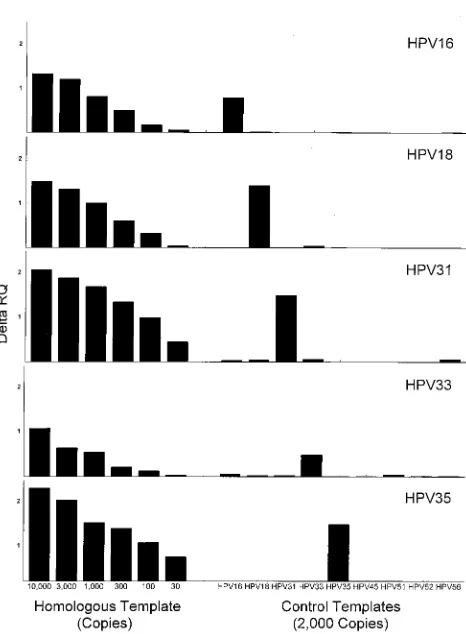
The sensitivities and specificities for all the fluorogenic
as-says are shown in Fig. 3. Each had a sensitivity limit of less than
100 copies of the homologous template per sample. The
HPV-FIG. 1. Schematic of HPV detection by fluorogenic probe assay. The circular HPV genome and the open reading frames are shown on the left. An approximately 470-bp region near the 39end of the L1 gene of HPV is amplified with type-specific primers, and the amplified product is detected with a fluorogenic probe that binds between the primers. Each HPV-type-specific probe is blocked at the 39end, contains a reporter (R) and quencher (Q) dye, and was selected to have only one binding site in the specific amplified product. During PCR amplification of HPV DNA, the 59339exonuclease activity ofTaqpolymerase degrades any probe bound to the template between the primers. Prior to degradation, the fluorescent spectrum of the probe (right) shows almost no emission in the 520-nm range, whereas after degradation, there is a large peak in this range. The intensity of the reporter fluorescence increases as the amount of specific product increases.
on May 15, 2020 by guest
http://jcm.asm.org/
16, -31, and -35 assays had sensitivities of less than 30 copies
per assay sample. The degree of cross-reactivity was low for all
the assays. The HPV-33 assay showed the greatest degree of
cross-reactivity: a signal equivalent to between 30 and 100
copies of HPV-33 was seen with 2,000 copies of HPV-16 or
HPV-51.
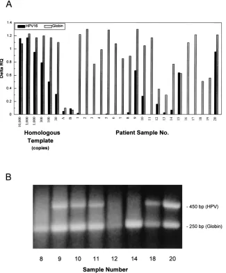
Analysis of patient samples.
To evaluate the performance of
the assay with actual patient specimens, 20 cervicovaginal
la-vage samples (12 from patients with known cervical disease
and 8 from women without disease) were tested in an
HPV-16–globin multiplex assay (Fig. 4). Patients 9, 10, 12, 15, and 20
were positive for HPV-16; the rest were negative. Comparison
of patient
D
RQ signals with the HPV-16 dilution series showed
that the positive patient samples contained between
,
30
cop-ies (patient 12) and about 1,000 copcop-ies (patient 20) per 10-
m
l
aliquot.
[image:3.612.58.555.82.311.2]The PCR products from the assay shown in Fig. 4A were
FIG. 2. Sensitivity and specificity of the HPV-16 fluorogenic probe assay. The homologous template was present in the amounts shown; the heterologous tem-plates were each present at 2,000 copies. Primers, probes, and amplification conditions were as described in Table 1 and Materials and Methods.
[image:3.612.321.554.372.689.2]FIG. 3. Sensitivities and specificities of various HPV fluorogenic probe as-says. Each assay included the indicated amounts of a homologous template and a set of heterologous templates (2,000 copies each). Other reaction conditions were as described in Table 1 and Materials and Methods.
TABLE 1. Primers and probes used in HPV fluorogenic assays
Gene Primer or probea Sequence
HPV-16 L1
U primer 6564
CCT TAT TGG TTA CAA CGA GCA C
L primer 7012
GCG TCC TAA AGG AAA CTG ATC TA
U probe 6862
CCC CAG GAG GCA CAC TAG AAG A(T)
bHPV-18 L1
U primer 6548
GTT ACA TAA GGC ACA GGG TCA T
L primer 6993
CGT CCA AGG GGA TAT TGA TC
U probe 6902
AAA GGA TGC TGC ACC GGC T
bHPV-31 L1
U primer 6490
GAT GCA ACG TGC TCA GGG A
L primer 6930
GCG ACC CAG TGG AAA CTG ATC TA
U probe 6852
CCC AAA AGC CCA AGG AAG AT
bC
HPV-33 L1
U primer 6490
GGT TAC TTC CGA ATC TCA GTT ATT T
L primer 6964
TCC CAA AGG AAA CTG ATC TAA A
L probe 6787
TGT TAA ACC AAA TTG CCA ATC TTC T
bHPV-35 L1
U primer 6518
ACG TGC ACA AGG CCA TAA TA
L primer 6947
CCA ACG GAA ACT GAT CTA AGT CT
U probe 6757
TGA ACC CGT CCA TTT TAG AGG AT
bGlobin
U primer 61992
GAA GAG CCA AGG ACA GGT AC
L primer 62240
CAA CTT CAT CCA CGT TCA CC
U probe 62049
CCC TAG GGT TGG CCA ATC TAC T
bC
aStrand sense and end nucleotide position (59terminus in U primers and probes and 39terminus in L primers and probes) as numbered in the GenBank reference
sequences. Accession numbers are as follows: HPV-16, K02718; HPV-18, X05015; HPV-31, J04353; HPV-33, A12360; HPV-35, M74117; and globin, U01317.
bT to which the TAMRA is coupled. Parentheses indicate that T was not present in the target DNA sequence.
888
SWAN ET AL.
J. C
LIN. M
ICROBIOL.
on May 15, 2020 by guest
http://jcm.asm.org/
[image:3.612.60.298.559.691.2]analyzed by agarose gel electrophoresis (Fig. 4B). Some
sam-ples contained an HPV-16-sized band but did not give a
pos-itive signal in the fluorogenic assay. For example, the sample
from patient 11 showed a strong ethidium bromide band but
was negative for HPV-16 (and also for HPV-18, -31, -33, and
-35; see below) by the fluorogenic assay. The sequence of this
product, determined by using consensus primers my09 and
my11, matched that of HPV-66. It was unexpected that an
HPV-66 template would be amplifiable with the
HPV-16-spe-cific primers since HPV-66 has several bases mismatched with
both the upper and lower primers. However, the fluorogenic
assay was still specific, since the probe was not degraded
de-spite the presence of a large amount of HPV-66 product. The
ethidium bromide staining band from sample 18 did not react
with the HPV-16 probe; HPV-31 is the predominant type in
this sample (see below). Sample 12 gave an appreciable
flu-orogenic signal for HPV-16 despite the fact that the product
was undetectable by ethidium bromide staining. The
homolo-gous template dilution series all contained 50 ng of human
placental DNA and showed a reproducible signal with the
globin probe. The differences in globin signal in the patient
samples could be due to differences in input DNA amount and
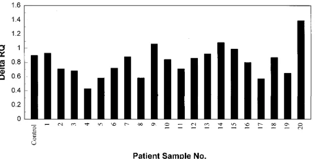
to inhibition by the extract. Samples 12 and 13 gave a low
globin signal in the multiplex assay but, when spiked with
HPV-16 DNA, showed a good HPV-16 signal (Fig. 5). Thus,
the original low globin signals in these samples presumably
reflected low DNA levels. There is, however, evidence of
in-hibition in some patient samples. For example, sample 4, when
spiked with 2,000 copies of HPV-16, showed a signal that
corresponded to only approximately 500 copies. Inhibitors in
patient samples have been reported previously (12). The range
of signals observed for samples 1 through 8 after addition of
2,000 HPV-16 DNA copies is not likely to be a problem since
the differences in amounts of DNA between normal and
dis-FIG. 4. (A) Analysis of patient samples by HPV-16–globin multiplex assay. Patient samples were amplified in a multiplex assay that contained the primers and probes for both HPV-16 and globin. Samples 1 through 8 were from women without cervical disease, and 9 through 20 were from patients with various levels of disease as indicated in Table 2. It was possible to detect each product independently since the HPV-16 and the globin probes were labeled with FAM and HEX, respectively. The sensitivity controls contained HPV-16 DNA in the amounts shown and a constant amount of human placental DNA (2,000 genome copies). Samples A and B were water blanks that were carried through the entire extraction procedure as contamination controls. The assay conditions are given in Materials and Methods. (B) Analysis of PCR products by agarose gel electrophoresis. Selected samples from panel A were analyzed by agarose gel electrophoresis and ethidium bromide staining. From the standard TaqMan reaction mixture, 40ml was applied to a 1% agarose gel containing 13TAE (10 mM Tris acetate [pH 7.4], 1 mM EDTA) and 0.25mg of ethidium bromide/ml. Gels were electrophoresed for 2 h at 80 V. The products were visualized on a UV light box and photographed.
on May 15, 2020 by guest
http://jcm.asm.org/
[image:4.612.147.472.71.464.2]eased specimens have been reported to be fivefold or greater
(20).
The patient samples that had been assayed for HPV-16
DNA were also assayed for HPV-18, -31, -33, and -35 DNAs.
Table 2 summarizes the results from these assays in relation to
pathological diagnosis. Eight of the 12 samples from women
with cervical disease were shown by fluorogenic assays to
con-tain detectable HPV. While it is possible that the four negative
samples contained no HPV DNA, they may have contained
HPV types for which we did not test (e.g., HPV-66; sample 11).
None of the samples from women without cervical disease was
positive for HPV-16, -18, -31, -33, or -35.
DISCUSSION
The HPV fluorogenic assay described here is a fast, simple
method for detecting and typing HPV DNA. It is a convenient,
relatively inexpensive assay for use in studies of large numbers
of specimens (fluorescent probes can be purchased for
approx-imately $0.10 per PCR test). The speed and simplicity of the
test result from the fact that the probe is present in the
reac-tion mixture during PCR and that after amplificareac-tion the
flu-orescence is measured without any further manipulations.
The combination of type-specific primers and probes was
used to make the assay both highly sensitive and specific (Fig.
3). The sensitivities of the assays for HPV-16, -18, -31, -33, and
-35 DNAs varied between approximately one copy of HPV in
100 cells and one copy in 1,000 cells for 50 ng (10,000 cells) of
input DNA. The sensitivity was approximately the same as that
obtained with some HPV PCR assays (14, 21) but was greater
than that of some primer-probe systems (9, 18, 19). For
exam-ple, Snijders et al. constructed primers and probes that were
capable of detecting almost all HPVs, but because of the
de-generacy of the oligonucleotides, the sensitivity was only about
7,000 copies of HPV DNA, even after overnight hybridization
with probes (19). The assays for HPV-16, -18, -31, and -35 gave
signals with 2,000 copies of heterologous templates that were
almost undetectable (equivalent to approximately 5 to 10
cop-ies of homologous templates). The HPV-33 assay is the least
specific, but even in this case, 2,000 copies of HPV-16 gave a
signal strength equivalent to only about 60 copies of the
ho-mologous template.
Development of protocols for HPV testing in cervical
dis-ease screening programs may require quantitative HPV DNA
detection assays followed by systematic evaluation of the
pre-dictive values for various levels of HPV DNA; this fluorogenic
assay would work well for that purpose (5, 7, 8, 10, 15). The
fluorogenic assay is not superior to a PCR dot blot
hybridiza-tion assay for quantitahybridiza-tion (9), but it has the advantage of being
simpler and being compatible with automatic data collection
techniques.
Inhibitors of amplification have been reported for some
pa-tient samples (16, 22). An assay with a
b
-globin probe would
allow the detection of low
b
-globin signals indicative of
poten-tially inhibitory specimens, and spiking such samples with
stan-dard templates should separate inhibitory specimens from
those with a low DNA content. Normalizing the HPV signal to
the
b
-globin signal should result in a better estimate of the
amount of HPV DNA present, independent of the cause of the
low
b
-globin signal.
All the HPV fluorogenic probes described here should be
useful with the commonly used my09-my11 consensus primer
system. For example, the HPV-16 fluorogenic probe gave
sen-sitivities and specificities approximately equal to those
de-scribed here when used with primers my09 and my11 (data not
shown). Fluorogenic screening for HPV-positive samples with
a consensus system followed by typing of the positive samples
with a type-specific assay would require fewer assays than
would a series of type-specific assays alone, but a good
[image:5.612.153.465.71.229.2]“pan-FIG. 5. Test for inhibitors in patient samples. Each of the patient samples used for Fig. 3 was retested for HPV-16 after the addition of 2,000 copies of an HPV-16 template.
TABLE 2. HPV types in lavage samples from patients with
cervical disease
Sample Diagnosisa HPV typeb
1–8
No disease
None
9
CIN I, CIS
16
10
CIN I, CIN III
16, 31
11
CIN III
None
c12
CIN I
16
13
CIV
None
14
CIN II, CIN III
31
15
CIN III
16
16
CIV
None
17
CIS
31
18
CIS
31
19
CIV
None
20
CIV
16
aCIS, carcinoma in situ; CIV, invasive carcinoma. For those patients in which
more than one biopsy sample was analyzed, the result for each is given.
bDetected by fluorogenic assay.
cHPV-66 identified by sequencing of the PCR product.
890
SWAN ET AL.
J. C
LIN. M
ICROBIOL.
on May 15, 2020 by guest
http://jcm.asm.org/
[image:5.612.59.298.553.693.2]HPV” fluorogenic probe is difficult to design (18). For
exam-ple, such a consensus probe for the region of the L1 gene
amplified by the L1-type-specific primers was found to require
nine degenerate positions, resulting in a mixture of 512
differ-ent probes. The alternative of screening for HPV positivity by
assays that involve electrophoretic separation of products
de-feats the purpose of having a simple HPV test.
The following protocol is suggested to determine DNA
am-plifiability by using the
b
-globin assay and to minimize the
number of reactions by using multiplexing. Sufficiently pure
templates for the fluorogenic assay would be obtained by
sim-ple, standard methods such as those described here. The first
multiplex PCR would include primers and probes for any HPV
type of interest and for
b
-globin. Subsequently,
b
-globin-pos-itive samples could be screened simultaneously for two HPV
types in a multiplex assay with two HPV probes, one labeled
with FAM and the other with HEX.
The simplicity of the fluorogenic assay makes it useful for
screening large numbers of samples for the detection of rare
events. For example, it is currently being adapted as a specific
test for integrated HPV, which may occur rarely in women with
low-grade cervical lesions (cervical intraepithelial neoplasia
[CIN] grades I and II).
REFERENCES
1.Bassler, H. A., S. J. A. Flood, K. J. Livak, J. Marmaro, R. Knorr, and C. A. Batt.1995. Use of a fluorogenic probe in a PCR-based assay for the detec-tion ofListeria monocytogenes. Appl. Environ. Microbiol.61:3724–3728. 2.Bauer, H. M., Y. Ting, C. E. Greer, J. C. Chambers, C. J. Tashiro, J.
Chimera, A. Reingold, and M. M. Manos.1991. Genital human papilloma-virus infection in female university students as determined by a PCR-based method. JAMA265:472–477.
3.Bosch, F. X., M. M. Manos, N. Mun˜oz, M. Sherman, A. M. Jansen, J. Peto, M. H. Schiffman, V. Moreno, R. Kurman, and K. V. Shah.1995. Prevalence of human papillomavirus in cervical cancer: a worldwide perspective. Inter-national biological study on cervical cancer (IBSCC) Study Group. J. Natl. Cancer Inst.87:796–802.
4.Cox, J. T., A. T. Lo¨rincz, M. H. Schiffman, M. E. Sherman, A. Cullen, and R. J. Kurman.1995. Human papillomavirus testing by hybrid capture ap-pears to be useful in triaging women with a cytologic diagnosis of atypical squamous cells of undetermined significance. Am. J. Obstet. Gynecol.172: 946–954.
5.Cuzick, J., A. Szarewski, G. Terry, L. Ho, A. Hanby, P. Maddox, M. Ander-son, G. Kocjan, S. T. Steele, and J. Guillebaud.1995. Human papillomavirus testing in primary cervical screening. Lancet345:1533–1536.
6.Fo¨rster, V. T. 1948. Zwischenmolekulare Energiewanderung und Flu-oreszenz. Ann. Phys.2:55–75.
7.Gibbs, A. C. C.1995. Comparison of ViraPap, Southern hybridization, and polymerase chain reaction methods for human papillomavirus identification in an epidemiological investigation of cervical cancer. J. Clin. Microbiol. 33:2229. (Letter.)
8.Guerrero, E., R. W. Daniel, F. X. Bosch, X. Castellsague´, N. Mun˜oz, M. Gili, P. Viladiu, C. Navarro, M. L. Zubiri, N. Ascunce, L. C. Gonzalez, L. Tafur,
I. Izarzugaza, and K. V. Shah.1992. Comparison of ViraPap, Southern hybridization, and polymerase chain reaction methods for human papillo-mavirus identification in an epidemiological investigation of cervical cancer. J. Clin. Microbiol.30:2951–2959.
9.Jacobs, M. V., A. M. de Roda Husman, A. J. C. van den Brule, P. J. F. Snijders, C. J. L. M. Meijer, and J. M. M. Walboomers.1995. Group-specific differentiation between high- and low-risk human papillomavirus genotypes by general primer-mediated PCR and two cocktails of oligonucleotide probes. J. Clin. Microbiol.33:901–905.
10. Kaufman, R. H., E. Adam, J. Icenogle, H. Lawson, N. Lee, K. O. Reeves, J. Irwin, T. Simon, M. Press, R. Uhler, C. Entman, and W. C. Reeves.1997. Relevance of HPV screening in management of cervical intraepithelial neo-plasia. Am. J. Obstet. Gynecol.92:87–100.
11. Kuypers, J. M., C. W. Critchlow, P. E. Gravitt, D. A. Vernon, J. B. Sayer, M. M. Manos, and N. B. Kiviat.1993. Comparison of dot filter hybridization, Southern transfer hybridization, and polymerase chain reaction amplification for diagnosis of anal human papillomavirus infection. J. Clin. Microbiol. 31:1003–1006.
12. Lampertico, P., J. S. Malter, M. Colombo, and M. A. Gerber.1990. Detec-tion of hepatitis B virus DNA in formalin-fixed, paraffin-embedded liver tissue by the polymerase chain reaction. Am. J. Pathol.137:253–258. 13. Livak, K. J., S. J. Flood, J. Marmaro, W. Giusti, and K. Deetz. 1995.
Oligonucleotides with fluorescent dyes at opposite ends provide a quenched probe system useful for detecting PCR product and nucleic acid hybridiza-tion. PCR Methods Applications4:357–362.
14. Manos, M. M., Y. Ting, D. K. Wright, A. J. Lewis, T. R. Broker, and S. M. Wolinsky.1989. Use of polymerase chain reaction amplification for the detection of genital human papillomaviruses. Cancer Cells7:209–214. 15. Mansell, M. E., L. Ho, G. Terry, A. Singer, and J. Cuzick.1994.
Semi-quantitative human papillomavirus DNA detection in the management of women with minor cytological abnormality. Br. J. Obstet. Gynaecol.101: 807–809.
16. Reddy, L. V., A. Kumar, and V. P. Kurup.1993. Specific amplification of Aspergillus fumigatus DNA by polymerase chain reaction. Mol. Cell. Probes 7:121–126.
17. Resnick, R. M., M. T. Cornelissen, D. K. Wright, G. H. Eichinger, H. S. Fox, J. ter Schegget, and M. M. Manos.1990. Detection and typing of human papillomavirus in archival cervical cancer specimens by DNA amplification with consensus primers. J. Natl. Cancer Inst.82:1477–1484.
18. Snijders, P. J., C. J. Meijer, and J. M. Walboomers.1991. Degenerate primers based on highly conserved regions of amino acid sequence in pap-illomaviruses can be used in a generalized polymerase chain reaction to detect productive human papillomavirus infection. J. Gen. Virol.72:2781– 2786.
19. Snijders, P. J., A. J. van den Brule, H. F. Schrijnemakers, G. Snow, C. J. Meijer, and J. M. Walboomers.1990. The use of general primers in the polymerase chain reaction permits the detection of a broad spectrum of human papillomavirus genotypes. J. Gen. Virol.71:173–181.
20. Terry, G., L. Ho, D. Jenkins, M. Hills, A. Singer, B. Mansell, and E. Beverley. 1993. Definition of human papillomavirus type 16 DNA levels in low and high grade cervical lesions by a simple polymerase chain reaction technique. Arch. Virol.128:123–133.
21. van den Brule, A. J., P. J. Snijders, R. L. Gordijn, O. P. Bleker, C. J. Meijer, and J. M. Walboomers.1990. General primer-mediated polymerase chain reaction permits the detection of sequenced and still unsequenced human papillomavirus genotypes in cervical scrapes and carcinomas. Int. J. Cancer 45:644–649.
22. Wiedbrauk, D. L., J. C. Werner, and A. M. Drevon.1995. Inhibition of PCR by aqueous and vitreous fluids. J. Clin. Microbiol.33:2643–2646.