0095-1137/96/$04.0010
Copyrightq1996, American Society for Microbiology
Identification of Staphylococci with a Self-Educating System
Using Fatty Acid Analysis and Biochemical Tests
RONALD J. BEHME,1,2* ROGER SHUTTLEWORTH,1ALAN MCNABB,1ANDW. D. COLBY1,2
Division of Microbiology, University Hospital,1and Department of Microbiology and Immunology,2
University of Western Ontario, London, Ontario, Canada
Received 29 January 1996/Returned for modification 2 May 1996/Accepted 10 September 1996
We characterized all of the 35 aerobic taxa of the genusStaphylococcusby using an objective, self-learning
system combining both whole-cell fatty acid (FA) analysis and the results of 35 biochemical tests. Isolates were compared with the type strain for each taxon to generate an FA profile library and a biochemical table of test
responses. Isolates were accepted into the system if they had a similarity index of>0.6 for a taxon within the
FA profile library and if they were identified as the same taxon by a computer program using a probability matrix constructed from the biochemical data. These stringent criteria led to acceptance of 1,117 strains assigned to legitimate taxa. Additional FA groups were assembled from selected strains that did not meet the inclusion criteria based on the type strains and were added to the system as separate entries. Currently, 1,512 isolates have been accepted into the system. This approach has resulted in a comprehensive table of biochem-ical test results and an FA profile library, which together provide a practbiochem-ical system for valid identifications.
The genusStaphylococcushas undergone extensive revision since 1975, when Schleifer and Kloos (29, 45) published the results of their investigation of staphylococci from human skin. There has since been a steady addition of species and subspe-cies that have been isolated from human, veterinary, and en-vironmental sources (26, 27), and there are now 33 species and 8 subspecies that have been validly described. The newer spe-cies include some, such asStaphylococcus lugdunensis(16, 46), which may be primary pathogens of humans and animals and should be routinely identified in clinical laboratories. Further-more, other coagulase-positive species, such asS. intermedius andS. schleiferisubsp.coagulans, which may be mistaken forS. aureus, have also been implicated in human infections (47, 48, 52).
The advent of nucleic acid methodologies has resulted in the recognition of new taxa which may not be identified by con-ventional methods (6), and older species descriptions have been changed by the division of a species into two or more subspecies, as in the cases ofS. cohnii(31) andS. capitis(3). Commercial identification systems often do not include newer species in their databases, and a significant proportion of iso-lates are not identifiable, presumably because they represent hitherto undescribed taxa.
Fatty acid (FA) analysis has been used for the identification of a wide range of bacteria, and the topic has been reviewed by Welch (55). In the present study an objective, self-learning system that combined FA analysis with a standardized bio-chemical procedure was used to characterize the aerobic staph-ylococci. All of the known aerobic taxa were represented, and strains from diverse locales and habitats were included. This approach has resulted in a library of FA profiles and a com-plete table of biochemical data which can be used in combi-nation to identify these important bacteria.
MATERIALS AND METHODS
Bacterial strains.The sources of the type strains, reference strains, and other
strains that were used to establish additional taxa defined by FA profiles are listed in Table 1. Other isolates were obtained from commercial culture collec-tions, the collections of other researchers, clinical specimens, and a variety of
animal and environmental sources in the field. The anaerobesS. saccharolyticus
andS. aureussubsp.anaerobiuswere excluded because their growth require-ments are not compatible with our methods.
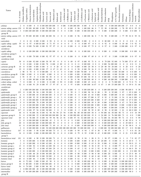
Biochemical tests.All of the strains were subjected to 35 biochemical tests, as
shown in Table 2. The test mixtures were incubated for 48 h at 358C except as
noted below.
The modified oxidase test (14) was performed with cultures cultivated for 24 h on horse blood agar. The tube coagulase test was performed with 0.5 ml of rabbit plasma (0803-46; Difco, Inc., Detroit, Mich.) and 0.1 ml of an overnight heart
infusion broth culture. The mixture was incubated for 4 h at 358C and then for
20 h at room temperature (18 to 248C). Growth on medium containing 6.5%
sodium chloride and resistance to 0.02% furazolidone (2) were both determined on P agar medium (30). The production of beta-glucosidase (56) was determined after incubation for 24 h.
Pyroglutamate aminopeptidase activity (22) was tested in 0.5-ml amounts of
Todd-Hewitt broth (11736; BBL) containing 0.01%L-pyroglutamic acidb
-naph-thylamide (P-5891; Sigma), as previously described (27).
Arginine hydrolysis and ornithine decarboxylase activity were tested in a me-dium containing 1.5 g of thiotone E peptone (12302; BBL), 0.76 g of beef extract (0126-01-8; Difco), 0.2 g of yeast extract (0127-01-7; Difco), 0.03 g of glucose, 6
ml of 1% phenol red solution, and 1.2% of the appropriateL-amino acid in 300
ml of distilled water. The pH was adjusted to 6.5, and the medium was dispensed in 1-ml amounts prior to sterilization. The medium was inoculated to give a heavy suspension and was overlaid with mineral oil. A deep red color was interpreted as a positive result, and an orange or red-orange color was interpreted as negative. This medium is more sensitive than Moeller medium, which accounts
for positive arginine reactions found with some species, e.g.,S. saprophyticus,
which are usually described as negative (27).
Acid production from carbohydrates was tested in phenol red broth base (11506; BBL) in which the indicator concentration was doubled to 0.036 g/liter. Carbohydrates were added to give a final concentration of 1.2%, and the pH was adjusted to 7.4. The medium (0.5-ml amounts) was inoculated with approxi-mately 0.1 ml of a heavy suspension of growth that was harvested from an overnight blood agar plate and suspended in 0.85% saline. Reactions were interpreted as positive (yellow), weak (yellow-orange), or negative (red-orange or red).
The remaining biochemical tests were performed as previously described (46). For data analysis, weak reactions were recorded as positive results.
Cellular FA analysis.The strains of staphylococci were subcultured on blood
agar at least twice at 24-h intervals in order to obtain organisms in a reproducible physiological state before analysis. The bacteria were then grown on Trypticase
soy agar (11043; BBL) containing 5% sheep blood incubated at 358C for 24 (6
1) h. The methyl esters of their FAs were extracted and analyzed as described by Miller and Berger (37). A Hewlett-Packard HP5890 series II gas chromatograph fitted with a flame ionization detector was used. Chromatograph control, peak
* Corresponding author. Mailing address: Division of Microbiology, University Hospital, 339 Windermere Rd., London, Ontario, Canada, N6A 5A5. Phone: (519) 663-3343. Fax: (519) 663-3743. Electronic mail address: [email protected].
3075
on May 15, 2020 by guest
http://jcm.asm.org/
naming, and data handling were performed with the MIS software (Microbial Identification, Inc., Newark, Del.).
Development of biochemical table and FA library.Initially, a table containing
biochemical test data (as in Table 2) was constructed solely from the results of type strains. Variability in the test results for a type strain was addressed by repeating the biochemical tests at least four times and expressing the data as the percentages of positive responses. The biochemical table served as a probability matrix for a computer program, RefID (4), which identifies unknown isolates by probability calculations as described by Lapage et al. (34).
The type strains were also subjected to at least six FA analyses, and a library entry for each taxon was constructed by using the MIS library generation soft-ware.
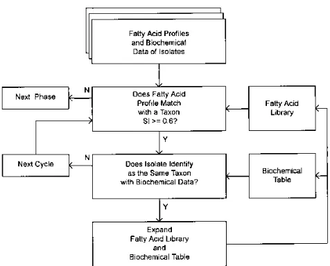
Further development of the biochemical table and the FA library was done in a heuristic fashion as shown in Fig. 1. The investigation was performed in numerous repetitive phases. New strains that met the inclusion criteria were added to the biochemical table and FA library with each iteration of the iden-tification system. For acceptance of a strain, the FA comparison required a similarity index value of at least 0.6 with the profile of a taxon within the library, and the same taxon was required to be the most likely identification when the biochemical results were analyzed with the RefID program.
At a later stage, additional FA groups were created for reference strains and other representative strains whose similarity index values were less than 0.6 with the type strain profiles and therefore did not meet the inclusion criteria for these groups. FA groups with uncertain affinities are referred to as unnamed groups. The ability to differentiate each of the taxa by biochemical tests was assessed with the RefID program. For each taxon in turn, a hypothetical organism, the centrostrain, was created from the probability matrix by determining the most common test result for each biochemical test. The test results for this centros-train were then used as if it were an unknown, and its identity was calculated. The identification score (relative probability) for the centrostrain decreases as the similarity with another taxon increases. An identification score of less than 98% for the centrostrain was considered unacceptable for differentiating the taxon represented by the centrostrain from the other taxa, since a score of below 98% indicates that there is no test that discriminates the taxon represented by the centrostrain from the next most similar taxon.
RESULTS
The biochemical and FA data for the aerobic members of the genusStaphylococcuswere accumulated by a heuristic pro-cedure that began with the type strains of the genus. After 25 iterations of the procedure, 1,117 isolates met the inclusion criteria for acceptance into the system. The biochemical and FA characteristics of these isolates are listed by species name in Tables 2 and 3, respectively.
Our stringent acceptance criteria excluded a significant pro-portion of isolates. Among the excluded isolates were refer-ence strains and other strains that were indistinguishable, in terms of their biotypes, from strains that had been accepted. Thus, a species may contain more than the one FA group represented by the type strain. Therefore, FA library entries and biochemical data were assembled for additional FA groups, and the identification procedure was continued.
[image:2.612.58.303.78.724.2]Some of the additional FA groups of a species contained isolates that were identical or very similar biochemically to isolates in the type strain group. Therefore, we could not dif-ferentiate between some of the FA groups of a species by biochemical tests. For this reason the biochemical data for two
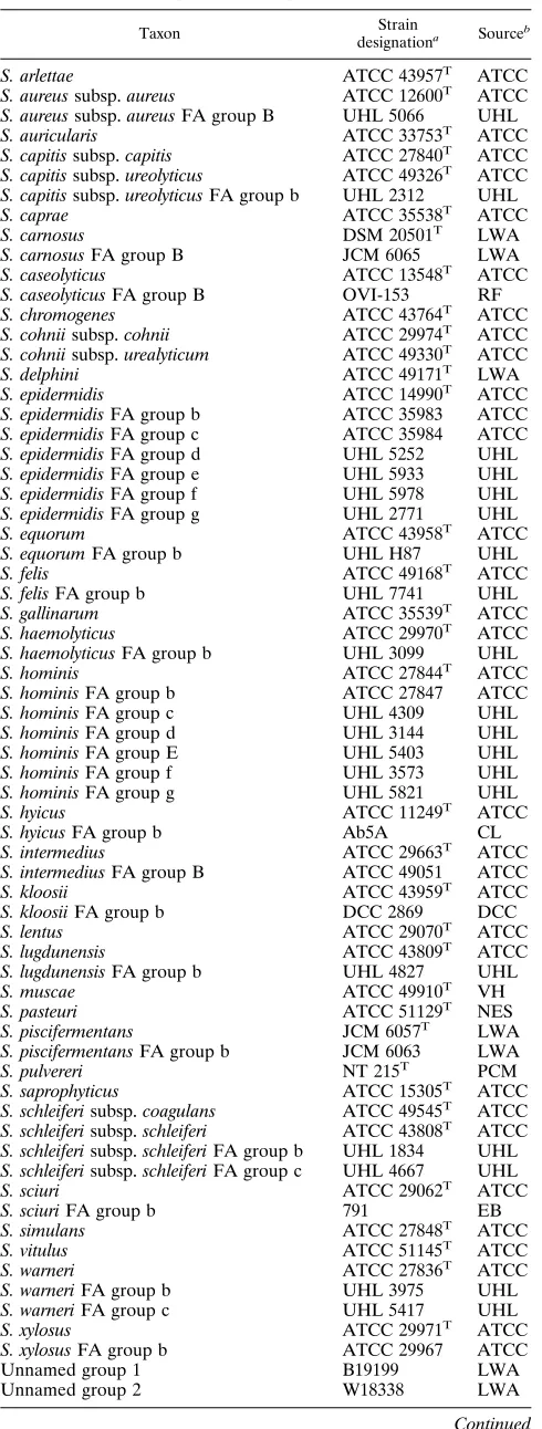
TABLE 1. Staphylococcal type and reference strains
Taxon Strain
designationa Sourceb
S. arlettae ATCC 43957T ATCC
S. aureussubsp.aureus ATCC 12600T ATCC
S. aureussubsp.aureusFA group B UHL 5066 UHL
S. auricularis ATCC 33753T ATCC
S. capitissubsp.capitis ATCC 27840T ATCC
S. capitissubsp.ureolyticus ATCC 49326T ATCC
S. capitissubsp.ureolyticusFA group b UHL 2312 UHL
S. caprae ATCC 35538T ATCC
S. carnosus DSM 20501T LWA
S. carnosusFA group B JCM 6065 LWA
S. caseolyticus ATCC 13548T ATCC
S. caseolyticusFA group B OVI-153 RF
S. chromogenes ATCC 43764T ATCC
S. cohniisubsp.cohnii ATCC 29974T ATCC
S. cohniisubsp.urealyticum ATCC 49330T ATCC
S. delphini ATCC 49171T LWA
S. epidermidis ATCC 14990T ATCC
S. epidermidisFA group b ATCC 35983 ATCC
S. epidermidisFA group c ATCC 35984 ATCC
S. epidermidisFA group d UHL 5252 UHL
S. epidermidisFA group e UHL 5933 UHL
S. epidermidisFA group f UHL 5978 UHL
S. epidermidisFA group g UHL 2771 UHL
S. equorum ATCC 43958T ATCC
S. equorumFA group b UHL H87 UHL
S. felis ATCC 49168T ATCC
S. felisFA group b UHL 7741 UHL
S. gallinarum ATCC 35539T ATCC
S. haemolyticus ATCC 29970T ATCC
S. haemolyticusFA group b UHL 3099 UHL
S. hominis ATCC 27844T ATCC
S. hominisFA group b ATCC 27847 ATCC
S. hominisFA group c UHL 4309 UHL
S. hominisFA group d UHL 3144 UHL
S. hominisFA group E UHL 5403 UHL
S. hominisFA group f UHL 3573 UHL
S. hominisFA group g UHL 5821 UHL
S. hyicus ATCC 11249T ATCC
S. hyicusFA group b Ab5A CL
S. intermedius ATCC 29663T ATCC
S. intermediusFA group B ATCC 49051 ATCC
S. kloosii ATCC 43959T ATCC
S. kloosiiFA group b DCC 2869 DCC
S. lentus ATCC 29070T ATCC
S. lugdunensis ATCC 43809T ATCC
S. lugdunensisFA group b UHL 4827 UHL
S. muscae ATCC 49910T VH
S. pasteuri ATCC 51129T NES
S. piscifermentans JCM 6057T LWA
S. piscifermentansFA group b JCM 6063 LWA
S. pulvereri NT 215T PCM
S. saprophyticus ATCC 15305T ATCC
S. schleiferisubsp.coagulans ATCC 49545T ATCC
S. schleiferisubsp.schleiferi ATCC 43808T ATCC
S. schleiferisubsp.schleiferiFA group b UHL 1834 UHL
S. schleiferisubsp.schleiferiFA group c UHL 4667 UHL
S. sciuri ATCC 29062T ATCC
S. sciuriFA group b 791 EB
S. simulans ATCC 27848T ATCC
S. vitulus ATCC 51145T ATCC
S. warneri ATCC 27836T ATCC
S. warneriFA group b UHL 3975 UHL
S. warneriFA group c UHL 5417 UHL
S. xylosus ATCC 29971T ATCC
S. xylosusFA group b ATCC 29967 ATCC
Unnamed group 1 B19199 LWA
Unnamed group 2 W18338 LWA
Continued
TABLE 1—Continued
Taxon Strain
designationa Sourceb
Unnamed group 3 F58366 LWA
Unnamed group 4 TNA 1078 LWA
a
A superscript T following the strain designation indicates a type strain.
b
ATCC, American Type Culture Collection, Bethesda, Md.; CL, C. La¨mmler,
Giessen, Germany; DCC, Delaware Culture Collection, Newark; EB, E. Bucher, Munich, Germany; LWA, Leona W. Ayers, Columbus, Ohio; NES, N. El Solh, Institut Pasteur, Paris, France; PCM, Polish Collection of Microorganisms, Wro-claw, Poland; RF, R. de la Fuente, Madrid, Spain; UHL, clinical isolates (except UHL H87 [from a horse] and UHL 7741 [from a cat]), University Hospital,
London, Ontario, Canada; VH, V. Ha´jek, Olomouc, Czechoslavakia.
on May 15, 2020 by guest
http://jcm.asm.org/
TABLE 2. Biochemical data
Taxon
No.
of
strains
% of positive reactionsa
Oxidase
(modified)
Coagulase
(tube)
Glucose
fermentation
Urea
hydrolysis
Arginine
hydrolysis
Ornithine
decarboxylase
Glucose
acid
Lactose
acid
Sucrose
acid
D
-Mannitol
acid
Salicin
acid
Sorbitol
acid
L
-Arabinose
acid
D
-Raf
finose
acid
Maltose
acid
D
-Xylose
acid
D
-Trehalose
acid
D
-Cellobiose
acid
D
-Fructose
acid
D
-Mannose
acid
D
-Galactose
acid
Xylitol
acid
D
-Melezitose
acid
Esculin
hydrolysis
Nitrate
reduction
PYR Phosphatase 6.5%
NaCl
growth
Novobiocin
resistance
Gelatinase DNase Tween
80
lipase
Furazolidone
resistance
Acetoin Beta-glucosidase
S. arlettae 2 0 0 100 0 0 0 100 100 100 100 0 0 100 0 100 100 100 0 100 0 0 0 17 100 0 0 100 100 100 100 17 0 0 0 0
S. aureussubsp.aureus130 0 99 100 80 100 0 100 79 100 94 2 0 0 0 100 0 96 1 100 100 86 0 7 52 98 0 100 100 1 78 99 83 0 95 32
S. aureussubsp.aureus
group B
3 0 100 100 100 100 0 100 100 100 100 0 0 0 0 100 0 100 0 100 100 100 0 0 0 100 0 100 100 0 0 100 0 0 100 0
S. aureussubsp.aureus
total
133 0 99 100 80 100 0 100 80 100 94 2 0 0 0 100 0 96 1 100 100 86 0 7 51 98 0 100 100 1 77 99 81 0 95 31
S. auricularis 2 0 0 10 0 100 0 100 0 50 0 0 0 0 0 100 0 100 0 100 0 0 0 0 0 80 100 0 10 0 0 0 0 0 0 0
S. capitissubsp.capitis 39 0 0 97 0 100 10 100 0 71 97 0 0 0 0 3 0 0 0 100 97 0 0 0 0 92 0 0 100 0 10 60 3 0 92 0
S. capitissubsp.
ureolyticus
34 0 0 100 76 100 0 100 91 97 97 0 0 0 0 100 0 0 0 100 97 73 0 0 0 97 0 3 100 0 100 100 0 0 97 0
S. capitissubsp.
ureolyticusgroup b
2 0 0 100 100 100 0 100 100 100 100 0 0 0 0 100 0 0 0 100 100 0 0 0 0 100 0 0 100 0 100 100 0 0 100 0
S. capitissubsp.
ureolyticustotal
36 0 0 100 78 100 0 100 92 97 97 0 0 0 0 100 0 0 0 100 97 69 0 0 0 97 0 3 100 0 100 100 0 0 97 0
S. caprae 38 0 0 100 92 100 0 100 50 95 95 0 0 0 0 89 0 97 0 100 95 71 0 0 0 79 100 92 100 0 74 100 37 0 87 0
S. carnosus 9 0 0 100 0 100 0 100 78 0 100 0 89 0 0 0 0 11 0 100 100 0 0 0 0 100 56 100 100 0 0 0 0 0 11 0
S. carnosusgroup B 10 0 0 100 0 100 0 100 0 0 0 20 0 0 0 0 0 100 0 100 20 30 0 0 20 80 50 20 100 0 0 20 0 0 0 20
S. carnosustotal 19 0 0 100 0 100 0 100 37 0 47 11 42 0 0 0 0 58 0 100 58 16 0 0 11 89 53 58 100 0 0 11 0 0 5 11
S. caseolyticus 2 90 0 70 0 0 0 100 100 0 0 0 0 0 0 100 0 100 70 100 0 70 0 0 0 100 100 0 100 50 100 0 0 0 100 0
S. caseolyticusgroup B 2 100 0 100 0 0 0 100 0 100 0 0 0 0 0 100 0 100 0 100 100 0 0 0 0 100 100 0 100 50 100 0 0 0 100 0
S. caseolyticustotal 4 95 0 85 0 0 0 100 50 50 0 0 0 0 0 100 0 100 35 100 50 35 0 0 0 100 100 0 100 50 100 0 0 0 100 0
S. chromogenes 5 0 0 100 80 100 0 100 84 100 0 0 0 0 0 20 0 96 0 100 100 40 0 0 0 100 60 100 100 0 100 100 0 0 0 0
S. cohniisubsp.cohnii 7 0 0 100 0 0 0 100 0 21 100 0 50 0 0 100 0 100 0 100 43 14 21 0 0 0 0 71 100 100 0 0 71 0 71 0
S. cohniisubsp.
urealyticum
32 0 0 100 91 29 0 100 97 17 97 47 6 0 3 97 0 100 0 100 100 16 47 0 47 3 69 53 100 100 75 0 59 0 31 46
S. delphini 2 0 100 100 100 100 0 100 100 100 90 0 0 0 0 100 0 0 0 100 100 100 0 0 0 100 100 100 100 0 100 90 100 0 0 38
S. epidermidis 155 0 0 100 98 94 1 100 99 100 2 0 1 0 1 99 0 3 0 100 98 94 0 88 1 99 0 92 100 3 87 1 85 0 100 0
S. epidermidisgroup b 6 0 0 100 100 83 0 100 100 100 0 0 17 0 0 100 0 0 0 100 83 83 0 100 0 100 0 100 100 0 83 0 100 0 100 0
S. epidermidisgroup c 39 0 0 100 92 95 0 100 100 100 3 0 3 0 0 100 0 3 0 100 97 100 0 97 0 95 0 31 100 0 54 0 64 0 100 0
S. epidermidisgroup d 3 0 0 100 100 100 0 100 100 100 0 0 0 0 0 100 0 0 0 100 100 100 0 100 0 67 0 100 100 0 100 0 67 0 100 0
S. epidermidisgroup e 9 0 0 100 100 78 0 100 89 100 0 0 22 0 0 100 0 0 0 100 100 89 0 89 0 100 0 100 100 0 67 0 78 0 100 0
S. epidermidisgroup f 2 0 0 100 100 100 0 100 50 100 0 0 0 0 0 100 0 0 0 100 100 100 0 100 0 100 0 100 100 0 100 0 100 0 100 0
S. epidermidisgroup g 3 0 0 100 100 100 0 100 100 100 0 0 0 0 0 100 0 0 0 100 100 100 0 0 0 100 0 100 100 0 100 0 33 0 100 0
S. epidermidistotal 217 0 0 100 97 94 0 100 98 100 2 0 3 0 0 100 0 2 0 100 98 95 0 89 0 98 0 82 100 2 81 0 80 0 100 0
S. equorum 48 0 0 94 100 56 0 100 92 98 94 94 23 94 0 100 96 88 48 100 100 50 0 8 94 100 46 50 100 98 71 0 0 0 10 99
S. equorumgroup b 21 0 0 100 100 52 0 100 100 100 100 100 24 86 0 100 100 100 62 100 100 19 0 0 24 100 14 76 100 100 71 0 0 0 5 100
S. equorumtotal 69 0 0 96 100 55 0 100 94 99 96 96 23 91 0 100 97 91 52 100 100 41 0 6 72 100 36 58 100 99 71 0 0 0 9 99
S. felis 21 0 0 100 100 100 0 100 100 43 5 0 0 0 0 0 0 100 0 100 100 100 0 0 0 100 100 100 100 0 0 48 95 0 0 0
S. felisgroup b 20 0 0 100 100 100 0 100 100 100 95 0 0 0 0 0 0 100 0 100 100 100 0 0 0 100 100 100 100 0 0 29 100 0 0 0
S. felistotal 41 0 0 100 100 100 0 100 100 71 49 0 0 0 0 0 0 100 0 100 100 100 0 0 0 100 100 100 100 0 0 39 98 0 0 0
S. gallinarum 2 0 0 100 100 100 0 100 50 100 100 100 100 100 100 100 100 50 100 100 100 0 0 0 100 100 50 100 100 100 83 17 0 0 50 100
S. haemolyticus 167 0 0 100 5 98 0 100 89 100 73 0 2 0 0 100 0 99 0 94 1 87 0 30 1 98 97 0 100 0 1 7 8 0 81 26
S. haemolyticus
group b
14 0 0 100 0 100 0 100 100 100 50 0 0 0 0 100 0 79 0 93 0 100 0 43 0 100 100 0 100 0 0 21 0 0 100 40
S. haemolyticustotal 181 0 0 100 4 98 0 100 90 100 71 0 2 0 0 100 0 98 0 94 1 88 0 31 1 98 97 0 100 0 1 8 7 0 82 27
S. hominis 5 0 0 100 100 0 0 100 20 100 0 0 0 0 0 100 0 100 0 100 0 20 0 45 20 100 0 0 100 0 0 0 65 0 5 0
S. hominisgroup b 2 0 0 100 100 0 0 100 63 100 0 0 0 0 0 100 0 100 0 100 0 63 0 50 0 100 0 0 100 0 0 0 50 0 13 0
S. hominisgroup c 13 0 0 100 92 0 0 100 62 100 38 0 0 0 0 100 0 15 0 100 0 38 0 85 0 92 8 0 100 77 0 8 0 0 15 0
S. hominisgroup d 48 0 0 100 77 4 0 100 85 100 29 0 0 0 0 100 0 65 2 92 0 83 2 92 0 98 10 0 100 2 0 0 46 0 38 0
S. hominisgroup E 2 0 0 100 100 0 0 100 100 100 0 0 0 0 0 100 0 0 0 100 0 100 0 100 0 100 0 0 100 100 0 0 0 0 100 50
S. hominisgroup f 5 0 0 100 60 20 0 100 80 100 20 0 0 0 0 100 0 80 0 100 0 80 0 80 0 100 20 0 100 0 0 0 25 0 40 0
S. hominisgroup g 10 0 0 100 100 10 0 100 50 100 0 0 0 0 0 100 0 80 0 100 0 60 0 60 0 100 0 0 100 0 0 0 30 0 50 0
S. hoministotal 85 0 0 100 84 5 0 100 73 100 24 0 0 0 0 100 0 61 1 95 0 70 1 83 1 98 8 0 100 15 0 1 36 0 35 0
S. hyicus 46 0 72 100 2 100 0 100 83 98 0 0 0 0 0 0 0 80 0 100 100 80 0 0 0 100 0 98 100 0 100 100 100 0 0 8
S. hyicusgroup b 10 0 80 100 0 100 0 100 100 100 0 0 0 0 0 0 0 100 0 100 100 90 0 0 0 100 0 100 100 0 100 100 100 0 0 0
S. hyicustotal 56 0 74 100 2 100 0 100 86 98 0 0 0 0 0 0 0 84 0 100 100 82 0 0 0 100 0 98 100 0 100 100 100 0 0 5
S. intermedius 1 0 100 100 100 20 0 100 100 100 100 0 0 0 0 100 0 100 0 100 100 0 0 0 0 100 100 100 100 0 100 100 100 0 0 0
S. intermedius
group B
85 0 94 100 100 100 0 100 100 100 12 0 0 0 0 100 0 100 0 100 100 100 0 0 0 100 99 100 100 0 100 100 98 0 1 4
Continued on following page
on May 15, 2020 by guest
http://jcm.asm.org/
FA groups were merged whenever the identification scores (relative probabilities) became less than 98% for their centro-strain biotypes. Merged FA groups were assigned a letter of the alphabet in lowercase; FA groups that did not merge were assigned uppercase letters. The FA groups included 395 addi-tional isolates, making a total of 1,512. Their biochemical and FA characteristics are listed in Tables 2 and 3, respectively.
DISCUSSION
In addition to conventional biochemical tests, various other methods for identifying staphylococci have been used, includ-ing the determination of bacteriolytic-activity patterns (53), sodium dodecyl sulfate-polyacrylamide gel electrophoresis of proteins (39), and several kinds of chromosomal analysis (7, 8, 17, 18, 51, 54). Commercial identification systems using
bio-chemical and enzyme tests are also available (26–28, 40), and their limitations and reliabilities have been discussed in a re-cent review by Kloos and Bannerman (27).
[image:4.612.60.559.83.534.2]FA analysis has been used for the identification of a wide range of microorganisms, including bacteria, yeasts, and fila-mentous fungi (55). Durham and Kloos (12) determined the FA compositions of 100 staphylococci representing 10 species and found that the FA compositions of all strains were quali-tatively similar but quantiquali-tatively different. Eerola and Leh-tonen (13) performed FA analysis on strains of various bacte-ria, including three species of staphylococci. They found that the occurrences and amounts of the various fatty acids were generally constant for members of the same species and that repeated analysis of the same strain produced essentially iden-tical results. Kotilainen et al. (32) extended that work to the analysis of seven species:S. epidermidis,S. warneri,S. hominis,
TABLE 2—Continued
Taxon
No.
of
strains
% of positive reactionsa
Oxidase
(modified)
Coagulase
(tube)
Glucose
fermentation
Urea
hydrolysis
Arginine
hydrolysis
Ornithine
decarboxylase
Glucose
acid
Lactose
acid
Sucrose
acid
D
-Mannitol
acid
Salicin
acid
Sorbitol
acid
L
-Arabinose
acid
D
-Raf
finose
acid
Maltose
acid
D
-Xylose
acid
D
-Trehalose
acid
D
-Cellobiose
acid
D
-Fructose
acid
D
-Mannose
acid
D
-Galactose
acid
Xylitol
acid
D
-Melezitose
acid
Esculin
hydrolysis
Nitrate
reduction
PYR Phosphatase 6.5%
NaCl
growth
Novobiocin
resistance
Gelatinase DNase Tween
80
lipase
Furazolidone
resistance
Acetoin Beta-glucosidase
S. intermediustotal 86 0 94 100 100 99 0 100 100 100 13 0 0 0 0 100 0 100 0 100 100 99 0 0 0 100 99 100 100 0 100 100 98 0 1 4
S. kloosii 2 0 0 100 100 30 0 100 100 90 100 0 0 100 0 100 0 100 0 100 0 20 0 0 50 0 100 100 100 100 0 0 0 0 30 0
S. kloosiigroup b 2 0 0 100 100 50 0 100 100 50 100 0 0 50 0 100 0 100 0 100 0 0 0 0 50 0 100 100 100 100 0 0 0 0 50 0
S. kloosiitotal 4 0 0 100 100 40 0 100 100 70 100 0 0 75 0 100 0 100 0 100 0 10 0 0 50 0 100 100 100 100 0 0 0 0 40 0
S. lentus 12 100 0 100 25 0 0 100 92 100 100 100 58 100 92 100 90 100 100 100 100 100 0 17 100 100 19 92 100 100 83 0 0 0 2 75
S. lugdunensis 105 0 0 100 56 0 100 100 72 100 1 0 0 0 0 100 0 99 0 100 100 72 0 7 0 98 99 2 100 0 15 96 0 0 99 43
S. lugdunensisgroup b 4 0 0 100 50 0 100 100 100 100 0 0 0 0 0 100 0 100 0 100 100 100 0 0 0 100 100 0 100 0 25 75 0 0 100 0
S. lugdunensistotal 109 0 0 100 56 0 100 100 73 100 1 0 0 0 0 100 0 99 0 100 100 73 0 6 0 98 99 2 100 0 16 95 0 0 99 43
S. muscae 2 0 0 100 0 0 0 100 0 100 0 0 0 0 0 0 100 100 0 100 0 0 0 0 0 100 0 100 100 0 0 100 100 0 0 67
S. pasteuri 2 0 0 100 100 100 0 100 0 100 100 0 0 0 0 100 0 100 0 100 50 100 0 88 0 100 0 0 100 0 88 0 50 0 100 100
S. piscifermentans 6 0 0 100 100 100 0 100 100 100 0 100 0 0 0 67 0 100 0 100 21 100 0 100 100 100 46 96 100 0 0 67 100 0 0 100
S. piscifermentansgroup b 2 0 0 100 100 100 0 100 100 100 0 100 0 0 0 100 0 100 0 100 0 50 0 50 100 100 50 100 100 0 0 100 100 0 50 100
S. piscifermentanstotal 8 0 0 100 100 100 0 100 100 100 0 100 0 0 0 75 0 100 0 100 16 88 0 88 100 100 47 97 100 0 0 75 100 0 13 100
S. pulvereri 5 100 0 100 0 0 0 100 0 100 100 0 0 0 0 0 100 100 0 100 100 0 0 0 20 100 0 0 100 100 100 0 0 0 0 0
S. saprophyticus 73 0 0 100 100 71 0 100 86 100 88 0 0 0 0 100 0 99 0 100 0 8 12 1 0 4 15 14 100 100 73 1 0 0 99 6
S. schleiferisubsp.coagulans 4 0 100 100 100 100 0 100 81 0 25 0 0 0 0 0 0 0 0 100 100 100 0 0 0 100 100 100 100 0 100 100 100 25 100 0
S. schleiferisubsp.schleiferi 8 0 0 100 0 100 0 100 13 0 0 0 0 0 0 0 0 88 0 100 100 100 0 3 0 100 100 100 100 0 100 100 48 0 100 0
S. schleiferisubsp.schleiferi
group b
17 0 0 100 0 100 0 100 0 0 0 0 0 0 0 0 0 88 0 100 100 100 0 0 0 94 100 100 100 0 82 100 88 0 100 14
S. schleiferisubsp.schleiferi
group c
6 0 33 100 0 100 0 100 0 0 0 0 0 0 0 0 0 100 0 100 100 100 0 0 0 100 100 100 100 0 100 100 83 0 100 0
S. schleiferisubsp.schleiferi
total
31 0 6 100 0 100 0 100 3 0 0 0 0 0 0 0 0 90 0 100 100 100 0 1 0 97 100 100 100 0 90 100 77 0 100 8
S. sciuri 27 100 0 100 0 0 0 100 81 100 100 100 96 67 0 100 0 100 100 100 100 52 0 22 100 100 4 100 100 100 100 59 0 0 0 100
S. sciurigroup b 2 100 0 100 0 0 0 100 50 100 100 100 100 100 0 100 0 100 100 100 100 100 0 100 100 100 0 100 100 100 100 100 0 0 0 100
S. sciuritotal 29 100 0 100 0 0 0 100 79 100 100 100 97 69 0 100 0 100 100 100 100 55 0 28 100 100 3 100 100 100 100 62 0 0 0 100
S. simulans 33 0 0 100 94 100 0 100 97 100 79 0 0 0 0 9 0 97 0 100 24 58 0 0 0 100 97 9 100 0 18 27 97 0 15 0
S. vitulus 13 100 0 100 0 0 0 100 0 100 100 31 52 0 0 23 65 94 69 100 100 0 0 0 77 100 0 4 100 100 100 0 0 0 0 25
S. warneri 76 0 0 100 92 95 4 100 8 100 64 0 0 0 0 100 0 100 0 97 7 11 0 9 0 43 1 1 100 0 63 0 89 0 91 58
S. warnerigroup b 4 0 0 100 100 100 0 100 0 100 0 0 0 0 0 100 0 100 0 100 0 0 0 0 0 100 0 0 100 0 75 0 100 0 75 50
S. warnerigroup c 5 0 0 100 80 80 0 100 20 100 60 0 0 0 0 100 0 100 0 100 0 20 0 20 0 80 0 0 100 0 40 0 100 0 100 75
S. warneritotal 85 0 0 100 92 94 4 100 8 100 61 0 0 0 0 100 0 100 0 98 6 11 0 9 0 48 1 1 100 0 62 0 91 0 91 59
S. xylosus 2 0 0 100 100 38 0 100 100 100 100 0 0 50 0 100 100 100 0 100 100 0 50 0 0 100 88 100 100 100 100 0 0 0 50 0
S. xylosusgroup b 31 0 0 100 100 65 0 100 100 100 100 0 19 87 0 100 100 97 0 100 100 19 14 0 7 97 83 100 100 100 65 3 0 0 30 13
S. xylosustotal 33 0 0 100 100 63 0 100 100 100 100 0 18 85 0 100 100 97 0 100 100 18 16 0 7 97 83 100 100 100 67 3 0 0 32 13
Unnamed group 1 2 0 0 100 13 0 0 100 0 100 0 0 0 0 0 0 0 0 0 88 0 0 0 0 0 100 25 88 100 0 0 0 0 0 25 0
Unnamed group 2 5 0 0 100 100 100 0 100 0 92 0 0 0 0 0 100 0 0 0 100 100 40 0 24 0 80 0 100 100 0 100 0 80 0 100 0 Unnamed group 3 3 0 0 100 0 100 0 100 0 100 67 0 0 0 0 100 0 100 0 100 0 0 0 0 0 100 100 0 100 0 0 33 0 0 100 17 Unnamed group 4 3 0 0 100 100 92 0 100 67 100 100 100 100 100 67 100 100 100 0 100 100 8 0 0 100 100 42 100 100 100 100 33 0 0 58 100
a
Multiple examinations of reference strains were reduced to a single data set prior to inclusion in the database. PYR, pyroglutamate aminopeptidase.
on May 15, 2020 by guest
http://jcm.asm.org/
S. haemolyticus, S. capitis, S. lugdunensis, and S. simulans. When the data were subjected to cluster analysis, they found that isolates belonging to the same species formed separate clusters, but the distinctions were not absolute; some strains were incongruently positioned in various clusters, and some strains identified as S. epidermidis and S. warneri remained outside all clusters.
Identification schemes in general suffer from the limitation that the reliability of the identification depends on the range of taxa represented in the database. Incorrect identifications can result if some taxa are omitted, and this is a particular weak-ness in probabilistic programs, where a high percent probabil-ity figure may be completely misleading if the database is restricted. For this reason, we felt it imperative to include all of the aerobic taxa in our system.
The FA analysis method produces a similarity index which is a measure of how much an unknown profile resembles “aver-age” profiles stored in the FA library. A perfect match gives a similarity index of 1.0. We chose a threshold value of 0.6 or greater for acceptance, which is considered a good match for an individual taxon (36). However, the profile of an unknown isolate may match the profiles of two or more taxa at a high level of similarity, in which case FA analysis cannot be relied upon alone (as with strains of S. capitis and S. caprae). An advantage of our system is that mistakes are less likely to occur, since isolates are identified by combining conventional bio-chemical tests with FA analysis.
The heuristic, or self-educating, approach which we devel-oped is an objective procedure that is based only on the char-acteristics of the type strains and the stringency of the accep-tance criteria. Preconceptions regarding the key features of a taxon are not needed.
Characteristics of selected species.The type strain group of
S. aureussubsp.aureuscontains human, animal, and environ-mental strains; the FA group B form of the subspecies contains three human strains only. One strain belonging to the type strain group was resistant to novobiocin, and occasional strains were negative in the tube coagulase test.
S. auricularisgrows slowly, which makes FA analysis prob-lematic and not reproducible. Only one strain (ATCC 33750) met the acceptance criteria; six other strains were biochemi-cally similar toS. auricularis, but they had FA similarity indices
of less than 0.6. Some carbohydrate reactions were weak, as was the glucose fermentation test. The species is characterized by high proportions (about 20%) of the C20:0fatty acid.
The reactions of the two subspecies ofS. capitisagreed well with published data (3, 29). DNase reactions were almost al-ways weak. Occasional maltose-positive strains of S. capitis subsp.capitismay be difficult to distinguish from urease-neg-ative strains ofS. capitissubsp.ureolyticus. These latter strains can be distinguished fromS. capitissubsp.capitisin that they produce acid from lactose, maltose, and galactose, and they produce gelatinase. Rare ornithine decarboxylase-positive strains of S. capitissubsp.capitismay be mistaken forS. lug-dunensis, but they are easily separated by other tests, andS. lugdunensisis usually yellow pigmented and beta-hemolytic on blood agar.
S. capraewas described on the basis of 10 strains isolated from goat’s milk (11), but strains have since been isolated from human clinical samples (25, 51), and these show some diver-gence with respect toSmaI-digested-DNA electrophoresis pat-terns (17). Vandenesch et al. (51) found eightEcoRI ribotypes among 26 strains from humans and goats, and they distin-guished human from goat ribotypes on the basis of a constant four-band pattern in the human isolates. Our strains include 4 goat strains and 34 human strains, many from bone and joint infections. Contrary to the original description, in our tests the type strain produced acid from sucrose, mannitol, and fructose and gave a negative result in the test for acetoin. Our type strain results agree (except for acetoin) with those found by Vandenesch et al. (51), as do the results for the human strains examined, but goat strains were more likely to give negative maltose reactions. All strains gave a strongly positive DNase reaction, but the positive acetoin reactions were weak. FA profiles are very similar forS. caprae, for both subspecies ofS. capitis, and, to a lesser extent, forS. haemolyticusandS. war-neri. For this reason, FA analysis should not be used alone to identifyS. caprae.
There are considerable biochemical differences between the S. carnosustype strain group andS. carnosusFA group B. The type strain group includes four commercial starter cultures for sausage production and three strains of unknown origin. All group B strains came from fermented fish in Japan. In both cases our results agree closely with those published previously (41, 49).
S. caseolyticus (9, 44) differs from other staphylococci in several respects, notably the positive oxidase and benzidine reactions, the occurrence of a class II Fructose-1,6-diphos-phate aldolase, and its possession of two types of cytochrome c. However, the results of DNA-rRNA hybridization tests sup-port its inclusion in the genus (44). Our type strain group contains only the type strain and one other strain of unknown origin. Group B contains two of the strains described by De La Fuente et al. (9). Four other lamb strains received from R. De La Fuente gave biochemical reactions similar to those of group B strains. Although their FA profiles were similar to those of accepted strains, they did not meet the acceptance criteria. The FA profile ofS. caseolyticusis quite different from that of other staphylococci, including the oxidase-positive S. sciuri species group. In particular, the extremely low levels of 15:0 anteiso, the presence of large amounts of 16:0, and the absence of 15:0 iso, 17:0 iso, and 17:0 anteiso FAs setS. caseolyticusapart from all other staphylococcal species.
Most of the biochemical characteristics that we list for S. cohnii agree with published data (31, 45). However, a clear distinction betweenS. cohniisubsp.cohniiandS. saprophyticus on the basis of acid production from sucrose is not supported by our data. A combination of tests (urea hydrolysis,
gelati-FIG. 1. Flow diagram for the development of the FA profile library and biochemical table. SI, similarity index; N, no; Y, yes.
on May 15, 2020 by guest
http://jcm.asm.org/
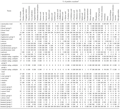
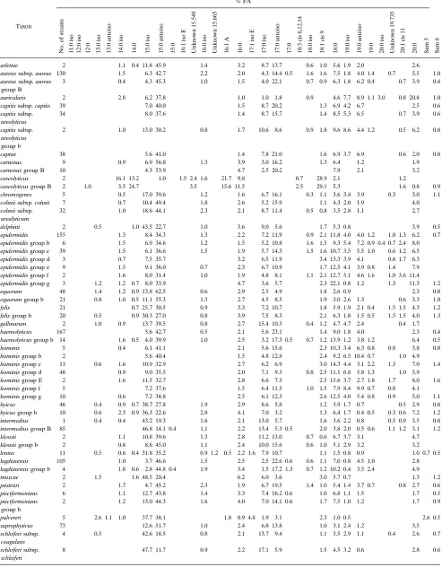
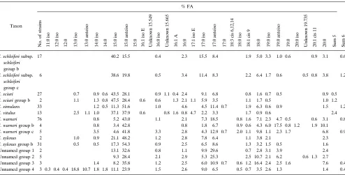
[image:5.612.60.298.68.260.2]TABLE 3. FA library entriesa
Taxon
% FA
No.
of
strains
11:0
iso
12:0
iso
12:0 13:0
iso
13:0
anteiso
14:0
iso
14:0 15:0
iso
15:0
anteiso
15:0 16:1
iso
E
Unknown
15.549
16:0
iso
Unknown
15.665
16:1
A
16:0 17:1
iso
E
17:0
iso
17:0
anteiso
17:0 18:3
cis
6,12,14
18:0
iso
18:1
cis
9
18:0 19:0
iso
19:0
anteiso
19:0 20:0
iso
Unknown
19.735
20:1
cis
11
20:0 Sum
5
Sum
6
S. arlettae 2 1.1 0.4 11.8 45.9 1.4 3.2 8.7 13.7 0.6 1.0 5.6 1.9 2.0 2.6
S. aureussubsp.aureus 130 1.5 6.3 42.7 2.2 2.0 4.3 14.4 0.5 1.6 1.6 7.5 1.8 4.0 1.4 0.7 5.5 1.0
S. aureussubsp.aureus
group B
3 0.4 4.3 45.3 1.0 1.5 4.0 22.1 0.7 0.9 6.3 1.8 6.2 0.4 0.7 3.9 0.4
S. auricularis 2 2.8 6.2 37.8 1.0 1.0 1.8 0.9 4.6 7.7 8.9 1.1 3.0 0.8 20.8 1.0
S. capitissubsp.capitis 39 7.0 40.0 1.5 8.7 20.2 1.3 6.9 4.2 6.7 2.5 0.6
S. capitissubsp.
ureolyticus
34 8.0 37.6 1.4 8.7 15.7 1.4 8.5 5.3 6.5 0.7 3.9 0.6
S. capitissubsp.
ureolyticus
group b
2 1.0 13.0 30.2 0.8 1.7 10.6 8.6 0.9 1.8 9.6 8.6 4.4 1.2 0.5 6.2 0.8
S. caprae 38 5.6 41.0 1.4 7.8 21.0 1.6 6.9 3.7 6.9 0.6 2.0 0.8
S. carnosus 9 0.9 6.9 56.8 1.3 3.9 3.0 16.2 1.3 6.4 1.2 1.9
S. carnosusgroup B 10 4.3 53.9 4.7 2.5 20.2 7.9 2.1 3.2
S. caseolyticus 2 16.1 13.2 1.0 1.5 2.4 1.6 21.7 9.8 0.7 28.9 2.1 1.2
S. caseolyticusgroup B 2 1.0 3.5 24.7 3.5 15.6 11.5 2.5 29.1 5.3 1.6 0.8 0.9
S. chromogenes 5 0.5 17.0 39.6 1.2 1.6 6.7 16.1 0.3 1.1 3.6 3.4 3.9 0.3 3.0 1.1
S. cohniisubsp.cohnii 7 0.7 10.4 49.4 1.8 2.6 5.2 15.9 1.1 4.3 2.0 1.9 4.0
S. cohniisubsp.
urealyticum
32 1.0 18.6 44.1 2.3 2.1 8.7 11.4 0.5 0.8 3.5 2.8 1.1 2.7
S. delphini 2 0.5 1.0 43.5 22.7 1.0 3.6 9.0 5.6 1.7 5.3 0.8 3.9 0.5
S. epidermidis 155 1.3 8.4 34.3 1.3 2.2 7.2 11.9 0.9 2.1 11.8 4.0 4.0 1.2 1.0 1.3 6.2 0.7
S. epidermidisgroup b 6 1.5 6.9 34.6 1.2 1.5 5.2 10.8 1.6 1.5 9.3 5.4 7.2 0.9 0.4 0.7 2.4 8.0
S. epidermidisgroup c 39 1.5 6.1 36.6 1.5 1.9 5.7 14.3 1.5 1.6 10.7 3.5 5.5 1.0 0.6 1.2 6.5
S. epidermidisgroup d 3 0.7 7.5 35.7 3.2 6.5 11.9 3.4 13.3 3.9 4.1 0.8 1.7 6.3
S. epidermidisgroup e 9 1.5 9.1 36.0 0.7 2.3 6.7 10.9 1.7 12.5 4.1 3.9 0.8 1.4 7.9
S. epidermidisgroup f 2 1.6 8.0 31.4 1.0 1.9 4.8 8.1 1.1 2.1 12.7 5.1 4.6 1.6 1.0 3.6 11.4
S. epidermidisgroup g 3 1.2 1.2 0.7 8.0 33.9 4.7 3.6 5.7 2.3 22.1 0.8 1.2 1.3 11.3 1.2
S. equorum 48 1.4 1.2 0.9 13.8 62.5 0.6 2.9 2.5 4.9 1.8 2.6 0.9 2.3 0.8
S. equorumgroup b 21 0.8 1.0 0.5 11.1 55.3 1.3 2.7 4.5 8.5 1.9 3.0 2.6 1.3 0.6 3.3 1.0
S. felis 21 0.7 25.7 30.5 0.9 3.3 7.2 10.7 1.8 5.9 1.9 2.1 0.4 1.5 1.5 4.3 1.2
S. felisgroup b 20 0.5 0.9 30.3 27.0 0.8 3.9 7.5 8.3 2.1 6.3 1.8 1.5 0.5 1.5 1.5 4.0 1.3
S. gallinarum 2 1.0 0.9 13.7 39.5 0.8 2.7 15.4 10.3 0.4 1.2 4.7 4.7 2.4 0.4 1.7
S. haemolyticus 167 5.6 42.7 0.5 2.1 5.6 23.1 1.4 9.0 1.8 4.0 2.3 0.4
S. haemolyticusgroup b 14 1.6 0.5 4.0 39.9 1.0 2.5 3.2 17.3 0.5 0.7 1.2 13.9 1.2 3.8 1.2 6.4 0.5
S. hominis 5 0.4 6.1 41.1 2.1 5.6 15.6 2.3 10.3 3.4 6.3 0.8 0.8 3.8 0.8
S. hominisgroup b 2 5.6 40.4 1.5 4.8 12.8 2.4 9.2 6.5 10.4 0.7 1.0 4.9
S. hominisgroup c 13 0.6 1.6 10.9 32.9 2.7 6.2 6.9 3.0 14.3 4.4 3.1 2.2 1.3 7.0 1.4
S. hominisgroup d 48 0.9 9.0 35.5 2.0 7.1 9.3 0.8 2.5 11.1 6.8 5.8 1.3 1.0 5.9
S. hominisgroup E 2 1.6 11.5 32.7 2.8 6.6 7.3 2.5 15.6 3.7 2.7 1.8 1.7 8.0 1.6
S. hominisgroup f 5 7.2 37.6 1.5 6.4 11.5 1.0 1.5 7.9 8.4 9.9 0.7 0.8 4.1
S. hominisgroup g 10 0.6 7.2 38.8 2.5 6.1 12.3 2.6 12.5 4.0 5.4 0.8 0.9 5.0 1.1
S. hyicus 46 0.4 0.9 0.7 38.7 27.8 1.9 2.9 8.6 5.8 1.2 3.9 1.7 0.7 0.5 2.9 0.8
S. hyicusgroup b 10 0.6 2.3 0.9 36.3 22.6 2.8 4.1 7.0 3.2 1.3 6.4 1.7 0.4 0.5 0.3 0.6 7.2 1.2
S. intermedius 1 0.4 0.4 43.2 19.3 1.6 2.1 13.0 5.7 1.6 3.6 2.2 0.8 0.5 0.9 3.5 0.8
S. intermediusgroup B 85 46.8 14.1 0.4 1.1 2.2 13.4 5.3 0.5 2.0 3.8 2.0 0.5 0.6 1.1 1.2 3.1 1.2
S. kloosii 2 1.1 10.8 39.6 1.3 2.8 11.2 13.0 0.7 0.6 6.7 3.7 3.1 4.7
S. kloosiigroup b 2 0.8 8.6 45.0 1.1 2.4 10.0 15.6 0.6 1.0 5.1 2.9 3.2 3.2
S. lentus 11 0.5 0.6 0.4 31.8 35.2 0.9 1.2 0.5 2.2 1.6 7.9 10.7 1.1 1.5 0.8 0.9 1.0 0.7 0.5
S. lugdunensis 105 1.0 3.7 46.6 1.5 2.5 2.5 22.6 0.6 0.6 1.1 7.0 0.8 4.5 1.0 2.8
S. lugdunensisgroup b 4 1.8 0.6 2.8 44.8 0.4 1.9 3.4 1.5 17.2 1.3 0.7 1.2 10.2 0.4 3.5 2.4 4.9
S. muscae 2 1.5 1.6 48.5 20.4 6.2 6.0 3.6 3.0 5.7 0.7 1.3 1.2
S. pasteuri 2 1.7 4.7 45.2 2.3 1.9 6.7 19.5 1.4 1.0 5.4 1.4 3.7 0.7 0.8 2.7 0.6
S. piscifermentans 6 1.1 12.7 43.8 1.4 3.3 7.4 16.2 0.6 1.0 6.8 1.1 1.5 1.7 0.5
S. piscifermentans
group b
2 1.2 13.0 44.3 1.6 4.0 7.0 14.1 0.6 1.7 7.5 1.0 1.2 1.7 0.9
S. pulvereri 5 2.6 1.1 1.0 37.7 38.1 1.8 0.9 4.8 1.9 3.1 2.3 1.0 0.5 2.6 0.5
S. saprophyticus 73 12.6 51.7 1.0 2.4 6.8 13.8 1.0 3.1 2.4 1.2 3.5
S. schleiferisubsp.
coagulans
4 0.5 42.6 18.5 0.8 2.1 13.7 9.4 1.1 3.5 2.9 1.1 0.4 2.6 0.7
S. schleiferisubsp.
schleiferi
8 47.7 11.7 0.9 2.2 17.1 5.9 1.5 4.5 3.2 0.6 2.8 0.6
Continued on following page
on May 15, 2020 by guest
http://jcm.asm.org/
nase, and acid production from lactose, sucrose, and mannose) is required to distinguish between these two taxa. S. cohnii subsp.urealyticumis more prevalent in human clinical samples thanS. cohniisubsp.cohnii, although neither is encountered frequently. All of our strains of S. cohnii subsp.urealyticum came from human samples. Urease-negative strains are other-wise typical.
Most of our strains ofS. epidermidiswere members of the type strain group. Generally, the lack of acid production from trehalose is a useful marker for this species, but trehalose-positive strains occur occasionally. Susceptibility to desferriox-amine (35) is also very useful for recognizing S. epidermidis. This test was not included at the beginning of the study, so it is not listed in Table 2. However, in our experience nearly all strains ofS. epidermidis andS. hominisare susceptible, while strains of other species are resistant. We encountered one strain each ofS. epidermidisandS. hoministhat were resistant and one strain each of S. warneriand S. cohniisubsp.cohnii which were susceptible. In addition, two of six strains of S. piscifermentanswere susceptible (5).
Isolates ofS. equorumwere first obtained from the skin of healthy horses (43). Our 68 strains include two of the original strains (DSM 20674 [ATCC 43958] and PA 219), two from German fish meal, three from healthy rabbits, one from an environmental sample, and one from a human clinical sample, and we collected the remaining 59 strains from healthy local horses.S. equorumis difficult to distinguish biochemically from S. xylosus, and our set of 35 tests did not always accomplish this. Colonial morphology is a useful indicator, forS. equorum forms small (,1-mm) colonies on blood agar plates incubated at 358C for 24 h, whereasS. xylosusforms much larger colonies which are often yellow pigmented. On P agar at 358C, colonies ofS. equorumare very small, are irregular in shape, and hardly increase in size with prolonged incubation. Colonies ofS.
xy-losus grow well on P agar. In addition, S. equorum is beta-glucosidase positive and gives a weak phosphatase reaction by the method described here, whereasS. xylosusis usually beta-glucosidase negative and gives a strong phosphatase reaction. Other quantitative differences are shown in Table 2. The two species were readily differentiated by FA analysis.
Of nine strains ofS. gallinarumthat we examined, only one strain, P57 of Devriese et al. (11), was accepted into the type strain group. The others gave similar biochemical reactions but did not fulfill the FA similarity criterion, even though the species was the first choice.
In general, strains ofS. haemolyticuswere readily identified by FA analysis and showed high similarity indices, but there was some ambiguity with S. capitissubsp.capitisand with S. caprae. These species are easily differentiated by biochemical tests. The biochemical resemblance between S. haemolyticus and unnamed group 3 is discussed below.
The FA results forS. hominisare variable, because of the low growth rate of the organisms. Similarity indices tend to be low, and few strains gave the value of 0.6 required for inclusion in the type strain group. The creation of six FA groups allowed the inclusion of many more strains. The biochemical results are generally similar for all of the groups, but the existence of novobiocin-resistant strains may easily cause confusion with other species.
Our two subgroups ofS. hyicus each contain strains from cattle and pigs, and the two groups show similar biochemical reactions which correspond to published data (10, 33). All trehalose-negative strains came from cattle, but not all of the bovine strains were trehalose negative. There is some hetero-geneity within this species as indicated by DNA-DNA hybrid-ization studies, possibly representing the inclusion of unnamed subspecies (10, 38), and FA analysis tends to support this.
There appears to be both genotypic and phenotypic
varia-TABLE 3—Continued
Taxon
% FA
No.
of
strains
11:0
iso
12:0
iso
12:0 13:0
iso
13:0
anteiso
14:0
iso
14:0 15:0
iso
15:0
anteiso
15:0 16:1
iso
E
Unknown
15.549
16:0
iso
Unknown
15.665
16:1
A
16:0 17:1
iso
E
17:0
iso
17:0
anteiso
17:0 18:3
cis
6,12,14
18:0
iso
18:1
cis
9
18:0 19:0
iso
19:0
anteiso
19:0 20:0
iso
Unknown
19.735
20:1
cis
11
20:0 Sum
5
Sum
6
S. schleiferisubsp.
schleiferi
group b
17 40.2 15.5 0.4 2.3 15.5 8.4 1.9 5.0 3.3 1.0 0.6 0.9 3.1 0.6
S. schleiferisubsp.
schleiferi
group c
6 38.6 19.8 0.5 3.4 11.4 8.3 2.2 6.4 1.7 0.6 0.5 0.8 3.8 1.2
S. sciuri 27 0.7 0.9 0.6 43.5 28.1 0.9 1.1 0.4 2.4 9.1 6.8 0.8 1.6 0.7 0.5 0.9 0.5
S. sciurigroup b 2 1.1 1.3 0.8 47.5 28.4 0.6 0.6 1.3 2.1 1.1 5.9 3.5 1.1 1.7 0.5 1.0 1.2
S. simulans 33 1.2 0.5 11.3 51.6 1.0 4.6 4.5 11.4 0.7 1.9 6.3 0.6 0.9 1.5 1.2
S. vitulus 13 2.5 1.1 1.0 37.1 37.9 0.6 0.8 1.6 0.8 4.7 2.2 3.3 1.7 0.9 0.6 2.4
S. warneri 76 0.8 5.2 43.0 1.1 2.1 7.3 18.5 0.8 1.6 7.1 2.3 4.7 0.5 0.6 3.1 0.8
S. warnerigroup b 4 0.8 3.4 42.8 0.8 1.8 6.7 0.9 0.6 4.3 6.0 17.5 0.8 1.2 1.9 10.1
S. warnerigroup c 5 3.5 4.6 41.8 3.3 2.8 4.3 12.9 0.7 2.0 1.1 9.8 1.1 2.3 1.7 6.8 0.9
S. xylosus 2 1.0 0.9 21.1 48.2 1.2 2.8 7.8 6.4 1.1 3.8 2.1 2.3
S. xylosusgroup b 31 0.5 0.5 17.3 54.3 0.9 2.5 6.5 8.6 1.3 3.2 1.5 0.5 1.6
Unnamed group 1 2 13.1 32.6 0.8 1.1 9.9 29.6 0.7 2.8 3.1 3.9 2.4
Unnamed group 2 5 9.3 28.4 2.1 2.9 5.3 25.3 2.5 10.7 2.1 6.2 0.6 1.3 2.7
Unnamed group 3 3 1.4 8.2 35.8 1.2 2.5 6.0 10.9 0.7 0.6 1.2 16.4 2.4 2.5 1.6 7.6 0.4
Unnamed group 4 3 0.3 0.4 0.4 18.8 10.7 1.8 1.8 11.1 23.9 1.5 2.6 9.0 6.5 0.5 0.7 3.5 2.6 1.3 1.4 0.4
aUnknown fatty acids are listed with their equivalent chain lengths. Summed feature 5 (sum 5) is a combination of 17:1 iso I and 17:1 anteiso B fatty acids, and
summed feature 6 (sum 6) is a combination of 18:2 cis 9, 12 and 18:0 anteiso fatty acids. The absence of an entry indicates that the fatty acid was not included in the library.
on May 15, 2020 by guest
http://jcm.asm.org/
[image:7.612.61.555.80.331.2]tion between strains of S. intermediusisolated from different animal hosts. De Buyser et al. (8) found that the type strain (a pigeon isolate) showed a ribotype different from that of strains isolated from carnivores, and biochemical differences have also been noted by Ha´jek et al. (19–21). In our system, the type strain gave results for acid production from mannitol and ga-lactose different from those for our other strains. For this reason, no strains were accepted into the type strain group. Contrary to the original description, we found that the type strain produced acid from maltose as well as from mannitol. The majority of our strains ofS. intermediuswere isolated from dogs; a few came from other animals and from animal bite wounds of humans. Our strains appear to belong to biotype 1 or 3, as described by Ha`jek et al. (20, 21), although only a minority were mannitol positive. The biochemical distinction betweenS. intermediusbiotype 2 andS. schleiferisubsp. coagu-lansremains problematic.
S. lugdunensis, first described in 1988 (16), has been shown to be a human pathogen capable of causing serious disease (23, 46), and it should be routinely identified in clinical laborato-ries. The species is well identified by FA analysis and appears to be homogeneous.
All of our strains ofS. piscifermentanscame from fermented fish or shrimp. The biochemical results are similar for the two FA groups that we established, and generally the results agree with those published previously (50). The DNase reaction was weak.
The biochemical results forS. schleiferi agree closely with those published previously (16, 24). As noted above, biochem-ical differentiation betweenS. schleiferisubsp.coagulansandS. intermediusbiotype 2 is difficult, but FA analysis usually sepa-rates these two groups. Occasional strains ofS. schleiferisubsp. schleiferiproduce a pseudo-coagulase phenomenon (52), as is shown in our FA group c. The strong DNase reaction may cause confusion with other species, particularlyS. aureus. Grat-tard et al. (18), usingHindIII endonuclease, divided 31 strains of S. schleiferi subsp. schleiferiinto six ribotypes. There was limited correlation between the ribotypes and our FA groups. When examined by the same method, two of our strains (one of FA group b and one of FA group c), established new ri-botypes (15).
The members of theS. sciurispecies group areS. sciuri,S. lentus, S. pulvereri, and S. vitulus (26, 42, 54, 57). They pos-sessed characteristic FA profiles that contained small amounts of 16:1 A (present otherwise only inS. caseolyticus) and 17:1 iso E (present only in this group) acids. Very small amounts of 20:0, if any, were present; this FA is characteristic for the other species of the genus.
The type strain group ofS. sciuricontains strains from hu-man, environmental, and animal (horse, cow, rat, and rabbit) sources. The two strains in FA group b were from barley straw. Almost all strains of S. lentus gave a strong positive phos-phatase reaction.
Webster et al. (54) described 11 strains ofS. vitulusobtained from animals and meat products. Our strains ofS. vituluswere all isolated from healthy horses.
The five strains of S. pulvereri described by Zakrzewska-Czerwinska et al. (57) fulfilled our acceptance criteria for S. vitulus, but becauseS. pulvereriwas described as a species, we show the data for the two groups separately. The biochemical and FA data are similar, and it is likely that they are synono-mous.S. vituluswas not included in the DNA-DNA hybridiza-tion experiments done with S. pulvereri, and further work is required to clarify the relationship.
Discrimination betweenS. pasteuriandS. warneriis difficult and currently requires the use of nucleic acid techniques (6).
We found that some strains which were identified asS. pasteuri by FA analysis were identified biochemically as S. warneri. Also, three of the five S. warneri FA group c strains were described by Chesneau et al. (6) asS. pasteuri(BM 10425, BM 10427, and BM 9363). It is likely that some of the other strains included asS. warneriare actuallyS. pasteuri. The one strain which was accepted asS. pasteuricame from an orangutan.
The type strain group ofS. xylosuscontains only one other strain isolated from salami in Germany. FA group b strains came from a variety of animal and environmental sources. We have never isolated S. xylosusfrom a human clinical sample. Both groups show similar biochemical profiles, but FA analysis suggests some heterogeneity within this species. It is interest-ing that De Buyser et al. (8) described the existence of two varieties within this species on the basis of an examination of rRNA gene restriction patterns. As indicated above,S. xylosus may be confused withS. equorumbiochemically.
We have recognized four unnamed FA groups that require further study. (i) Unnamed group 1 consists of two strains of human origin which have distinct biochemical and FA patterns. These correspond to those noted recently by Ayers and So-lomon (1). (ii) Unnamed group 2 consists of four strains of human origin. Their biochemical pattern is similar to that ofS. epidermidis, but the FA profile revealed a much larger amount of 17:0 anteiso and a smaller amount of 20:0 FAs than in S. epidermidis. (iii) Unnamed group 3 consists of two strains of human origin and one of bovine origin. The biochemical pat-tern resembles that of S. haemolyticus, but acid production from lactose and from galactose is consistently negative. The FA profile is closer to those ofS. epidermidisandS. hominis. (iv) The three strains in unnamed group 4 exhibit biochemical results which are similar to those forS. gallinarum. Two strains were from bovine sources, and the other was a contaminant in a culture ofS. gallinarumsent to us. Their FA profile was quite different from that of S. gallinarumand indeed was different from those of all other staphylococci, with major differences in the contents of 13:0 iso, 13:0 anteiso, 17:0 iso, and 17:0 anteiso fatty acids. The DNase reaction is weak or negative, and col-onies on P agar after 5 days are irregular in outline, 6 to 9 mm in diameter, flat, matt, and pale yellow.
Our examination of the aerobic species of Staphylococcus represents a new attempt to standardize the identification of these important bacteria. The combination of FA analysis and biochemical testing overcomes most of the identification errors that would result from using either method alone. The meth-odology is useful for reference laboratories and adaptable for use in the clinical laboratory. The heuristic system we have described is an objective approach for characterizing recog-nized species of bacteria, and it also serves well as a research tool for distinguishing undescribed taxa.
ACKNOWLEDGMENTS
We thank R. G. E. Murray at the University of Western Ontario for his encouragement, invaluable advice, and suggestions for improving the manuscript; G. Durat, University Hospital, London, Ontario, for technical assistance in characterizing the isolates; Leona W. Ayers, Ohio State University Hospitals, for invaluable advice and gifts of strains; and all of the other numerous individuals who provided us with strains.
REFERENCES
1.Ayers, L. W., and M. C. Solomon.1994. Recognition of a new species of
Staphylococcusby phenotypic and cellular fatty acid subgrouping and
ri-botyping, abstr. C-131, p. 513.InAbstracts of the 94th General Meeting of
the American Society for Microbiology 1994. American Society for Micro-biology, Washington, D.C.
2.Baker, J. S.1984. Comparison of various methods for differentiation of
on May 15, 2020 by guest
http://jcm.asm.org/
staphylococci and micrococci. J. Clin. Microbiol.19:875–879.
3.Bannerman, T. L., and W. E. Kloos.1991.Staphylococcus capitis subsp.
ureolyticussubsp. nov. from human skin. Int. J. Syst. Bacteriol.41:144–147.
4.Behme, R. J., A. McNabb, and R. Shuttleworth.1993. RefID, a computer
program to identify clinically relevant bacteria, abstr. R-23, p. 297.In
Ab-stracts of the 93rd General Meeting of the American Society for Microbi-ology 1993. American Society for MicrobiMicrobi-ology, Washington, D.C.
5.Behme, R. J., R. Shuttleworth, A. S. McNabb, and W. D. Colby.Unpublished
data.
6.Chesneau, O., A. Morvan, F. Grimont, H. Labischinski, and N. El Solh.
1993.Staphylococcus pasteurisp. nov., isolated from human, animal, and
food specimens. Int. J. Syst. Bacteriol.43:237–244.
7.De Buyser, M.-L., A. Morvan, S. Aubert, F. Dilasser, and N. El Solh.1992.
Evaluation of a ribosomal RNA gene probe for the identification of species
and subspecies within the genusStaphylococcus. J. Gen. Microbiol.138:889–
899.
8.De Buyser, M.-L., A. Morvan, F. Grimont, and N. El Solh.1989.
Character-ization ofStaphylococcusspecies by ribosomal RNA gene restriction
pat-terns. J. Gen. Microbiol.135:989–999.
9.De La Fuente, R., G. Suarez, J. A. Ruiz Santa Quiteria, H. Meugnier, M. Bes,
J. Freney, and J. Fleurette.1992. Identification of coagulase negative
staph-ylococci isolated from lambs asStaphylococcus caseolyticus. Comp. Immun.
Microbiol. Infect. Dis.15:47–52.
10. Devriese, L. A., V. Ha`jek, P. Oeding, S. A. Meyer, and K. H. Schleifer.1978.
Staphylococcus hyicus(Sompolinsky 1953) comb. nov. andStaphylococcus hyicussubsp.chromogenessubsp. nov. Int. J. Syst. Bacteriol.28:482–490.
11. Devriese, L. A., B. Poutrel, R. Kilpper-Ba¨lz, and K. H. Schleifer.1983.
Staphylococcus gallinarumandStaphylococcus caprae, two new species from
animals. Int. J. Syst. Bacteriol.33:480–486.
12. Durham, D. R., and W. E. Kloos.1978. Comparative study of the total
cellular fatty acids ofStaphylococcusspecies of human origin. Int. J. Syst.
Bacteriol.28:223–228.
13. Eerola, E., and O.-P. Lehtonen.1988. Optimal data processing procedure for
automatic bacterial identification by gas-liquid chromatography of cellular
fatty acids. J. Clin. Microbiol.26:1745–1753.
14. Faller, A., and K. H. Schleifer.1981. Modified oxidase and benzidine tests
for separation of staphylococci from micrococci. J. Clin. Microbiol.13:1031–
1035.
15. Fleurette, J.1993. Personal communication.
16. Freney, J., Y. Brun, M. Bes, H. Meugnier, F. Grimont, P. A. D. Grimont, C.
Nervi, and J. Fleurette.1988.Staphylococcus lugdunensissp. nov. and
Staph-ylococcus schleiferisp. nov., two species from human clinical specimens. Int.
J. Syst. Bacteriol.38:168–172.
17. George, C. G., and W. E. Kloos.1994. Comparison of theSmaI-digested
chromosomes ofStaphylococcus epidermidisand the closely related species
Staphylococcus capitisandStaphylococcus caprae. Int. J. Syst. Bacteriol.44: 404–409.
18. Grattard, F., J. Etienne, B. Pozzetto, F. Tardy, O. C. Gaudin, and J.
Fleu-rette.1993. Characterization of unrelated strains ofStaphylococcus schleiferi
by using ribosomal DNA fingerprinting, DNA restriction patterns, and
plas-mid profiles. J. Clin. Microbiol.31:812–818.
19. Ha´jek, V.1976.Staphylococcus intermedius, a new species isolated from
animals. Int. J. Syst. Bacteriol.26:401–408.
20. Ha´jek, V., and J. Balusek.1988. Biochemical profiles and differentiation of
coagulase-positive staphylococci from rooks and gulls. Res. Vet. Sci.44:242–
246.
21. Ha´jek, V., J. Balusek, V. Hora´k, and D. Koukalova´.1991. Characterization of
coagulase-positive staphylococci isolated from free-living birds. J. Hyg.
Epi-demiol. Microbiol. Immunol.35:407–418.
22. He´bert, G. A., C. G. Crowder, G. A. Hancock, W. R. Jarvis, and C.
Thorns-berry.1988. Characteristics of coagulase-negative staphylococci that help
differentiate these species and other members of the familyMicrococcaceae.
J. Clin. Microbiol.26:1939–1949.
23. Herchline, T. E., and L. W. Ayers.1991. Occurrence of Staphylococcus
lugdunensisin consecutive clinical cultures and relationship of isolation to
infection. J. Clin. Microbiol.29:419–421.
24. Igimi, S., E. Takahashi, and T. Mitsuoka.1990.Staphylococcus schleiferi
subsp.coagulanssubsp. nov., isolated from the external auditory meatus of
dogs with external ear otitis. Int. J. Syst. Bacteriol.40:409–411.
25. Kanda, K., E. Suzuki, K. Hiramatsu, T. Oguri, H. Miura, T. Ezaki, and T.
Yokota.1991. Identification of a methicillin-resistant strain ofStaphylococcus
capraefrom a human clinical specimen. Antimicrob. Agents Chemother. 35:174–176.
26. Kloos, W. E., and T. L. Bannerman.1994. Update on clinical significance of
coagulase-negative staphylococci. Clin. Microbiol. Rev.7:117–140.
27. Kloos, W. E., and T. L. Bannerman.1995.StaphylococcusandMicrococcus,
p. 282–298.InP. R. Murray, E. J. Baron, M. A. Pfaller, F. C. Tenover, and
R. H. Yolken (ed.), Manual of clinical microbiology, 6th ed. American Society for Microbiology, Washington, D.C.
28. Kloos, W. E., and C. G. George.1991. Identification ofStaphylococcus
spe-cies and subspespe-cies with the MicroScan Pos ID and Rapid Pos ID panel
systems. J. Clin. Microbiol.29:738–744.
29. Kloos, W. E., and K. H. Schleifer.1975. Isolation and characterization of
staphylococci from human skin. II. Description of four new species:
Staph-ylococcus warneri,Staphylococcus capitis,Staphylococcus hominis, and Staph-ylococcus simulans. Int. J. Syst. Bacteriol.25:62–79.
30. Kloos, W. E., T. G. Tornabene, and K. H. Schleifer.1974. Isolation and
characterization of micrococci from human skin, including two new species:
Micrococcus lylaeandMicrococcus kristinae. Int. J. Syst. Bacteriol.24:79–101.
31. Kloos, W. E., and J. F. Wolfshohl.1991.Staphylococcus cohniisubspecies:
Staphylococcus cohniisubsp.cohniisubsp. nov. andStaphylococcus cohnii
subsp.urealyticumsubsp. nov. Int. J. Syst. Bacteriol.41:284–289.
32. Kotilainen, P., P. Huovinen, and E. Eerola.1991. Application of gas-liquid
chromatographic analysis of cellular fatty acids for species identification and
typing of coagulase-negative staphylococci. J. Clin. Microbiol.29:315–322.
33. La¨mmler, C.1991. Characterization ofStaphylococcus hyicuswith the ATB
32 Staph system and with conventional tests. J. Clin. Microbiol.29:1221–
1224.
34. Lapage, S. P., S. Bascomb, W. R. Willcox, and M. A. Curtis.1970. Computer
identification of bacteria, p. 1–22.InA. Baillie and R. J. Gilbert (ed.),
Automation, mechanization and data handling in microbiology. Academic Press Inc., New York.
35. Lindsay, J. A., and T. V. Riley.1991. Susceptibility to desferrioxamine: a new
test for the identification ofStaphylococcus epidermidis. J. Med. Microbiol.
35:45–48.
36. Microbial ID, Inc.1992. Microbial Identification System operating manual,
version 4. Microbial ID, Inc., Newark, Del.
37. Miller, L., and T. Berger.1985. Bacteria identification by gas
chromatogra-phy of whole cell fatty acids. Application note 228-41. Hewlett-Packard, Inc., Avondale, Pa.
38. Phillips, W. E., Jr., and W. E. Kloos.1981. Identification of
coagulase-positiveStaphylococcus intermediusandStaphylococcus hyicussubsp.hyicus
isolates from veterinary clinical specimens. J. Clin. Microbiol.14:671–673.
39. Pierre, J., L. Gutmann, M. Bornet, E. Bergogne-Berezin, and R. Williamson.
1990. Identification of coagulase-negative staphylococci by electrophoretic profile of total proteins and analysis of penicillin-binding proteins. J. Clin.
Microbiol.28:443–446.
40. Renneberg, J., K. Rieneck, and E. Gutschik.1995. Evaluation of Staph ID 32
system and Staph-Zym system for identification of coagulase-negative
staph-ylococci. J. Clin. Microbiol.33:1150–1153.
41. Schleifer, K. H., and U. Fischer.1982. Description of a new species of the
genusStaphylococcus:Staphylococcus carnosus. Int. J. Syst. Bacteriol.32:
153–156.
42. Schleifer, K. H., U. Geyer, R. Kilpper-Ba¨lz, and L. A. Devriese.1983.
Ele-vation ofStaphylococcus sciurisubsp.lentus(Kloos et al.) to species status:
Staphylococcus lentus(Kloos et al.) comb. nov. Syst. Appl. Microbiol.4:382– 387.
43. Schleifer, K. H., R. Kilpper-Ba¨lz, and L. A. Devriese.1984.Staphylococcus
arlettaesp. nov.,S. equorum sp. nov. andS. kloosii sp. nov.: three new coagulase-negative, novobiocin resistant species from animals. Syst. Appl.
Microbiol.5:501–509.
44. Schleifer, K. H., R. Kilpper-Ba¨lz, U. Fischer, A. Faller, and J. Endl.1982.
Identification of “Micrococcus candidus” ATCC 14852 as a strain of
Staph-ylococcus epidermidisand of “Micrococcus caseolyticus” ATCC 13548 and
Micrococcus variansATCC 29750 as members of a new species, Staphylo-coccus caseolyticus. Int. J. Syst. Bacteriol.32:15–20.
45. Schleifer, K. H., and W. E. Kloos.1975. Isolation and characterization of
staphylococci from human skin. I. Amended descriptions ofStaphylococcus
epidermidisandStaphylococcus saprophyticusand descriptions of three new
species:Staphylococcus cohnii,Staphylococcus haemolyticus, and
Staphylo-coccus xylosus. Int. J. Syst. Bacteriol.25:50–61.
46. Shuttleworth, R., and W. D. Colby.1992.Staphylococcus lugdunensis
endo-carditis. J. Clin. Microbiol.30:1948–1952.
47. Talan, D. A., E. J. C. Goldstein, D. Staatz, and G. D. Overturf.1989.
Staphylococcus intermedius: clinical presentation of a new human dog bite
pathogen. Ann. Emerg. Med.18:410–413.
48. Talan, D. A., D. Staatz, A. Staatz, E. J. C. Goldstein, K. Singer, and G. D.
Overturf.1989.Staphylococcus intermediusin canine gingiva and
canine-inflicted human wound infections: laboratory characterization of a newly
recognized zoonotic pathogen. J. Clin. Microbiol.27:78–81.
49. Tanasupawat, S., Y. Hashimoto, T. Ezaki, M. Kozaki, and K. Komagata.
1991. Identification ofStaphylococcus carnosusstrains from fermented fish
and soy sauce mash. J. Gen. Appl. Microbiol.37:479–494.
50. Tanasupawat, S., Y. Hashimoto, T. Ezaki, M. Kozaki, and K. Komagata.
1992.Staphylococcus piscifermentanssp. nov., from fermented fish in
Thai-land. Int. J. Syst. Bacteriol.42:577–581.
51. Vandenesch, F., S. J. Eykyn, M. Bes, H. Meugnier, J. Fleurette, and J.
Etienne.1995. Identification and ribotypes ofStaphylococcus capraeisolates
isolated as human pathogens and from goat milk. J. Clin. Microbiol.33:888–892.
52. Vandenesch, F., C. Lebeau, M. Bes, G. Lina, B. Lina, T. Greenland, Y.
Benito, Y. Brun, J. Fleurette, and J. Etienne.1994. Clotting activity in
Staphylococcus schleiferisubspecies from human patients. J. Clin. Microbiol. 32:388–392.
53. Varaldo, P. E., G. Satta, G. Grazi, and C. A. Romanzi.1978. Grouping of
on May 15, 2020 by guest
http://jcm.asm.org/
staphylococci on the basis of their bacteriolytic-activity patterns: a new
ap-proach to the taxonomy of theMicrococcaceae. Int. J. Syst. Bacteriol.28:
141–147.
54. Webster, J. A., T. L. Bannerman, R. J. Hubner, D. N. Ballard, E. M. Cole,
J. L. Bruce, F. Fiedler, K. Schubert, and W. E. Kloos.1994. Identification of
theStaphylococcus sciurispecies group withEcoRI fragments containing
rRNA sequences and description ofStaphylococcus vitulussp. nov. Int. J.
Syst. Bacteriol.44:454–460.
55. Welch, D. F.1991. Applications of cellular fatty acid analysis. Clin.
Micro-biol. Rev.4:422–438.
56. White, D. G., R. J. Harmon, and B. E. Langlois.1990. Fluorogenic assay for
differentiatingStaphylococcus warneriandStaphylococcus hominisstrains of
bovine origin. J. Clin. Microbiol.28:602.
57. Zakrzewska-Czerwinska, J., A. Gaszewska-Mastalarz, B. Lis, A. Gamian,
and M. Mordarski.1995.Staphylococcus pulvererisp. nov., isolated from
human and animal specimens. Int. J. Syst. Bacteriol.45:169–172.