0095-1137/96/$04.0010
Copyrightq1996, American Society for Microbiology
Prevalence and Varieties of
Helicobacter
Species in Dogs from
Random Sources and Pet Dogs: Animal and
Public Health Implications
K. A. EATON,1* F. E. DEWHIRST,2B. J. PASTER,2N. TZELLAS,2B. E. COLEMAN,2
J. PAOLA,1ANDR. SHERDING1
Department of Veterinary Biosciences, Ohio State University, Columbus, Ohio 43210,1and
Department of Molecular Genetics, Forsyth Dental Center, Boston, Massachusetts 021152
Received 2 April 1996/Returned for modification 28 June 1996/Accepted 23 July 1996
Gastric bacteria of a variety of ultrastructural morphologies have been identified in or isolated from domes-tic carnivores, but their prevalence in different populations of animals and their clinical significance are still unknown. The purposes of this study were (i) to evaluate the prevalence and morphologic types of gastric bacteria in three different populations of dogs; (ii) to determine which of the organisms were culturable, and if the cultured organisms were morphologically similar to the organisms seen in situ; (iii) to identify the isolated organisms; and (iv) to determine if gastric bacteria were associated with gastritis. Three groups of dogs were examined: healthy laboratory dogs, healthy dogs from an animal shelter, and pet dogs with various nongastric illnesses. Of these, 100% of laboratory and shelter dogs and 67% of pet dogs were colonized by large, tightly coiled gastric spiral bacteria morphologically similar to Gastrospirillum hominis orHelicobacter felis
(referred to as gastrospirilla). Regardless of the presence or density of gastric bacteria, all of the dogs in the study except one had mild to moderate gastritis.Helicobacterspp. were isolated from only 6 of 39 stomachs cultured, and only three of the organisms isolated were morphologically similar to the bacteria seen in situ. Five helicobacters were identified by 16S rDNA (genes coding for rRNA) sequence analysis. Three were strains ofH. felis, one wasH. bilis, and one was a novel helicobacter morphologically similar to “Flexispira rappini.” Gastrospirilla are almost universal in the stomachs of domestic dogs, and in most infected dogs, they do not appear to be associated with clinical signs or histologic lesions compared with uninfected dogs. Nongastro-spirillum helicobacters are rare in dogs and are not histologically detectable. Helicobacter pylori was not isolated from domestic dogs.
The discovery ofHelicobacter pylori in human beings (18) and its relationship to gastritis, peptic ulcer, and gastric neo-plasia (22) has led to renewed interest in the incidence and clinical significance of gastric bacteria in domestic pets, specif-ically cats and dogs. The resulting research has focused on two specific areas of significance. First, it has been suggested that domestic animals could be a source of infection for human beings (21, 31, 32). Part of evaluating this possibility requires examination of the prevalence and types of gastric bacteria in dogs and cats. Second, the clinical significance of gastric heli-cobacters and related organisms in dogs and cats is of interest from a veterinary perspective as well as in terms of the devel-opment of animal models of human disease.
Research over the past 100 years has indicated that gastric bacteria are common in dogs, cats, and other mammals (6, 11, 12, 16, 21, 34 [for review, see reference 5]). The most common gastric organisms described are 7- to 10-mm-long tightly coiled spiral bacteria which live deep in the gastric glands and in the parietal cell canaliculi. These organisms, originally called gas-tric spirilla or spirilliform bacteria (11, 34), are now variously referred to asGastrospirillumsp.,Gastrospirillum hominis, and
Gastrospirillum suis (19, 20, 30). Two unique human species
were identified by 16S rRNA sequence analysis (30) and given the name “Helicobacter heilmannii.” These species have been
given the designations “Gastrospirillum hominis1” (GenBank accession no. L10079) and “G. hominis2” (GenBank accession number L10080). Recently, 10 strains were isolated from dogs and given the nameHelicobacter bizzozeronii(8). It is probable that the large, tightly coiled gastric spiral bacteria seen in many mammalian species represent several different Helicobacter
species. In this report, we will use the term gastrospirilla for this morphologically defined group of mammalian gastric or-ganisms. Among the helicobacters, the gastrospirilla are most similar toHelicobacter felisin their 16S rRNA sequences, large size (7 to 10mm), and habit of growth deep within the gastric glands and gastric parietal cell canaliculi (24, 30). UnlikeH. fe-lis, however, they lack superficial periplasmic fibers.
“Flexispira rappini” is a morphologic type of helicobacter
characterized by fusiform shape, periplasmic fibers, and several amphitrichous flagella and found in rats, mice, sheep, and humans (5, 25, 29, 33). Examination of over a dozen “F. rap-pini” isolates indicates that there are at least six species with this morphology, includingHelicobacter bilis(GenBank acces-sion number U51873) and “F. rappini” (GenBank accession number M88138) (1). In this report, we will use the term flexispiras to refer to helicobacters with “F. rappini” morphol-ogy.
Despite the frequent occurrence of gastric bacteria in dogs and cats, there have been few studies examining the prevalence of these bacteria in different populations of animals, evaluating the presence of helicobacter-like bacteria other than gastrospi-rilla in domestic carnivores, or evaluating the pathogenicity of these species. Studies of laboratory cats and dogs with either naturally occurring (11, 34) or experimentally induced (2, 4, 7,
* Corresponding author. Mailing address: Department of Veteri-nary Biosciences, Ohio State University, 1925 Coffey Rd., Columbus, OH 43210. Phone: (614) 292-9667. Fax: (614) 292-6473. Electronic mail address: keaton@magnus.acs.ohio-state.edu.
3165
on May 15, 2020 by guest
http://jcm.asm.org/
15, 21, 26) infections with gastrospirilla, H. felis, orH. pylori
demonstrated mild histologic lesions but no clinical signs. Some clinical studies with pet dogs and cats have suggested that gastrospirilla may be associated with clinical signs or his-tologic gastritis, but the almost universal presence of these bacteria in both sick and healthy animals precludes adequate evaluation of uninfected controls (6, 12).
The purposes of this study were first, to evaluate the prev-alence and morphologic types of gastric bacteria in three pop-ulations of dogs; second, to determine what fraction of these organisms were culturable and whether the cultured organisms were morphologically similar to the organisms seen in situ; third, to identify the isolated organisms on the basis of mor-phology, urease and catalase testing, and sequence analysis of rDNA genes (genes coding for rRNA); and, finally, to deter-mine if naturally occurring gastric bacteria were associated with gastritis in dogs.
MATERIALS AND METHODS
Animals.Dogs from three sources were evaluated. Group A consisted of 31 dogs from a commercial supplier of random-source laboratory dogs. These dogs were male and female mixed-breed dogs which ranged in age from 6 months to 4 years. They were all clinically healthy. Group B consisted of eight dogs from a local animal shelter (four males and four females). These dogs were all clinically healthy young and mature adults. Group C consisted of 15 male and female dogs which were presented to the pathology service of the Ohio State University School of Veterinary Medicine. These dogs ranged in age from 6 weeks to 16 years and had died or had been euthanatized for various nongastric illnesses.
Examination procedures.Group A dogs were examined endoscopically. Bi-opsy samples for histologic examination and culture were taken from cardiac, fundic, and antral areas of the stomach (one biopsy sample from each site). Group B and C dogs were examined by necropsy. Group B dogs were necropsied within 1 h of death, and group C dogs were necropsied within 24 h of death. Only tissue sections in which autolysis was absent or minimal (i.e., did not interfere with the interpretation of mild superficial gastritis) were included in group C. At necropsy, stomachs were removed, opened along the greater curvature, and examined. Samples from the cardia, fundus, and pyloric antrum were removed for urease testing, culture, and histopathologic examination. Mucosal samples for the urease test (2 to 4 mm in diameter) were immediately immersed in urease indicator medium (10). They were scored positive if the indicator turned red within 3 h and negative if there was no color change within that time. Samples for histologic examination were immersed in 10% neutral buffered formalin.
Cultures.Only stomachs from dogs which had been dead for 1 h or less (that is, dogs in groups A and B) were cultured. Samples for culture were inoculated onto blood agar plates containing Skirrow’s supplement (vancomycin, 0.01%; trimethoprim, 0.05%; and polymyxin B, 2.5 IU/ml). The biopsy samples were spread over the entire surface of the plate, and care was taken to ensure close contact of the mucosal surface with the agar. Plates were incubated in 5% O2–10% CO2–85% nitrogen at 378C and 90% humidity and examined after 4
days. Growth suggestive of aHelicobactersp. was identified either as small (1 mm or less in diameter), clear, dome-shaped colonies or as a fine, translucent lawn. Such growth was harvested with a sterile cotton-tipped applicator, tested for urease activity, subcultured onto fresh blood agar plates without an antibiotic supplement, and incubated for a further 4 days. Plates which showed no obvious growth were swabbed with a sterile cotton-tipped applicator, spread onto a fresh blood agar plate, and incubated as described above. Once pure cultures were
achieved, they were examined for urease and catalase activity by Gram staining, by examination of wet mounts, and by electron microscopic examination.
Electron microscopy.Bacteria were harvested by scraping the surface of blood agar plates with a wire loop. They were suspended in 10% neutral buffered formalin and collected by centrifugation in a microcentrifuge for 1 min. Bacterial pellets were post-fixed in osmium tetroxide, embedded, stained with uranyl acetate, and examined with a Phillips 300 electron microscope (3).
Histologic examination.Formalin-fixed sections from the cardia, antrum, and fundus were paraffin embedded and cut in 6-mm sections. They were stained with hematoxylin and eosin and Warthin-Starry stains and examined for the presence of gastritis and bacteria. Hematoxylin-eosin-stained sections were scored on a six-point scale for severity of lymphoplasmacytic inflammation as follows: 0, no inflammation; 1, mild multifocal inflammation; 2, mild, widespread inflamma-tion; 3, mild, widespread, and moderate multifocal inflammainflamma-tion; 4, moderate, widespread inflammation; 5, moderate, widespread, and multifocal severe in-flammation; and 6, severe, widespread inflammation. Warthin-Starry-stained sections were scored for the density and distribution of bacteria with the same six-point scale. In addition, the presence or absence of mucosal lymphoid folli-cles, granulocytes, intraepithelial leukocytes, fibrosis, and tortuosity of gastric glands was recorded. All sections were scored blind, without knowledge of their source. Gastritis and density of bacterial colonization were scored separately for the cardia, fundus, and antrum. Unless otherwise noted, the scores reported for each dog are the highest of the three values.
Amplification of 16S rDNA cistrons.For isolation of DNA, bacteria were harvested from blood agar plates and resuspended in phosphate-buffered saline. DNA was extracted by protease digestion and phenol-chloroform extraction (28). The 16S rRNA cistrons were amplified with primers 1 and 2, shown in Table 1. PCRs were performed in thin-walled tubes with a Perkin-Elmer 480 thermal cycler. One microliter of DNA was combined with 0.6mM primers and other reagents in the Hot Start protocol suggested by Perkin-Elmer. The following conditions were used for amplification: denaturation at 728C for 45 s, annealing at 608C for 45 s, and elongation at 728C for 90 s, with 5 s added for each elongation step. A total of 30 cycles were performed, followed by a final elon-gation step at 728C for 15 min. The purity of the product was determined by electrophoresis in a 1% agarose gel. DNA was stained with ethidium bromide and viewed under long-wavelength UV light.
Purification of PCR products.The amplified DNA was purified by precipita-tion with polyethylene glycol 8000 (14). After removal of Ampliwax, 0.6 volume of 20% polyethylene glycol 8000 (Sigma, St. Louis, Mo.) in 2.5 M NaCl was added, and the mixture was incubated at 378C for 10 min. The sample was centrifuged for 15 min at 15,0003g, and the pellet was washed with 80% ethanol and pelleted as described before. The pellet was air dried and dissolved in 30ml of distilled water and used for cycle sequencing as described below.
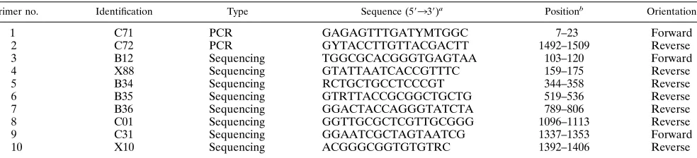
Sequencing methods.The DNA sample from the PCR was directly sequenced with a cycle sequencing kit (fmolDNA Sequencing System; Promega Corp., Madison, Wis.). The manufacturer’s protocol was followed. The eight sequencing primers are given in Table 2. Primers 3 and 4 were used for sequencing only those strains which possessed an intervening sequence inserted into the 16S rDNA at
Escherichia coliposition 210. Primers were end labeled with33P (NEN/Dupont)
according to the manufacturer’s protocol. Approximately 100 ng of purified DNA from the PCR was used for sequencing. Reaction products were loaded onto 8% polyacrylamide–urea gels, electrophoresed, and detected by exposure to X-ray film for 24 h.
16S rRNA data analysis.A program set for data entry, editing, sequence alignment, secondary structure comparison, similarity matrix generation, and dendrogram construction for 16S rRNA data was written in Microsoft Quick BASIC for use on IBM PC-AT and compatible computers (23). RNA sequences were entered and aligned as previously described (23). Our sequence database contains approximately 500 sequences determined in our laboratory and another 400 obtained from GenBank or the Ribosomal Database Project (17). Similarity matrices were constructed from the aligned sequences by using only those
se-TABLE 1. Oligonucleotide primers used for PCR amplification and sequencing of 16S rDNA
Primer no. Identification Type Sequence (59339)a Positionb Orientation
1 C71 PCR GAGAGTTTGATYMTGGC 7–23 Forward
2 C72 PCR GYTACCTTGTTACGACTT 1492–1509 Reverse
3 B12 Sequencing TGGCGCACGGGTGAGTAA 103–120 Forward
4 X88 Sequencing GTATTAATCACCGTTTC 159–175 Reverse
5 B34 Sequencing RCTGCTGCCTCCCGT 344–358 Reverse
6 B35 Sequencing GTRTTACCGCGGCTGCTG 519–536 Reverse
7 B36 Sequencing GGACTACCAGGGTATCTA 789–806 Reverse
8 C01 Sequencing GGTTGCGCTCGTTGCGGG 1096–1113 Reverse
9 C31 Sequencing GGAATCGCTAGTAATCG 1337–1353 Forward
10 X10 Sequencing ACGGGCGGTGTGTRC 1392–1406 Reverse
a
Base codes are standard International Union of Biochemistry codes for bases and ambiguity.
b
Numbering based on theE. colisequence.
on May 15, 2020 by guest
http://jcm.asm.org/
[image:2.612.64.556.81.193.2]quence positions from which 90% of the strains had data. The similarity matrices were corrected for multiple base changes at single positions by the method of Jukes and Cantor (13). Phylogenetic trees were constructed by the neighbor-joining method of Saitou and Nei (27).
Nucleotide sequence accession number.The GenBank accession numbers of all strains examined in this study are included in Fig. 3.
RESULTS
Prevalence and type of bacteria.The histologic examination and urease test revealed gastric bacteria in 100% of the dogs in groups A (31 of 31) and B (8 of 8) and 67% of the dogs in group C (10 of 15). In all cases, the bacteria were large, tightly coiled spiral organisms which were morphologically consistent with gastrospirilla. They were present in the surface mucus, gastric pits, gastric glands, and parietal cells and in cardiac, fundic, and antral regions of the stomach. Bacteria morpho-logically consistent withH. pylori, flexispiras, or other nongas-trospirilla were not seen in any dog.
Culture of gastric helicobacters, as determined by the pres-ence of urease-positive, slowly growing colonies or lawns, was successful for only six dogs: five from group A and one from group B. Of these, only five strains could be isolated in pure culture. All organisms cultured were urease positive and grew either as small, translucent colonies or as a translucent lawn on blood agar plates, and all were obligate microaerophiles. Cata-lase activity, colony morphology, and bacterial morphology varied, however (Table 2 and Fig. 1). Three were long, loosely coiled spiral organisms (7 to 10mm long) with bipolar flagellar tufts. These bacteria most resembledH. felis, except that two isolates did not contain periplasmic fibers. Subculture of the isolate with periplasmic fibers resulted in loss of the fibers (Fig. 1). The two remaining isolates were long, straight rods with numerous periplasmic fibers, characteristic of flexispiras.
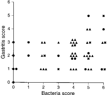
Most dogs had moderate to large numbers of bacteria in the gastric glands and on the epithelial surface (bacterial scores of 4 to 6). Group C was the only group in which there were uninfected dogs (5 of 15 dogs) and the only group in which there were dogs with bacterial scores of 1 (2 of 15 dogs). Otherwise, there were no differences in bacterial density be-tween the groups (Fig. 2).
Histologic examination.Most of the dogs had mild to mod-erate lymphocytic gastritis (scores of 1 to 3) consisting of scat-tered lymphocytes and a few plasma cells in the superficial mucosa (Fig. 2). One dog (group C) had no evidence of gas-tritis. Five dogs (two in group A, one in group B, and two in group C) had gastritis scores of 4 or 5. None of the dogs had widespread severe gastritis. Inconsistent findings were mucosal lymphoid follicles (21 of 54 dogs), increased tortuosity of glands (9 of 54 dogs, all in group A), and microerosions (1 dog in group A). The gastritis scores did not differ among the cardia, fundus, or antrum (not shown).
The five dogs with the highest gastritis score (4 to 5) had bacterial scores of 5 to 6, and the dog with the lowest gastritis score (0) had no bacteria (Fig. 2). In the other dogs, there was
no correlation between the bacterial density score and the gastritis score. Most of the dogs had gastritis scores of 2 or 3 and corresponding bacterial scores of 4 or 5, but 11 dogs had minimal gastritis (score of 1 or 2) with large numbers of bac-teria (scores of 5 to 6), and 4 dogs had few bacbac-teria (scores of 0 to 2) but moderate gastritis (score of 3). The gastritis scores of the five dogs with no bacteria ranged from 0 to 3. Finally, there was no correlation between the presence or density of bacteria and the presence of mucosal lymphoid follicles, tor-tuous glands, or other epithelial changes.
16S rDNA sequence analysis.The full sequences (bases 24 to 1491 according toE. colinumbering) were determined for each of the five dog isolates. Analysis of 16S rDNA sequences allowed placement of all five isolates into the genus
Helico-bacter. A similarity matrix was constructed by using the
se-quences of the 5 dog strains, 16 reference helicobacter strains, 1 reference campylobacter strain, and 1 reference arcobacter strain. A phylogenetic tree constructed from this matrix is shown in Fig. 3. Dog strains Dog-1, Dog-2, and Dog-3, which were morphologically similar to H. felis, were identified as
H. felis on the basis of sequence analysis. The sequences of
each of the dog strains isolated in this study were more closely related to the previously described dog strain sequence than to the cat strain sequence (24). This may reflect slight genotypic differences betweenH. felisstrains isolated from cats and those isolated from dogs. The two strains which had flexispira mor-phology represent two species. Strain Dog-5 was identified as
H. bilis(99.3% similarity). Thus,H. biliscan infect both mice
and dogs. The sequence of strain Dog-4 differs from the other helicobacter sequences in our database by about 2%, indicat-ing that it represents a novel species.
DISCUSSION
The results of this study confirm those of other studies sug-gesting that the presence of gastrospirilla is almost universal in dogs (6, 11, 12, 34). In addition, we found, as did other inves-tigators, that these large gastric spiral bacteria are the most common type of organism in dogs. Even when flexispiras were cultured from dog stomachs, gastrospirilla predominated on the basis of histologic examination. Organisms with flexispira morphology were not seen in the stomach of any dog.
It is interesting that of the 39 stomachs cultured, helico-bacters were recovered from only 6, and helicohelico-bacters mor-phologically similar to those seen histologically were isolated from only 3. This finding is consistent with previous studies suggesting that in most cases, gastrospirilla are not culturable by routine laboratory methods (16, 19, 30), although some species or strains may be recovered in culture (8, 9). The culture results suggest that in this study, the overwhelming majority of the gastric organisms in these dogs were uncultur-able gastrospirilla.
The culture of two strains ofH. felislacking in periplasmic fibers somewhat blurs the distinction betweenH. felisand
gas-TABLE 2. Morphologic and biochemical features of bacteria isolated from dog stomachs
Bacterial strain Source
Activity of: Morphology
Urease Catalase Colony Bacterial
Dog-1 Group A 1 1 Pinpoint colonies H. felis-like without periplasmic fibers
Dog-2 Group A 1 1 Pinpoint colonies H. felis-like without periplasmic fibers
Dog-3 Group B 1 1 Pinpoint colonies H. felis-like without periplasmic fibers
Dog-4 Group A 1 2 Lawn Flexispira
Dog-5 Group A 1 2 Lawn Flexispira
on May 15, 2020 by guest
http://jcm.asm.org/
[image:3.612.58.555.82.163.2]trospirilla such as H. bizzozeronii, “G. hominis 1,” and
“G. hominis2.” It is usually thought that H. felis and other
gastrospirilla may be distinguished by culturability and the presence or absence of periplasmic fibers. However, in this study, two strains ofH. felis with no periplasmic fibers were isolated, suggesting that this anatomic feature is not necessarily characteristic of H. felis. The subsequent loss of periplasmic fibers from one isolate further suggests that the presence of these fibers may not be useful for identification of these bac-teria.
Strikingly, H. pylori or H. pylori-like organisms were not
cultured or identified histologically in any of the 54 dogs in this study. This is consistent with other studies in which H. pylori
[image:4.612.57.558.68.563.2]was not described for dogs (6, 11, 12, 34). These findings are important, because they indicate that pet dogs do not represent a source of H. pylori for the human population, at least in central Ohio. Cats were not examined in this study, but other studies suggest that the prevalence of gastrospirilla and the absence ofH. pyloriin cats are similar to the findings for dogs (6, 12, 21). The only description ofH. pyloriin cats or dogs was of a single specific-pathogen-free laboratory cat colony in which gastrospirilla were absent (7). It is most likely that these
FIG. 1. Transmission electron micrograph of gastric bacteria cultured from dog stomachs. (A)H. felis-like organism with spiral morphology and periplasmic fibers (arrows). Cross-sections of flagella are visible (arrowhead). Bar, 0.25mm. (B)H. felis-like organism without periplasmic fibers. Note polar tuft of flagella (arrow). Bar, 0.35mm. (C) Flexispira-like organism with rod morphology, numerous periplasmic fibers, and polar flagella. Bar, 0.2mm.
on May 15, 2020 by guest
http://jcm.asm.org/
animals were susceptible to H. pylori because of the lack of normal gastric flora and that they became colonized by contact with a human caretaker.
The risk of transmission of gastrospirilla between pets and owners in the developed world is also likely to be low. Gastro-spirilla are found in 0.3% or less of gastroscopies, compared
with H. pylori, which is found in between 30 and 100% of
gastroscopies (31). This is in spite of the frequent occurrence of these organisms in dogs and cats, the large number of pets, and their close contact with their human companions. Thus, there is a large reservoir of gastrospirilla in close contact with
the human population, but human infection remains extremely rare.
The clinical significance of gastrospirilla in dogs was not determined with certainty by this study. Almost all of the an-imals studied were infected, and therefore, the number of negative controls was insufficient to allow comparison. This is consistent with other published studies which suggest that gas-trospirilla are almost universally present in cats and dogs (6, 11, 12, 16, 21, 34). The dogs in this study demonstrated histo-logic gastritis consistent with similar descriptions for dogs and cats in other studies (6, 11, 12, 21, 34). The lesions were similar to those described for cats experimentally infected with gas-trospirilla from cheetahs (2). It is important to note, however, that in most dogs, the lesions were mild despite the very large number of gastrospirilla in some animals. The five dogs in this study with moderate to severe gastritis also had large numbers of gastrospirilla. Thus, it is possible that these organisms in-duce gastritis in some individual animals or that certain bac-terial strains induce gastritis in dogs. However, most dogs had many bacteria and only mild gastritis, suggesting that in these animals, bacteria did not induce histologically evident disease. Finally, all of the dogs in this study either were clinically healthy or had clinical conditions which were unrelated to gastric disease. Thus, whether or not the bacteria were respon-sible for the mild gastritis seen, it is likely that large numbers of gastrospirilla are not clinically significant in healthy dogs without underlying disease.
ACKNOWLEDGMENTS
F.E.D. and B.J.P. are supported in part by NIH grants DE-07009 and DE-10374.
We thank Tom Wakefield for excellent technical assistance.
REFERENCES
1.Dewhirst, F. E., and B. J. Paster.Unpublished information.
2.Eaton, K. A., M. J. Radin, and S. Krakowka.1993. Animal models of bacterial gastritis: the role of host, bacterial species and duration of infection on severity of gastritis. Int. J. Med. Microbiol. Parasitol. Infect. Dis.280:28– 37.
3.Eaton, K. A., M. J. Radin, L. Kramer, R. Wack, R. Sherding, S. Krakowka, J. G. Fox, and D. R. Morgan.1993. Epizootic gastritis associated with gastric spiral bacilli in cheetahs (Acinonyx jubatus). Vet. Pathol.30:55–63. 4.Fox, J. G., M. Batchelder, R. Marini, L. Yan, L. Handt, X. Li, B. Shames, A.
Hayward, J. Campbell, and J. C. Murphy.1995.Helicobacter pylori-induced gastritis in the domestic cat. Infect. Immun.63:2674–2681.
5.Fox, J. G., and A. Lee.1989. Gastric campylobacter-like organisms: their role in gastric disease of laboratory animals. Lab. Anim. Sci.39:543–553. 6.Geyer, C., F. Colbatzky, J. Lechner, and W. Hermanns.1993. Occurrence of
spiral-shaped bacteria in gastric biopsies of dogs and cats. Vet. Rec.133:18– 19.
7.Handt, L. K., J. G. Fox, F. E. Dewhirst, G. J. Fraser, B. J. Paster, L. L. Yan, H. Rozmiarek, R. Rufo, and I. H. Stalis.1994.Helicobacter pyloriisolated from the domestic cat: public health implications. Infect. Immun.62:2367– 2374.
8.Ha¨nninen, M.-L., I. Happonen, S. Saari, and K. Jalava.1996. Culture and characteristics ofHelicobacter bizzozeronii, a new canine gastricHelicobacter
sp. Int. J. Syst. Bacteriol.46:160–166.
9.Hanninen, M. L., K. Jalava, S. Saari, J. Happonen, and E. Westermarck.
1995. Culture of “Gastrospirillum” from gastric biopsies of dogs. Eur. J. Clin. Microbiol. Infect. Dis.14:145–146.
10. Hazell, S. L., T. J. Borody, A. Gal, and A. Lee.1987.Campylobacter pyloridis
gastritis I: detection of urease as a marker of bacterial colonization and gastritis. Am. J. Gastroenterol.82:292–296.
11. Henry, G. A., P. H. Long, J. L. Burns, and D. L. Charbonneau.1987. Gastric spirillosis in beagles. Am. J. Vet. Res.48:831–836.
12. Hermanns, W., K. Kregel, W. Breuer, and J. Lechner.1995. Helicobacter-like organisms: histopathological examination of gastric biopsies from dogs and cats. J. Comp. Pathol.112:307–318.
13. Jukes, T. H., and C. R. Cantor.1969. Evolution of protein molecules, p. 21–132.InH. N. Munro (ed.), Mammalian protein metabolism, vol. 3. Academic Press, New York.
14. Kusukawa, N., T. Uemori, K. Asada, and I. Kato.1990. Rapid and reliable FIG. 2. Relationship between presence and density of bacteria and severity
[image:5.612.69.289.71.265.2]of gastritis. Each point represents a single dog in group A (å), B (X), or C (F).
FIG. 3. Phylogenetic tree showing the genetic relationships between the bac-teria isolated in this study and closely related helicobacter species.
on May 15, 2020 by guest
http://jcm.asm.org/
protocol for direct sequencing of material amplified by the polymerase chain reaction. BioTechniques9:66–72.
15. Lee, A., S. Krakowka, J. G. Fox, G. Otto, K. A. Eaton, and J. C. Murphy.
1992. Role ofHelicobacter felis in chronic canine gastritis. Vet. Pathol.
29:487–494.
16. Lee, A., and J. O’Rourke.1993. Gastric bacteria other thanHelicobacter pylori. Gastroenterol. Clin. North Am.22:21–42.
17. Maidak, B. L., N. Larsen, M. J. McCaughey, R. Overbeek, G. J. Olsen, K. Fogel, J. Blandy, and C. R. Woese.1994. The ribosomal database project. Nucleic Acids Res.22:3485–3487.
18. Marshall, B. J., and J. R. Warren.1984. Unidentified curved bacilli in the stomach of patients with gastritis and peptic ulceration. Lanceti:1311–1315. 19. McNulty, C. A., J. C. Dent, A. Curry, J. S. Uff, G. A. Ford, M. W. Gear, and S. P. Wilkinson.1989. New spiral bacterium in gastric mucosa. J. Clin. Pathol.42:585–591.
20. Mendes, E. N., D. M. Queiroz, G. A. Rocha, S. B. Moura, V. H. Leite, and M. E. Fonseca.1990. Ultrastructure of a spiral micro-organism from pig gastric mucosa (“Gastrospirillum suis”). J. Med. Microbiol.33:61–66. 21. Otto, G., S. H. Hazell, J. G. Fox, C. R. Howlett, J. C. Murphy, J. L. O’Rourke,
and A. Lee.1994. Animal and public health implications of gastric coloni-zation of cats byHelicobacter-like organisms. J. Clin. Microbiol.32:1043– 1049.
22. Parsonnet, J., D. Vandersteen, J. Goates, R. K. Sibley, J. Pritikin, and Y. Chang.1991. Helicobacter pylori infection in intestinal- and diffuse-type gastric adenocarcinomas. J. Natl. Cancer Inst.83:640–643.
23. Paster, B. J., and F. E. Dewhirst.1988. Phylogeny of the campylobacters, wolinellas,Bacteroides gracilis, andBacteroides ureolyticusby 16S ribosomal ribonucleic acid sequencing. Int. J. Syst. Bacteriol.38:56–62.
24. Paster, B. J., A. Lee, J. G. Fox, F. E. Dewhirst, L. A. Tordoff, G. J. Fraser, J. L. O’Rourke, N. S. Taylor, and R. Ferrero.1991. Phylogeny ofHelicobacter
felissp. nov.,Helicobacter mustelae, and related bacteria. Int. J. Syst. Bacte-riol.41:31–38.
25. Phillips, M. W., and A. Lee.1983. Isolation and characterization of a spiral bacterium from the crypts of rodent gastrointestinal tracts. Appl. Environ. Microbiol.45:675–683.
26. Radin, M. J., K. A. Eaton, S. Krakowka, D. R. Morgan, A. Lee, G. Otto, and J. Fox.1990.Helicobacter pylorigastric infection in gnotobiotic beagle dogs. Infect. Immun.58:2606–2612.
27. Saitou, N., and M. Nei.1987. The Neighbor-Joining method: a new method for reconstructing phylogenetic trees. Mol. Biol. Evol.4:406–425. 28. Sambrook, J., E. F. Fritsch, and T. Maniatis.1989. Molecular cloning: a
laboratory manual, 2nd ed. Cold Spring Harbor Laboratory Press, Cold Spring Harbor, N.Y.
29. Schauer, D. B., N. Ghori, and S. Falkow.1993. Isolation and characterization of “Flexispira rappini” from laboratory mice. J. Clin. Microbiol.31:2709– 2714.
30. Solnick, J. V., J. O’Rourke, A. Lee, B. J. Paster, F. E. Dewhirst, and L. S. Tompkins.1993. An uncultured gastric spiral organism is a newly identified Helicobacter in humans. J. Infect. Dis.168:379–385.
31. Stolte, M., E. Wellens, B. Bethke, M. Ritter, and H. Eidt.1994.Helicobacter heilmannii(formerlyGastrospirillum hominis) gastritis: an infection trans-mitted by animals? Scand. J. Gastroenterol.29:1061–1064.
32. Thomson, M. A., P. Storey, R. Greer, and G. J. Cleghorn.1994. Canine-human transmission of Gastrospirillum hominis. Lancet343:1605–1607. 33. Vandamme, P., E. Falsen, R. Rossau, B. Hoste, P. Segers, R. Tytgat, and J.
De Ley.1991. Revision ofCampylobacter,Helicobacter, andWolinella taxon-omy: emendation of generic descriptions and proposal ofArcobactergen. nov. Int. J. Syst. Bacteriol.41:88–103.
34. Weber, A. F., O. Sasa, and J. H. Sautter.1958. Some observations concern-ing the presence of spirilla in the fundic glands of dogs and cats. Am. J. Vet. Res.19:677–680.