Amendment history:
Correction
(May 1998)
HLA-DQB1 polymorphism determines
incidence, onset, and severity of
collagen-induced arthritis in transgenic mice.
Implications in human rheumatoid arthritis.
D S Bradley, … , H S Luthra, C S David
J Clin Invest.
1997;
100(9)
:2227-2234.
https://doi.org/10.1172/JCI119760
.
Certain HLA-DR alleles have been associated with predisposition to human rheumatoid
arthritis (RA). There is also evidence that certain HLA-DQ alleles may also be important in
determining susceptibility to RA. We have previously demonstrated that mice transgenic for
HLA-DQ8, a DQ allele associated with susceptibility to RA, develop severe arthritis after
type II collagen immunization. To investigate the influence of polymorphic difference at the
DQ loci on susceptibility to arthritis, we generated mice transgenic for HLA-DQ6, an allele
associated with a nonsusceptible haplotype. The DQ6 mice were found to be resistant to
collagen-induced arthritis. We also assessed the combined effect of an RA-susceptible and
an RA nonassociated DQ allele by producing double-transgenic mice expressing DQ6 and
DQ8 molecules, representing the more prevalent condition found in humans where
heterozygosity at the DQ allele is common. The double-transgenic mice developed
moderate CIA when immunized with CII when compared with the severe arthritis observed
in DQ8 transgenic mice, much like RA patients bearing both susceptible and
nonsusceptible HLA haplotypes. These studies support a role for HLA-DQ polymorphism in
human RA.
Research Article
Find the latest version:
The Journal of Clinical Investigation
Volume 100, Number 9, November 1997, 2227–2234 http://www.jci.org
HLA-DQB1 Polymorphism Determines Incidence, Onset, and Severity of
Collagen-induced Arthritis in Transgenic Mice
Implications in Human Rheumatoid Arthritis
David S. Bradley,* Gerald H. Nabozny,* Shen Cheng,* Paul Zhou,* Marie M. Griffiths,§ Harvinder S. Luthra,‡
and Chella S. David*
*Department of Immunology and ‡Department of Rheumatology, Mayo Clinic and Medical School, Rochester, Minnesota 55905; and §Research Service, Veterans Affairs Medical Center, and Department of Medicine, Division of Rheumatology, University of Utah,
Salt Lake City, Utah 84132
Abstract
Certain HLA-DR alleles have been associated with predis-position to human rheumatoid arthritis (RA). There is also evidence that certain HLA-DQ alleles may also be important in determining susceptibility to RA. We have previously dem-onstrated that mice transgenic for HLA-DQ8, a DQ allele associated with susceptibility to RA, develop severe arthritis after type II collagen immunization. To investigate the in-fluence of polymorphic difference at the DQ loci on suscep-tibility to arthritis, we generated mice transgenic for HLA-DQ6, an allele associated with a nonsusceptible haplotype. The DQ6 mice were found to be resistant to collagen-induced ar-thritis. We also assessed the combined effect of an RA-sus-ceptible and an RA nonassociated DQ allele by producing double-transgenic mice expressing DQ6 and DQ8 mole-cules, representing the more prevalent condition found in humans where heterozygosity at the DQ allele is common. The double-transgenic mice developed moderate CIA when immunized with CII when compared with the severe arthri-tis observed in DQ8 transgenic mice, much like RA patients bearing both susceptible and nonsusceptible HLA haplo-types. These studies support a role for HLA-DQ polymor-phism in human RA. (J. Clin. Invest. 1997. 100:2227–2234.)
Key words: autoimmune disease • HLA class II •
rheuma-toid arthritis • transgenic mice • type II collagen
Introduction
Certain MHC class II alleles are linked to human autoimmune diseases such as RA (1–3), insulin-dependent (type I) diabetes mellitus (2–7), systemic lupus erythematosus (1, 8–11), Sjög-ren’s syndrome (1, 10, 11), multiple sclerosis (12), Grave’s
dis-ease (5–7), and Addison’s disdis-ease (13). Although the mecha-nism(s) by which expression of certain MHC class II alleles predispose an individual to a particular autoimmune disease are not known, it is clear that increased susceptibility is associ-ated with the polymorphic regions unique to the predisposing alleles. The polymorphic differences may influence susceptibil-ity during thymic education of T cells by selection of poten-tially autoreactive T cells. Conversely, nonsusceptible class II alleles may either negatively select or fail to positively select autoreactive T cells, thereby reducing the risk of developing autoimmunity. In the periphery, the unique specificity of a class II molecule derived from a susceptible allele may be able to recognize and present an autoantigenic peptide, stimulating an undeleted group of autoimmune T cells. On the other hand, T cells directed against a bacterial or viral antigen may cross-react with an autoantigen (14). We have recently proposed that the presentation of self peptides derived from class II mol-ecules may play a role in shaping the T cell repertoire (15). This mechanism is particularly intriguing in light of evidence that a significant percentage of the naturally processed pep-tides presented by class II molecules are derived from endoge-nous class II molecules (16, 17).
Human RA is an autoimmune disease in which the associa-tion between expression of certain HLA class II alleles and the predisposition to disease has been extensively studied. Expres-sion of certain HLA-DR4 subtypes: DRB1*0401 (Dw4), DRB1*0404/0408 (Dw14), and DRB1*0405 (Dw15) have been linked with susceptibility to RA, while another HLA-DR4 subtype, DRB1*0402 (Dw10), has been shown not to be asso-ciated with RA (18–20). In addition, a DR1 subtype, DRB1* 0101 (Dw1), has also been linked to RA (21). Gregersen et al. and Winchester et al. (22, 23) observed that each of these DR alleles associated with RA contain significant homology within their third hypervariable regions (HV3), positions 67–74, and thus hypothesized that the presence of this region, termed the shared epitope region, was responsible for the predisposition to RA associated with these alleles. The mechanism by which the shared epitope region is able to predispose an individual to RA is still unclear. Additionally, further analyses of HLA class II associations with RA have shown inconsistencies to this hy-pothesis, including the association of the DR alleles DRB1* 0301 (Dw3), DRB1*0901 (Dw23), and DRB1*1001 with RA, which encodes different sequences within the shared epitope region (23–26).
Recent studies have suggested that the HLA-DQ molecule may also play a role in susceptibility to autoimmunity. Because of linkage disequilibrium, expression of certain DR and DQ alleles is frequently linked (27). This phenomenon has led to the finding that certain DQ alleles, expressed with previously identified DR alleles, are more closely associated with
suscep-The current addresses of three of the authors are as follows: G.H. Nabozny, Division of Immunological Diseases, Boehringer Ingelheim Pharmaceuticals, Inc., Ridgefield, CT 06877; S. Cheng, DEKALB Poultry Research, Inc., 3100 Sycamore Rd., Dekalb, IL 60115; and P. Zhou, Laboratory of Oral Medicine, National Institute of Dental Re-search, National Institutes of Health, 9000 Rockville Dr., Bethesda, MD 20892.
Address correspondence to Dr. Chella S. David, Department of Immunology, Mayo Clinic, Rochester, MN 55905. Phone: 507-284-8182; FAX: 507-284-1637; E-mail: david.chella@mayo.edu
tibility, or resistance, to Sjögren’s syndrome (28), myasthenia gravis (29), and insulin-dependent (type I) diabetes (30, 31). Examination of the association of DQ alleles with RA has also suggested a preferential usage of certain alleles. Two studies have reported an increased number of RA patients expressing DQ7 (32, 33) that is in linkage disequilibrium with DR4 (22). In addition, analysis of HLA class II linkage in the Asian In-dian population has shown that 100% of RA patients studied expressed the DQ8 allele (34), also in linkage disequilibrium with DR4, compared to one-third of healthy controls.
Type II collagen (CII)1 constitutes a major component
of hyaline cartilage in the joints (35). As a triple helix with a unique repeat sequence, CII could harbor several sequestered determinants, and thus harbor a potential autoantigen. Studies in RA patients have shown antibody and T cell activity di-rected to intact and denatured CII (36–38). Furthermore, in-jection of heterologous CII into rats, mice, and monkeys re-sults in joint inflammation and pathology with considerable similarity to human RA (35, 39, 40). Collagen-induced arthritis (CIA) is linked to MHC class II genes, similar to RA (41).
Recent findings by Nabozny et al. (42) using HLA-DQ8 transgenic mice in CIA also support a role for DQ in the pre-disposition to RA. These mice express DQ8 transgenes in the absence of endogenous murine class II expression, and devel-oped severe polyarthritis after immunization with CII. To determine whether polymorphism within the DQB1 locus de-termines susceptibility to CIA similar to human RA, we gener-ated transgenic mice expressing HLA-DQ6 (DQA1*0103/ DQB1*0601) gene, which is not associated with RA in hu-mans. Furthermore, since many RA patients are heterozygous at the HLA DQ loci, the influence of the nonsusceptible HLA class II molecules on an autoimmune response has not been well-defined. We generated double-transgenic mice expressing DQ6 and DQ8 molecules to simulate a heterozygous human haplotype.
Methods
Mice. Mice used in this study were bred within the pathogen-free barrier facility, and were maintained in our clean conventional area of the Mayo Immunogenetics Mouse Colony (Rochester, MN). Mice expressing HLA-DQ6 and HLA-DQ8 (42) transgenes were pro-duced by our transgenic laboratory as previously described (43). In brief, cosmid H11A, containing DQA1*0301 and DQB1*0302 genes (44), and cosmid X10A, containing DQB1*0302, were microinjected into (CBA/J 3 B10.M)F2 embryos, resulting in B10.M-DQ8
trans-genic mice. Similarly, a 40-kb construct containing DQB1*0601, de-rived from a cosmid isolate from the AKIBA cell line, was microin-jected into (CBA/J 3 B10.M)F2 embryos, producing B10.M-DQ6b
transgenic mice (45), and a construct containing the DQA1*0103 gene, also derived from the AKIBA cell line, was microinjected into (CBA/J 3 B10.M)F2 embryos, resulting in B10.M-DQ6a transgenic
mice. Each strain was bred onto a mouse class II–deficient (Ab0) background (46). The DQ6a (Ab0) and DQ6b (Ab0) mice were then mated to obtain a DQ6ab strain. The DQ6b (Ab0) mice were also mated to the DQ8ab (Ab0) mice to generate the DQ6b/8ab (Ab0) double-transgenic mice. All mice were between 6 and 12 wk of age when experiments were initiated. Both male and female mice were equally represented in each experimental cohort.
Flow cytometry. Expression of HLA-DQ and H-2 class II mole-cules on the surface of peripheral blood lymphocytes (PBLs) was an-alyzed by flow cytometry. Lymphocytes were isolated from freshly obtained whole blood on a Ficoll-Hypaque gradient, and were washed in phosphate buffered saline containing 1% BSA and 0.1% sodium azide. The cells were incubated with mAb specific for HLA-DQa (IVD12; [47]), HLA-DQ6b (L227; [48]), H-2Aab (7–16.7; [49]),
or H-2Ebb (Y-17; [50]) at 48C for 30 min, washed, and incubated with
FITC-conjugated goat Fab92 fragment specific for mouse IgG (Accu-rate Chemical & Science Corp., Westbury, NY) at 48C for 30 min. The T cell receptor (TCR) Vb usage of CD41 T cells was determined on PBLs, extensively washed, and incubated for 30 min with one of the following mAb specific for: Vb2 (B20.6.5; [51]), Vb4 (KT4; [52]), Vb5.1,2 (MR 9.4; [53]), Vb5.1 (MR 9.8; [53]), Vb6 (44.22.1; [54]), Vb7 (TR.310; [55]), Vb8.1,2,3 (F23.1: [56]), Vb8.1,2 (KJ16-133; [57]), Vb8.2 (F23.2; [58]), Vb9 (MR 10.2), Vb11 (RR-153; [59]), Vb14 (14.2; [60]), or Vb17 (KJ23a; [61]). The cells were washed and incubated with FITC-conjugated Fab92 of anti–rat IgG for 30 min. The cells were washed again and incubated with phycoerytherin-conjugated mouse anti-CD4 mAb for 30 min. All stained cells were fixed in 1% leuko-cyte formalin and analyzed using a FACS® vantage flow cytometer
(Becton Dickinson, Mountain View, CA).
Assessment of CII-specific T cell proliferation. Mice were primed with 200 mg of bovine CII emulsified 1:1 in CFA (Mycobacterium tu-berculosis H37 Ra; Difco Laboratories Inc., Detroit, MI), 100 mg in-tradermally at the base of the tail, and 50 mg in each of the hind foot-pads. 10 d later, the mice were killed, and draining lymph nodes were removed. Lymph node cells (LNCs) were purified and resuspended, 1 3
107 cells/ml, in complete medium (RPMI 1640, 5% heat inactivated
horse serum, 25 mM Hepes, 2 mM glutamine, 100 U/ml penicillin, and 100 mg/ml streptomycin; Gibco Laboratories, Grand Island, NY). 100 ml of the LNCs suspension (1 3 106 cells) was added in triplicate
to flat-bottomed microtiter plates (Corning Glass Works, Corning, NY), and was challenged with 5 mg/well heat-denatured bovine CII. Triplicate control wells were also included with LNCs cultured in me-dia alone. The cells were incubated for 2 d at 378C in 5% CO2, pulsed
with 1.8 mCi of [3H]thymidine, and incubated for an additional 18 h.
The cells were harvested, and [3H]thymidine uptake was measured as
cpm in a liquid scintillation counter (Beckman Instruments, Palo Alto, CA). To compare proliferative responses between strains of mice with different innate background cpm, proliferation was stan-dardized to a stimulation index, and calculated as follows: mean cpm of triplicate cultures with denature bovine CII 4 mean cpm of tripli-cate cultures with medium alone. Only stimulation index values of greater than 2.5 were considered to be evidence of a significant prolif-erative response.
For blocking studies, mAb specific for DQ (IVD12), HLA-DQ6b (L227), H-2Aab (7-17.7), H-2Ebb (Y-17), CD4 (GK1.5), and
CD8 (53.7.72), or an irrelevant isotype-matched mAb specific for HLA-A, -B, -C (MB40.5), was added (20 ml of culture supernatant/ well) to cultures of LNCs stimulated with bovine CII. The effect of blocking was reported as the percent of inhibition: 1 2 (Dcpm of cul-tures 1 experimental mAb) 4 (Dcpm of cultures 1 irrelevant control mAb) 3 100.
Induction of CIA. CIA was induced as previously described (42). In brief, lyophilized bovine CII (62) was reconstituted (2 mg/ml) in 0.01 N acetic acid and emulsified, 1:1, in CFA. Mice were immunized with 100 ml of the bovine CII emulsion intradermally at the base of the tail, and then boosted 28 d later with 100 mg of bovine CII emulsi-fied in incomplete Freund’s Adjuvant. Experimental mice were mon-itored at least three times a week for 12 wk after immunization, for the development of clinical arthritis. The severity of arthritis was scored for each limb as previously described (41): 0, normal, no in-flammation; 11, swelling of digits; 12, severe swelling of foot, ankle, and/or joint deformity; or 13, ankylosis of the ankle joint. Thus, each mouse had a possible score of 0 to 12.
Anti CII antibody analysis. Anti–bovine and anti–mouse CII IgG levels were measured by ELISA (63) in sera obtained 35 and 84 d af-1. Abbreviations used in this paper: AU, antibody units; CII, type II
ter bovine CII immunization. Microtiter plates were coated with ei-ther bovine or mouse CII, 6 mg/well, in KPO4 buffer, pH 7.6, and
were incubated overnight at 48C. The plates were washed with PBS-Tween (PBS 1 0.5% Tween 20 1 0.2 M NaCl) and blocked with 1% BSA in PBS-Tween. Sample sera were diluted serially, fourfold from 1:100 to 1:65,000, added to the wells in duplicate, and incubated over-night at 48C. A series of dilutions of a high titer anti-CII positive se-rum were run in parallel with all assays. The plates were washed with PBS-Tween, peroxidase-conjugated goat anti–mouse IgG (Organon Teknika Corp., West Chester, PA) was added, and the plates were incubated overnight at 48C. After washing plates with PBS-Tween, O-phenlenediamine was added, and the colorimetric change was de-termined at 410 nm. Levels of CII antibody are expressed as anti-body units (AU), calculated by comparison with the optical density ob-tained from high titer standard sera, arbitrarily determined to equal 100 AU/ml.
To determine the CII specificity of the anti–bovine CII antibod-ies, a similar ELISA technique (63) was performed with plates coated with one of the following cyanogen bromide (CB) fragments of bo-vine CII: CB8, CB9/7, CB10, CB11, or CB12. The level of anti-CII IgG specific for a particular CB fragment was expressed as percentage of the total CII antibody response: (1 2 [OD of the CB specific IgG] 4
[OD of total anti–bovine CII IgG]) 3 100.
Statistical analysis.Statistical comparisons of the arithmetic means of the arthritic severity, onset of clinical arthritis, and antibody levels were determined by the Student’s t test.
Results
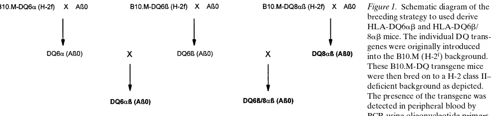
Generation of transgenic HLA-DQ6ab (Ab0) and double transgenic HLA-DQ6b/8ab (Ab0) mice. We generated mice transgenic for HLA-DQ6, an allele not associated with predis-position to RA in humans, to investigate the influence of poly-morphism in the HLA-DQ loci on susceptibility to arthritis. As outlined in Fig.1, DQ6a (Ab0) and DQ6b (Ab0) mice were mated, and offspring were screened for the presence of DQ6a and b transgenes by PCR of peripheral blood, and the expression of DQ6 molecules by flow cytometeric analyses of PBLs. Only mice bearing both DQ6a and b genes expressed DQ6 molecules on their PBLs (z 37%) (Table I). The paren-tal DQ6a and DQ6b strains are capable of expressing H-2Aab
and H-2Ebb chains within the cytoplasm (46), and to insure
that the DQ6 molecules detected in the DQ6ab mice did not result from the formation of hybrid molecules of endogenous H-2 class II chains with the corresponding DQ chain such as Aab-DQ6b or DQ6a-Ebb, DQ6 PBLs were screened for the
sur-face expression H-2Aab and H-2Ebb chains. As shown in Table
I, neither H-2Aab nor H-2Ebb molecules were undetected on
DQ6ab PBLs. Thus, DQ6ab mice express DQ6 molecules on
the surface of their PBLs in the absence of endogenous mouse class II surface expression.
A second objective of these studies was to examine the in-fluence of simultaneous expression of two DQ alleles, one as-sociated with the predisposition to RA, and one asas-sociated with resistance to RA. To this end, DQ6b (Ab0) mice were mated with DQ8ab (Ab0) mice (42) as depicted in Fig. 1. The resulting litters were screened by PCR for the presence of DQ6b, DQ8a, and DQ8b genes, and by flow cytometry for ex-pression of DQ6 and DQ8 molecules. Mice positive for the products of both transgenes were then intercrossed to develop the HLA-DQ6b/8ab (Ab0) strain (DQ6b/8ab). Representa-tive surface expression of DQ molecules on DQ6b8ab PBLs is shown in Table I. Approximately 40% of the PBLs from DQ6b/ 8ab mice express DQ molecules, comparable to DQ expres-sion on DQ6ab and DQ8ab PBLs. This result is also equiva-lent to I-Ab expression detected on B10.EaK PBLs. The DQ6
molecule was detected on 48% of PBLs from DQ6b/8ab mice, expression equivalent to DQ6ab transgenic mice. Analysis of DQ6-specific surface expression was not quantitative, and therefore, levels of total DQ and DQ6 expression cannot be compared. Neither H-2Aab nor H-2Ebb molecules were
de-tected on DQ6b/8ab PBLs (Table I), indicating the lack of both endogenous class II expression and the formation of hy-brid molecules.
Each of the DQ transgenic strains expressed a small
popu-Figure 1. Schematic diagram of the breeding strategy to used derive HLA-DQ6ab and HLA-DQ6b/ 8ab mice. The individual DQ trans-genes were originally introduced into the B10.M (H-2f) background.
[image:4.612.314.556.83.189.2]These B10.M-DQ transgene mice were then bred on to a H-2 class II– deficient background as depicted. The presence of the transgene was detected in peripheral blood by PCR using oligonucleotide primers specific for the designated DQ chain. The expression of DQ molecules and the absence of endogenous H-2 class II was determined by flow cy-tometry of PBLs, using the mAbs identified in Methods. The three strains of importance to these studies are distinguished in bold lettering.
Table I. Representative Percentages of PBLs Expressing MHC Class II Molecules in HLA-DQ Transgenic Mice
Strain
[image:4.612.60.559.597.714.2]% DQa (IVD12)*
% DQ6b (L227)
% H-2Aab
(7–16.7)
% H-2Ebb
(Y17)
HLA-DQ6ab 37.2 52.3 2‡ 2
HLA-DQ8ab 34.5 14.8 2 2
HLA-DQ6b/8ab 39.1 47.8 2 2
H-2Ab0 2 2 2 2
B10 (H-2b) 2 2 52.6 2
B10.Eak (H-2Ab/EakEbb) 2 2 38.6 29.8
LNCs were purified and assessed for the surface expression of HLA-DQ and H-2 molecules by flow cytometry as described in Methods. The values displayed are the result of pooled lymph node cells from two mice/strain, and are representative of numerous analyses. *mAb used for primary staining. ‡“2” indicates that the percentage of cells
lation of CD41 PBLs, 5.0, 6.2, and 9.3% in DQ6ab, DQ6b/ 8ab, and DQ8ab mice, respectively, compared with 12.4% in B10.T(6R) mice. Although there was a considerable difference between the size of CD41 populations detected in DQ6ab and
DQ8ab mice, the DQ6ab mice do contain sufficient numbers of CD41 cells to mount CD4-mediated immune responses di-rected against various antigens, including house dust mite, rag-weed, and rye grass allergens (64) as well as GAD peptides (65). There were differences in the percentage expression of individual Vb TCR families between these strains. Total Vb8 TCR expression was equivalent in DQ6ab, DQ6b/8ab, and DQ8ab mice, while expression of the Vb 8.1 subfamily was undetected in DQ6ab and DQ6b/8ab mice (Fig. 2).
CII-specific proliferative response by HLA-DQ6ab and DQ6b/8ab mice. To assess the role of HLA-DQ6 molecule in the recognition and presentation of bovine CII, in vitro prolif-erative assays were performed. In three separate assays, LNCs derived from bovine CII–primed DQ6ab and DQ6b/8ab mice were challenged with bovine CII. One representative prolifer-ative assay is depicted in Fig. 3. The DQ6ab LNCs did not re-spond to bovine CII. On the other hand, DQ6b/8ab LNCs did mount a significant CII-specific proliferative response, equiva-lent to DQ8ab transgenic LNCs.
The proliferative response induced by bovine CII in DQ6b/ 8ab mice was DQ8-specific and driven by CD41 cells, as shown in Fig. 4. Blocking of either DQ or CD4 molecules in-hibited CII-specific proliferation, while anti-DQ6b, anti– H-2Aab, anti–H-2Ebb, and anti-CD8 mAb had no effect on the
CII-induced response. Therefore, although the DQ6 molecule did not adequately recognize and/or present CII, its presence did not inhibit the ability of DQ8 molecules to induce a CII-specific cell-mediated response of DQ6b/8ab mice in vitro.
[image:5.612.55.299.56.289.2]Clinical parameters of CIA in HLA-DQ6ab and HLA-DQ6b/8ab mice. Three independent experiments pooled in
Figure 2.The TCR Vb8 usage in HLA-DQ6ab and HLA-DQ6b/8ab
[image:5.612.313.556.419.641.2]mice. PBLs of three DQ6ab, DQ6b/8ab, DQ8ab, and B10.T(6R) mice were assessed individually for TCR Vb 8 expression by flow cy-tometry. The expression of individual V8 subfamilies was determined subtractively from Vb expression determined by anti-Vb8.1,2,3 (F23.1), Vb8.1,2 (KJ16-133), and Vb8.2 (F23.2).
Figure 3. CII-specific proliferative response of HLA-DQ6ab and HLA-DQ6b/8ab mice. Purified LNCs of bovine CII–primed mice, pooled from three DQ6ab, DQ6b/8ab, DQ8ab, or DQ6b/8ab nega-tive mice, were challenged with bovine CII, and proliferation was de-termined by [3H]thymidine incorporation as described in Methods.
The results are displayed as a standardized stimulation index to allow for comparison of proliferation between LNCs of different strains with differing background proliferation, and are representative of three separate assays (only proliferative responses of a stimulation in-dex $ 2.5 were considered significant.)
Figure 4. Inhibition of CII-specific HLA-DQ6b/8ab LNCs prolifera-tive response. Purified LNCs from three bovine CII–primed DQ6b/ 8ab mice were cocultured with bovine CII and mAb specific for the DQa, DQ6b, H-2Aab, H-2Ebb, CD4, or CD8 molecules, or an
[image:5.612.56.300.451.632.2]Table II, containing a total of 21 DQ6ab, 14 DQ6ab negative littermates, and 19 CIA-susceptible B10.T(6R) (H-2Aq) mice
were immunized with bovine CII in CFA on day 0 and boosted with bovine CII in IFA 28 d later. The DQ6ab mice were resis-tant to CIA showing a mean incidence of 14% compared with 95% incidence in CIA-prototypic B10.T(6R) mice (P, 0.01). Only one of the three DQ6ab mice displaying disease exhib-ited severe arthritis, and the mean severity of arthritic DQ6ab
was considerably less than that of the B10.T(6R) group. To determine the effect of the expression of HLA-DQ6 molecules on HLA-DQ8–mediated arthritis, CIA was induced in cohorts of DQ6b/8ab and DQ6b/8ab-negative littermates, as well as in DQ8ab and B10.T(6R) mice. The pooled results of three separate experiments are shown in Table III. The mean incidence of arthritis in the DQ6b/8ab mice was slightly reduced compared with the DQ8ab and B10.T(6R) groups. The day of onset was delayed in DQ6b/8ab mice by an aver-age of 6 d relative to DQ8ab mice. The severity of disease was significantly reduced in DQ6b/8ab when compared with DQ8ab
mice with (P , 0.05). Although the presence of DQ6 mole-cules had no effect on in vitro CII presentation, it did cause a reduction in the incidence and severity of disease.
Anti-CII antibody levels in HLA-DQ6ab and HLA-DQ6b/ 8ab mice after CII immunization. Anti-CII IgG levels were determined in the sera of DQ6ab on days 35 and 84 after bo-vine CII immunization to determine the influence of the HLA-DQ6 molecule on the humoral CII response. An anti–bovine CII-specific IgG response was detected in DQ6ab mice (Ta-ble IV). This response, however, was significantly lower than the levels of anti–bovine CII IgG present in B10.T(6R) mice (P, 0.01 at day 35 and P, 0.10 at day 84). Immunization of DQ6ab mice with heterologous (bovine) CII also induced anti–self (mouse) CII-specific antibodies. The anti–mouse CII IgG response was also significantly less in B10.T(6R)mice (P,
0.01).
To assess the effect of expression of HLA-DQ6 in conjunc-tion with HLA-DQ8 on the strong DQ8-induced anti-CII IgG response, anti-CII levels were also measured in DQ6b/8ab
mice. Elevated levels of anti–bovine CII IgG were detected in DQ6b/8ab mice at both time points (Table IV). These levels, however, were significantly lower than antibovine CII levels in the DQ8ab and B10.T(6R) groups (P, 0.02 and P, 0.10, re-spectively). Immunization of DQ6b/8ab mice with heterolo-gous CII also induced anti–mouse CII specific antibodies.
To determine the CII specificity of the anti–bovine CII antibodies detected in DQ6ab and DQ6b/8ab mice, the sera was tested against the CB fragments of bovine CII. As with CII-immunized DQ8ab mice (42), the principle anti-CII IgG response in DQ6ab and DQ6b/8ab mice was specific for the CB10 fragment (Fig. 5). This is contrary to a predominately anti-CB11 response in B10.T(6R) mice. It is interesting that only 44% of the total anti–bovine CII response was directed against CB10 in DQ6b/8ab mice while 55% was CB10 specific in the DQ8 group, a significant reduction (P, 0.05). Thus,
z 60% of the reduction of the anti–bovine CII IgG response in
DQ6b/8ab relative to DQ8ab mice was due to a diminished anti-CB10 antibody.
Discussion
The DQ6ab mice were resistant to arthritis, with only 3 of the 21 mice immunized with bovine CII developing disease (2 mild/ 1 severe). The HLA-DQ6 gene is in linkage disequilibrium with DR2. We have shown that expression of an
HLA-Table II. Incidence, Onset, and Severity of Arthritis in CII-Immunized HLA-DQ6ab Mice
Mouse strain
Incidence of arthritis
Day of onset6SE
Peak arthritic severity6SE Arthritic/
total mice
Percent arthritic
HLA-DQ6ab 3/21‡ 14.3‡ 41611 3.060.3
HLA-DQ6ab neg 0/14 0.0 2 2
Ab0 0/5 0.0 2 2
B10.T(6R) 18/19‡ 94.7‡ 4264 6.861.1
[image:6.612.55.295.83.184.2]CIA was induced as described in Methods. Mice were monitored for 12 wk for the onset and severity of disease. Only arthritic mice were in-cluded in the computation of arthritic severity, with potential scores of 0–12/mouse. ‡P, 0.01.
Table III. Incidence, Onset, and Severity of Arthritis in CII-Immunized HLA-DQ6b/8ab Mice
Mouse strain
Incidence of arthritis
Day of onset6SE
Peak arthritic severity6SE Arthritic/
total mice
Percent arthritic
HLA-DQ6b/8ab 18/30 60.0 4966 3.461.0* HLA-DQ6b/8ab neg 0/27 0.0 2 2
HLA-DQ8ab 23/29 79.3 4365 6.060.9* B10.T(6R) 23/26 88.5 4764 4.761.0
[image:6.612.314.556.83.186.2]CIA was induced as described in Methods. Mice were monitored for 12 wk for the onset and severity of disease. Only arthritic mice were in-cluded in the computation of arthritic severity, with potential scores of 0–12/mouse. *P, 0.05.
Table IV. Anti-CII Antibody Levels in HLA-DQ6ab, HLA-DQ6b/8ab, and HLA-DQ8ab Transgenic Mice
Mouse strain
Anti–bovine CII IgG (AU/ml)
Anti–mouse CII IgG (AU/ml)
day 35 day 84 day 35 day 84
HLA-DQ6ab 41631‡ 110623§ 36625i 60613¶
HLA-DQ6ab neg 060 262 060 665 B10.T(6R) 136619‡ 170626§ 160626i 144621¶
HLA-DQ6b/8ab 136622**‡‡ 106617§§ii 5264¶¶ 130623***
HLA-DQ6b/8ab neg 563 260 160 360 HLA-DQ8ab 210622‡‡ 160623§§ 7567 153617
B10.T(6R) 196626** 174616ii 109617¶¶ 269625***
[image:6.612.317.555.529.663.2]DRB1*1502 (DR2) transgene can mediate protection to CIA (66). Taken together, these results may explain why HLA-DQ6/DR2 individuals are not predisposed to RA. We pre-dicted that double-transgenic DQ6/DR2 mice would be com-pletely resistant to CIA. There was a dichotomy between the DQ6ab humoral and cell-mediated CII-specific responses. The DQ6ab T cells failed to proliferate when challenged with bovine CII, suggesting an inability of DQ6 either to recognize or present bovine CII–derived peptides. DQ6ab mice, how-ever, did produce both heterologous and self CII–specific IgG. Therefore, even though DQ6ab B cells were stimulated by bo-vine CII, the essential contribution of T cells necessary to in-duce arthritis was lacking in DQ6ab mice.
This finding would suggest that allelic differences between DQ6 and DQ8 molecules may result in a qualitative difference in the recognition or presentation of bovine CII. One possibil-ity is that the presence of the DQ6 molecule may result in ac-tive deletion of T cells necessary for pathogenesis. This dele-tion may be mediated through the DQ6-induced deledele-tion of one or more Vb TCR families. It would be of interest to deter-mine whether deletion of the Vb 8.1 TCR subgroup in DQ6ab
mice plays a role in CIA resistance. A more plausible explana-tion for the resistance to arthritis in DQ6ab mice is that, while DQ8 molecules are able to recognize and present one or more arthritogenic epitope(s) of bovine CII, resulting in both hu-moral and cell-mediated CII-specific responses and disease, DQ6 is incapable of recognizing or presenting epitopes critical for arthritis. Thus, the expression of the DQ6 molecule would achieve resistance through a neutral or passive mechanism.
The epitope mapping of DQ6 and DQ8 transgenic mice with type II collagen is currently underway, and may be insightful as to the arthritogenic epitopes of CII.
The majority of humans are not homozygous at the HLA-DQ loci. Thus, many individuals express both a DQ allele ated with predisposition to disease, and a second allele associ-ated with resistance. We have recreassoci-ated this situation in this study. Our findings are similar to human studies, which have shown lesser severity of RA in heterozygous individuals of an RA-susceptible and nonsusceptible haplotype (67–69). The double-transgenic mice are an excellent tool with which to in-vestigate the role of the polymorphic differences in DQ in sus-ceptibility to RA, not only of CII, as described here, but also of other putative autoantigens such as proteoglycan or heat-shock proteins. These findings suggest that HLA-DQ polymorphism may play an important role in predisposition to human RA. In human RA, DQ4, 7, 8, and 9 are in linkage disequilibrium with the susceptible DRB1 alleles. All of these molecules contain an identical P1 pocket that could present an arthritogenic epitope in contrast to the nonassociated alleles, such as DQ6. Our findings suggest that the DQa chain does not play a major role. We are currently generating DQ8b/6a transgenic mice to determine whether disease is specific to DQ8ab.
On the basis of our findings so far, we propose the follow-ing model to explain how the DQ and DR genes play a role in CIA, and by deduction, human RA. The HLA class II genes expressed in the mouse in the absence of endogenous class II, shape the CD4 T cell repertoire in these mice. The mouse TCR can now interact with HLA class II 1 peptide in the periphery similar to T cells in the human. The DQ8 molecule presents an arthritogenic CII peptide, while the DQ6 molecule does not. Thus, the resistance of DQ6 is passive. We have previously re-ported that the DR2 molecule can downregulate CIA by gen-erating a self peptide, contributing to negative selection of an autoreactive T cell (66). In human RA, presentation of DRB1 HV3-derived peptide by the DQ molecules may shape the T cell repertoire (70). For example, the DRB1*0401 (QKRAA) will positively select a self-reactive T cell, while DRB1*0402 (DERAA) will negatively select the T cell. In the periphery, DQ presentation of infectious antigens will expand the autore-active T cells to initiate the inflammatory disease. In addition, the DR molecules may present heat-shock protein antigens to augment the disease (71). Thus, RA predisposition and sus-ceptibility is probably mediated by interaction of DQ/DR mol-ecules. We are currently generating double-transgenic DQ/DR mice that will further simulate a human haplotype and reveal how DQ/DR interaction may play a role in the disease process. This humanized CIA model can potentially unravel many in-triguing mysteries of how HLA class II molecules predispose individuals to RA, what may be the trigger for the onset of the disease, what determines severity, and, in the long term, poten-tial insight for therapy.
Acknowledgments
The authors thank Dr. Hidetoshi Inoko for the HLA-DQ6a and b
[image:7.612.58.299.57.296.2]cosmids, Dr. Jack Strominger for the HLA DQ8 cosmids, and Dr. Chris Benoist for the Ab0 mice. We are indebted to Dr. Jeanine Baisch for generation of the DQ.Ab0 mice, Julie Hanson and the ani-mal care technicians for the breeding and care of the mice, Michelle Smart for technical assistance, and Mary Brandt for secretarial assis-tance.
Figure 5. Specificity of anti-CII antibody responses in HLA-DQ6ab
and HLA-DQ6b/8ab mice. The levels of anti–bovine CII IgG specific for CB fragments of bovine CII were determined by ELISA on sera collected 35 d after CII immunization of DQ6ab, DQ6b/8ab, DQ8ab, and B10.T(6R) mice. The percent of total anti-CII response was calculated by the following equation: (1 2 [OD of anti-CB spe-cific response/OD of total anti-bovine CII response]) 3 100. *P,
These studies were supported by National Institutes of Health grants AI 14764 and AR 30752. M.M. Griffiths is supported by re-search funds from the Department of Veteran’s Affairs.
References
1. Goldstein, R., and F.C. Arnett. 1987. The genetics of rheumatic disease in man. Rheum. Dis. Clin. N. Am. 13:487–510.
2. Nepom, G.T. 1989. Determinants of genetic susceptibility in HLA-associ-ated autoimmune disease. Clin. Immunol. Immunopathol. 53(2 Pt 2):S53–S62.
3. Torfs, C.P., M.C. King, B. Huey, J. Malmgren, and F.C. Grumet. 1986. Genetic interrelationship between insulin-dependent diabetes mellitus, the au-toimmune thyroid diseases, and rheumatoid arthritis. Am. J. Hum. Genet. 38: 170–187.
4. Chaplin, D.D., and M.E. Kemp. 1988. The major histocompatibility com-plex and autoimmunity. Year. Immunol. 3:179–198.
5. Badenhoop, K. 1990. Immunogenetic markers for autoimmune diseases of the endocrine system. Klin. Wochenschr. 68(Suppl. 21):10–14.
6. Skanes, V.M., J. Barnard, N. Farid, W.H. Marshall, L. Murphy, D. Ride-out, R. Taylor, G. Xidos, and B. Larsen. 1986. Class III alleles and high-risk MHC haplotypes in type I diabetes mellitus, Graves’ disease and Hashimoto’s thyroiditis. Mol. Biol. Med. 3:143–157.
7. Farid, N.R., and C. Thompson. 1986. HLA and autoimmune endocrine disease 1985. Mol. Biol. Med. 3:85–97.
8. Arnett, F.C., and J.D. Reveille. 1992. Genetics of systemic lupus erythe-matosus. Rheum. Dis. Clin. North Am. 18:865–892.
9. Christiansen, F.T., W.J. Zhang, M. Griffiths, S.A. Mallal, and R.L. Daw-kins. 1991. Major histocompatibility complex (MHC) complement deficiency, ancestral haplotypes and systemic lupus erythematosus (SLE): C4 deficiency explains some but not all of the influence of the MHC. J. Rheumatol. 18:1350– 1358.
10. Skarsvag, S., K.E. Hansen, A. Holst, and T. Moen. 1992. Distribution of HLA class II alleles among Scandinavian patients with systemic lupus erythe-matosus (SLE): an increased risk of SLE among non[DRB1*03,DQA1*0501, DQB1*0201] class II homozygotes? Tissue Antigens. 40:128–133.
11. Arnett, F.C., W.B. Bias, and J.D. Reveille. 1989. Genetic studies in Sjogren’s syndrome and systemic lupus erythematosus. J. Autoimmun. 2:403–413. 12. Oksenberg, J.R., A.B. Begovich, H.A. Erlich, and L. Steinman. 1993. Genetic factors in multiple sclerosis. JAMA. 270:2362–2369.
13. Maclaren, N.K., and W.J. Riley. 1986. Inherited susceptibility to autoim-mune Addison’s disease is linked to human leukocyte antigens-DR3 and/or DR4, except when associated with type I autoimmune polyglandular syndrome. J. Clin. Endocrinol. Metab. 62:455–459.
14. Wilson, C., A. Ebringer, K. Ahmadi, J. Wrigglesworth, H. Tiwana, M. Fielder, A. Binder, C. Ettelaie, P. Cunningham, C. Joannou, and S. Bansal. 1995. Shared amino acid sequences between major histocompatibility complex class II glycoproteins, type XI collagen and Proteus mirabilis in rheumatoid ar-thritis. Ann. Rheum. Dis. 54:216–220.
15. Zanelli, E., C.J. Krco, J.M. Baisch, S. Cheng, and C.S. David. 1996. Im-mune response of HLA-DQ8 transgenic mice to peptides from the third hyper-variable region of HLA-DRB1 correlates with predisposition to rheumatoid ar-thritis. Proc. Natl. Acad. Sci. USA. 93:1814–1819.
16. Vogt, A.B., H. Kropshofer, H. Kalbacher, M. Kalbus, H.G. Ram-mensee, J.E. Coligan, and R. Martin. 1994. Ligand motifs of HLA-DRB5*0101 and DRB1*1501 molecules delineated from self-peptides. J. Immunol. 153: 1665–1673.
17. Chicz, R.M., W.S. Lane, R.A. Robinson, M. Trucco, J.L. Strominger, and J.C. Gorga. 1994. Self-peptides bound to the type I diabetes associated class II MHC molecules HLA-DQ1 and HLA-DQ8. Int. Immunol. 6:1639–1649.
18. Nepom, G.T., and H. Erlich. 1991. MHC class-II molecules and autoim-munity. Annu. Rev. Immunol. 9:493–525.
19. Wordsworth, B.P., J.S. Lanchbury, L.I. Sakkas, K.I. Welsh, G.S. Panayi, and J.I. Bell. 1989. HLA-DR4 subtype frequencies in rheumatoid arthritis indi-cate that DRB1 is the major susceptibility locus within the HLA class II region. Proc. Natl. Acad. Sci. USA. 86:10049–10053.
20. Ollier, W., and W. Thomson. 1992. Population genetics of rheumatoid arthritis. Rheum. Dis. Clin. North Am. 18:741–759.
21. Gregersen, P.K., M. Shen, Q.L. Song, P. Merryman, S. Degar, T. Seki, J. Maccari, D. Goldberg, H. Murphy, J. Schwenzer, et al. 1986. Molecular diver-sity of HLA-DR4 haplotypes. Proc. Natl. Acad. Sci. USA. 83:2642–2646.
22. Gregersen, P.K., J. Silver, and R.J. Winchester. 1987. The shared epi-tope hypothesis. An approach to understanding the molecular genetics of sus-ceptibility to rheumatoid arthritis. Arthritis Rheum. 30:1205–1209.
23. Winchester, R., E. Dwyer, and S. Rose. 1992. The genetic basis of rheu-matoid arthritis. The shared epitope hypothesis. Rheum. Dis. Clin. North Am. 18:761–783.
24. Willkens, R.F., G.T. Nepom, C.R. Marks, J.W. Nettles, and B.S. Ne-pom. 1991. Association of HLA-Dw16 with rheumatoid arthritis in Yakima In-dians. Further evidence for the “shared epitope” hypothesis. Arthritis Rheum. 34:43–47.
25. Yelamos, J., J.R. Garcia-Lozano, I. Moreno, I. Aguilera, M.F. Gonzalez, A. Garcia, A. Nunez-Roldan, and B. Sanchez. 1993. Association of HLA-DR4-Dw15 (DRB1*0405) and DR10 with rheumatoid arthritis in a Spanish popula-tion. Arthritis Rheum. 36:811–814.
26. Sattar, M.A., M. al-Saffar, R.T. Guindi, T.N. Sugathan, and K. Behbe-hani. 1990. Association between HLA-DR antigens and rheumatoid arthritis in Arabs. Ann. Rheum. Dis. 49:147–149.
27. Begovich, A.B., G.R. McClure, V.C. Suraj, R.C. Helmuth, N. Fildes, T.L. Bugawan, H.A. Erlich, and W. Klitz. 1992. Polymorphism, recombination, and linkage disequilibrium within the HLA class II region. J. Immunol. 148: 249–258.
28. Harley, J.B., M. Reichlin, F.C. Arnett, E.L. Alexander, W.B. Bias, and T.T. Provost. 1986. Gene interaction at HLA-DQ enhances autoantibody pro-duction in primary Sjogren’s syndrome. Science (Wash. DC). 232:1145–1147.
29. Bell, J., L. Rassenti, S. Smoot, K. Smith, C. Newby, R. Hohlfeld, K. Toyka, H. McDevitt, and L. Steinman. 1986. HLA-DQ beta-chain polymor-phism linked to myasthenia gravis. Lancet. 1:1058–1060.
30. Todd, J.A., J.I. Bell, and H.O. McDevitt. 1987. HLA-DQ beta gene con-tributes to susceptibility and resistance to insulin-dependent diabetes mellitus. Nature (Lond.). 329:599–604.
31. Baisch, J.M., T. Weeks, R. Giles, M. Hoover, P. Stastny, and J.D. Capra. 1990. Analysis of HLA-DQ genotypes and susceptibility in insulin-dependent diabetes mellitus. N. Eng. J. Med. 322:1836–1841.
32. Singal, D.P., M. D’Souza, B. Reid, W.G. Bensen, Y.B. Kassam, and J.D. Adachi. 1987. HLA-DQ beta-chain polymorphism in HLA-DR4 haplotypes as-sociated with rheumatoid arthritis. Lancet. 2:1118–1120.
33. Lanchbury, J.S., L.I. Sakkas, S.G. Marsh, J.G. Bodmer, K.I. Welsh, and G.S. Panayi. 1989. HLA-DQ beta 3.1 allele is a determinant of susceptibility to DR4-associated rheumatoid arthritis. Hum. Immunol. 26:59–71.
34. Taneja, V., N.K. Mehra, A.N. Chandershekaran, R.K. Ahuja, Y.N. Singh, and A.N. Malaviya. 1992. HLA-DR4-DQw8, but not DR4-DQw7 haplo-types, occur in Indian patients with rheumatoid arthritis. Rheumatol. Int. 11: 251–255.
35. Stuart, J.M., A.S. Townes, and A.H. Kang. 1984. Collagen autoimmune arthritis. Annu. Rev. Immunol. 2:199–218.
36. Stuart, J.M., E.H. Huffstutter, A.S. Townes, and A.H. Kang. 1983. Inci-dence and specificity of antibodies to types I, II, III, IV, and V collagen in rheu-matoid arthritis and other rheumatic diseases as measures by 125I-radioimmu-noassay. Arthritis Rheum. 26:832–840.
37. Trentham, D.E. 1982. Collagen arthritis as a relevant model for rheuma-toid arthritis. Arthritis Rheum. 25:911–916.
38. Trentham, D.E., G.M. Kammer, W.J. McCune, and J.R. David. 1981. Autoimmunity to collagen: a shared feature of psoriatic and rheumatoid arthri-tis. Arthritis Rheum. 24:1363–1369.
39. Trentham, D.E., A.S. Townes, and A.H. Kang. 1977. Autoimmunity to type II collagen: an experimental model of arthritis. J. Exp. Med. 146:857–868.
40. Yoo, T.J., S.Y. Kim, J.M. Stuart, R.A. Floyd, G.A. Olson, M.A. Cremer, and A.H. Kang. 1988. Induction of arthritis in monkeys by immunization with type II collagen. J. Exp. Med. 168:777–782.
41. Wooley, P.H., H.S. Luthra, J.M. Stuart, and C.S. David. 1981. Type II collagen–induced arthritis in mice. I. Major histocompatibility complex (I re-gion) linkage and antibody correlates. J. Exp. Med. 154:688–700.
42. Nabozny, G.H., J.M. Baisch, S. Cheng, D. Cosgrove, M.M. Griffiths, H.S. Luthra, and C.S. David. 1996. HLA-DQ8 transgenic mice are highly sus-ceptible to collagen-induced arthritis: a novel model for human polyarthritis. J. Exp. Med. 183:27–37.
43. Wei, B.Y., J. Martin, S. Savarirayan, R. Little, and C.S. David. 1990. Transgenic mice and mutants in MHC research. I.K. Egorov and C.S. David, editors. Springer-Verlag, Berlin, Heidelberg, Germany. 237–246.
44. Okada, K., J.M. Boss, H. Prentice, T. Spies, R. Mengler, C. Auffray, J. Lillie, D. Grossberger, and J.L. Strominger. 1985. Gene organization of DC and DX subregions of the human major histocompatibility complex. Proc. Natl. Acad. Sci. USA. 82:3410–3414.
45. Zhou, P., G.D. Anderson, S. Savarirayan, H. Inoko, and C.S. David. 1991. Thymic deletion of V beta 111, V beta 51 T cells in H-2E negative, HLA-DQ beta1 single transgenic mice. J. Immunol. 146:854–859.
46. Cosgrove, D., D. Gray, A. Dierich, J. Kaufman, M. Lemeur, C. Benoist, and D. Mathis. 1991. Mice lacking MHC class II molecules. Cell. 66:1051–1066.
47. Giles, R.C., G. Nunez, C.K. Hurley, A. Nunez-Roldan, R. Winchester, P. Stastny, and J.D. Capra. 1983. Structural analysis of a human I-A homologue using a monoclonal antibody that recognizes an MB3-like specificity. J. Exp. Med. 157:1461–1470.
48. Lampson, L.A., and R. Levy. 1980. Two populations of Ia-like mole-cules on a human B cell line. J. Immunol. 125:293–299.
49. Beck, B.N., J.M. Buerstedde, C.J. Krco, A.E. Nilson, C.G. Chase, and D.J. McKean. 1986. Characterization of cell lines expressing mutant I-Ab and I-Ak molecules allows the definition of distinct serologic epitopes on A alpha and A beta polypeptides. J. Immunol. 136:2953–2961.
50. Lerner, E.A., L.A. Matis, C.A. Janeway, Jr., P.P. Jones, R.H. Schwartz, and D.B. Murphy. 1980. Monoclonal antibody against an Ir gene product? J. Exp. Med. 152:1085–1101.
Au-phan, A. Millward, A.M. Schmitt-Verhulst, and B. Malissen. 1991. Engineered secreted T-cell receptor alpha beta heterodimers. Proc. Natl. Acad. Sci. USA. 88:8077–8081.
52. Tomonari, K., E. Lovering, and S. Spencer. 1990. Correlation between the V beta 41 CD81 T-cell population and the H-2d haplotype. Immunogenet-ics. 31:333–339.
53. Kanagawa, O., Y. Utsunomiya, J. Bill, E. Palmer, M.W. Moore, and F.R. Carbone. 1991. Conformational difference of T cell antigen receptors re-vealed by monoclonal antibodies to mouse V beta 5 T cell receptor for antigen determinants. J. Immunol. 147:1307–1314.
54. Acha-Orbea, H., R.M. Zinkernagel, and H. Hengartner. 1985. Cytotoxic T cell clone-specific monoclonal antibodies used to select clonotypic antigen-specific cytotoxic T cells. Eur. J. Immunol. 15:31–36.
55. Okada, C.Y., B. Holzmann, C. Guidos, E. Palmer, and I.L. Weissman. 1990. Characterization of a rat monoclonal antibody specific for a determinant encoded by the V beta 7 gene segment. Depletion of V beta 71 T cells in mice with Mls-1a haplotype. J. Immunol. 144:3473–3477.
56. Staerz, U.D., H.G. Rammensee, J.D. Benedetto, and M.J. Bevan. 1985. Characterization of a murine monoclonal antibody specific for an allotypic de-terminant on T cell antigen receptor. J. Immunol. 134:3994–4000.
57. Haskins, K., C. Hannum, J. White, N. Roehm, R. Kubo, J. Kappler, and P. Marrack. 1984. The antigen-specific, major histocompatibility complex-restricted receptor on T cells. VI. An antibody to a receptor allotype. J. Exp. Med. 160:452–471.
58. Kappler, J.W., U. Staerz, J. White, and P.C. Marrack. 1988. Self-toler-ance eliminates T cells specific for Mls-modified products of the major histo-compatibility complex. Nature (Lond.). 332:35–40.
59. Bill, J., O. Kanagawa, D.L. Woodland, and E. Palmer. 1989. The MHC molecule I-E is necessary but not sufficient for the clonal deletion of V beta 11-bearing T cells. J. Exp. Med. 169:1405–1419.
60. Liao, N.S., J. Maltzman, and D.H. Raulet. 1989. Positive selection deter-mines T cell receptor V beta 14 gene usage by CD81 T cells. J. Exp. Med. 170: 135–143.
61. Kappler, J.W., N. Roehm, and P. Marrack. 1987. T cell tolerance by
clonal elimination in the thymus. Cell. 49:273–280.
62. Griffiths, M.M., E.J. Eichwald, J.H. Martin, C.B. Smith, and C.W. DeWitt. 1981. Immunogenetic control of experimental type II collage induced arthritis. Arthritis Rheum. 24:781–789.
63. Griffiths, M.M., G.H. Nabozny, J. Hanson, D.S. Harper, S. McCall, K.G. Moder, G.W. Cannon, H.S. Luthra, and C.S. David. 1994. Collagen-induced ar-thritis and TCRs in SWR and B10.Q mice expressing an Ek alpha transgene. J. Immunol. 153:2758–2768.
64. Krco, C.J., S. Chapoval, J. Harders, T. Neeno, and C.S. David. 1996. Identification of HLA-DQ restricted T cell epitopes on dust mite, grass, and weed allergens using HLA-DQ6 transgenic mice. Hum. Immunol. 49:16. (Abstr.)
65. DeSouza, N.F., E. Zanelli, B. Wilson, J.L. Strominger, S.R. Munn, and C.S. David. 1996. Definition of antigenic determinants on glutamic acid decar-boxylase molecule in HLA-DQ transgenic mice. Hum. Immunol. 47:151. (Abstr.) 66. Gonzalez-Gay, M.A., E. Zanelli, S.D. Khare, C.J. Krco, P. Zhou, M.M. Griffiths, H.S. Luthra, and C.S. David. 1996. Human leukocyte antigen-DRB1*1502 (DR2Dw12) transgene reduces incidence and severity of arthritis in mice. Hum. Immunol. 50:54–60.
67. Deighton, C.M., G. Cavanagh, A.S. Rigby, H.L. Lloyd, and D.J. Walker. 1993. Both inherited HLA-haplotypes are important in the predisposition to rheumatoid arthritis. Br. J. Rheumatol. 32:893–898.
68. Wordsworth, P., K.D. Pile, J.D. Buckely, J.S. Lanchbury, B. Ollier, M. Lathrop, and J.I. Bell. 1992. HLA heterozygosity contributes to susceptibility to rheumatoid arthritis. Am. J. Hum. Genet. 51:585–591.
69. Weyand, C.M., K.C. Hicok, D.L. Conn, and J.J. Goronzy. 1992. The in-fluence of HLA-DRB1 genes on disease severity in rheumatoid arthritis. Ann. Intern. Med. 117:801–806.
70. Zanelli, E., C.J. Krco, and C.S. David. 1997. Critical residues on HLA-DRB1*0402 HV3 peptide for HLA-DQ8-restricted immunogenicity: implica-tions for rheumatoid arthritis predisposition. J. Immunol. 158:3545–3551.


![Figure 5. Specificity of anti-CII antibody responses in HLA-DQ6cific response/OD of total anti-bovine CII response]) was calculated by the following equation: (1 DQ8and HLA-DQ6for CB fragments of bovine CII were determined by ELISA on sera collected 35 d a](https://thumb-us.123doks.com/thumbv2/123dok_us/8217044.820518/7.612.58.299.57.296/specificity-responses-response-calculated-following-fragments-determined-collected.webp)
