51
CHAPTER 5
APPLIED OF IMAGE PROCESSING TECHNIQUE
ON SEMI-AUTO COUNT OF SKIN SPOT
Anis Izura Abu Zaki Nabilah Ibrahim
Suhaila Sari Lina Farhana Mahadi
Faculty of Electrical and Electronic Engineering, Universiti Tun Hussein Onn Malaysia, 86400 Batu Pahat, Johor, Malaysia
5.0 INTRODUCTION
Skin is the biggest organ in the human body and works to separate the inner body part from outer environment. In the skin, there are sebaceous glands found inside the pores of the skin. They are at all over the body except for the palms of the hands and the feet soles. There are more sebaceous glands on the face and scalp than elsewhere. Sebaceous gland secretes an oily protective skin surface, sebum, which is against pathogens and also help to slow down the skin ageing process [1]. They can help to maintain the moisture of the skin. However, the sebaceous glands become overactive sometimes, thus, producing too much sebum and the pores can get clogged together with dead skin [2][3]. This will results in having blackheads along with other factors. Blackhead is one of an acne vulgaris type [4]. It is a small dark spots on the skin that sometimes hard to be seen under a naked eye. If the clogged pores infect the glands, the accumulated sebum may form a sac and slowly increase in size. Lack of sebum production can also provide unsatisfied result that could cause dry skin, which makes the skin, looks rough and dull.
52
cheeks as the region of interest will be recorded to measure the type of the skin where the sebum clogs the pores. If the skin type is oily domain, it is assume that the production of sebum is high. Although there are several analysis related to the skin had been done using image processing, there are still a few analyses about the blackhead to be specified [5]–[9]. Previously, dermatological disease was diagnosed using color skin-images. This work has two steps, which are detecting the abnormality and identifying the diseases using color image processing technique, k-means clustering and color gradient technique [6]. This method however is not able to fully detect the tiny disease, blackheads or new acne formation, in the region of interest. Thus, an effective technique was proposed to develop a system of acne detection [8]. It was focused on binary thresholding applied to facial images with various types, shapes or amount of acne. This method is more effective than the manual counting by the dermatologist. Unfortunately, it was found to have influence of manual decision on the first stage of binary thresholding method, which could lead to the argumentation results. Thus, this work is focus on the detection of blackhead and the tendency of the production on the oily skin type and others factor such as the daily sugar intake by using image-processing techniques.
5.1 RELATION BETWEEN SEBUM AND BLACKHEAD
53
enzymes in the skin and sexual organs. More androgens that are active stimulate sebaceous gland cells to produce more sebum.
While, blackheads are small, dark lesions that appear on the skin, often on the face but they can also appear on the following body parts such as back, chest and neck. Blackhead somehow is one of an acne vulgaris type [4]. They form before bacteria invade the pores of a person skin. A blackhead can develop into a pimple, which is also known as a papule or pustule [15]. Blackhead, usually develop after puberty, when hormone levels surge and reach the skin. The presence of higher levels of hormones in the skin triggers the stimulation of the sebaceous glands, which produce oily substances, especially sebum. The sebaceous glands that produce too much sebum will lead to the accumulation of the oily substance in the pores, into the dead cells. When the clogged oil or sebum is exposed to air, it becomes black, which known as blackhead. Other than the hormone and sebum relation, there are a lot of factors that contribute to the formation of blackhead, such as sugar and dairy intake, and smoking habit.
5.2 MORPHOLOGICAL OPERATION
The morpohological operation functions in detecting the blackhead by forming its shape. Basically, the morphological operation use dilation and erosion techniques. Dilation operates by adding pixels to the boundaries of objects in an image, while erosion removes pixels on object boundaries, as shown in Figures 5.1 and 5.2. Dilation and erosion are usually used in combination for specific image pre-processing applications, such as filling holes or removing small objects. Each pixel in the image is adjusted based on the value of other pixels in their ‘neighborhood’. It works by choosing the shape and size of the ‘neighborhood’ that is needed. For example, the shape needed for the detection of blackhead is the disk shape. The structure element or ‘strel’ size of the disk is varied depending on the size of the blackhead. If the size of blackhead in the image is small, the size value of ‘strel’ would be small so that the small image of blackhead can fit into the size of ‘strel’ value.
54
Figure 5.1: Dilation of binary image
Figure 5.2: Erosion of binary image
5.3 IMAGE REGION PROPERTIES
[image:4.420.85.338.205.315.2]55
[image:5.420.63.356.33.193.2]Figure 5.3: Objects labelling using Region Properties
5.4 DATA ANALYSIS OF SKIN IMAGE
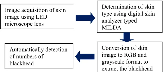
There are few steps in detecting the blackhead of skin. As shown in the flowchart of work in Figure 5.4, firstly, a phone LED microscope lens is used for images acquisition, which focused on nose and cheeks of subject face. The lens provide 60×100 magnification, which able to get a clear image of skin. Three samples of image are collected from a subject faces, which are the nose and the cheeks parts. Before taking the sample of images, subjects are advised to clean their faces an hour before the image is recorded in order to obtain the standard condition of human skin.
Figure 5.4 Flowchart of work to detect the blackhead of human skin
Image acquisition of skin image using LED microscope lens
Determination of skin type using digital skin analyzer typed MILDA
Conversion of skin image to RGB and grayscale format to extract the blackhead Automatically detection
[image:5.420.79.350.356.476.2]56
The next step is proceed with the analysing of skin face type. This step is aim to study which skin types have the tendency to produce more blackhead in skin face. Using the digital skin analyser MILDA, the analyser could detect the skin moisture content, oil content and the elasticity of the skin. The results of the skin type is displayed on the LCD screen, which the device is straightforward and simple to use besides, it only takes a short time to display the results with different colour of the background that indicate the skin condition. However, this skin analyser is very sensitive to different part of human skin. In this work, the region of interest is being narrowed by only focusing on nose and cheeks area. Thus, in order to get the accurate measurement results, the analysis must be taken in room temperature condition where the subject is controlled not to be in the sweaty and on make-up conditions.
5.5 BLACKHEAD SKIN DETECTION
Figure 5.5 shows the recorded skin images by using the LED microscope lens. Left and middle images are skin images at nose with clearly blackhead, while the right skin image is the cheeks skin image with no blackhead. All these raw images were then been converted to the RGB and grayscale images to extract the blackhead and reducing the background noise using the thresholding at level 0.56, as shown in Figure 5.6. The level was determined based on the optimum blackhead detected to avoid the false isolated blackhead.
[image:6.420.79.350.358.454.2]
57
Figure 5.6 RGB layer after thresholding
58
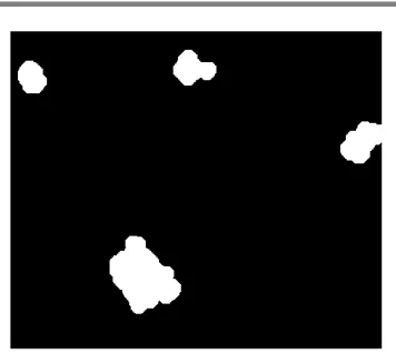
Figure 5.7 Filled detected blackhead using morphological operation
To appear the blackhead looks clearer and easily identified, a bounding box is drawn with the use of ‘regionprops’ function. This function was applied to count automatically the numbers of blackhead, simultaneously crop the region to be identified, as shown in Figure 5.8.
[image:8.420.140.315.304.459.2]59
5.6 DISCUSSION
[image:9.420.95.356.215.451.2]All the female subjects were required to clean their faces an hour before the skin type analysis and the image of skin samples were taken, while for the male subjects, the skin analysis and the image of skin samples were taken straightaway. The significant result between the female and male subjects can be seen from the number of blackhead detected on both female and male subjects as shown in Tables 5.1 and 5.2.
Table 5.1. Blackhead detected on various level of oily skin type female subject
Num Skin type oily
percentage (%) Smoking habit Blackhead detected
1 30.4 No -
2 23.3 No -
3 24.9 No -
4 26.1 No -
5 24.3 No -
6 29.6 No -
7 28.1 No -
8 27.0 No -
9 24.6 No -
10 28.6 No -
11 28.1 No -
12 29.4 No -
13 25.3 No -
14 30 No -
60
Table 5.2. Blackhead detected on various level of oily skin type male subject
Num Skin type oily
percentage (%) Smoking habit Blackhead detected
1 28.2 Yes 4
2 33.7 No -
3 26.8 Yes -
4 29.9 Yes -
5 31.4 No -
6 27.8 No -
7 31.3 No -
8 34.3 Yes 2
9 29.4 Yes -
10 27.5 No -
11 31.4 Yes -
12 35.3 No -
13 26.9 No -
14 31.3 Yes -
15 28.7 Yes -
By comparing the number of blackheads detected between female and male subjects, male subjects are more likely to have blackhead. However, the most considerable parameter is the cleaning face before the images taken. Male subjects did not clean their faces while the female subjects did. This reason could be the significance procedure that produce such the different result. In addition, there are some factors that possible be discussed which the female subjects are having a normal skin condition and they are also not a smoker, compare to male subjects who have opposite conditions. Result from Table 5.2 shows that two subjects were detected with blackheads which are the first and the eighth subjects. Both the subjects are smoking and significantly has high percentage of oily skin 34.3% for eighth subject.
61
sebum secreted. Since the data collected from the numbers of blackhead detected is not good enough which there is not much blackhead detected from all the subjects, there is a problem identified in the image acquisition method. The used of phone LED microscope lens is inadequate since it can only capture a small area of the skin. It also has disturbance in light intensity which affecting a few sample of images.
The morphological function helped to detect the blackhead in the sample of image. Moreover, the region properties function lead the detected blackhead to be counted automatically. As for the validation of the proposed technique, study are still on going to justify the values of thresholding level for RGB and the value of area of structuring element. However, the proposed technique to detect and count blackhead automatically, in this work is found to be simpler than the previous method.
ACKNOWLEDGEMENTS
This research was supported by the Ministry of Education Malaysia under Fundamental Research Grant Scheme (FRGS), Vot K048 and GPPS Vot U955.
REFERENCES
[1] M. Picardo, A. Mastrofrancesco, and T. Bíró, “Sebaceous gland-a major player in skin homoeostasis,” Exp. Dermatol., vol. 24, no. 7, pp. 485–486, 2015.
[2] M. Picardo, M. Ottaviani, E. Camera, and A. Mastrofrancesco, “Sebaceous gland lipids,” Dermatoendocrinol, 1 (2), pp. 68–71, 2009. [3] “Acne” Young Women Health Organization, September 2017. [4] J. Huamyun and A. S. Malik, “Multispectral and thermal images for acne vulgaris classification,” 2011 Natl. Postgrad. Conf. - Energy Sustain. Explor. Innov. Minds, NPC 2011, pp. 1–4, 2011.
62
images,” Proc. - Int. Conf. Mach. Learn. Cybern., vol. 5, pp. 1675–1680, 2012.
[7] G. Maroni, M. Ermidoro, and F. Previdi, “Automated Detection, Extraction and Counting of Acne Lesions for Automatic Evaluation and Tracking of Acne Severity,” 2017.
[8] T. Chantharaphaichi, B. Uyyanonvara, C. Sinthanayothin, and A. Nishihara, “Automatic acne detection for medical treatment,” 2015 6th Int. Conf. Inf. Commun. Technol. Embed. Syst. IC-ICTES 2015. [9] A. Sparavigna and R. Marazzato, “An image processing analysis of skin textures,” no. 1, pp. 1–9.
[10] Emanuela Camera, Matteo Ludovici, Sara Tortorella,Jo-Linda Sinagra, Bruno Capitanio, Laura Goracci, and Mauro Picardo, “Use of Lipidomics to Investigate Sebum Dysfunction in Juvenile Acne”, J. Lipid Res., 57 (6), pp. 1051-1058, 2016.
[11] Martini/bartholomew, “The System Introduction to the Integumentary System,” 2010.
[12] S. Titus and J. Hodge, “Diagnosis and treatment of acne,” Postgrad. Med., 86 (8), pp. 734–740, 2012.
[13] P. E. Pochi, J. S. Strauss, and D. T. Downing, “Age-related changes in sebaceous gland activity,” J. Invest. Dermatol, 73 (1), pp. 108–111, 1979.
[14] C. C. Zouboulis and A. Boschnakow, “Chronological and photoaging of the human sebaceous gland,” H+G Zeitschrift fur Hautkrankheiten, 76 (10), pp. 639–645, 2001.
[15] Ertuğrul H. Aydemir, “Acne Vulgaris”, Turk. Pediatri Ars., 49
(1), pp. 13-16, 2014.