Harnessing endogenous stem/progenitor cells
for tendon regeneration
Chang H. Lee, … , Guodong Yang, Jeremy J. Mao
J Clin Invest.
2015;125(7):2690-2701. https://doi.org/10.1172/JCI81589.
Current stem cell–based strategies for tissue regeneration involve ex vivo manipulation of
these cells to confer features of the desired progenitor population. Recently, the concept that
endogenous stem/progenitor cells could be used for regenerating tissues has emerged as a
promising approach that potentially overcomes the obstacles related to cell transplantation.
Here we applied this strategy for the regeneration of injured tendons in a rat model. First, we
identified a rare fraction of tendon cells that was positive for the known tendon stem cell
marker CD146 and exhibited clonogenic capacity, as well as multilineage differentiation
ability. These tendon-resident CD146
+stem/progenitor cells were selectively enriched by
connective tissue growth factor delivery (CTGF delivery) in the early phase of tendon
healing, followed by tenogenic differentiation in the later phase. The time-controlled
proliferation and differentiation of CD146
+stem/progenitor cells by CTGF delivery
successfully led to tendon regeneration with densely aligned collagen fibers, normal level of
cellularity, and functional restoration. Using siRNA knockdown to evaluate factors involved
in tendon generation, we demonstrated that the FAK/ERK1/2 signaling pathway regulates
CTGF-induced proliferation and differentiation of CD146
+stem/progenitor cells. Together,
our findings support the use of endogenous stem/progenitor cells as a strategy for tendon
regeneration without cell transplantation and suggest this approach warrants exploration in
other tissues.
Research Article
Stem cells
Find the latest version:
Introduction
Stem cell–based therapies have received tremendous attention in the hope of regenerating defective tissues or organs. Current stem cell–based regenerative therapies predominantly involve isolation and sorting, ex vivo culture expansion, and transplanta-tion with or without directed differentiatransplanta-tion (1–4). Despite being a valid approach, cell transplantation has encountered crucial barriers in therapeutic translation, including immune rejection; pathogen transmission; potential tumorigenesis; issues associ-ated with packaging, storage, and shipping; and difficulties in clinical adoption and regulatory approval (2, 5–8). Accordingly, a second potential therapeutic approach has been suggested by recent works showing the existence of endogenous stem cells with regenerative capacity (1, 3, 6). We and others reported promising findings that healing, repair, or regeneration can be achieved by recruiting, activating, and/or differentiating either tissue-resident or circulating stem cells, instead of stem cell transplantation necessitating ex vivo manipulation (1, 3, 6, 9). For example, synovial joint condyles were successfully regenerated in rabbits by recruiting BM and synovium mesenchymal stem cells into implanted scaffolds incorporated with growth factors (6). With a similar approach, a complex multiphase knee meniscus was regenerated in sheep by recruiting endogenous stem/progen-itor cells (9). In another study, endogenous latent transforming growth factor–β1 (TGF-β1) activated by a low-power laser suc-cessfully promoted dental pulp regeneration by differentiating
endogenous stem cells (1). Taken together, regeneration by har-nessing the regenerative potential of endogenous stem cells may serve as a straightforward strategy for regenerative medicine that may overcome the current translational hurdles associated with cell transplantation (1, 3, 10).
Here we tested the emerging idea of regeneration by endoge-nous stem/progenitor cells for treating tendon injury. Tendons are dense connective tissues with the primary function of transferring mechanical forces from muscle to bone. Tendon injuries — caused by laceration, contusion, or tensile overload — are highly preva-lent, accounting for about half of the 33 million musculoskeletal injuries in the USA (11–14). More than 30% of Americans over 60 years of age experience rotator cuff injuries, with over 50,000 of those patients undergoing surgical repair each year (15–17). Achilles tendinopathy affects 11% of regular runners (15), and 5 million new cases of tennis elbow (lateral epicondylitis) occur annually in the USA (15). Undoubtedly, tendon injuries represent an acute healthcare burden in the USA, with a total cost exceed-ing $30 billion per year (15, 18). However, tendon trauma in adults does not spontaneously heal, and scar-like tissue is frequently formed with somewhat high cellularity and disarrayed collagen fibers, failing to restore structural integrity, mechanical proper-ties, or functionality (14, 19).
Several approaches have been investigated to improve tendon healing. Natural or synthetic biomaterials have been applied as a structural tendon substitute (15, 20). Biological augmentation of tendon healing has been attempted by delivering growth factors and cytokines, including IGF-1, VEGF, bFGF, TGF-β, PDGF, GDF-5, and platelet-rich plasma (PRP). Tissue engineering strategies have also been applied to tendon healing using various cell types, Current stem cell–based strategies for tissue regeneration involve ex vivo manipulation of these cells to confer features of the
desired progenitor population. Recently, the concept that endogenous stem/progenitor cells could be used for regenerating tissues has emerged as a promising approach that potentially overcomes the obstacles related to cell transplantation. Here we applied this strategy for the regeneration of injured tendons in a rat model. First, we identified a rare fraction of tendon cells that was positive for the known tendon stem cell marker CD146 and exhibited clonogenic capacity, as well as multilineage differentiation ability. These tendon-resident CD146+ stem/progenitor cells were selectively enriched by connective tissue
growth factor delivery (CTGF delivery) in the early phase of tendon healing, followed by tenogenic differentiation in the later phase. The time-controlled proliferation and differentiation of CD146+ stem/progenitor cells by CTGF delivery successfully
led to tendon regeneration with densely aligned collagen fibers, normal level of cellularity, and functional restoration. Using siRNA knockdown to evaluate factors involved in tendon generation, we demonstrated that the FAK/ERK1/2 signaling pathway regulates CTGF-induced proliferation and differentiation of CD146+ stem/progenitor cells. Together, our findings
support the use of endogenous stem/progenitor cells as a strategy for tendon regeneration without cell transplantation and suggest this approach warrants exploration in other tissues.
Harnessing endogenous stem/progenitor cells
for tendon regeneration
Chang H. Lee,1 Francis Y. Lee,2 Solaiman Tarafder,1 Kristy Kao,1,3 Yena Jun,1,3 Guodong Yang,3 and Jeremy J. Mao2,3
1Regenerative Engineering Laboratory, 2Department of Orthopedic Surgery, and 3Center for Craniofacial Regeneration, Columbia University Medical Center, New York, New York, USA.
Conflict of interest: The authors have declared that no conflict of interest exists. Submitted: February 20, 2015; Accepted: April 30, 2015.
showed multipotentiality and the ability to form ectopic tendon-like tissue upon transplantation in vivo (35). In this study, we tar-geted the TSCs selected by the surface expression of CD146 to promote endogenous tendon regeneration. A profibrogenic cue, connective tissue growth factor (CTGF), selectively enriched the endogenous CD146+ TSCs, followed by directed tenogenic
dif-ferentiation via FAK/ERK1/2 signaling that consequently led to regeneration of transected rat patellar tendon (PT). The regener-ated tendons exhibited reorganized collagen fibers reminiscent of native tendon, substantiated by fully restored mechanical properties. Our data collectively demonstrate that the regenera-tive capacity of TSCs can be harnessed by a single growth factor delivery that may represent a simple and straightforward strat-egy for tendon regeneration by avoiding the obstacles associated with cell transplantation.
including mesenchymal stem/progenitor cells (MSCs), tenocytes, ligament fibroblasts, and dermal fibroblasts (21–28). Despite the promising improvements in healing, the previous and existing approaches somewhat failed to achieve functional restoration of ruptured tendons (29–32) or suffered from the limited availability of a potent cell source (21, 22, 33).
[image:3.585.40.396.55.532.2]Tendons in adulthood are sparsely populated by cells referred to as tenocytes, which only account for approximately 5% of the total tissue volume (15, 18, 33, 34). The primary function of teno-cytes is to maintain tissue homeostasis (33, 34). Recently, a rare population of cells in tendons was identified to possess stem/ progenitor cell properties (35, 36). The rarity of stem/progenitor cells in tendons can be appreciated in that they likely account for <1% of all cells, which together represent only 5% of total tissue volume. Culture-expanded tendon stem/progenitor cells (TSCs)
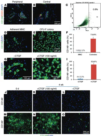
Figure 1. CD146+ cells in tendons. Immunofluorescence revealed CD146+
cells surrounding blood vessels in rat PT (A and B). Flow cytometry showed that
approximately 0.8% of isolated cells from rat PT are highly positive for CD146 (C). Approximately 1% of adherent MNCs
from PT were CD146+ (D and F), whereas
approximately 72% of CFU-F were CD146+
(n = 6 per group, P < 0.0001) (E and F).
Treatment with 100 ng/ml of CTGF sig-nificantly increased the number of CD146+
cells (G–I). In addition, in vitro fate of
CD146 expression was regulated by CTGF. CTGF treatment for 4 weeks failed to induce any CD146 expression in the sorted CD146– tendon cells (J–L). Sorted CD146+
tendon cells maintained CD146 expression with CTGF treatment by 4 weeks (M and N). However, CD146 expression was
dimin-ished in CD146+ tendon cells cultured for
4 weeks without CTGF treatment (O). All
CTGF delivery orchestrates the promoted healing of rat PT. Upon full transection of the rat PT following a well-established proto-col (Supplemental Figure 3, A–D; ref. 39), we delivered 100 ng/ml CTGF in a 200 μl fibrin gel (50 mg/ml fibrinogen and 50 U/ml thrombin), which releases 100% loaded CTGF in 5 days (Supple-mental Figure 3E). The joint was then stabilized using a cerclage suture through the tibia and quadriceps (Supplemental Figure 3D; ref. 39). Fibrin gel alone (without CTGF) led to high cellularity in the tissue that bridged the 2 transected patellar ends at 2 days (Figure 2, E and L), leading to scar-like healing at 1 week and 2 weeks post-operative (Figure 2, A, C, F, G, M, and N). Strikingly, CTGF deliv-ery led to dense and eventually aligned collagen fibers by 1 week (Figure 2, B and I) and 2 weeks (Figure 2, D and J), similar to native tissue (Figure 2K), and only modest numbers of putative inflamma-tory cells at 2 days postoperative (Figure 2H). Masson’s trichrome staining (Figure 2, L–R) showed a paucity of collagen fibers without CTGF delivery (Figure 2, L–N), but densely aligned collagen fibers were observed at 2 weeks with CTGF delivery (Figure 2Q), corre-sponding to native PT (Figure 2R). By 4 weeks postoperative, the scar-like tissue with disorganized collagen in the fibrin-alone group remained (Supplemental Figure 4A), whereas the CTGF-regener-ated tendon exhibited dense and aligned collagen structure (Sup-plemental Figure 4B) reminiscent of native tissue. Picrosirious Red staining of sections followed by polarized light microscopy further demonstrated that native-like collagen orientation was achieved in the CTGF-regenerated tendon (Supplemental Figure 4, C and D). The collagen fiber orientation was further analyzed using a digital image processing technique, as per our prior method (40). Angular deviation (AD) of collagen fibers in the CTGF-regenerated tendon was at a level similar to that seen in native tendon; in both cases, AD was much smaller than that seen in scar-like tendon healing without CTGF delivery (Supplemental Figure 4E) (n = 6 per group, P < 0.001). Macroscopically, the 4 weeks–harvested PT without CTGF showed an increase in volume as compared with healed PT with CTGF (Supplemental Figure 5). Upon mechanical testing of the 4 weeks–harvested PT (Figure 2S) at 0.25 mm/sec displace-ment, we found that CTGF delivery yielded rat PTs with a tensile stiffness on par with the native PTs’ tensile stiffness (Figure 2T), both of which were significantly higher than that of PTs treated with fibrin gel alone (without CTGF) (Figure 2T) (n = 6 tissue sam-ples per group, P < 0.05 compared with the other groups). In addi-tion, there was no significant difference between the maximum tensile force of CTGF-regenerated (39.1 ± 6.21 N) and native PT (42.1 ± 3.8 N), whereas maximum tensile force of PT healed with-out CTGF yielded 15.12 ± 8.21 N (mean ± SD, n = 6 tissue samples per group; P < 0.001). These data suggest that CTGF is pivotal and sufficient for regenerating tendons with aligned collagen fibers and tensile mechanical properties restored to the native level.
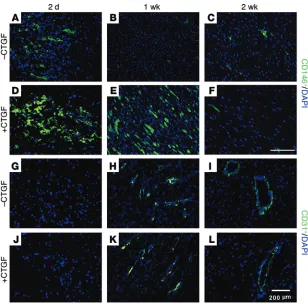
CTGF contributes to tendon regeneration by inducing a transient increase in CD146+ stem/progenitor cells. CTGF delivery induced
more CD146+ stem/progenitor cells populating the regenerating
PT by both the 2-day and 1-week timeframes following CTGF-fibrin gel delivery (Figure 3, D and E) in comparison with the fibrin gel–alone group (Figure 3, A–C). Importantly, the abun-dance of CD146+ cells was diminished by 2 weeks postoperative
(Figure 3F). Quantitatively, total cell density was significantly higher in the fibrin-alone control than in the CTGF-delivered
ten-Results
CD146+ tendon cells are clonogenic and enrichable by clonal
selec-tion and CTGF stimulaselec-tion. CD146 is one of the markers that identifies TSCs (35). In PT of skeletally mature (12-week-old) Sprague-Dawley rats, CD146+ cells are mainly perivascular,
surrounding CD31+ vascular endothelial linings both in
periph-eral (Figure 1A) and central regions (Figure 1B). FACS analysis showed that CD146+ cells in fresh-isolated rat PT only accounted
for approximately 0.8% of the total mononucleated cell (MNC) population (Figure 1C). Consistently, CD146+ cells were rare in
adherent MNCs from rat PT (Figure 1D). Interestingly, CFU of tendon cells (CFU-F) overwhelmingly expressed CD146, rep-resenting an approximately 70-fold increase over the parent MNC population in rat PT (Figure 1, E and F) (n = 6 per group, P < 0.0001). Since clonogenicity is one of the key indicators of stemness, CD146+ cells in rat PT are likely a fraction of the stem/
progenitor cells. Consistently, sorted CD146+ cells also showed
positive expression of TSC markers, including CD44 and CD90 (Supplemental Figure 1; supplemental material available online with this article; doi:10.1172/JCI81589DS1). In addition, CD146+
cells increased robustly when WT MNCs in rat PT (Figure 1G) were stimulated with 100 ng/ml CTGF for 1-week (Figure 1H), representing an approximately 20-fold increase (Figure 1I) and suggesting that CTGF selectively enriches CD146+ cells (n = 10
biological replicates per group, P < 0.001). However, CTGF treatment fails to induce CD146 expression in sorted CD146–
cells up to 4 weeks (Figure 1, J–L), suggesting that CD146+ cells
are not further differentiated cells from CD146– cells. CD146+
cells maintained CD146 expression for 2 weeks in vitro culture with 100 ng/ml CTGF treatment, whereas they lost CD146 expression without CTGF (Figure 1, M–O). We selected CTGF as a bioactive cue stimulating CD146+ cells, given our previous
works showing its capacity to induce fibroblastic differentiation of MSCs (37) and a pilot study showing its superior capacity to induce tenogenic differentiation of CD146+ tendon cells.
CD146+ tendon cells are multipotent. Isolated and
culture-expanded tendon cells and their selected fractions were cultured in osteogenic, chondrogenic, and adipogenic differentiation medium for 4 weeks and stained with Alizarin Red (AR), Safranin O (Saf-O), and Oil-Red O (ORO), respectively, following our prior methods (37, 38). The parent, WT MNCs in the rat PT differen-tiated into osteogenic cells (Supplemental Figure 2A) and adipo-genic cells (Supplemental Figure 2C), but hardly into chondroadipo-genic cells (Supplemental Figure 2B). Interestingly, colony-yielding cells of the rat PT not only showed robust chondrogenesis (Supplemen-tal Figure 2E), but also enhanced osteogenesis (Supplemen(Supplemen-tal Fig-ure 2D) and adipogenesis (Supplemental FigFig-ure 2F). CD146– cells
behaved similarly to the parent, WT MNCs from the rat PT, with modest ability to differentiate into chondrocytes (Supplemental Figure 2H). Strikingly, CD146+ cells acquired similar chondrogenic
capacity as CFU-F, colony-forming cells (Supplemental Figure 2K) in addition to their ability toward osteogenic (Supplemental Fig-ure 2J) and adipogenic (Supplemental FigFig-ure 2L) differentiation. The control represents WT rat PT cells exposed to growth medium and showed a lack of any differentiation (Supplemental Figure 2, M–O). Thus, CD146+ cells are clonogenic and multipotent,
Figure 2. CTGF-enhanced PT healing: H&E in low mag-nification, high magmag-nification, and Masson’s trichrome.
Low-magnification H&E shows scar-like tissue formation in the healing region (HR) without CTGF (A and C) whereas
the CTGF-delivered group promoted healing (B and D)
by 2 weeks. Arrows indicate uninjured tendon regions. Consistently in higher magnification, inflammatory matrix with high cell numbers was formed without CTGF (E and L), whereas CTGF attenuated inflammation (H and O) at 2
days. By 1 week, CTGF induced dense alignment of collagen fibers (I and P), in contrast to collagen-lacking scar tissue
formed without CTGF (F and M). Native-like highly aligned
collagen fibers were formed after 4 weeks CTGF delivery (J
and Q), in contrast to scar-like matrix without CTGF (G and N). Native PT sections are shown in K and R. Furthermore,
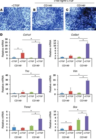
tative PCR (qPCR) demonstrated that CD146+
cells treated by 100 ng/ml CTGF for 2 weeks expressed significantly more collagen type I (Col1a1), collagen type III (Col3a1), tenascin-C (Tnc), vimentin (Vim), tenomodulin (Tnmd), and scleraxis (Scx) than CTGF-treated CD146– cells
(Figure 4D) (n = 6 biological replicates per group, P < 0.05). The tendon-related gene expression in CTGF-stimulated CD146+ cells were
approxi-mately 3- to 25-fold of CTGF-stimulated CD146–
cells, suggesting that CD146+ cells are stem/
progenitor cells that readily differentiate into tenocyte-like cells with CTGF.
CTGF promotes in vivo proliferation and dif-ferentiation of CD146+ stem/progenitor cells in
a timely manner. Our in vivo PT healing study provided an initial clue showing that CTGF may promote proliferation of CD146+ cells,
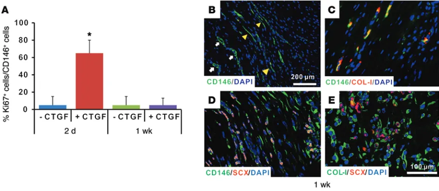
fol-lowed by differentiation. Proliferative Ki67+/
CD146+ cells were drastically increased in the
CTGF- delivered group by 2 days, in compar-ison with the fibrin-alone control (Figure 5A, P < 0.001; Supplemental Figure 8, n = 10 ran-domly selected slides per group), consistent with our in vitro data of CD146+ cell
prolifera-tion promoted by CTGF, as shown in Supple-mental Figure 7. However, the number of Ki67+/
CD146+ cells was on the level of the control
group by 1 week (Figure 5A). Interestingly, a fraction of CD146+ tendon cells was observed to
have a spindle-shaped, tenocyte-like phenotype at 1 week postoperative in the CTGF-delivered group (Figure 5B). The spindle-shaped CD146+
tendon cells co-expressed COL1A1 and SCX (Figure 5, C–E), sug-gesting that CD146+ tendon cells differentiate into tenocyte-like
cells in vivo. These data are consistent with findings shown in Figures 3 and 4 above, showing the proliferation and differenti-ation of CD146+ cells by CTGF. Although it stimulates
prolifera-tion and differentiaprolifera-tion of CD146+ TSCs, CTGF does not affect
migration of CD146+ TSCs by 24 hours CTGF (100 ng/ml)
stim-ulation, demonstrated by a boyden chamber–based migration assay (Supplemental Figure 9).
CTGF expression in CTGF-treated CD146– cells. Interestingly,
the abundant CD146– cells expressed CTGF at 1 week
postoper-ative with CTGF delivery, in contrast to the undetected CTGF expression in the fibrin-alone group (Supplemental Figure 10B). Consistently, in vitro cultured CD146– cells expressed over
3,000 times more Ctgf mRNA at 7 days after treatment with 100 ng/ml CTGF, followed by a gradual decrease over time (Sup-plemental Figure 10C), supporting our in vivo data of delayed CTGF expression in CD146– cells. Then, the paracrine effects
of CTGF-treated CD146– tendon cells on tenogenic
differentia-tion of CD146+ TSCs were confirmed by a Transwell coculture
experiment. CD146– cells treated by CTGF for 1 day were plated
in Transwell inserts equipped with 6 well plates where CD146+
TSCs were cultured. After 2 weeks, tendon-related gene expres-sion — including Col1a1, Col3a1, Tnc, Vim, Scx, and Tnmd — don by 4 weeks postoperative (Supplemental Figure 6A) (n = 10
per group: randomly selected tissue section slides, P < 0.001). At 4 weeks postoperative, total cell density in the CTGF-regenerated PT reached the level of cell density of the native PT (Supplemen-tal Figure 6A). The number of blood vessels positive to CD31 increased by 1 week, with increasing diameter by 2 weeks; there was no significant difference between groups with and without CTGF delivery (Figure 3, G–L, and Supplemental Figure 6B) (n = 10 per group: randomly selected tissue section slides, P < 0.001), sug-gesting that abundant CD146+ stem/progenitor cells upon CTGF
delivery by 1 week (Figure 3E) acted in roles beyond angiogenesis in tendon regeneration.
CTGF stimulates proliferation and tenogenic differentiation of CD146+ stem/progenitor cells. To understand the roles of the
CD146+ cells in CTGF-promoted tendon healing, we performed
in vitro studies for the proliferation and tenogenic differentiation of CD146+ cells upon CTGF treatment. During 5 days culture with
100 ng/ml CTGF, the number of CD146+ TSCs was significantly
increased, in comparison with CD146– cells (Supplemental
Fig-ure 7) (n = 6 biological replicates per group; P < 0.001). CD146+
stem/progenitor cells isolated from rat PT cells showed increased collagen deposition upon treatment with 100 ng/ml CTGF for 2 weeks (Figure 4C), in comparison with CD146– cells (Figure
4B) and MNCs without CTGF treatment (Figure 4A).
Quanti-Figure 3. Fraction of CD146+ tendon cells in vivo upon transection, followed by CTGF delivery. At 2 days and 1 week, CTGF increased the number of CD146+ cells in the healing region (D and E),
compared with without CTGF (A and B). However, the number of CD146+ cells was decreased by 2
weeks both with and without CTGF delivery (C and F). The number of blood vessels increased by 1
week after transection and decreased by 2 weeks (G–L). There was no obvious difference in blood
[image:6.585.37.345.54.358.2]was significantly increased in CD146+ TSCs by coculturing with
CTGF-treated CD146– cells, as compared with coculturing with
untreated CD146– cells (Supplemental Figure 10D) (n = 6 per
group, P < 0.001). Given the fast release of growth factors from fibrin gel (<5 days; Supplemental Figure 3E), it is postulated that CTGF-treated CD146– cells may serve as a paracrine source of
CTGF to regulate tenogenic differentiation of CD146+ cells
shown in Figure 5, B–E.
Transplantation of CTGF-pretreated CD146+ stem/progenitor
cells leads to tendon regeneration. Given the transient increase of CD146+ cells in healing tendon with CTGF (Figure 3, D–F), the
iso-lated CD146+ PT cells with or without 100 ng/ml CTGF
pretreat-ment for 1 week were delivered in the PT healing model. A total of 5 × 10⁵ cells were delivered via fibrin gel after PT transection as
described above. At 2 weeks postoperative, the transplantation of CD146+ cells pretreated by CTGF for 1 week resulted in realigned
collagen structure (Supplemental Figure 11, C and F), similar to the CTGF-regenerated tendon (Figure 2, J and Q). However,
trans-plantation of CD146+ cells without CTGF
pre-treament and CD146– cells failed to reconstruct
collagen orientation (Supplemental Figure 11, A, B, D, and E). Consistently, the tensile property of healed tendon with CTGF-treated CD146+ cells
was on the level of the native tendon, in contrast to the suboptimal properties that resulted in the groups with CD146– and untreated CD146+ cells
(Supplemental Figure 11G).
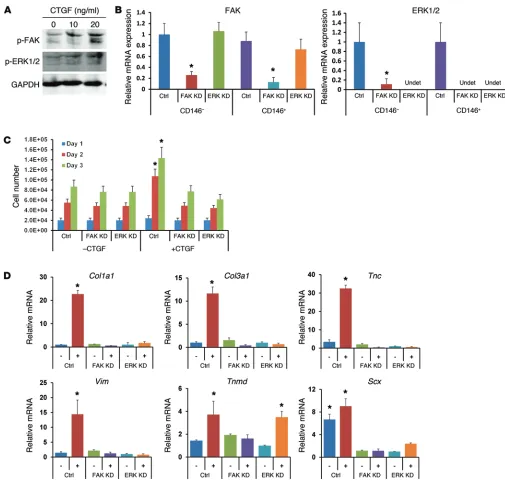
CTGF-induced proliferation and tenogenic dif-ferentiation of CD146+ cells are regulated by FAK
and ERK1/2 signaling. We have performed signal-ing studies, targetsignal-ing the FAK and ERK1/2 path-way that have been reported to be involved in CTGF-induced cell proliferation and matrix syn-thesis (41). Western blotting showed that exoge-nous CTGF treatment in CD146+ cells increased
phosphated FAK and phosphate ERK1/2 at 12 hours (Figure 6A). Then, qPCR analysis con-firmed the successful knockdown (KD) of Fak and Erk1/2 by Silencer siRNA (100 nM) and Neon Transfection System (Invitrogen) with preopti-mized electroporation conditions (1,400 V; 20 ms; 2 pulses) (Figure 6B). FAK and ERK1/2 KD by siRNA significantly reduced the proliferation of CD146+ cells promoted by 100 ng/ml CTGF
treatment (Figure 6C). CTGF-elevated expres-sion of tendon-related genes — including Col1a1, Col3a1, Tnc, Vim, Tnmd, and Scx — was significantly diminished by FAK and ERK1/2 KD by 1 week, except for Tnmd (Figure 6D) (n = 6 biological replicates per group; P < 0.01).
Discussion
Our data represent the first demonstration of tendon regenera-tion by coaxing tissue-resident stem/progenitor cells. TSCs were first identified as clonogenic and multipotent cells that can form tendon-like tissue upon ectopic implantation (35). TSCs express some MSC markers — including Stro-1, CD44, CD90, and CD146 — but exhibit distinct aspects from MSCs (35). In this study, we identified TSCs using a single surface marker expression of CD146 to minimize the technical complexity of tracking innate stem/pro-genitor cells in the tendon-healing process. Despite being less spe-cific than the previously identified TSCs, CD146+ tendon cells are
[image:7.585.46.352.60.505.2]extremely rare (~0.8%) in tendon; they exhibit clonogenic capac-ity and play essential roles in tendon regeneration. Accordingly, the CD146-based cell selection is a valid approach in identifying
Figure 4. Tenogenic differentiation of CD146+ tendon
cells. CD146+ tendon cells showed increased collagen
deposition upon CTGF treatment, compared with CD146– cells (A–C). Expression of tendon/ligament
fibroblasts–related genes — including Col1a1, Col3a1, Tnc, Vim, Tnmd, and Scx — was drastically elevated in CD146+ cells upon CTGF treatment in comparison with
CD146– cells (D) (n = 6 biological replicates per group;
innate TSCs involved in CTGF-improved tendon healing. More importantly, CD146+ TSCs are selectively enriched and
differenti-ated by a single growth factor, CTGF, which enables tendon regen-eration by a simple approach.
Fibrin gel selected as the CTGF delivery vehicle in this study is a biologically derived and FDA-approved hydrogel (42). Due to the fast gelling process from mixing fibrinogen and thrombin, fibrin gel has been applied for in situ delivering of cells and/or bio-chemical factors. As a blood plasma–derived protein, fibrin gel is biocompatible, biodegradable, and favorable for cell adhesion and infiltration (42). Despite these advantages, one limitation of fibrin gel as a delivery vehicle is the fast release rate of loaded growth factors (42). We showed that the total amount of CTGF loaded in fibrin gel was released within 5 days in vitro (Supplemental Figure 3E). In general, a growth factor loaded in fibrin gel diffuses in a few days, but the binding between fibrin’s heparin-binding domain and growth factors provide a prolonged release (43) that may affect CTGF’s release from fibrin (44). Nonetheless, a few days of release is meaningfully shorter than the 2–4 weeks or longer required for tendon healing time. Moreover, the release rate is faster in vivo than in vitro due to the enzymatic digestion of fibrin via fibrinolysis (42). Despite the short-term release, CTGF delivery induced the drastic proliferation of CD146+ TSCs in 2–7 days of in vivo tendon
healing, followed by directed tenogenic differentiation starting from 7 days (Figure 3, A–F, and Figure 5, A–E). Given the fibrin’s fast growth factor release rate, our in vitro experiments demonstrating that CTGF promotes the doubling rate and tenogenic differentia-tion of CD146+ stem/progenitor cells (Figure 4 and Supplemental
Figure 7) are not sufficient to explain the timely regulated prolif-eration and differentiation observed in the in vivo healing model.
To provide a potential explanation for the timely controlled proliferation and differentiation in vivo, we investigated a paracrine role of CD146– cells and effects of one-time CTGF stimulation in
CD146+ cells on tendon healing. Interestingly, CD146– tendon cells
stimulated by CTGF drastically increased CTGF expression both in vitro and in vivo (Supplemental Figure 10, A–C). Consistently, coculturing with CD146– cells once treated with CTGF
success-fully enhanced expression of tenogenic markers in CD146+ TSCs,
suggesting that CD146– tendon cells may act as a paracrine source
for CD146+ TSCs’ tenogenic differentiation in the later healing
phase. In addition, transplantation of CD146+ TSCs treated by
CTGF short-term was able to induce tendon regeneration (Supple-mental Figure 11, C and F), whereas scar-like tissue with disrupted collagens formed when CD146– or untreated CD146+ tendon cells
were delivered (Supplemental Figure 11, A, B, E, and F). Accord-ingly, it is postulated that CTGF stimulates proliferation of CD146+
TSCs, as well as stimulates their commitment into tendon progen-itor cells from an early time point until 7 days. Directed tenogenic differentiation of CD146+ TSCs is possibly further stimulated by
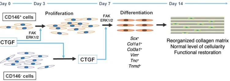
CTGF secreted from CD146– tendon cells, as summarized in
Fig-ure 7. CTGF’s delayed paracrine effect is consistent with previous findings in multiple connective tissues (45–49) but researchers pre-viously lacked an understanding of the mechanism.
The TGF-β pathway has been reported to play critical roles in tendon development (50). A recent in vitro study suggested a potential link between CTGF and BMP12 in tenogenic differenti-ation of rat flexor TSCs (51). Although we have demonstrated that the FAK/ERK1/2 pathway regulates CTGF’s function in CD146+
[image:8.585.66.498.56.244.2]TSCs using siRNA KD, it is unclear how CTGF-regulated tenogenic differentiation is linked with signaling mediated by TGF-β and/or BMPs. Follow-up investigation of CTGF’s signaling pathway and its potential crosstalk with TGF-β will likely provide an in-depth understanding of the connection between CTGF’s roles in regen-eration and development. The incomplete description of CTGF’s signaling pathway and its link with other signaling pathways is a limitation of the present study that warrants additional research. Figure 5. Proliferation and differentiation of CD146+ cells in vivo tendon healing. Ki67+ proliferative CD146+ cells were significantly increased with CTGF
delivery by 2 days in comparison with fibrin alone, but there was no difference at 1 week (A). Starting at 1 week, a fraction of CD146+ cells was observed to
have be spindle-shaped with aligned collagen fibers in the CTGF-delivered group (B), suggesting that CD146+ cells may undergo tenogenic differentiation.
The spindle-shaped tenocyte-like cells derived from CD146+ cells co-expressed COL1A1 and SCX, as demonstrated by immunofluorescence (C–E). (n = 10
tendinopathy successfully recapitulated pathological conditions in various tendons, featured by hypercellularity, disorganized collagen fibers, increased cartilaginous matrix, decreased mechanical prop-erties, and impaired healing upon acute injury (54, 55). Accordingly, follow-up studies with a pathological tendon model will be neces-sary for the translation of the present strategy into a therapy for the predominant pathological condition in human patients.
Our collective data suggest a strategy for tendon regenera-tion by harnessing the regenerative potential of tissue-resident Despite being a reproducible model for tendon regeneration
[image:9.585.39.544.52.533.2]with bioactive cues or tissue engineered constructs (52), the rat PT healing model in this study is a healthy tendon with an acute injury that may not represent a clinically relevant model (52). In addition, the incident of PT rupture is relatively low in human patients, in comparison with other tendons (53). To more closely replicate the chronic effects of tendinopathy in tendon physiology and healing, a tendinopathic condition has been created by mechanical over-use with or without an acute injury (54). The mechanically induced
Figure 6. Signaling study in CTGF-treated CD146+ tendon cells. Western Blot shows that CTGF treatment initiated FAK and ERK1/2 signaling (A). FAK and ERK1/2 were successfully KD using 100 nM Silencer siRNA with Neon system (Invitrogen) (B). FAK and ERK1/2 KD significantly attenuated the
proliferation of CD146+ cells promoted by CTGF (C). FAK and ERK1/2 KD attenuated the elevated tendon-related gene expression by CTGF — including
Col1a1, Col3a1, Tnc, Vim, and Scx — by 1 week. However, Tnmd expression was elevated with ERK1/2 KD (D). Control (Ctrl) indicates scrambled siRNA
CD146 were colabeled to identify proliferative CD146+ cells in vivo.
All primary antibodies and secondary antibodies were purchased from Abcam, Santa Cruz Biotechnology, or Invitrogen. To achieve high-quality antibody staining, the procedures for antigen retrieval, antibody dilution, and incubation duration were preoptimized as described in our prior works (6, 9, 37), and specificity of each antibody was confirmed by positive and negative controls. All images were acquired using an inverted fluorescence microscope (Olympus IX73).
CFU-F assay. For assays of colony-forming efficiency, we cultured single-cell suspensions of tendon-derived cells in a 100-mm culture dish (500 cells/dish). After 10 days, the established colonies were stained with crystal violet, and the colony-forming rates were calcu-lated. As per our prior methods (35), established clones were harvested by local application of trypsin for multilineage differentiation study.
Multilineage differentiation. For the differentiation study, P2–P3 rat tendon cells were plated in 6 wells (50,000 cells/well). Osteogenic, chondrogenic, and adipogenic differentiation were induced by applying differentiation media, as per our prior methods (6, 37, 38). Osteogenic differentiation medium contained 100 nM dexamethasone, 10 mM
β-glycerophosphate, and 0.05 mM l-ascorbic acid 2-phosphate (Sigma-
Aldrich) per our prior methods (12, 51). Chondrogenic medium was
sup-plemented with 10 ng/ml transforming growth factor–β3 (TGF-β3) (R&D
Systems). Adipogenic differentiation medium consisted of basal medium
supplemented with 0.5 μM dexamethasone, 0.5 μM
isobutylmethylxan-thine (IBMX), and 50 μM indomethacin, per our prior methods. After 4
weeks, AR, Saf-O, and ORO staining were performed to evaluate osteo-genic, chondroosteo-genic, and adipogenic differentiation, respectively.
In vitro cell proliferation. To study the effect of CTGF on cell
proliferation, sorted CD146+/– rat PT cells were plated in 6 wells at
20,000 cells/well. For up to 5 days, CD146+/– cells were cultured with
or without 100 ng/ml, and total live cell numbers were counted daily using a hemocytometer.
Animal surgery. A well-established surgical model for rat PT repair (39) was adopted, following IACUC approved protocol. Briefly, a 10-mm longitudinal incision was made just medial to the knee. Upon exposure of the PT, a full-thickness transverse incision was made using a no. 11 blade scalpel. Fibrin glue, prepared by mixing 1:1 of fibrinogen (50 mg/ml) and thrombin (50 U/ml) with or without 100 ng/ml CTGF was applied on the transection site using Fibrijet dual injector. For cell transplantation groups, the isolated and sorted
stem/progenitor cells. The extremely rare TSC population was successfully enriched by the short-term release of a single growth factor, subsequently undergoing tenogenic differentiation lead-ing to tendon regeneration via the FAK/ERK1/2 pathway. The CTGF-regenerated tendon fully restored native-like mechancial properties, collagen structure/orientation, and cellularity without cell transplantation. In conclusion, tendon regeneration by CTGF stimulation to CD146+ TSCs may represent a simple and
translat-able approach that circumvents the difficulties associated with cell transplantation and a complex delivery system.
Methods
Cell isolation and sorting. PT obtained from 12-week-old Sprague-Daw-ley rats was cleaned of the surrounding adipose tissue, leaving all other parts intact. The harvested PT was then minced and digested in 2 mg/ml collagenase at 37°C for 4 hours. The digest was centrifuged, and the pel-let was resuspended in Dulbecco’s Modified Eagle Medium-Low Glu-cose (DMEM-LG; Sigma-Aldrich) containing 10% fetal bovine serum (FBS; Atlanta Biologicals) and 1% antibiotic (1× antibiotic- antimycotic, including 10 units/l penicillin G sodium, 10 mg/ml streptomycin
sul-fate, and 0.25 μg/ml amphotericin B) (Invitrogen). At 80%–90%
con-fluence, cells were trypsinized, centrifuged, resuspended in growth
medium as passage 1 (P1) cells, and incubated in 5% CO2 at 37°C, with
fresh medium changes every 3–4 days. CD146+ cells were sorted using
BD LSR II (BD Biosciences) with P0 rat PT cells (1 × 10⁶) labeled with
1 μg of anti-CD146 antibody (ab75769) and Alexa Fluor 488 secondary
antibody (ab150077), per our prior methods (37, 56).
Immunofluorescence. CD146+ cells in ex vivo culture or tissues
were labeled using immunofluorescence following our established protocols (37, 56) using monoclonal anti-rat antibodies and iso-type-matched Alexa Fluor secondary antibodies, with nucleus
label-ing with DAPI. All the tissue sections were made in 5 μm thickness,
and the antigen retrieval procedures were performed following the manufacturer’s protocols. CD31 was labeled to identify blood ves-sels in healing tendon by 2 weeks postoperative following the same protocol. CD146 (ab75769), SCX (sc-87425), COL1A1 (ab6308), and DAPI were colabeled with multiple fluorescent secondary
antibod-ies to track tenogenic differentiation of CD146+ cells by 1 week
[image:10.585.102.478.55.187.2]post-operative. CTGF (ab5097) and CD146 (ab24577) were colabeled to identify CTGF-expressing cells in healing tendon. Similarly, Ki67 and
Figure 7. Tendon healing process by CTGF and endogenous CD146+ TSCs. Our data collectively suggest that CTGF-treated CD146+ TSCs undergo a robust
proliferation phase in 2–7 days of CTGF-initiated healing via the FAK/ERK1/2 signaling pathway. Then, the CD146+ TSCs differentiated into tenocyte-like
cells starting at 7 days, which presumably is stimulated by secreted CTGF from CD146– tendon cells. The CD146+ TSCs differentiated by CTGF consequently
(Corning, Costar) was utilized. Briefly, a total of 100,000 cells/insert
of CD146– cells treated by CTGF for 1 day were plated in Trans well
inserts (0.4 μm pores) equipped with 6 well plates where 80%–90%
confluent CD146+ TSCs were cultured. After 2 weeks culture with
media change every 3 days, total RNA was isolated from CD146+ TSCs,
and tendon-related gene expression — including Col1a1, Col3a1, Tnc, Vim, Scx, and Tnmd — was measured using qPCR as described above.
Coculturing with untreated CD146– cells was used as the control.
FAK and ERK1/2 signaling. Western blotting was first per-formed to confirm phosphorylation of FAK and ERK1/2 upon CTGF
treatment. Briefly, CD146+/– tendon cells treated with 10 or 20 ng/
ml CTGF were trypsinized and washed with ice-cold PBS. Cellular protein was extracted in RIPA Lysis Buffer (Thermo Fisher Scien-tific Inc.) with Protease/Phosphatase Inhibitor Cocktail (Cell Sig-naling Technology). Proteins were separated by SDS-PAGE, trans-ferred to nitrocellulose membrane (Bio-Rad), and detected with antiphosphorylated FAK (ab81298), antiphosphorylated ERK1/2 (sc-292838), and anti-GAPDH antibodies (sc-25778). Images were then developed with fluorescent secondary antibodies and an infrared fluorescence imaging system (Odyssey; LI-COR). Then FAK and ERK1/2 signaling were KD using Silencer siRNA (100 nM) and Neon transfection system (Invitrogen) with preoptimized electroporation conditions (1,400 V; 20 ms; 2 pulses), following the manufacturer’s protocol. Scrambled siRNA was used as the
neg-ative control. FAK and ERK1/2 KD efficiency in CD146+/– tendon
cells was confirmed using qPCR by measuring mRNA expression of Fak and Erk1/2 with commercially available primers (Invitrogen).
Then, the CD146+/– tendon cells with FAK or ERK1/2 KD were used
for in vitro proliferation and tenogenic differentiation experiments as described above.
Statistics. For all quantitative data, following confirmation of normal data distribution, 1-way ANOVA with post-hoc Tukey hon-est significant difference (HSD) thon-ests were used with P value of 0.05. Sample sizes for all quantitative data were determined by power anal-ysis with 1-way ANOVA using a level of 0.05, power of 0.8, and effect size of 1.50 that were chosen to assess matrix synthesis, gene expres-sion, and mechanical properties in the regenerated meniscus tissues and controls upon verification of normal data distribution. In case of skewed data distribution, a nonparametric test, Kruskal-Wallis 1-way ANOVA, was performed using the sample parameters. Power analy-sis performed using PASS (NCSS) indicated necessary sample sizes of 6 and 8 to achieve a power value of 0.8 for the in vitro and in vivo experiments, respectively. Expected SD and means were entered on the basis of our previous work for soft-tissue regeneration and MSC differentiations (6, 37, 38).
Study approval. All animal procedures in this study were approved by Columbia University IACUC.
Acknowledgments
We would like to thank R. Kopec for administrative assistance, and C. Chandhanayingyong and A. Hsu for surgical assistance.
Address correspondence to: Chang H. Lee, Assistant Professor and Director, Regenerative Engineering Laboratory, Columbia University Medical Center, 630 W. 168 St., VC12-230, New York, New York 10032, USA. Phone: 212.305.2480; E-mail: chl2109@ cumc.columbia.edu.
CD146– cells, CD146+ cells, and CD146+ cells that had pretreatment
with 100 ng/ml for 1 week were resuspended with thrombin solution (500,000 cells/sample) and applied into the transected site along with fibrinogen. A 2-0 Ethibond suture (Ethicon) was then passed through the tibia and quadriceps in a cerclage technique. The surgi-cal site was then closed using 4.0 absorbable (continuous stitch) for the s.c. layer, and 4.0 PDS and monocryl (interrupted stitches) for the skin closure. The power analysis described below estimated 8 as the sample number for each group and time point. Thus, 160 animals total were used for the present study.
Histology and histomorphometry. Rat PT harvested at 2 days, 1 week, 2 weeks, and 4 weeks postoperative were fixed in formalin, embedded, and sectioned for histological analysis and
immunofluo-rescence. The tissue sections were stained with H&E and Masson’s
trichrome staining, as per our prior methods (6, 9, 37, 38). Native PT from 12-week-old Sprague-Dawley rats was used as the positive con-trol. From randomly selected slides with immunofluorescence (n = 10 per group), the total number of cells, total CD31-lined blood vessels,
and number of Ki67+/CD146+ cells were counted using an imaging
processing, following our existing protocol (6, 9, 37, 38).
Collagen fiber orientation was analyzed in Picrosirious Red stained tissue sections using a digital image processing technique, as per our prior method (40). Briefly, the automated image process-ing method has been used to estimate local directionality and AD in images of oriented textiles, as well as in biological tissues and cultured cells (40). The analysis of each image yielded a distribution of fiber orientations, ranging from –90° to 90°, where 0° was defined as the vertical direction. The degree of collagen fiber alignment was quan-tified using the AD. The value of the AD was calculated using circular statistics (40). The algorithm for this process was implemented using MATLAB (The MathWorks Inc.).
In vitro tenogenic differentiation. A total of 100,000 cells/well
of CD146+/– cells were plated in 6 wells and treated with 100 ng/ml
CTGF, with media containing 50 μg/ml ascorbic acid, per our prior
methods (37). After 2 weeks, cells were fixed with formalin, and col-lagen deposition was observed by Masson’s trichrome staining. qPCR was performed to measure tendon-related gene expression — includ-ing Col1a1, Col3a1, Tnc, Vim, Tnmd, and Scx — usinclud-ing commercially available primers and StepOne System (Invitrogen).
Tensile properties of regenerated tendon. All mechanical tests were performed using Electroforce Biodynamic test system (Bose Corpora-tion) following established protocols (57, 58). The 4 weeks–harvested quadriceps-PT-tibia complexes were prepared, clamped with tensile jigs, and preconditioned for 10 cycles at 0.1 Hz between 5N and 10N while maintaining 100% humidity. Then, a constant displacement rate at 0.25 mm/sec was applied until failure. Elongation was measured by the embedded displacement sensor and a Digital Video Extensometer (DVE), and force was recorded. Then, the stiffness (N/mm) of each sample was calculated from the force-displacement curve. Native PT and healed PT without CTGF delivery served as controls.
Elevated CTGF expression in CTGF-treated CD146– cells affecting tenogenic differentiation of CD146+ TSCs. Sorted CD146– tendon cells
were plated in 6 wells (100,000 cells/well) and treated with 100 ng/ml CTGF in vitro for 1 week. Then qPCR was performed at 1, 3, 7, 10, and 14 days to measure CTGF mRNA expression, as described above. To
confirm the paracrine effects of CTGF-treated CD146– tendon cells on
1. Arany PR, et al. Photoactivation of endogenous latent transforming growth factor-beta1 directs dental stem cell differentiation for regeneration.
Sci Transl Med. 2014;6(238):238ra269.
2. Chen FM, Wu LA, Zhang M, Zhang R, Sun HH. Homing of endogenous stem/progenitor cells for in situ tissue regeneration: Promises, strategies, and translational perspectives. Biomaterials. 2011;32(12):3189–3209.
3. Miller FD, Kaplan DR. Mobilizing endogenous stem cells for repair and regeneration: are we there yet? Cell Stem Cell. 2012;10(6):650–652. 4. Vanden Berg-Foels WS. In situ tissue
regen-eration: chemoattractants for endogenous stem cell recruitment. Tissue Eng Part B Rev. 2014;20(1):28–39.
5. Fodor WL. Tissue engineering and cell based therapies, from the bench to the clinic: the poten-tial to replace, repair and regenerate. Reprod Biol
Endocrinol. 2003;1:102.
6. Lee CH, Cook JL, Mendelson A, Moioli EK, Yao H, Mao JJ. Regeneration of the articular surface of the rabbit synovial joint by cell homing: a proof of concept study. Lancet. 2010;376(9739):440–448.
7. Mao JJ, et al. Craniofacial tissue engineering by stem cells. J Dent Res. 2006;85(11):966–979. 8. Prockop DJ. Repair of tissues by adult stem/
progenitor cells (MSCs): controversies, myths, and changing paradigms. Mol Ther. 2009;17(MSCs):939–946.
9. Lee CH, Rodeo SA, Fortier LA, Lu C, Erisken C, Mao JJ. Protein-releasing polymeric scaffolds induce fibrochondrocytic differentiation of endogenous cells for knee meniscus regeneration in sheep. Sci Transl Med. 2014;6(266):266ra171. 10. Mao J, et al. Facial reconstruction by biosurgery:
cell transplantation versus cell homing. Tissue
Eng Part B Rev. 2010;16(2):257–262.
11. Fleming BC, Spindler KP, Palmer MP, Magarian EM, Murray MM. Collagen-platelet composites improve the biomechanical properties of healing anterior cruciate ligament grafts in a porcine model. Am J Sports Med. 2009;37(8):1554–1563. 12. Spindler KP, Murray MM, Devin C, Nanney LB, Davidson JM. The central ACL defect as a model for failure of intra-articular healing. J Orthop Res. 2006;24(3):401–406.
13. Tozer S, Duprez D. Tendon and ligament: devel-opment, repair and disease. Birth Defects Res C
Embryo Today. 2005;75(3):226–236.
14. Voleti PB, Buckley MR, Soslowsky LJ. Tendon healing: repair and regeneration. Annu Rev
Biomed Eng. 2012;14:47–71.
15. Chen J, Xu J, Wang A, Zheng M. Scaffolds for ten-don and ligament repair: review of the efficacy of commercial products. Expert Rev Med Devices. 2009;6(1):61–73.
16. Milgrom C, Schaffler M, Gilbert S, van Holsbeeck M. Rotator-cuff changes in asymptomatic adults. The effect of age, hand dominance and gender.
J Bone Joint Surg Br. 1995;77(2):296–298.
17. Tempelhof S, Rupp S, Seil R. Age-related prevalence of rotator cuff tears in asymp-tomatic shoulders. J Shoulder Elbow Surg. 1999;8(4):296–299.
18. Kew SJ, et al. Regeneration and repair of tendon and ligament tissue using collagen fibre
bioma-terials. Acta Biomater. 2011;7(9):3237–3247. 19. Thomopoulos S, Williams GR, Gimbel JA, Favata
M, Soslowsky LJ. Variation of biomechanical, structural, and compositional properties along the tendon to bone insertion site. J Orthop Res. 2003;21(3):413–419.
20. Kuo CK, Marturano JE, Tuan RS. Novel strat-egies in tendon and ligament tissue engineer-ing: advanced biomaterials and regeneration motifs. Sports Med Arthrosc Rehabil Ther Technol. 2010;2:20.
21. Fan H, Liu H, Toh SL, Goh JC. Anterior cruciate ligament regeneration using mesenchymal stem cells and silk scaffold in large animal model.
Bio-materials. 2009;30(28):4967–4977.
22. Fan H, Liu H, Wong EJ, Toh SL, Goh JC. In vivo study of anterior cruciate ligament regeneration using mesenchymal stem cells and silk scaffold.
Biomaterials. 2008;29(23):3324–3337.
23. Juncosa-Melvin N, Boivin GP, Galloway MT, Gooch C, West JR, Butler DL. Effects of cell-to-collagen ratio in stem cell-seeded con-structs for Achilles tendon repair. Tissue Eng. 2006;12(4):681–689.
24. Juncosa-Melvin N, et al. Effects of cell-to-col-lagen ratio in mesenchymal stem cell-seeded implants on tendon repair biomechanics and histology. Tissue Eng. 2005;11(3):448–457. 25. Juncosa-Melvin N, et al. Effects of mechanical
stimulation on the biomechanics and histol-ogy of stem cell-collagen sponge constructs for rabbit patellar tendon repair. Tissue Eng. 2006;12(8):2291–2300.
26. Nirmalanandhan VS, et al. Improving linear stiffness of the cell-seeded collagen sponge constructs by varying the components of the mechanical stimulus. Tissue Eng Part A. 2008;14(11):1883–1891.
27. Nourissat G, et al. Mesenchymal stem cell ther-apy regenerates the native bone-tendon junction after surgical repair in a degenerative rat model.
PLoS One. 2010;5(8):e12248.
28. Ouyang HW, et al. Mesenchymal stem cell sheets revitalize nonviable dense grafts: implications for repair of large-bone and tendon defects.
Trans-plantation. 2006;82(2):170–174.
29. Albano JJ, Alexander RW. Autologous fat grafting as a mesenchymal stem cell source and living bioscaffold in a patellar tendon tear. Clin J Sport
Med. 2011;21(4):359–361.
30. Awad HA, et al. Autologous mesenchymal stem cell-mediated repair of tendon. Tissue Eng. 1999;5(3):267–277.
31. Awad HA, et al. In vitro characterization of mes-enchymal stem cell-seeded collagen scaffolds for tendon repair: effects of initial seeding density on contraction kinetics. J Biomed Mater Res. 2000;51(2):233–240.
32. Bullough R, Finnigan T, Kay A, Maffulli N, Forsyth NR. Tendon repair through stem cell intervention: cellular and molecular approaches.
Disabil Rehabil. 2008;30(20):1746–1751.
33. Hsu SL, Liang R, Woo SL. Functional tissue engi-neering of ligament healing. Sports Med Arthrosc
Rehabil Ther Technol. 2010;2:12.
34. Woo SL, Jia F, Zou L, Gabriel MT. Functional tissue engineering for ligament healing: poten-tial of antisense gene therapy. Ann Biomed Eng.
2004;32(3):342–351.
35. Bi Y, et al. Identification of tendon stem/ progenitor cells and the role of the extra-cellular matrix in their niche. Nat Med. 2007;13(10):1219–1227.
36. Matsumoto T, et al. Isolation and characteriza-tion of human anterior cruciate ligament- derived vascular stem cells. Stem Cells Dev. 2012;21(6):859–872.
37. Lee CH, Shah B, Moioli EK, Mao JJ. CTGF directs fibroblast differentiation from human mesenchy-mal stem/stromesenchy-mal cells and defines connective tissue healing in a rodent injury model. J Clin
Invest. 2010;120(9):3340–3349.
38. Lee CH, Marion NW, Hollister S, Mao JJ. Tissue formation and vascularization in anatomically shaped human joint condyle ectopically in vivo.
Tissue Eng Part A. 2009;15(12):3923–3930.
39. Spang JT, et al. Platelet concentrate vs. saline in a rat patellar tendon healing model. Knee Surg
Sports Traumatol Arthrosc. 2011;19(3):495–502.
40. Lee CH, et al. Nanofiber alignment and direction of mechanical strain affect the ECM produc-tion of human ACL fibroblast. Biomaterials. 2005;26(11):1261–1270.
41. Radhakrishnan SS, et al. Effect of connective tissue growth factor on protein kinase expression and activity in human corneal fibroblasts. Invest
Ophthalmol Vis Sci. 2012;53(13):8076–8085.
42. Spicer PP, Mikos AG. Fibrin glue as a drug deliv-ery system. J Control Release. 2010;148(1):49–55. 43. Martino MM, Briquez PS, Ranga A, Lutolf
MP, Hubbell JA. Heparin-binding domain of fibrin(ogen) binds growth factors and pro-motes tissue repair when incorporated within a synthetic matrix. Proc Natl Acad Sci U S A. 2013;110(12):4563–4568.
44. Ball DK, Rachfal AW, Kemper SA, Brigstock DR. The heparin-binding 10 kDa fragment of con-nective tissue growth factor (CTGF) containing module 4 alone stimulates cell adhesion. J
Endo-crinol. 2003;176(2):R1–7.
45. Faherty N, et al. TGFβ and CCN2/CTGF mediate actin related gene expression by differential E2F1/ CREB activation. BMC Genomics. 2013;14:525. 46. Guney MA, et al. Connective tissue growth
factor acts within both endothelial cells and beta cells to promote proliferation of devel-oping beta cells. Proc Natl Acad Sci U S A. 2011;108(37):15242–15247.
47. Huang G, Brigstock DR. Regulation of hepatic stellate cells by connective tissue growth factor.
Front Biosci (Landmark Ed). 2012;17:2495–2507.
48. Koitabashi N, et al. Increased connective tissue growth factor relative to brain natriuretic peptide as a determinant of myocardial fibrosis.
Hyper-tension. 2007;49(5):1120–1127.
49. Sonnylal S, et al. Connective tissue growth factor causes EMT-like cell fate changes in vivo and in vitro. J Cell Sci. 2013;126(pt 10):2164–2175. 50. Pryce BA, Watson SS, Murchison ND, Staverosky
JA, Dunker N, Schweitzer R. Recruitment and maintenance of tendon progenitors by TGFbeta signaling are essential for tendon formation.
Development. 2009;136(8):1351–1361.
cells and signaling. Cell Physiol Biochem. 2015;35(5):1831–1845.
52. Shearn JT, et al. Tendon tissue engineering: prog-ress, challenges, and translation to the clinic. J
Mus-culoskelet Neuronal Interact. 2011;11(2):163–173.
53. Clayton RA, Court-Brown CM. The epidemiology of musculoskeletal tendinous and ligamentous injuries. Injury. 2008;39(12):1338–1344. 54. Lake SP, Ansorge HL, Soslowsky LJ. Animal
models of tendinopathy. Disabil Rehabil. 2008;30(20):1530–1541.
55. Sharma P, Maffulli N. Tendon injury and tendi-nopathy: healing and repair. J Bone Joint Surg Am. 2005;87(1):187–202.
56. Yang R, Chen M, Lee CH, Yoon R, Lal S, Mao JJ. Clones of ectopic stem cells in the regen-eration of muscle defects in vivo. PLoS One. 2010;5(10):e13547.
57. Altman GH, et al. Silk matrix for tissue engi-neered anterior cruciate ligaments. Biomaterials. 2002;23(20):4131–4141.
58. Donahue TL, Gregersen C, Hull ML, Howell SM. Comparison of viscoelastic, structural, and material properties of double-looped anterior cruciate ligament grafts made from bovine digi-tal extensor and human hamstring tendons.