http://dx.doi.org/10.4236/ojo.2015.54011
Combined Use of Percutaneous Canulated
Screws and External or Internal Fixation
for Less Invasive Treatment of Tibial
Plateaux Fractures
Konstantinos C. Xarchas1, Georgios Kyriakopoulos1, Dimitrios Mavrolias1,
Leon Oikonomou1, Ioannis Petropoulos2
1Department of Orthopaedics, Athens General Hospital Georgios Gennimatas, Athens, Greece 2Orthopaedic Surgeon, Athens, Greece
Email: drkcxr@yahoo.com
Received 25 November 2014; accepted 8 April 2015; published 14 April 2015
Copyright © 2015 by authors and Scientific Research Publishing Inc.
This work is licensed under the Creative Commons Attribution International License (CC BY).
http://creativecommons.org/licenses/by/4.0/
Abstract
The classic way of treatment of tibial plateau fractures with an extensive approach, opening of the knee and compressive internal fixation can lead to major complications such as infection, skin ne-crosis and knee stiffness. Here we present a less invasive and thus safer surgical technique, and its indications and results. Twenty patients with various types of fractures according to Schatzker’s classification (mainly V and VI) were treated during a time period of seven years. Surgical treat-ment usually consisted of a combination of percutaneous canulated screws with a hybrid external fixator. In three cases canulated screws were combined with a laterally applied anatomic locking plate. Patients were followed up for six months to three years postoperatively. Indications as well as intra and postoperative parameters such as surgical time, stability of fixation, blood loss, wound healing, infection, fracture healing and final result were studied. No major complications were recorded either early or later. The use of external οr less invasive internal fixation in com-bination with percutaneous canulated screws appears to be an adequate method for the treatment of most types of these fractures.
Keywords
Tibial Plateau Fractures, Less Invasive Surgery
1. Introduction
K. C. Xarchas et al.
by dissociation of the shaft from the condyles. Open management of these fractures, especially those classified as Schatzker VI, is usually followed by severe damage of the soft tissues, which may lead to wound complica-tions after internal fixation [1]-[3]. The use of an external hybrid ring fixator in those trauma situations is espe-cially attractive for six basic reasons: less damage of anatomical structures (the fracture is aligned and stabilized without extensive soft tissue dissection), sufficient access for debridement and other procedures, mechanical ef-fectiveness of the fixation, minimal loss of average of joint motion, early weight bearing and overall less com-plications [4]. Furthermore, in cases where a hybrid fixator cannot be used, a combination of canulated screws with a lateral anatomic locking plate applied, according to the rules of less invasive internal fixation, can be a good option [5].
2. Patients and Methods
Between 2006 and 2013, we treated twenty patients (fifteen men and five women—mean age forty years old) with various types of fractures according to Schatzker’s classification. Ten fractures were type VI, five were type V, three type IV and two type II. There were 4 open and 16 closed fractures. Open fractures were classified according to the criteria of Gustillo, three open fractures were classified as type II and one as type III a. All open fractures were treated with immediate irrigation and debridement. There were no cases with vascular damage in this series, but two suffered of peroneal nerve neurapraxia that recovered spontaneously. Ten fractures were op-erated on within eight hours after admission, six within the first 48 hours and 4 patients that suffered of exces-sive edema were operated on six days after admission. In the meantime, the limb was kept elevated in a plaster back slab and closely inspected. In all patients low molecular weight heparin was commenced on admission and continued for one month postoperatively.
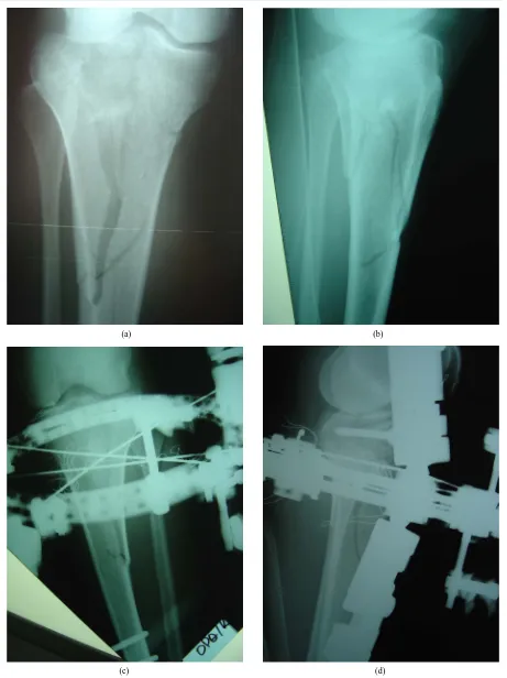
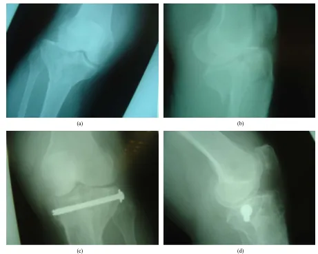
Operation was performed under epidural anesthesia. Preoperative assessment of radiographs and compute-rized tomography scans helped us achieve a good perspective of the fracture components. A tourniquet was ap-plied only if the open method was to be used. Initially, when necessary, the knee hematoma was aspirated. Op-eration was then carried out under II control and always started with the application of one or more 6.5 millime-ter canulated screws with washers, usually from the lamillime-teral side, parallel and as close to the articular surface as possible. At this stage, if needed, the articular surface was elevated by the use of a guide pin. Thus, the tibial plateau were reduced and compressed. Fixation was then completed with application of a hybrid external fixator (bridging the knee if required) or less invasive internal fixation with a limited lateral approach and an anatomic locking plate. Manual traction and various instruments (an owl or a large tenaculum reduction forceps or a trac-tor or use of a k-w like a joystick) were used to reduce the condyles [6]. Varus or valgus angulation was as-sessed very carefully and corrected. We did not perform ligament or meniscus repairs. All fractures were re-duced with the knee closed. In case of instability, the knee was bridged with an extension of the hybrid to the lateral side of the femur [7]. We used a full ring, parallel to the articular surface and centered over the tibia, al-lowing close to 90˚ of knee flexion. We always applied two wires forming an x in axial viewing. These wires were placed within anatomically safe zones at the typical positions (posterolateral to anteromedial tibia through the head of the fibula and anterolateral to posteromedial). Then a third wire was inserted, usually between the first two and parallel to the articular surface in the AP view, but occasionally parallel and above the second one.. If an olive wire were used, it was inserted until the olive contacted the bone surface to apply extra compression. The wires were then fixed to the ring and tensioned. Three peripheral pins were applied, the fixator mounted and the fracture finally manipulated, reduced and stabilized (Figures 1(a)-1(d), Figures 2(a)-2(d)).
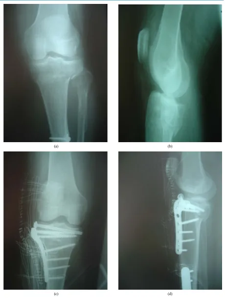
In three of our cases a hybrid fixator could not be used because of previous tibial shaft fractures that had been treated with open reduction and internal fixation—the presence of metal ware forbidding application of peri-pheral pins. In these cases, we chose to combine canulated screws with an anatomic lateral plate. Here, the ca-nulated screws were applied open, through a lateral incision, but extremely close to the articular surface to allow correct positioning of the anatomic locking plate. The knee was once more not opened and the plate applied to unite the condyles to the shaft. Careful correction of varus-valgus malalignement and accurate canulated screw positioning were always checked with the II (Figures 3(a)-3(d)).
(a) (b)
[image:3.595.83.544.78.694.2]
(c) (d)
Figure 1.(a), (b): Scatzker VI fracture. Pre operative X-rays AP and Lateral; (c), (d): Same fracture treated with
K. C. Xarchas et al.
(a) (b)
[image:4.595.80.541.80.449.2](c) (d)
Figure 2.(a), (b): Schatzker IV fracture. Pre operative X-rays AP and Lateral; (c), (d): The fracture was treated with
combina-tion of hybrid fixator with one canulated screw. X-rays AP and Lateral three years later before removal of canulated screw.
removed after complete fracture healing was confirmed, both radiologically and clinically. All fractures had united between the 12th and 16th postoperative week, at which time vigorous physiotherapy to achieve full range of knee motion was advised. Patients were usually discharged six months postoperatively.
3. Results
The study included patients from three different hospitals (two of the third level and one of the second level), and was approved by the hospitals. All patients were operated on by the same surgeon and all of them were in-vestigated retrospectively. Mean surgical time was 75 minutes (1 - 2 hours). In all cases blood loss was minimal and no blood transfusion was required because of the tibial fracture. In the three cases treated with a plate, the skin healed uneventfully. In two cases of open fractures, final skin wound closure was delayed and performed safely on the fifth postoperative day. No serious infection was observed. There were two cases with superficial pin tract infections, treated with local debridement and oral antibiotics. Two peroneal nerve palsies due to origi-nal trauma recovered on their own between three and six months after the accident. After reduction and place-ment of the fixator, an intra-articular gap or step-off usually existed, that averaged four millimeters (between 0 and 6 mm). Alignment of the shaft was within 6 degrees of the neutral. The average duration to union was from in twelve to sixteen weeks. After removal of the external fixator, there was no late tibial angulation.
(a) (b)
[image:5.595.81.541.77.687.2]
(c) (d)
Figure 3.(a), (b): Schatzker VI fracture. Pre operative X-rays AP and Lateral; (c), (d): The fracture was treated with
K. C. Xarchas et al.
and assessed anteroposterior stability with the Lachman test. Results were compared to those of the contralateral leg. The active and the passive range of motion were measured with a goniometer. All knees were grossly stable in the medio-lateral examination even those suffering of an injury of the medial collateral ligament that had re-quired bridging of the knee. Anteroposterior instability though, was quite frequent with a positive Lachman test for three, posterior drawer for one knee and one with hyperextension of 5 degrees. Average knee motion was between −5˚ (0 to −10) of extension and 110˚ of flexion (100 to 120). All three knees that were treated with the open technique, scored above average. Pain was not a major problem as no patient changed his occupation and habits because of it.
4. Discussion
The purpose of this study was to investigate whether our methods of less invasive treatment of severe tibial pla-teau fractures were as effective as the traditional ones. If such was the case, we thought that our approach would compare favorably to them, because it caused less damage to the anatomical structures, provided sufficient access for debridement and other procedures together with a mechanically effective fixation, allowed minimal loss of joint motion with early weight bearing, and overall caused less complications [8] [9]. Traditionally, open reduction with restoration of the articular surface has been the most common treatment for displaced fractures of the tibial plateau [10]-[12]. Later on, hybrid or Ilizarov ring fixation has been introduced and extensively studied for the treatment of such fractures [4] [9] [12]. On the contrary, only a handful of studies have recently described the combined usage of hybrid external fixator and minimal internal fixation with canulated screws as well as the combined use of an anatomical plate with canulated screws [13]-[17]. We used canulated screws together with a hybrid ring fixator or, if this was not possible, with an anatomic plate for reduction and adequate fixation. Common partially threaded cancellous screws can be used, but placement of canulated screws offers safety and precision. External fixation offers an atraumatic approach for the treatment of these severe fractures whereas a laterally applied anatomic locking plate offers the least invasive option available. In both cases treatment of the articular surface is with closed reduction and canulated screws, a technique that is well accepted in the literature
[18]. The metaphyseal and diaphyseal portion of the fracture can then be reduced and fixed with either external fixation or minimal internal fixation [19].
Collection of data from series mainly comprising severe fractures has shown that our results compare well both with published reports of more traditional methods [2] [3] [10] [11] and the latest minimal invasive trends
[4] [9] [12] [20]-[25]. When compared to the classical unilateral fixator, the hybrid fixator is considered better because it provides greater stability and allows for better purchase on small bone fragments [8] [13]. Addition-ally, canulated screws are impossible to be put properly together with half pins. Furthermore, the studies by Watson et al. [26] and Ali et al. [27] have shown the hybrid external fixator to be equal biomechanically to dual plating. A disadvantage of this technique is the difficulty to reset pieces of bone in the articular surface. Also, central depressions are difficult to reduce, turning the method inappropriate for the treatment of Schatzker type III fractures [6]. Indeed, in some of our patients, reset of the articular surface was difficult, ending to postopera-tive gaps or steps that were usually within acceptable limits. It is most important though to understand that we cannot be certain with the relationship between the quality of radiographic reduction or radiographic findings of osteoarthritis and the occurrence of clinically significant post traumatic osteoarthritis [6] [16] [17] [28]. Adding to that point Mattiassich et al. [29] have suggested that short term FU has little prognostic value with regards to future osteoarthritic changes , and Ahearn et al. [16] show that ORIF with locking plates has similar radio-graphic results compared to hybrid fixation with limited osteosynthesis.
Early and full weight bearing is a new dimension of comfort and freedom and some patients have been able to return to demanding occupations with the fixator in place. Once the patient was bearing full weight, load trans-mission across the fracture was progressively increased by liberating the compression mechanism of the device. Thus bone healing was accelerated.
5. Conclusion
sup-port our belief that the use of external οr less invasive internal fixation in combination with percutaneous canu-lated screws is an adequate method for the treatment of most types of tibial plateaux fractures.
References
[1] Fernandez, D.L. (1998) Anterior Approach to the Knee with Osteotomy of the Tibial Tubercle for Bicondylar Tibial
Fractures. The Journal of Bone and Joint Surgery (American Volume), 70, 208-219.
[2] Lachiewicz, P.F. and Funcik, T. (1990) Factors Influencing the Results of Open Reduction and Internal Fixation of
tibial Plateau Fractures. Clinical Orthopaedics and Related Research, 259, 210-215.
[3] Papagelopoulos, P.J., Partsinevelos, A.A., Themistocleous, G.S., Mavrogenis, A.F., Korres, D.S. and Soucacos, P.N.
(2006) Complications after Tibia Plateau Fracture Surgery. Injury, 37, 475-484.
http://dx.doi.org/10.1016/j.injury.2005.06.035
[4] Canadian Orthopaedic Trauma Society (2006) Open Reduction and Internal Fixation Compared with Circular Fixator
Application for Bicondylar Tibial Plateau Fractures. Results of a Multicenter, Prospective, Randomized Clinical Trial.
The Journal of Bone and Joint Surgery (American Volume), 88, 2613-2623. http://dx.doi.org/10.2106/JBJS.E.01416
[5] Egal, K.A., Su, E., Tejwani, N.C., Sims, S.H., Kummer, F.J. and Koval, K.J. (2004) Treatment of Tibial Plateau
Frac-tures Using the Less Invasive Stabilization System Plate: Clinical Experience and a Laboratory Comparison with
Double Plating. Journal of Trauma-Injury Infection & Critical Care, 57, 340-346.
http://dx.doi.org/10.1097/01.TA.0000112326.09272.13
[6] Koval, K.J., Sanders, R., Borrelli, J., Helfet, D., DiPasquale, T. and Mast, J.W. (1992) Indirect Reduction and
Percuta-neous Screw Fixation of Displaced Tibial Plateau Fractures. Journal of Orthopaedic Trauma, 6, 340-346.
http://dx.doi.org/10.1097/00005131-199209000-00012
[7] Katsenis, D.L., Dendrinos, G.K. and Kontos, S.J. (2006) High Energy Tibial Plateau Fractures Treated with Hybrid
Fixation: Is Knee Bridging Necessary? Orthopedics, 29, 355-361.
[8] Kataria, H., Sharma, N. and Kanojia, R.K. (2007) Small Wire External Fixation for High-Energy Tibial Plateau
Frac-tures. Journal of Orthopaedic Surgery, 15, 137-143.
[9] Aggarwal, A.K. and Nagi, O.N. (2006) Hybrid External Fixation in Periarticular Tibial Fractures. Good Final Outcome
in 56 Patients. Acta Orthopedica Belgica, 72, 434-440.
[10] Benirschke, S.K., Agnew, S.G., Mayo, K.A., Santoro, V.M. and Henley, M.B. (1992) Immediate Internal Fixation of
Open, Complex Tibial Plateau Fractures: Treatment by a Standard Protocol. Journal of Orthopaedic Trauma, 6, 78-86.
[11] Savoie, F.H., Vander Griend, R.A., Ward, E.F. and Hughes, J.L. (1987) Tibial Plateau Fractures. A Review of
Opera-tive Treatment Using AO Technique. Orthopedics, 10, 745-750.
[12] Mahadeva, D., Costa, M.L. and Gaffey, A. (2008) Open Reduction and Internal Fixation versus Hybrid Fixation for
Bicondylar/Severe Tibial Plateau Fractures: A Systematic Review of the Literature. Archives of Orthopaedic and
Trauma Surgery, 128, 1169-1175. http://dx.doi.org/10.1007/s00402-007-0520-7
[13] Babis, G.C., Evangelopoulos, D.S., Kontovazenitis, P., Nikolopoulos, K. and Soucacos, P.N. (2011) High Energy Tibial
Plateau Fractures Treated with Hybrid External Fixation. Journal of Orthopaedic Surgery and Research, 6, 35.
http://dx.doi.org/10.1186/1749-799X-6-35
[14] Ali, A.M. (2013) Outcomes of Open Bicondylar Tibial Plateau Fractures Treated with Ilizarov External Fixator with or
without Minimal Internal Fixation. European Journal of Orthopaedic Surgery & Traumatology, 23, 349-355.
http://dx.doi.org/10.1007/s00590-012-0989-9
[15] Malakasi, A.1, Lallos, S.N., Chronopoulos, E., Korres, D.S. and Efstathopoulos, N.E. (2013) Comparative Study of
In-ternal and Hybrid ExIn-ternal Fixation in Tibial Condylar Fractures. European Journal of Orthopaedic Surgery &
Trau-matology, 23, 97-103. http://dx.doi.org/10.1007/s00590-011-0911-x
[16] Ahearn, N., Oppy, A., Halliday, R., Rowett-Harris, J., Morris, S.A., Chesser, T.J. and Livingstone, J.A. (2014) The
Outcome Following Fixation of Bicondylar Tibial Plateau Fractures. TheBone & Joint Journal, 96-B, 956-962.
http://dx.doi.org/10.1302/0301-620X.96B7.32837
[17] Pun, T.B., Krishnamoorthy, V.P., Poonnoose, P.M., Oommen, A.T. and Korula, R.J. (2014) Outcome of Schatzker
type V and VI Tibial Plateau Fractures. Indian Journal of Orthopaedics, 48, 35-41.
http://dx.doi.org/10.4103/0019-5413.125490
[18] Keogh, P., Kelly, C., Cashman, W.F., McGuinness, A.J. and O’Rourke, S.K. (1992) Percutaneous Screw Fixation of
Tibial Plateau. Injury, 23, 387-389
[19] Subasi, M., Kapukaya, A., Arslan, H., Ozkul, E. and Cebesoy, O. (2007) Outcome of Open Comminuted Tibial Plateau
Fractures Treated Using an External Fixator. Journal of Orthopaedic Science, 12, 347-353.
K. C. Xarchas et al.
[20] Hall, J.A., Beuerlein, M.J. and McKee, M.D. (2009) Open Reduction and Internal Fixation Compared with Circular
Fixator Application for Bicondylar Tibial Plateau Fractures. Surgical Technique. The Journal of Bone and Joint
Surgery (American Volume), 91, S74-S88. http://dx.doi.org/10.2106/JBJS.G.01165
[21] Kumar, A. and Whittle, A.P. (2000) Treatment of Complex (Schatzker Type VI) Fractures of the Tibial Plateau with
Circular Wire External Fixation: Retrospective Case Review. Journal of Orthopaedic Trauma, 14, 339-344.
http://dx.doi.org/10.1097/00005131-200006000-00006
[22] Gaudinez, R.F., Mallik, A.R. and Szporn, M. (1996) Hybrid External Fixation of Comminuted Tibial Plateau Fractures.
Clin Orthopaedics and Related Research, 328, 203-210. http://dx.doi.org/10.1097/00003086-199607000-00032
[23] El-Alfy, B., Othman, A. and Mansour, E. (2011) Indirect Reduction and Hybrid External Fixation in Management of
Comminuted Tibial Plateau Fractures. Acta Orthopedica Belgica, 77, 349-354.
[24] Ariffin, H.M., Mahdi, N.M., Rhani, S.A., Baharudin, A. and Shukur, M.H. (2011) Modified Hybrid Fixator for
High-Energy Schatzker V and VI Tibial Plateau Fractures. Strategies in Trauma and Limb Reconstruction, 6, 21-26.
http://dx.doi.org/10.1007/s11751-011-0105-4
[25] Krupp, R.J., Malkani, A.L., Roberts, C.S., Seligson, D., Crawford III, C.H. and Smith, L. (2009) Treatment of Bicondylar
Tibia Plateau Fractures Using Locked Plating versus External Fixation. Orthopedics, 32.
http://dx.doi.org/10.3928/01477447-20090624-11
[26] Watson, J.T., Ripple, S., Hoshaw, S.J. and Fhyrie, D. (2002) Hybrid External Fixation for Tibial Plateau Fractures:
Clinical and Biomechanical Correlation. Orthopaedic Clinics of North America, 33, 199-209.
http://dx.doi.org/10.1016/S0030-5898(03)00080-4
[27] Ali, A.M., Yang, L., Hashmi, M. and Saleh, M. (2001) Bicondylar Tibial Plateau Fractures Managed with the Sheffield
Hybrid Fixator. Biomechanical Study and Operative Technique. Injury, 32, S86-S91.
http://dx.doi.org/10.1016/S0020-1383(01)00165-6
[28] El Barbary, H., Abdel Ghani, H., Misbah, H. and Salem, K. (2005) Complex Tibial Plateau Fractures Treated with
Ili-zarov External Fixator with or without Minimal Internal Fixation. International Orthopaedics, 29, 182-185.
[29] Mattiassich, G., Foltin, E., Scheurecker, G., Schneiderbauer, A., Kröpfl, A. and Fischmeister, M. (2014) Radiographic
and Clinical Results after Surgically Treated Tibial Plateau Fractures at Three and Twenty Two Years Postsurgery.
In-ternational Orthopaedics, 38, 587-594. http://dx.doi.org/10.1007/s00264-013-2174-0
[30] Katsenis, D., Dendrinos, G., Kouris, A., Savas, N., Schoinochoritis, N. and Pogiatzis, K. (2009) Combination of Fine
Wire Fixation and Limited Internal Fixation for High-Energy Tibial Plateau Fractures: Functional Results at Minimum